1
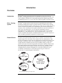
pYES2.1-E and pYC2-E
Echo™-Adapted Expression
Vectors
For cloning of the gene of interest using the Echo™
Cloning System and expression in Saccharomyces
cerevisiae
Catalog nos. ET200-XX, ET210-XX
Version J
29 December 2010
25-0340
Corporate Headquarters
Invitrogen Corporation
1600 Faraday Avenue
Carlsbad, CA 92008
T: 1 760 603 7200
F: 1 760 602 6500
E: [email protected]
For country-specific contact information visit our web site at www.invitrogen.com
User Manual
ii
Table of Contents
Kit Contents and Storage .....................................................................................................................................iv
Accessory Products.............................................................................................................................................vii
Introduction ................................................................................................................... 1
Overview ..............................................................................................................................................................1
Methods ......................................................................................................................... 5
Recombining Your Gene into pYES2.1-E or pYC2-E .........................................................................................5
Transforming the Recombination Reaction ..........................................................................................................6
Yeast Transformation .........................................................................................................................................10
Expression of Your Recombinant Protein ..........................................................................................................12
Appendix...................................................................................................................... 16
Small-Scale Yeast Transformation .....................................................................................................................16
Recipes ...............................................................................................................................................................17
Maps of pYES2.1-E and pYC2-E.......................................................................................................................21
Features of pYES2.1-E and pYC2-E ..................................................................................................................23
Map of pYES2.1-E/Uni-lacZ..............................................................................................................................24
Map of pYC2-E/Uni-lacZ...................................................................................................................................25
Technical Service ...............................................................................................................................................26
Purchaser Notification ........................................................................................................................................27
Product Qualification..........................................................................................................................................28
References ..........................................................................................................................................................29
iii
Kit Contents and Storage
Shipping and
Storage
The pYES2.1-E or pYC2-E Echo™-Adapted Expression Vectors are shipped on dry ice.
The INVSc1 stab is shipped at room temperature. Upon receipt, store the reagents as
follows:
•
•
•
Types of Kits
pYES2.1-E or pYC2-E reagents at -20°C
One Shot® Chemically Competent E. coli at -80°C
INVSc1 stab at +4°C
Several pYES2.1-E and pYC2-E Echo™ Cloning System Kits are available. The table
below lists the kits that include the pYES2.1-E or pYC2-E Echo™-Adapted Expression
Vectors.
Kit
™
Reagents Supplied
Catalog nos.
pYES2.1-E Echo -Adapted
Expression Vector Kit
pYES2.1-E vector
Expression Control vector
Cre Recombinase
10X Recombinase buffer
T7 Forward Sequencing Primer
INVSc1
ET200-01
pYC2-E Echo™-Adapted
Expression Vector Kit
pYC2-E vector
Expression Control vector
Cre Recombinase
10X Recombinase buffer
T7 Forward Sequencing Primer
INVSc1
ET210-01
pYES2.1-E Echo™-Adapted
Expression Vector Kit with a
choice of Donor Vector Kit and
One Shot® TOP10 Chemically
Competent E. coli (see page viii
for more information on donor
vectors)
pUni/V5-His TOPO TA Cloning® Kit
ET200-10C
pUniBlunt/V5-His TOPO® Cloning Kit
ET200-20C
pUni/V5-His A, B, and C
ET200-30C
pUniD/V5-His TOPO® Cloning Kit
ET200-40C
pUni/V5-His TOPO TA Cloning® Kit
ET210-10C
pUniBlunt/V5-His TOPO® Cloning Kit
ET210-20C
pUni/V5-His A, B, and C
ET210-30C
pUniD/V5-His TOPO® Cloning Kit
ET210-40C
pYC2-E Echo™-Adapted
Expression Vector Kit with a
choice of Donor Vector Kit and
One Shot® TOP10 Chemically
Competent E. coli (see page viii
for more information on donor
vectors)
Continued on next page
iv
Kit Contents and Storage, continued
pYES2.1-E and
pYC2-E Reagents
pYES2.1-E and pYC2-E reagents are listed below. Store at -20°C.
Item
INVSc1
S. cerevisiae
Strain
Concentration
Amount
pYES2.1-E or pYC2-E
Supercoiled, lyophilized in TE, pH 8
20 µg
Cre recombinase
Please check the label on the tube for
exact concentration of the enzyme
Enzyme supplied in:
50 mM Tris-HCl, pH 8.0
5 mM EDTA
1 mM EGTA
10 mM β-mercaptoethanol
20% Glycerol
15 µl
10X Recombinase Buffer
500 mM Tris-HCl, pH 7.5
100 mM MgCl2
300 mM NaCl
1.0 mg/ml BSA
25 µl
T7 Forward Sequencing
Primer (20 mer)
Lyophilized in TE Buffer, pH 8
2 µg
(5´-TAATACGACTCACTATAGGG-3´) (327 pmoles)
Expression control
(pYES2.1-E/Uni-lacZ or
pYC2-E/Uni-lacZ)
Supercoiled, lyophilized in TE, pH 8
20 µg
INVSc1
Yeast strain
1 stab
INVSc1 is a fast-growing, easily transformed diploid strain. Store at +4°C.
Genotype:
MATα his3∆1 leu2 trp1-289 ura3-52
MATa his3∆1 leu2 trp1-289 ura3-52
Continued on next page
v
Kit Contents and Storage, continued
One Shot® TOP10
E. coli
The table below describes the items included in the One Shot® TOP10 Chemically
Competent E. coli kit. Store at -80°C.
Item
Genotype of
TOP10
vi
Composition
Amount
SOC Medium
(may be stored at room
temperature or +4°C)
2% Tryptone
0.5% Yeast Extract
10 mM NaCl
2.5 mM KCl
10 mM MgCl2
10 mM MgSO4
20 mM glucose
6 ml
TOP10 E. coli
--
11 x 50 µl
pUC19 Control DNA
10 pg/µl in 5 mM Tris-HCl, 50 µl
0.5 mM EDTA, pH 8
Use this strain for transformation of the fusion vector. Note: This strain cannot be used
for transformation and growth of donor vectors.
F- mcrA ∆(mrr-hsdRMS-mcrBC) Φ80lacZ∆M15 ∆lacΧ74 recA1 araD139 ∆(araleu)7697 galU galK rpsL (StrR) endA1 nupG
Accessory Products
Products
Available
Separately
Many of the reagents in the pYES2.1-E and pYC2-E Echo™-Adapted Expression Vector
Kits, and additional reagents that may be used with the Echo™ Cloning System are
available separately. In addition, reagents for expression, detection, and purification of
your protein of interest from your pYES2.1-E or pYC2-E fusion vector are available from
Invitrogen. Ordering information is provided below.
™
Transformation Kit The S.c. EasyComp Transformation Kit (Catalog no. K5050-01) is designed for rapid
preparation of transformation-competent Saccharomyces cerevisiae cells.
Echo™ Cloning
Products
Many of the reagents supplied in the pYES2.1-E or pYC2-E Echo™-Adapted Expression
Vectors, as well as additional reagents that may be used for Echo™ Cloning, are available
separately from Invitrogen. Ordering information is provided below.
Product
Amount
Catalog No.
2 µg
N560-02
®
21 x 50 µl
C4040-03
®
11 x 50 µl
C1010-10
®
One Shot PIR2 Chemically Competent E. coli
11 x 50 µl
C1111-10
Cre Recombinase
10 reactions
R100-10
T7 Forward Primer
One Shot TOP10 Chemically Competent E. coli
One Shot PIR1 Chemically Competent E. coli
Continued on next page
vii
Accessory Products, continued
Donor Vectors
The table below lists a variety of donor vectors currently available from Invitrogen to
facilitate cloning of your gene of interest for use with Echo™ Cloning System.
Product
Application
pUniD/V5-His-TOPO
Cloning Kit
®
Quantity
Directional cloning of blunt PCR
products
10 reactions
ET004-10
pUni/V5-His-TOPO TA
Cloning® Kit
Cloning A-tailed PCR products
10 reactions
ET001-10
pUniBlunt/V5-His-TOPO®
Cloning Kit
Cloning blunt PCR products
10 reactions
ET002-10
pUni/V5-His A, B, and C
Cloning DNA fragments using
restriction enzymes
10 reactions
ET003-10
Detection and
Purification
After expressing your protein of interest from your pYES2.1-E or pYC2-E fusion vector,
use the reagents below to detect and purify your protein of interest.
Product
Amount
Catalog No.
Anti-His(C-term) Antibody
50 µl*
R930-25
Anti-His(C-term)-HRP Antibody
50 µl*
R931-25
Anti-V5 Antibody
50 µl*
R960-25
Anti-V5-HRP Antibody
50 µl*
R961-25
™
6 purifications
K850-01
™
50 ml
R801-01
150 ml
R801-15
50
R640-50
ProBond Purification System
ProBond Metal-Binding Resin
(precharged resin provided as a 50% slurry in 20%
ethanol)
Purification Columns
(10 ml polypropylene columns)
*Enough for 25 Westerns.
viii
Catalog no.
Introduction
Overview
Introduction
The Echo™ Cloning System allows direct recombination of your gene of interest
downstream of an appropriate promoter for expression in the host system of choice. The
vectors pYES2.1-E and pYC2-E are specifically designed for regulated expression of
recombinant proteins in S. cerevisiae. The GAL1 promoter regulates expression in yeast.
Echo™ Cloning
System
The Echo™ Cloning System is based on the univector plasmid-fusion system (UPS)
described by Elledge and coworkers to quickly and easily recombine a gene of interest
into a series of recipient (acceptor) vectors (Liu et al., 1998; Liu et al., 1999). The system
consists of the univector (donor vector) containing the gene of interest and recipient
(acceptor) vectors containing various regulatory sequences for expression in the host of
choice. The system utilizes the cre-lox site-specific recombination system of
bacteriophage P1 to recombine the gene of interest into the acceptor vector of choice
(Abremski et al., 1983; Sternberg et al., 1981). The product of the cre gene is a sitespecific recombinase that catalyzes recombination between two 34 bp loxP sequences to
resolve P1 dimers generated by replication of circular lysogens.
Plasmid Fusion
The donor vector (pUni) and the acceptor vector (i.e. pYES2.1-E or pYC2-E) each
contain a single lox site. The donor vector contains a loxP site while pYES2.1-E and
pYC2-E each contain a loxH site (for more information on loxP and loxH, please see the
next page). Construct the donor vector containing the gene of interest via the TOPO®
Cloning method or traditional restriction enzyme-mediated cloning (see page viii).
pYES2.1-E and pYC2-E contain the appropriate transcription regulatory sequences that
will control expression of the gene of interest in Saccharomyces. A unique loxH site is
located downstream of these sequences in both vectors. By mixing the donor vector
containing the gene of interest with pYES2.1-E or pYC2-E in the presence of Cre
recombinase, a plasmid fusion is created that expresses the gene of interest in
Saccharomyces. A generic diagram is shown below.
KanR
r
ote
om
r
P
Kg
X
lox*
e
C
or
i
pU
C
i
Cre
recombinase
pAcceptor
(2.5 to 5.8 kb)
or
Recombinant
Plasmid
(4.8 kb + gene to
8.1 kb + gene)
pU
r
ote
om
r
P
gen
R
loxP
gene
Kan
or
i
lox*
R6
Kg
R6
pUni
(2.3 kb + gene)
AmpR
AmpR
P
lox
lox* = loxP or loxH depending on acceptor vector
Continued on next page
1
Overview, continued
loxP or loxH Sites
The sequence of the loxP site is shown below. The loxP site consists of a 34 bp sequence
containing two 13 bp inverted repeats (see underlined bases) separated by an 8 bp spacer
(Hoess et al., 1982). The inverted repeats may form a stem and loop structure that may
reduce expression of the gene of interest in some cases. A variation of the loxP site (loxH,
see below) was created to eliminate the formation of a stem and loop structure and improve
expression. Mutated bases are shown in boldface. Please note that some acceptor vectors
including pYES2.1-E and pYC2-E contain a loxH site. Cre-mediated recombination can
still occur between a loxP and a loxH site although the efficiency may be slightly reduced.
•
loxP: ATA ACT TCG TAT AGC ATA CAT TAT ACG AAG TTA T
•
loxH: ATT ACC TCA TAT AGC ATA CAT TAT ACG AAG TTA T
Cre Recombinase
Cre recombinase (MW = 35 kDa) is a site-specific recombinase that binds to specific
sequences (loxP sites), brings together the target sites, cleaves, and covalently attaches to
the DNA. Recombination occurs following two pairs of strand exchanges and ligation of
the DNAs in a novel (recombinant) form. A nucleophilic hydroxylated tyrosine initiates
the DNA cleavage event by attack on a specific phosphodiester bond followed by the
covalent attachment of the recombinase to the target sequence through a phosphoamino
acid bond (Abremski and Hoess, 1992; Argos et al., 1986). The reaction does not require
any host factors or ATP, but does require Mg2+ or spermidine for activity (Abremski et
al., 1983). Recombination between two supercoiled substrates, each containing a loxP
site, results in a supercoiled dimer. The extent of the reaction is 10-20% under optimal
conditions (Abremski and Hoess, 1984; Abremski et al., 1983).
Selection of
Recombinants
By fusing the two plasmids, kanamycin resistance from the donor vector is now linked to
the pUC origin of replication. The recombination reaction is transformed into TOP10
E. coli and recombinants selected by plating the transformation reaction onto plates
containing kanamycin. Because the donor plasmid carries the R6Kγ origin of replication,
it will not propagate in TOP10. In addition, the acceptor vector, which carries the
ampicillin resistance gene, will not be selected. Therefore every colony that is selected on
kanamycin will represent a recombined fusion plasmid.
Continued on next page
2
Overview, continued
pYES2.1-E and
pYC2-E
pYES2.1-E or pYC2-E allow you to induce expression of your protein of interest in
S. cerevisiae. Both pYES2.1-E and pYC2-E contain the following features:
•
•
•
•
Yeast GAL1 promoter for high-level inducible protein expression in yeast by
galactose and repression by glucose (Giniger et al., 1985; West et al., 1984) (see
page 12 for more information)
A loxH site for plasmid fusion
URA3 auxotrophic marker for selection of yeast transformants
Ampicillin resistance for selection in E. coli
The vectors differ in their mechanism of replication.
•
•
pYES2.1-E contains the 2µ origin for episomal maintenance and high copy
replication.
pYC2-E contains the CEN6/ARSH4 sequence for non-integrative centromeric
maintenance and low copy replication.
For more information and a map of each vector, please see pages 21-23.
Other acceptor vectors are available separately and are provided with their own
manuals. For more information on other available acceptor vectors, please visit our
Web site (www.invitrogen.com) or call Technical Service (see page 26).
2µ Origin
The pYES2.1-E vector contains the 2µ origin for maintenance and replication in yeast. The
sequence containing the 2µ origin was originally isolated from the naturally-occurring yeast
2µ plasmid (Hartley and Donelson, 1980). When placed in a heterologous expression
plasmid (i.e. pYES2.1-E), the presence of the 2µ origin allows the plasmid to be episomally
maintained and replicated at high copy number (generally 10-40 copies per cell).
CEN6/ARSH4
Sequence
The pYC2-E vector contains the CEN6/ARSH4 sequence for maintenance and replication
in yeast (Sikorski and Hieter, 1989). The CEN6/ARSH4 sequence is a 518 bp hybrid
DNA fragment that contains a yeast centromere sequence (CEN) and an autonomously
replicating sequence (ARS) (Sikorski and Hieter, 1989). The CEN6 sequence is derived
from the CEN6 locus of yeast chromosome 6 (Panzeri and Philippsen, 1982) while the
ARSH4 sequence is derived from the yeast histone H4-associated ARS (Bouton and
Smith, 1986). When placed in a heterologous expression plasmid (i.e. pYC2-E), the
presence of the CEN6/ARSH4 sequence allows non-integrative centromeric maintenance
and low copy number replication of the plasmid (generally 1-2 copies per cell).
Continued on next page
3
Overview, continued
Experimental
Outline
The table below describes the general steps needed to recombine, transform, and express
your protein of interest.
Step
4
Action
Page
1
Perform the recombination reaction using your donor vector and
pYES2.1-E or pYC2-E.
2
Transform the recombination reaction into competent TOP10 E. coli and 6
select recombinants on LB plates containing 50 µg/ml kanamycin.
3
Pick transformants and analyze by restriction digestion.
7
4
Isolate plasmid DNA, transform into INVSc1, and select for uracil
prototrophy.
10-11
5
Induce with galactose to express your gene of interest.
12-13
6
Assay for expression of your protein.
14
7
Purify your protein, if desired.
15
5
Methods
Recombining Your Gene into pYES2.1-E or pYC2-E
Introduction
At this point you should have a plasmid preparation of your donor vector in addition to
pYES2.1-E or pYC2-E. Please review the information below and on the next page before
performing the recombination reaction.
Preparing
pYES2.1-E or
pYC2-E
To prepare pYES2.1-E or pYC2-E for use, add 20 µl of sterile, deionized water to the
lyophilized plasmid. This will yield a 1 µg/µl stock solution. You can further dilute a
small aliquot or use as is. Store at -20°C when you are finished.
If you wish to propagate this plasmid or prepare more plasmid DNA, you may transform
this plasmid into TOP10 E. coli as described on pages 6-7. Use 10-100 ng plasmid for
transformation and select on LB plates containing 50-100 µg/ml ampicillin.
Before Starting
You will need the following reagents and equipment.
•
100 ng of your donor vector
•
100 ng of pYES2.1-E or pYC2-E
•
Microcentrifuge tubes
•
Heat blocks set at 37°C and 65°C
•
Ice bucket with ice
•
Cre recombinase (included in the kit)
•
10X Recombinase buffer (included in the kit)
Recombination
Reaction
1.
2.
3.
4.
Set up each 20 µl recombination reaction on ice as follows.
Donor vector (100 ng)
x µl
pYES2.1-E or pYC2-E (100 ng)
y µl
10X Recombinase buffer
2 µl
Deionized water
add to a total volume of 19 µl
Cre recombinase
1 µl
Final volume
20 µl
Incubate at 37°C for 20 minutes.
Incubate at 65°C for 5 minutes to inactivate the recombinase.
Place tube on ice and proceed to Transformation, next page. If you run out of time,
you may store the recombination reaction at +4°C or -20°C overnight. Longer
storage times have not been tested.
5
Transforming the Recombination Reaction
Introduction
Once you have performed the recombination reaction you are ready to transform your
E. coli host. We recommend TOP10 E. coli for transformation but other strains may be
used. Strains should be endA and recA to ensure quality plasmid preparations and reduce
the chances of recombination, respectively.
Materials Supplied In addition to general microbiological supplies (i.e. plates, spreaders), you will need the
following reagents and equipment.
by the User
•
•
•
•
Important
Preparing for
Transformation
42°C water bath
LB plates containing 50 µg/ml kanamycin (see Important, below)
37°C shaking and non-shaking incubator
SOC (included in the One Shot® kit)
It is important to select for the fusion plasmid using kanamycin. Remember that the
donor vector contains the R6Kγ origin. This origin can only be maintained in E. coli
strains containing the pir gene. By fusing the plasmids, kanamycin is now linked to the
pUC origin, allowing the fusion to be maintained in strains that do not contain the pir
gene (i.e. TOP10). By selecting on kanamycin, you ensure that only colonies that contain
the fusion vector are selected.
The following transformation protocol is for use with the One Shot® TOP10 available with
the kit. If you are using other competent cells, please follow the manufacturer’s protocol.
For each transformation, you will need one vial of competent cells and two selective plates.
One Shot®
Transformation
Reaction
•
•
•
•
Equilibrate a water bath to 42°C.
Thaw the vial of SOC medium from the One Shot® kit and bring to room temperature.
Warm LB plates containing 50 µg/ml kanamycin at 37°C for 30 minutes.
Thaw on ice 1 vial of One Shot® TOP10 E. coli for each transformation.
1.
Add 5 µl of the recombination reaction to a vial of One Shot® TOP10 E. coli and mix
gently. Do not mix by pipetting up and down.
2.
Heat-shock the cells for 30 seconds at 42°C without shaking.
3.
Immediately transfer the tubes to ice.
4.
Add 500 µl of room temperature SOC medium.
5.
Cap the tube tightly and shake the tube horizontally at 37°C for 45 minutes.
6.
Spread 50 µl from each transformation on a pre-warmed plate. Pellet the remaining
cells and resuspend the cell pellet in 50 µl SOC and plate. Incubate overnight at 37°C.
7.
An efficient recombination reaction will produce hundreds of colonies. Pick
~5 colonies for analysis.
Continued on next page
6
Transforming the Recombination Reaction, continued
Analyzing Positive 1.
Clones
Culture 5 colonies overnight at 37°C in 2-5 ml LB or SOB medium containing
50 µg/ml kanamycin.
2.
Isolate plasmid DNA using your method of choice. If you need ultra-pure plasmid
DNA for automated or manual sequencing, we recommend the S.N.A.P.™ MiniPrep
Kit (Catalog no. K1900-01) or the S.N.A.P.™ MidiPrep Kit (Catalog no. K1910-01).
3.
Analyze the plasmids by restriction analysis. Use an enzyme or enzymes that cut
once in the donor vector and once in the acceptor vector to yield two fragments that
are distinguishable from one another. Please note that other strategies are possible.
4.
(Optional) To sequence the fusion plasmid to confirm the fusion junctions, we
recommend using the T7 Forward and Uni1 Forward sequencing primers. Refer to
the diagram on the following page for the sequences around the pYES2.1-E or
pYC2-E loxH site. Refer to the donor vector manual for the sequence around the
donor vector loxP site.
If you need help with setting up restriction enzyme digests or DNA sequencing, refer to
general molecular biology texts (Ausubel et al., 1994; Sambrook et al., 1989).
Continued on next page
7
Transforming the Recombination Reaction, continued
Sequencing Your
Construct in
pYES2.1-E or
pYC2-E
Aside from the origin of replication, the pYES2.1-E and pYC2-E sequences are identical.
Recombination into either vector produces the same sequence upstream of your insert.
This sequence is shown below. Unique restriction sites are labeled for your convenience.
Note that the complete sequence of pYES2.1-E and pYC2-E can be downloaded from our
Web site (www.invitrogen.com) or requested from Technical Service (page 26).
GAL1 promoter
TATA box
300
TTAACAGATA TATAAATGCA AAAACTGCAT AACCACTTTA ACTAATACTT TCAACATTTT
start of transcription
360
CGGTTTGTAT TACTTCTTAT TCAAATGTAA TAAAAGTATC AACAAAAAAT TGTTAATATA
GAL1 forward priming site
420
3´ end of GAL1 promoter
CCTCTATACT TTAACGTCAA GGAGAAAAAA CCCCGGATCG GACTACTAGC AGCTGTAATA
T7 promoter/priming site
480
Hind III
loxH site
CGACTCACTA TAGGGAATAT TAAGCTT ATT ACC TCA TAT AGC ATA CAT TAT ACG AAG
Uni1 Forward priming site
537
TTA T
RBS from
donor vector
Gene of
interest
C-terminal tag
(optional)
donor vector
Pme I
loxP site
GTTTAAACCC GCTGATCCTA GAGGGCCGCA TCATGTAATT AGTTATGTCA
Continued on next page
8
Transforming the Recombination Reaction, continued
Fusion Vector
Analysis
It should be clear from restriction analysis that you have a dimer fusion plasmid consisting
of the donor vector and pYES2.1-E or pYC2-E. Occasionally, trimers will result. Trimers
usually consist of two donor vector molecules and one acceptor molecule. Please note that
trimers usually express as well as the dimer product.
In theory, trimers may result from two sequential fusion events or a single fusion event
between a pre-existing monomeric substrate and a dimeric substrate. The production of
trimers can be eliminated if gel-purified monomeric supercoiled DNA is used in the
recombination reaction.
Preparing Glycerol Once you have identified the correct clone, prepare a glycerol stock for long-term
storage. In addition, store a stock of plasmid DNA at -20°C.
Stock for LongTerm Storage
1. Streak out the original colony on LB plates containing 50 µg/ml kanamycin.
2.
Select a single colony and inoculate into 1-2 ml of LB containing 50 µg/ml
kanamycin.
3. Grow overnight until culture is saturated.
4. Mix 0.85 ml of culture with 0.15 ml of sterile glycerol and transfer to a cryovial.
5.
Store at -80°C.
9
Yeast Transformation
Introduction
In this section, you will use a small-scale yeast transformation protocol to transform your
pYES2.1-E or pYC2-E construct into the INVSc1 yeast host strain included with each
vector. After transformation, induce expression of your recombinant fusion protein from
pYES2.1-E or pYC2-E using galactose.
Basic Yeast
Molecular Biology
The user should be familiar with basic yeast molecular biology and microbiological
techniques. Please refer to Current Protocols in Molecular Biology, Unit 13 (Ausubel et
al., 1994) and the Guide to Yeast Genetics and Molecular Biology (Guthrie and Fink,
1991) for information on preparing yeast media and handling yeast.
Genotype/
Phenotype of
INVSc1
The genotype and phenotype of the INVSc1 host strain are provided below.
Genotype: MATα/MATa his3∆1/his3∆1 leu2/leu2 trp1-289/trp1-289 ura3-52/ura3-52
Phenotype: His-, Leu-, Trp-, UraNote that INVSc1 is a diploid strain that is auxotrophic for histidine, leucine,
tryptophan, and uracil. The strain will not grow in SC minimal medium that is deficient
in histidine, leucine, tryptophan, or uracil. A recipe for preparation of SC minimal
medium is provided in the Appendix, page 18.
The INVSc1 strain is a suitable strain to use for expression purposes, but should not
be used for genetic analyses because it does not sporulate well.
Initiating INVSc1
Culture
To initiate a culture of INVSc1 from the stab provided with the kit, streak a small
amount from the stab on a YPD plate (see Appendix for recipe, page 19) and incubate
at 30°C. Once growth is established, you may check the phenotype of the strain by
streaking a single colony on an SC minimal plate supplemented with the appropriate
amino acids. INVSc1 will not grow in SC minimal medium that is deficient in histidine,
leucine, tryptophan, or uracil.
Be sure to make glycerol stocks of the strain. Store glycerol stocks at -80°C. If you plan
to use the strain directly from plates, be sure that the plates are less than 4 days old.
Plasmid Isolation
Isolate plasmid DNA from E. coli for yeast transformation using your method of choice.
We recommend the S.N.A.P.™ MiniPrep Kit (Catalog no. K1900-01) or the S.N.A.P.™
MidiPrep Kit (Catalog no. K1910-01). Other resin-based methods are suitable.
Positive Control
The pYES2.1-E and pYC2-E are supplied with a corresponding positive control vector
(pYES2.1-E/Uni-lacZ and pYC2-E/Uni-lacZ, respectively) to help you optimize
expression conditions for your protein. The gene encoding β-galactosidase is expressed
in yeast cells under the control of the GAL1 promoter. Successful transformation and
galactose induction will result in β-galactosidase expression that can be easily assayed
(see next page).
Continued on next page
10
Yeast Transformation, continued
Assay for
β-galactosidase
Activity
You may assay for β-galactosidase expression by activity assay using cell-free lysates
(Miller, 1972). Invitrogen offers the β-Gal Assay Kit (Catalog no. K1455-01) for fast
and easy detection of β-galactosidase expression.
Reagents for
Yeast
Transformation
Many protocols are suitable for the preparation of competent INVSc1 yeast cells. The
S.c. EasyComp™ Kit (Catalog no. K5050-01) provides a quick and easy method for the
preparation of competent yeast cells that can be used immediately or stored frozen for
future use. Transformation efficiency is guaranteed at >103 transformants per µg DNA.
A small-scale yeast transformation protocol is included in the Appendix (see page 16)
for your convenience. Alternatively, there are published references for other small-scale
transformation methods (Gietz et al., 1992; Gietz et al., 1995; Hill et al., 1991; Schiestl
and Gietz, 1989).
Transforming
Yeast
Use one of the methods described above (or one of your own choosing) to transform your
pYES2.1-E or pYC2-E fusion vector into competent INVSc1. We recommend that you
include the appropriate control vector (see the previous page) as a positive control for
expression and a sample with no DNA as a negative control for transformation.
Select for transformants on SC minimal media lacking uracil (SC-U). Transformants
should exhibit the uracil prototrophy. See the Appendix, page 18 for a recipe to prepare
SC minimal media.
Once you have identified a transformant, be sure to purify the colony and make a glycerol
stock for long-term storage.
Maintaining
Transformants
Maintain yeast cells containing your pYES2.1-E or pYC2-E fusion vector in SC-U
medium containing 2% glucose or 2% raffinose (see the next page).
Note: The growth rate of yeast strains varies with the carbon source. Yeast strains
typically exhibit the fastest growth in medium containing glucose.
11
Expression of Your Recombinant Protein
Introduction
Once you have obtained a transformant containing your pYES2.1-E or pYC2-E fusion
vector, you are ready to induce expression of your recombinant fusion protein of
interest. This section provides information on how to induce and assay for expression of
your protein of interest.
GAL1 Promoter
In INVSc1, transcription from the GAL1 promoter is repressed in the presence of glucose
(West et al., 1984). Removing glucose and adding galactose as a carbon source induces
transcription (Giniger et al., 1985). Maintaining cells in glucose gives the most complete
repression and the lowest basal transcription of the GAL1 promoter. Transferring cells
from glucose- to galactose-containing medium causes the GAL1 promoter to become
de-repressed and allows transcription to be induced.
Alternatively, cells may be maintained in medium containing raffinose as a carbon source.
The presence of raffinose does not repress or induce transcription from the GAL1
promoter. Addition of galactose to the medium induces transcription from the GAL1
promoter even in the presence of raffinose. Induction of the GAL1 promoter by galactose
is more rapid in cells maintained in raffinose when compared to those maintained in
glucose.
You may choose to grow cells containing your pYES2.1-E or pYC2-E fusion vector in
glucose or raffinose depending on how quickly you want to obtain your expressed protein
after induction with galactose and on the toxicity of the expressed protein. For more
information about expression in yeast, please refer to the Guide to Yeast Genetics and
Molecular Biology (Guthrie and Fink, 1991).
For a protocol to induce expression of your fusion protein with galactose, proceed to
Time Course of Protein Induction by Galactose on the next page.
Continued on next page
12
Expression of Recombinant Protein, continued
Time Course of
Protein Induction
by Galactose
To induce expression of your protein of interest from the GAL1 promoter, galactose is
added to the medium. For cells that have been maintained in glucose, recombinant fusion
protein can be detected in as little as 4 hours after galactose induction. Recombinant fusion
protein can be detected in cells that have been cultured in raffinose by 2 hours after
galactose induction.
If you are assaying for expression of your recombinant fusion protein for the first time, we
recommend that you perform a time course to optimize expression of your recombinant
protein (e.g. 0, 4, 8, 12, 16, 24 hours after galactose induction). A standard protocol is
provided below to perform a time course experiment. Other protocols are suitable.
1.
2.
3.
4.
5.
Inoculate a single colony of INVSc1 containing your pYES2.1-E or pYC2-E fusion
vector into 15 ml of SC-U selective medium containing 2% glucose or 2% raffinose.
Grow overnight at 30°C with shaking.
Determine the OD600 of your overnight culture. Calculate the amount of overnight
culture necessary to obtain an OD600 of 0.4 in 50 ml of induction medium (SC-U
selective medium containing 2% galactose).
Example: Assume that the OD600 of an overnight culture is 3 OD600 per ml. Then, the
amount of overnight culture needed to inoculate a 50 ml culture to OD600 = 0.4 is
(0.4 OD/ml) (50 ml) = 6.67 ml
3 OD/ml
Remove the amount of overnight culture as determined in Step 2 and pellet the cells at
1500 x g for 5 minutes at room temperature. Discard the supernatant.
Resuspend the cells in 50 ml of induction medium. Grow at 30°C with shaking.
Harvest an aliquot of cells at 0, 4, 8, 12, 16, and 24 hours after addition of cells to the
induction medium. For each time point, remove 5 ml of culture from the flask and
determine the OD600 of each sample. You will use this information when assaying for
your recombinant fusion protein (see Step 3 on the next page).
6.
Centrifuge the cells at 1500 x g for 5 minutes at +4°C.
7.
Decant the supernatant. Resuspend cells in 500 µl of sterile water.
8.
Transfer cells to a sterile microcentrifuge tube. Centrifuge samples for 30 seconds at
top speed in the microcentrifuge.
9.
Remove the supernatant.
10. Store the cell pellets at -80°C until ready to use. Proceed to the next section to prepare
cell lysates to detect your recombinant protein (see the next page).
Continued on next page
13
Expression of Recombinant Protein, continued
Detection of
Recombinant
Fusion Protein
To detect expression of your recombinant fusion protein by western blot (see below), you
may use the Anti-V5 or the Anti-His(C-term) antibodies available from Invitrogen (see
page viii for ordering information) or an antibody to your protein of interest.
You will also need to prepare a cell lysate from your yeast transformant. A general
protocol for small-scale preparation of cell lysates using acid-washed glass beads is
provided below for your convenience. Other protocols are suitable. Please refer to Current
Protocols in Molecular Biology, Unit 13.13 (Ausubel et al., 1994) for more information.
For large-scale preparations (culture volumes over 1 liter), see Scale-up on the next page.
Materials Needed:
•
Breaking buffer (50 mM sodium phosphate, pH 7.4, 1 mM EDTA, 5% glycerol, 1
mM PMSF) (please refer to Appendix, page 19 for instructions to prepare the sodium
phosphate stock buffer).
•
Acid-washed glass beads (0.4-0.6 mm size; Sigma-Aldrich, Catalog no. G8772).
Protocol:
1.
You may prepare cell lysates from either frozen cells or fresh cells.
Reminder: You will need to know the OD600 of your cell sample(s) before beginning
(see Step 5, previous page).
2.
Resuspend fresh or frozen cell pellets in 500 µl of breaking buffer. Centrifuge at
1500 x g for 5 minutes at +4°C to pellet cells.
3.
Remove supernatant and resuspend the cells in a volume of breaking buffer to obtain
an OD600 of 50-100. Use the OD600 determined in Step 5, previous page, to calculate
the appropriate volume of breaking buffer to use.
4.
Add an equal volume of acid-washed glass beads.
5.
Vortex mixture for 30 seconds, followed by 30 seconds on ice. Repeat four times for
a total of four minutes to lyse the cells. Cells will be lysed by shear force. You can
check for the extent of lysis by checking a small aliquot under the microscope.
6.
Centrifuge in a microcentrifuge for 10 minutes at maximum speed.
7.
Remove supernatant and transfer to a fresh microcentrifuge tube. Assay the lysate for
protein concentration using BSA as a standard.
8.
Add SDS-PAGE sample buffer to a final concentration of 1X and heat the sample for
5 minutes at 70°C.
9.
Load 20 µg of lysate onto an SDS-PAGE gel and electrophorese. Use the appropriate
percentage of acrylamide to resolve your recombinant protein.
If you cloned your PCR product in frame with the C-terminal peptide, this will increase
the size of your protein by ~3 kDa.
Continued on next page
14
Expression of Recombinant Protein, continued
Scale-up of
Expression for
Purification
Once you have determined the optimal induction time necessary to obtain maximal protein
expression, you may increase the protein yield by scaling up the procedure described on
page 13. If you plan to use ProBond™ resin to purify your recombinant fusion protein,
please see the Note below. To prepare cell lysates from culture volumes over 1 liter, we
recommend that you use a bead beater (Biospec Products, Bartlesville, OK) to lyse the
cells. Please refer to Current Protocols in Molecular Biology, Unit 13.13 (Ausubel et al.,
1994) for a suitable protocol to lyse cells with a bead beater.
Purification
For help with purification of your recombinant fusion protein, please refer to the ProBond™
Purification System manual. You can download the manual from our Web site
(www.invitrogen.com) or request a copy from Technical Services (see page 26).
If you are using another type of resin, please refer to the manufacturer’s recommendations.
15
Appendix
Small-Scale Yeast Transformation
Introduction
A small-scale yeast transformation protocol for routine transformations is provided
below. Other protocols are suitable. The S.c. EasyComp™ Transformation Kit (Catalog
no. K5050-01) is available from Invitrogen for rapid preparation of transformationcompetent yeast cells. Please visit our Web site (www.invitrogen.com) or call Technical
Service for more information (see page 26).
Materials Needed
Be sure to have the following reagents on hand before starting.
Protocol
•
YPD liquid medium (see Recipe, page 19)
•
1X TE (see Recipe, page 19)
•
1X LiAc/0.5X TE (see Recipe, page 20)
•
Denatured salmon sperm DNA (Sigma-Aldrich, Catalog no. D9156)
•
Fusion vector construct (or other plasmid DNA to be transformed)
•
1X LiAc/40% PEG-3350/1X TE (See Recipe, page 20)
•
DMSO
•
Selective plates
1.
Inoculate 10 ml of YPD medium with a colony of INVSc1 and shake overnight at
30°C.
2.
Determine the OD600 of your overnight culture. Dilute culture to an OD600 of 0.4 in
50 ml of YPD medium and grow an additional 2-4 hours.
3.
Pellet the cells at 1500 x g and resuspend the pellet in 40 ml 1X TE.
4.
Pellet the cells at 1500 x g and resuspend the pellet in 2 ml of 1X LiAc/0.5X TE.
5.
Incubate the cells at room temperature for 10 minutes.
6.
For each transformation, mix together 1 µg plasmid DNA and 100 µg denatured
sheared salmon sperm DNA with 100 µl of the yeast suspension from Step 5.
7.
Add 700 µl of 1X LiAc/40% PEG-3350/1X TE and mix well.
8.
Incubate solution at 30°C for 30 minutes.
9.
Add 88 µl DMSO, mix well, and heat shock at 42°C for 7 minutes.
10. Centrifuge in a microcentrifuge for 10 seconds and remove supernatant.
11. Resuspend the cell pellet in 1 ml 1X TE and re-pellet.
12. Resuspend the cell pellet in 50-100 µl 1X TE and plate on a selective plate.
To calculate the number of yeast cells, assume that 1 OD600 unit = ~2.0 x 10⁷ yeast
cells/ml.
16
Recipes
LB (Luria-Bertani)
Medium and
Plates
Composition:
1.0% Tryptone
0.5% Yeast Extract
1.0% NaCl
pH 7.0
1.
For 1 liter, dissolve 10 g tryptone, 5 g yeast extract, and 10 g NaCl in 950 ml
deionized water.
2.
Adjust the pH of the solution to 7.0 with NaOH and bring the volume up to 1 liter.
3.
Autoclave on liquid cycle for 20 minutes at 15 psi. Allow solution to cool to 55°C
and add antibiotic if needed.
4.
Store at room temperature or at +4°C.
LB agar plates
1.
Prepare LB medium as above, but add 15 g/L agar before autoclaving.
2.
Autoclave on liquid cycle for 20 minutes at 15 psi.
3.
After autoclaving, cool to ~55°C, add antibiotic (50 µg/ml of kanamycin), and pour
into 10 cm plates.
4.
Let harden, then invert and store at +4°C, in the dark.
SOB Medium (with SOB (per liter)
Kanamycin)
2% Tryptone
0.5% Yeast Extract
0.05% NaCl
2.5 mM KCl
10 mM MgCl2
1.
Dissolve 20 g tryptone, 5 g yeast extract, and 0.5 g NaCl in 950 ml deionized water.
2.
Make a 250 mM KCl solution by dissolving 1.86 g of KCl in 100 ml of deionized
water. Add 10 ml of this stock KCl solution to the solution in Step 1.
3.
Adjust pH to 7.5 with 5 M NaOH and add deionized water to 1 liter.
4.
Autoclave this solution, cool to ~55°C, and add 10 ml of sterile 1 M MgCl2. You may
also add kanamycin to 50 µg/ml.
5.
Store at +4°C. Medium is stable for only ~1 month.
Continued on next page
17
Recipes, continued
SC Minimal
Medium and
Plates
SC is synthetic minimal defined medium for yeast.
0.67% yeast nitrogen base (without amino acids but with ammonium sulfate)
2% carbon source (i.e. glucose or raffinose)
0.01% (adenine, arginine, cysteine, leucine, lysine, threonine, tryptophan, uracil)
0.005% (aspartic acid, histidine, isoleucine, methionine, phenylalanine, proline, serine,
tyrosine, valine)
2% agar (for plates)
1.
Dissolve the following reagents in 900 ml deionized water (800 ml if preparing
medium containing raffinose). Note: We make medium and plates as we need them
and weigh out each amino acid. Many researchers prepare 100X solutions of each
amino acid that they need.
Reminder: Omit uracil to make selective plates for growing pYES2.1-E or pYC2-E
fusion vector transformants.
6.7 g Yeast Nitrogen Base
2.
Induction Medium
0.1 g each
0.05 g each
adenine
aspartic acid
arginine
histidine
cysteine
isoleucine
leucine
methionine
lysine
phenylalanine
threonine
proline
tryptophan (W)
serine
uracil (U)
tyrosine
valine
If you are making plates, add the agar after dissolving the reagents above.
3.
Autoclave at 15 psi, 121°C for 20 minutes.
4.
Cool to 50°C and add 100 ml of filter-sterilized 20% glucose or 200 ml of filtersterilized 10% raffinose.
5.
Pour plates and allow to harden. Invert the plates and store at +4°C. Plates are
stable for 6 months.
If you are making induction medium, follow Steps 1-3 above except dissolve the
reagents in 800 ml of deionized water. Cool the medium to 50°C and add 100 ml of
filter-sterilized 20% galactose and 100 ml of filter-sterilized 10% raffinose to the
medium. Note: Raffinose is included to increase growth rate.
When making stock solutions of raffinose, do not autoclave the stock solution.
Autoclaving the solution will convert the raffinose to glucose. Filter-sterilize the stock
solution.
Continued on next page
18
Recipes, continued
YPD
Yeast Extract Peptone Dextrose Medium (1 liter)
1% yeast extract
2% peptone
2% dextrose (D-glucose)
1.
Dissolve the following in 1000 ml of water:
10 g yeast extract
20 g peptone
20 g dextrose (see note below if making plates)
2.
Optional: Add 20 g agar, if making plates.
3.
Autoclave for 20 minutes on liquid cycle.
4.
Store medium at room temperature or cool the medium and pour plates. The shelf life
is approximately one to two months.
Note: If making plates, omit dextrose from Step 1. Autoclaving agar and dextrose
together will cause the dextrose to caramelize. Prepare a separate stock solution of 20%
dextrose and autoclave or filter-sterilize. After the YPD broth (900 ml volume) has been
autoclaved, add 100 ml of 20% dextrose to the medium.
0.1 M Sodium
Phosphate, pH 7.4
10X TE
Before beginning, have the following reagents on hand.
•
•
Sodium phosphate, monobasic (NaH2PO4·H2O; Sigma-Aldrich S9638)
Sodium phosphate, dibasic (Na2HPO4·7H2O; Sigma-Aldrich S9390)
1.
Prepare 100 ml of 1 M NaH2PO4·H2O by dissolving 13.8 g in 90 ml of deionized
water. Bring volume up to 100 ml. Filter-sterilize.
2.
Prepare 100 ml of 1 M Na2HPO4·7H2O by dissolving 26.81 g in 90 ml of deionized
water. Bring volume up to 100 ml. Filter-sterilize.
3.
For 1 liter of 0.1 M sodium phosphate, pH 7.4, mix together 22.6 ml of 1 M
NaH2PO4 and 77.4 ml of 1 M Na2HPO4. Bring up the volume to 1 liter with sterile
water.
4.
Filter-sterilize and store at room temperature.
100 mM Tris, pH 7.5
10 mM EDTA
1.
For 100 ml, dissolve 1.21 g of Tris base and 0.37 g of EDTA in 90 ml of deionized
water.
2.
Adjust the pH to 7.5 with concentrated HCl and bring the volume up to 100 ml.
3.
Filter-sterilize and store at room temperature.
Alternatively, you can make the solution using 1 M Tris-HCl, pH 7.5 and 0.5 M EDTA,
pH 8.0.
1X TE
10 mM Tris, pH 7.5
1 mM EDTA
Dilute 10X TE 10-fold with sterile water.
Continued on next page
19
Recipes, continued
10X LiAc
1X LiAc
1 M Lithium Acetate, pH 7.5
1.
For 100 ml, dissolve 10.2 g of lithium acetate in 90 ml of deionized water.
2.
Adjust pH to 7.5 with dilute glacial acetic acid and bring up the volume to 100 ml.
3.
Filter-sterilize and store at room temperature.
100 mM Lithium Acetate, pH 7.5
Dilute 10X LiAc solution 10-fold with sterile, deionized water.
1X LiAc/0.5X TE
1X LiAc/40% PEG3350/1X TE
20
100 mM Lithium Acetate, pH 7.5
5 mM Tris-HCl, pH 7.5
0.5 mM EDTA
1.
For 100 ml, mix together 10 ml of 10X LiAc and 5 ml of 10X TE.
2.
Add deionized water to 100 ml.
3.
Filter-sterilize and store at room temperature.
100 mM Lithium Acetate, pH 7.5
40% PEG-3350
10 mM Tris-HCl, pH 7.5
1 mM EDTA
1.
Prepare solution immediately prior to use. For 100 ml, mix together 10 ml of 10X
LiAc, 10 ml of 10X TE, and 40 g of PEG-3350.
2.
Add deionized water to 100 ml and dissolve the PEG. You may have to heat the
solution to fully dissolve the PEG.
3.
Autoclave at 121°C, 15 psi for 20 minutes. Store at room temperature.
Maps of pYES2.1-E and pYC2-E
The map below shows the features of pYES2.1-E (5825 bp). The complete sequence of
the vector is available for downloading from our Web site (www.invitrogen.com) or
from Technical Service (page 26).
1
P GAL
lox
H
f1
ori
40pUC
TV
TS
C1
CY
or
i
1
f1
pYES2.1-E
5825 bp
in
pi
cil
rig
li n
2m o
Features of pYES2.1-E
5825 nucleotides
m
pYES2.1-E Map
U RA3
A
GAL1 promoter: bases 1-451
GAL1 promoter priming site: bases 414-437
T7 promoter priming site: bases 475-494
loxH site: bases 507-540
CYC1 transcriptional termination region: bases 570-823
CYC1 reverse priming site: bases 587-605
pUC origin: bases 1007-1680
Ampicillin resistance gene (bla): base 1825-2685 (C)
URA3 gene: 2703-3810 (C)
2 micron origin: bases 3814-5285
f1 origin: bases: 5353-5808
(C) = complementary
Continued on next page
21
Maps of pYES2.1-E and pYC2-E, continued
pYC2-E Map
The map below shows the features of pYC2-E (4489 bp). The complete sequence of the
vector is available for downloading from our Web site (www.invitrogen.com) or
from Technical Service (page 26).
P
r0igin
SCVo4
pU
1
CYC
1T
T f1
GA
L1
loxH
pYC2-E
pi
RS
cil
6/ A
li n
C EN
4489 bp
H
m
4
Features of pYC2-E
4489 nucleotides
U RA3
GAL1 promoter: bases 1-451
GAL1 Forward priming site: bases 414-437
T7 promoter priming site: bases 475-494
loxH site: bases 507-540
CYC1 transcriptional termination region: bases 570-823
CYC1 Reverse priming site: bases 587-605
pUC origin: bases 1007-1680
Ampicillin resistance gene: bases 1825-2685 (C)
URA3 gene: bases 2703-3810 (C)
CEN6/ARSH4: bases 3823-4341
f1 origin:bases 4342-4447
(C) = complementary strand
22
A
Features of pYES2.1-E and pYC2-E
Features
The important elements of pYES2.1-E and pYC2-E are described in the following table.
All features have been functionally tested.
Feature
Benefit
GAL1 promoter
Permits galactose-inducible expression of genes cloned
into pYES2/CT (West et al., 1984)
GAL1 forward priming site
Allows sequencing through the insert
T7 promoter/priming site
Allows for in vitro transcription in the sense orientation
and sequencing through the insert
loxH site
Allows recombination between the donor vector and
pYES2.1-E and pYC2-E (Hoess et al., 1982)
CYC1 transcription
termination signal
Permits efficient termination and stabilization of mRNA
CYC1 reverse priming site
Allows sequencing through the insert
pUC origin
Allows maintenance and high copy replication in E. coli
Ampicillin resistance gene
Allows selection of transformants in E. coli
URA3 gene
Permits selection of yeast transformants in uracildeficient medium
2µ origin (pYES2.1-E)
Permits episomal maintenance and high copy replication
in yeast
CEN6/ARSH4 sequence
(pYC2-E)
Permits non-integrative centromeric maintenance and
low copy replication in yeast (Sikorski and Hieter, 1989)
f1 origin
Allows rescue of single-stranded DNA
23
Map of pYES2.1-E/Uni-lacZ
Description
pYES2.1-E/Uni-lacZ is a 11194 bp control vector expressing β-galactosidase. The lacZ
gene was amplified and TOPO® Cloned into pUni/V5-His/Gene-TOPO®. The resulting
vector was recombined with pYES2.1-E using Cre recombinase to create pYES2.1-E/
Uni-lacZ. Note: pUni/V5-His/Gene-TOPO® is similar to pUni/V5-His-TOPO® TA
except that it contains additional DNA between the TOPO® Cloning site and the V5
epitope.
Map of
Control Vector
The figure below summarizes the features of the pYES2.1-E/Uni-lacZ vector. The
complete nucleotide sequence for pYES2.1-E/Uni-lacZ is available for downloading
from our Web site (www.invitrogen.com) or by contacting Technical Service (see
page 26).
loxH
L1
P GA
Lac
s
ZV
5-H
i
B
f1
p
GH
or
i
24
K a na m y c i n
or i
Kg
ll
ci
pi
Am
GAL1 promoter: bases 1-451
GAL1 forward priming site: bases 414-437
T7 promoter priming site: bases 475-494
in
loxH site: bases 507-540
pU
lacZ ORF: bases 561-3617
Co
ri
V5 epitope: bases 3630-3671
6xHis tag: bases 3681-3698
pUni Reverse priming site: bases 3760-3781
BGH polyadenylation region: bases 3779-3987
T7 transcriptional termination region: bases 4002-4130
Kanamycin resistance gene: bases 4309-5103 (C)
kan promoter region: bases 5104-5241 (C)
R6Kg origin: bases 5464-5855
pUni Forward priming site: bases 5823-5841
loxP site: bases 5876-5909
CYC1 transcription termination region: bases 5939-6192
CYC1 Reverse priming site: bases 5956-5974
pUC origin: bases 6376-7049
Ampicillin resistance gene (bla): bases 7194-8054 (C)
URA3 gene: bases 8072-9179 (C)
2 micron origin: bases 9183-10654
f1 origin: bases 10722-11177
(C) = complementary strand
R6
2m origin
A
11194 bp
UR A 3
Features of pYES2.1-E/Uni-lacZ
11194 nucleotides
pYES2.1-E/Uni-lacZ
xP
lo
CYC1 TT
Map of pYC2-E/Uni-lacZ
Description
pYC2-E/Uni-lacZ is a 9858 bp control vector expressing β-galactosidase. The lacZ gene
was amplified and TOPO® Cloned into pUni/V5-His/Gene-TOPO®. The resulting vector
was recombined with pYC2-E using Cre recombinase to create pYC2-E/Uni-lacZ.
Note: pUni/V5-His/Gene-TOPO® is similar to pUni/V5-His-TOPO® TA except that it
contains additional DNA between the TOPO® Cloning site and the V5 epitope.
Map of Control
Vector
The figure below summarizes the features of the pYC2-E/Uni-lacZ vector. The
complete nucleotide sequence for pYC2-E/Uni-lacZ is available for downloading
from our Web site (www.invitrogen.com) or by contacting Technical Service (see
page 26).
L1
P GA
loxH
Lac
ZV
n
lli
ci
pi
Am
K a na m y c i n
go
ri
pYC2-E/Uni-lacZ
9858 bp
GAL1 promoter: bases 1-451
GAL1 Forward priming site: bases 414-437
pU
T7 promoter priming site: bases 475-494
Co
ri
loxH site: bases 507-540
lacZ ORF: bases 561-3617
V5 epitope: bases 3630-3671
6xHis tag: bases 3681-3698
pUni Reverse priming site: bases 3760-3781
BGH polyadenylation region: bases 3779-3987
T7 transcription termination region: bases 4002-4130
Kanamycin resistance gene (ORF): bases 4309-5103 (C)
kan promoter: bases 5104-5241 (C)
R6Kg origin: bases 5464-5855
pUni Forward priming site: bases 5823-5841
loxP site: bases 5876-5909
CYC1 transcriptional termination region: bases 5939-6192
CYC1 Reverse priming site: bases 5956-5974
pUC origin: bases 6576-7049
Ampicillin resistance gene (bla) ORF: bases 7181-8041 (C)
URA3 gene: bases 8072-9179 (C)
CEN6/ARS4: bases 9192-9710
f1 origin: bases 9711-9816
(C) = complementary strand
R6
K
U RA3
Features of pYC2-E/Uni-lacZ
9858 nucleotides
A
Hp
BG
CE
N6
/A
RS
H4
s
5-H
i
xP
lo
C Y C 1 TT
25
Technical Service
Web Resources
Contact Us
Visit the Invitrogen Web site at www.invitrogen.com for:
•
Technical resources, including manuals, vector maps and sequences, application
notes, MSDSs, FAQs, formulations, citations, handbooks, etc.
•
Complete technical service contact information
•
Access to the Invitrogen Online Catalog
•
Additional product information and special offers
For more information or technical assistance, call, write, fax, or email. Additional
international offices are listed on our Web page (www.invitrogen.com).
Corporate Headquarters:
Invitrogen Corporation
1600 Faraday Avenue
Carlsbad, CA 92008 USA
Tel: 1 760 603 7200
Tel (Toll Free): 1 800 955 6288
Fax: 1 760 602 6500
E-mail: [email protected]
Japanese Headquarters:
Invitrogen Japan
LOOP-X Bldg. 6F
3-9-15, Kaigan
Minato-ku, Tokyo 108-0022
Tel: 81 3 5730 6509
Fax: 81 3 5730 6519
E-mail: [email protected]
European Headquarters:
Invitrogen Ltd
Inchinnan Business Park
3 Fountain Drive
Paisley PA4 9RF, UK
Tel: +44 (0) 141 814 6100
Tech Fax: +44 (0) 141 814 6117
E-mail: [email protected]
Material Data
Safety Sheets
(MSDSs)
MSDSs are available on our Web site at www.invitrogen.com. On the home page, click
on Technical Resources and follow instructions on the page to download the MSDS for
your product.
Limited Warranty
Invitrogen is committed to providing our customers with high-quality goods and services.
Our goal is to ensure that every customer is 100% satisfied with our products and our
service. If you should have any questions or concerns about an Invitrogen product or
service, contact our Technical Service Representatives.
Invitrogen warrants that all of its products will perform according to specifications stated
on the certificate of analysis. The company will replace, free of charge, any product that
does not meet those specifications. This warranty limits Invitrogen Corporation’s liability
only to the cost of the product. No warranty is granted for products beyond their listed
expiration date. No warranty is applicable unless all product components are stored in
accordance with instructions. Invitrogen reserves the right to select the method(s) used to
analyze a product unless Invitrogen agrees to a specified method in writing prior to
acceptance of the order.
Invitrogen makes every effort to ensure the accuracy of its publications, but realizes that
the occasional typographical or other error is inevitable. Therefore Invitrogen makes no
warranty of any kind regarding the contents of any publications or documentation. If you
discover an error in any of our publications, please report it to our Technical Service
Representatives.
Invitrogen assumes no responsibility or liability for any special, incidental, indirect
or consequential loss or damage whatsoever. The above limited warranty is sole and
exclusive. No other warranty is made, whether expressed or implied, including any
warranty of merchantability or fitness for a particular purpose.
26
Purchaser Notification
Limited Use Label
License No. 119:
Echo™ Cloning
Products
No license is conveyed to use this product with any recombination sites other than those purchased
from Invitrogen Corporation or its authorized distributor. The buyer cannot modify the
recombination sequence(s) contained in this product for any purpose.
Limited Use Label
License No. 141:
Expression of
Polypeptides in
Yeast
This product is the subject of U.S. and foreign patents. Rights to use this product are limited to
academic research use only. Non-academic entities are required to obtain a separate license from
Washington Research Foundation to utilize this product for any use. Washington Research
Foundation, 2815 Eastlake Avenue East, Suite 300, Seattle, Washington 98102. Tel:
206-336-5600. Fax: 206-336-5615.
Information for
European
Customers
The INVSc1 yeast strain is genetically modified and carries the auxotrophic reporter gene HIS3. As
a condition of sale, this product must be in accordance with all applicable local legislation and
guidelines including EC Directive 90/219/EEC on the contained use of genetically modified
organisms.
27
Product Qualification
Vectors
pYES2.1-E, pYES2.1-E/Uni-lacZ, pYC2-E, and pYC2-E/Uni-lacZ are qualified by
restriction digest. The restriction enzymes and the expected fragments are listed below.
Restriction
Enzyme
pYES2.1-E
pYES2.1-E/
Uni-lacZ
pYC2-E
pYC2-E/ UnilacZ
ApaL I
4579 bp
1246 bp
Not tested
3243 bp
1246 bp
Not tested
EcoR V
Not tested
4634 bp,
4103 bp
2457 bp
Not tested
4634 bp
2767 bp
2457 bp
Hind III
5725 bp
6120 bp
5074 bp
4489 bp
5074 bp
4784 bp
Drd I
Not tested
Not tested
4489 bp
Not tested
BamH I
Not tested
Not tested
Not tested
9858 bp
Pme I
5825 bp
Not tested
4489 bp
Not tested
Primers
The T7 Forward Sequencing primer has been lot-qualified by DNA sequencing
experiments using the dideoxy chain termination technique.
INVSc1
The INVSc1 yeast strain is tested for growth on YPD medium.
Cre Recombinase
Purity: >95% homogeneity
Endonuclease activity: Negative
Exonuclease activity: Negative
Functional Assay: Cre recombinase is qualified using the assay on page 5 of this manual.
The donor vector is pUni/lacZ and the acceptor vector is pcDNA3.1-E. Five microliters of
the recombination reaction is transformed into 50 µl One Shot® TOP10 competent E. coli
using the protocol on page 6. Twenty-five microliters of the transformation reaction is plated
on LB plates containing 50 µg/ml kanamycin and X-gal (performed in duplicate). One
microliter of Cre recombinase should yield >500 blue, kanamycin-resistant transformants.
One Shot® TOP10
Competent E. coli
All competent cells are qualified as follows:
• Cells are tested for transformation efficiency using the control plasmid included in the
kit. Transformed cultures are plated on LB plates containing 100 µg/ml ampicillin and
the transformation efficiency is calculated. Test transformations are performed in
duplicate. Transformation efficiency should be ~1 x 10⁹ cfu/µg DNA for chemically
competent cells.
• To verify the absence of phage contamination, 0.5-1 ml of competent cells are added to
LB top agar and poured onto LB plates. After overnight incubation, no plaques should
be detected.
• Untransformed cells are plated on LB plates 100 µg/ml ampicillin, 25 µg/ml
streptomycin, 50 µg/ml kanamycin, or 15 µg/ml chloramphenicol to verify the absence
of antibiotic-resistant contamination.
28
References
Abremski, K., and Hoess, R. (1984). Bacteriophage P1 Site-Specific Recombination. Purification and Properties of
the Cre Recombinase Protein. J. Biol. Chem. 259, 1509-1514.
Abremski, K., Hoess, R., and Sternberg, N. (1983). Studies on the Properties of P1 Site-Specific Recombination:
Evidence for Topologically Unlinked Products Following Recombination. Cell 32, 1301-1311.
Abremski, K. E., and Hoess, R. H. (1992). Evidence for a Second Conserved Arginine Residue in the Integrase
Family of Recombination Proteins. Protein Eng. 5, 87-91.
Argos, P., Landy, A., Abremski, K., Egan, J. B., Haggard-Ljungquist, E., Hoess, R. H., Kahn, M. L., Kalionis, B.,
Narayana, S. V. L., Pierson III, L. S., Sternberg, N., and Leong, J. M. (1986). The Integrase Family of Site-Specific
Recombinases: Regional Similarities and Global Diversity. EMBO J. 5, 433-440.
Ausubel, F. M., Brent, R., Kingston, R. E., Moore, D. D., Seidman, J. G., Smith, J. A., and Struhl, K. (1994).
Current Protocols in Molecular Biology (New York: Greene Publishing Associates and Wiley-Interscience).
Bouton, A. H., and Smith, M. M. (1986). Fine-Structure Analysis of the DNA Sequence Requirements for
Autonomous Replication of Saccharomyces cerevisiae Plasmids. Mol. Cell. Biol. 6, 2354-2363.
Gietz, D., Jean, A. S., Woods, R. A., and Schiestl, R. H. (1992). Improved Method for High-Efficiency
Transformation of Intact Yeast Cells. Nuc. Acids Res. 20, 1425.
Gietz, R. D., Schiestl, R. H., Willems, A. R., and Woods, R. A. (1995). Studies on the Transformation of Intact
Yeast Cells by the LiAc/SS-DNA/PEG Procedure. Yeast 11, 355-360.
Giniger, E., Varnum, S. M., and Ptashne, M. (1985). Specific DNA Binding of GAL4, a Positive Regulatory Protein
of Yeast. Cell 40, 767-774.
Guthrie, C., and Fink, G. R. (1991) Guide to Yeast Genetics and Molecular Biology. In Methods in Enzymology,
Vol. 194. (J. N. Abelson and M. I. Simon, eds.) Academic Press, San Diego, CA.
Hartley, J. L., and Donelson, J. E. (1980). Nucleotide Sequence of the Yeast Plasmid. Nature 286, 860-865.
Hill, J., Donald, K. A., and Griffiths, D. E. (1991). DMSO-Enhanced Whole Cell Yeast Transformation. Nucleic
Acids Res. 19, 5791.
Hoess, R. H., Ziese, M., and Sternberg, N. (1982). P1 Site-Specific Recombination: Nucleotide Sequence of the
Recombining Sites. Proc. Natl. Acad. Sci USA 79, 3398-3402.
Liu, Q., Li, M. Z., Leibham, D., Cortez, D., and Elledge, S. (1998). The Univector Plasmid-Fusion System, a
Method for Rapid construction of Recombinant DNA Without Restriction Enzymes. Current Biology 8, 1300-1309.
Liu, Q., Li, M. Z., Liu, D., and Elledge, S. J. (1999). Rapid Construction of Recombinant DNA by the Univector
Plasmid-Fusion System. Methods in Enzymology, in press.
Miller, J. H. (1972). Experiments in Molecular Genetics (Cold Spring Harbor, New York: Cold Spring Harbor
Laboratory).
Panzeri, L., and Philippsen, P. (1982). Centromeric DNA from Chromosome VI in Saccharomyces cerevisiae
Strains. EMBO 1, 1605-1611.
Continued on next page
29
References, continued
Sambrook, J., Fritsch, E. F., and Maniatis, T. (1989). Molecular Cloning: A Laboratory Manual, Second Edition
(Plainview, New York: Cold Spring Harbor Laboratory Press).
Schiestl, R. H., and Gietz, R. D. (1989). High Efficiency Transformation of Intact Cells Using Single Stranded
Nucleic Acids as a Carrier. Curr. Genet. 16, 339-346.
Sikorski, R. S., and Hieter, P. (1989). A System of Shuttle Vectors and Yeast Host Strains Designed for Efficient
Manipulation of DNA in Saccharomyces cerevisiae. Genetics 122, 19-27.
Sternberg, N., Hamilton, D., Austin, S., Yarmolinsky, M., and Hoess, R. (1981). Site-Specific Recombination and
its Role in the Life Cycle of P1. CSH Symp. Quant. Biol. 45, 297-309.
West, R. W. J., Yocum, R. R., and Ptashne, M. (1984). Saccharomyces cerevisiae GAL1-GAL10 Divergent
Promoter Region: Location and Function of the Upstream Activator Sequence UASG. Mol. Cell. Biol. 4, 2467-2478.
©1999-2006, 2010 Invitrogen Corporation. All rights reserved.
For research use only. Not for any animal or human therapeutic or diagnostic use.
30
Corporate Headquarters
Invitrogen Corporation
1600 Faraday Avenue
Carlsbad, CA 92008
T: 1 760 603 7200
F: 1 760 602 6500
E: [email protected]
For country-specific contact information visit our web site at www.invitrogen.com
User Manual