1
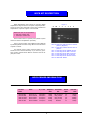
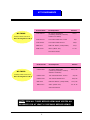
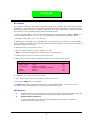
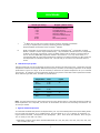
780 Dubuque Avenue
So. San Francisco, CA 94080, U.S.A.
Tel: (800) 989-6296 / Fax:(650)871-2857
http://www.maximbio.com
E-mail: [email protected]
MPCR Kit for Mouse Chemokine Genes Set-1
Cat No. MP-70068: 50 reactions
Cat No. MP-70070: 100 reactions
INSTRUCTION
MANUAL
ID-M10087
Revised August 8, 2002
*These products are designed and sold for use in the Multiplex PCR (MPCR) covered by patent # 5,582,989. Use of the MPCR process requires a license. A
limited, non-automated research field license under the patent to use only this amount of the product to practice the MPCR process is conveyed to the
purchaser by the purchase of this product.
The Polymerase Chain Reaction (PCR) process is covered by patents owned by Hoffman-LaRoche. Use of the PCR process requires a license. A license for
diagnostic purposes may be obtained from Roche Molecular System. A license for research may be obtained by the purchase and the use of authorized
reagents and DNA thermocyclers from the Perkin-Elmer Corporation or by negotiating a license with Perkin-Elmer.
This product is intended for research use only and not for diagnostic purposes.
INTRODUCTION
Cytokines play important roles in orchestrating and
controlling inflammation and sepsis process. The cytokines
IL-1, IL-6, and TNF are sometimes referred to as the ‘inflammatory triad’ since they mediate both local and systemic inflammatory responses which are believed to have
survival value (1, 2). IL-1 and TNF are rapidly produced by
monocytes and macrophages in response to a number of
stimuli such as endotoxins, muramyl dipeptides, lectins,
immune complexes and other noxious agents. Of these bacteria, endotoxin has frequently been used both in vivo and
in vitro to stimulate the production of IL-1 and TNF and to
study their activity. IL-6 is produced by a wide variety of
lymphoid and non-lymphoid cells both constitutively and in
response to many stimuli including other cytokines. For example, both IL-1 and TNF are potent inducers of IL-6 and
once IL-6 is induced cytokines may be distributed to a large
number of activity sites via the circulation system. Thus,
their activity may be represented in both local and systemic
inflammatory events.
Inflammatory cytokines can be divided into two
groups: those involved in acute inflammation and those responsible for chronic inflammation. Cytokines in acute inflammation include IL-1, TNF-alpha, IL-6, IL-11, IL-8 and
other chemokines, G-CSF, and GM-CSF. The cytokines in
chronic inflammation can be subdivided into cytokines mediating humoral responses such as IL-4, IL-5, IL-6, IL-7,
and IL-13, and those mediating cellular responses such as
IL-1, IL-2, IL-3, IL-4, IL-7, IL-9, IL-10, IL-12, interferons,
transforming growth factor-s, and tumor necrosis factor alpha. Some cytokines, such as IL-1, significantly contribute
to both acute and chronic inflammation (3,4). While the
acute production of these cytokines is beneficial, excessive
or sustained production can be deleterious and result in
immunopathology.
eral inflammatory diseases of the central nervous system.
Expression of a variety of checmokines, including MCP-1
beta, RANTES, MIP-1 alpha, and IL-8, and receptors, including CXCR-4, CCR-1, CCR-3 and CCR-5, have been shown
to be increased in HIV encephalitis brain tissue, particularly
in areas of neuroglial reaction. Their expression pattern
supported their involvement in the recruitment of inflammatory infiltrates and formation of microglial nodules. Additionally, presence of chemokine receptors on neurons may
be involved in the pathogenesis of neurologic damage in
AIDS patients (6).
Analysis of the temporal and spatial distribution of
RNA expression provides researchers with important clues
about the function of apoptosis regulating genes in their
own systems. Northern Blot and RNase Protection Assay
are the most widely used procedures for determining the
abundance of a specific mRNA in a total or poly(A) RNA
sample. RT-MPCR provides an alternate and accurate method
to detect multiple gene expression by amplifying all the genes
under the same conditions (8, 9, 10). Variations in RNA
isolation, initial quantitation errors or tube-to-tube variations in RT and PCR can be compensated by including a
house-keeping gene, such as GAPDH, in MPCR. Alternatively, a parallel RT-PCR using the same cDNA, PCR conditions and primers for one of house-keeping genes may be
run to offset any variations. Differences in gene expression
can be determined by normalizing its expression against
GAPDH expression.
Maxim's Mouse Chemokine MPCR kits have been designed to detect the expression of Mouse IP-10, Rantes,
MCP-1, MCP-2 and GAPDH genes. The PCR primers have
similar Tm and no obvious 3'-end overlap to enhance multiple amplification in a single tube; The kit will yield 532
bp(GAPDH), 198 bp(IP-10), 176 bp(Rantes), 233 bp(MCP1) and 275 bp (MCP-2) PCR products with RNA from human
cells or positive controls from kit. The gene expression of
these genes can be analyzed and compared with GAPDH
gene expression.
The most notable of these recent discoveries is that
certain chemokine receptors function as co-receptors for
HIV-1. Moreover, mutations in these receptors can result in
host resistance to infection and also affect the progression
of disease (9,10,11). For example, monocyte chemotatic
protein (MCP)-1, RANTES , macrophage inflammatory protein (MIP)-1 alpha, MIP-1 beta, and lnterleukin-8 (IL-8) have
been found to be more highly expressed in HIV-associated
dementia (5). In addition, chemokines are involved in the
migration of leukocytes and have been implicated in sev-
2
PCR PRODUCT QUANTITATION
II: Non-Radioactive Quantitation
I: Radioactive Quantitation
Nonradioactive quantitation methods include the use
of biotinylated or digoxygenin-labeled primers in conjunction with the appropriate detection methods (8), use of a
bioanalyzer or WAVE. For an in-depth discussion of the various methods of PCR product quantitation, refer to the review article by Bloch (9).
In our experience, visual inspection of an EtBr-stained
agarose gel is sensitive and precise enough to detect changes
as low as two-fold. If greater discrimination is necessary,
several methods are available. The simplest procedure is to
add a radioactively labeled dNTP to the PCR reaction. After
gel analysis, the band may be excised and counted in a
scintillation counter. Alternatively the gel may be dried and
an autoradio-gram may be generated which can be scanned
in a densitometer. Another method is to label the 5’ end of
one or both of the primers with 32P, which is incorporated
into the PCR products and then assayed for radioactivity
(7).
In addition to the above methods, several companies
now offer gel video systems which can scan and quantitate
EtBr-stained gel bands in much the same way a densitometer does. Lab-on-a-chip (BioAnalyzer), CE, HPLC, and WAVE
may also be used to analyze MPCR products and quantitate
simultaneously.
Southern blot hybridization with synthetic DNA probes
may also be performed to verify and quantitate PCR generated products, either by densitometry of an autoradiogram
or by excising and counting the signal from a hybridization
membrane. This method also quantitates only the target
product without interference from nontarget products or
primer-generated artifacts.
COMPARISON OF MPCR WITH RPA
RPA
(RNase Protection Assay)
MPCR
(Multiplex Polymerase Chain Reaction)
√
√
√
√
√
√
√
Non-isotope method with high sensitivity
0.1-1µg total RNA per MPCR
√
Whole process takes only a few hours
√
Detect Multiple Genes Simultaneously &
Quantitatively
√
Signal can be quantified directly from gel
if isotope is included in MPCR. Additional
techniques can be used to quantify MPCR
product (using Bioanalyzer, HPLC, and WAVE.)
√
Non-specific products can be eliminated by using
probes and southern hybridization.
Ready-to-use
√
3
Isotope or Non-Isotope methods
1-20 µg total RNA per RPA assay
Whole process takes two days
Detect Multiple Genes Simultaneously &
Quantitatively
Signal can be quantified directly from gel
Non-specific signal can be generated
by either low stringent conditions or
high-secondary-structure template.
Make own "hot" RNA probes
MPCR KIT DESCRIPTION
Figure 1
MPCR Amplification Kits include all necessary MPCR
amplification reagents with the exception of Taq Polymerase.
These kits have been designed to direct the simultaneous
amplification of specific regions of human DNA.
N
M
1
2
3
4
5
6
MPCR Kits come in two quantities:
• 50X 50µL reaction kits
• 100X 50µL reaction kits
Each kit offers Maxim’s optimal primer/buffer system
which will enhance amplification specificity.
Figure 1 shows quality control MPCR results obtained
by following MPCR kit manual using different concentrations
of positive control.
Lane N: PCR using mCK1G Primers without
positive (Negative)
Lane 1: PCR using mCK1G Primers with 1X
positive
Lane 2: PCR using Mouse GAPDH Primers
Lane 3: PCR using Mouse MCP-1 Primers
Lane 4: PCR using Mouse MCP-2 Primers
Lane 5: PCR using Mouse IP-10 Primers
Lane 6: PCR using Mouse Rantes Primers
Lane M: DNA M.W. Marker
For optimal results, please read and follow the instructions in this manual carefully. If you have any questions, please contact Maxim Biotech Customer Service at
(650) 871-1919.
MPCR PRIMER INFORMATION
Product
Code
Gene
mCK1G-IP10
mCK1G-RAN
mCK1G-MCP2
mCK1G-MCP1
mCK1G-GAP
Mouse
Mouse
Mouse
Mouse
Mouse
IP-10
Rantes
MCP2
MCP1
GAPDH
5’/3’ Tm
Amplicon
Size
Accession
No.
Intron
Span
68oC/68oC
68oC/69oC
67oC/67oC
67oC/67oC
70oC/67oC
198bp
176bp
233bp
275bp
532bp
NM_0212714
NM_013653
NM_021443
NM_011333
M32599
yes
no
yes
yes
no
4
Genomic
Size
391bp
176bp
1336bp
1344bp
532bp
KIT COMPONENTS
MP-70068
50X50µL MPCR reaction kit
Store all reagents at -20°C
Product Code
Kit Component
Amount
mCK1G-B001
2X mCK1G MPCR Buffer
(containing chemicals, enhancer,
stabilizer and dNTPs)
mCK1G-C001
10X mCK1G MPCR Pos. Control
mCK1G-P001
10X mCK1G MPCR Primers
MRB-0014
DNA M.W. Marker (100bp Ladder)
100 µl
MRB-0011P
ddH20 (DNase free)
2.0 ml
1250 µl
50µl
250µl
Instruction Manual
Product Code
Kit Component
mCK1G-B001
2X mCK1G MPCR Buffer
(containing chemicals, enhancer,
stabilizer and dNTPs)
mCK1G-C001
10X mCK1G MPCR Pos. Control
mCK1G-P001
10X mCK1G MPCR Primers
MRB-0014
DNA M.W. Marker (100bp Ladder)
MRB-0011P
ddH20 (DNase free)
MP-70070
100X50µL MPCR reaction kit
Store all reagents at -20°C
Amount
1250 µl X2
50µl X2
250µl X2
100 µl X2
2.0 ml X2
Instruction Manual
NOTE: SPIN ALL TUBES BEFORE USING AND VORTEX ALL
REAGENTS FOR AT LEAST 15 SECONDS BEFORE USING!!
5
PROCEDURE
RT Protocol:
The isolation of undegraded, intact RNA is an essential prerequisite for successful first strand synthesis and PCR
amplification. Care should be taken to avoid RNase contamination of buffers and containers used for RNA work by
pretreating with DEPC, autoclaving, and baking. Always wear sterile gloves when handling reagents. Use cDNA
derived from 105 cells (1µg cDNA) and apply them to each MPCR reaction.
1. Prepare total RNA, mRNA or use the control GAPDH RNA which is provided in Maxim's MPCR kit. NOTE: It is
best to use cDNA derived from 0.5-1 x 10⁵ cells ( 0.5-1µg cDNA derived from RNA) for each MPCR reaction.
2. Equilibrate 3 water baths: 37°C, 70°C and 95°C.
3. On ice, pipet 1-2 µg mRNA or 10 µg total RNA (from 106 cells) dissolved in pure water or 2 µl control GAPDH
RNA into a RNAase free reaction vial. We strongly recommend including a positive control reaction when setting up
an RT-PCR reaction for the first time.
4. Add sterile water to a final volume of 14.5 µl.
5. Add 4 µl random hexamer (50 mM) or Oligo(dT) (50 mM).
NOTE: The hexamer and Oligo(dT) RT reactions may be run simultaneously.
6. Incubate tube(s) at 70°C for 5 minutes and quickly chill on ice.
7. Begin your RT reaction by adding the following reagents to your hexamer or Oligo mixture:
Reagent
Description
RNase Inhibitor
5 X RT buffer
130U/µl
250mM Tris-HCl (pH8.3)
375mM KCl, 15mM MgCl2, 50mMDTT
1mM each
250U/µl
dNTPs
MMLV RT
Volume per Reaction
0.5µl
10µl
20µl
1µl
8. Incubate the RT mixture at 37°C for 60 minutes.
9. Then, heat RT mixture at 95°C for 10 minutes and quickly chill on ice.
10. Add another 50 µl water or 0.1X TE buffer.
11. 2-5 µl of above cDNA is sufficient for most genes in a standard MPCR reaction. However, more or less
DNA may be needed in PCR depending on the copy number of the specific gene.
PCR Protocol:
1.
2.
Taq DNA polymerase from Perkin-Elmer or its derivatives are highly recommended for MPCR. Ampli-Taq
Gold, however, is not recommended because its own optimal buffer system is required.
Reaction Mixture Preparation:
A. Set up MPCR reactions with the test samples and MPCR buffers provided in the MPCR kit
according to the table on the next page:
6
PROCEDURE
Volume (Per assay)
Reagent (Add in order)
25.0 µl
5.0µl
0.5µl
5.0µl
2X MPCR BufferMixture
10X MPCR Primers
Taq DNA Polymerase(5U/ml)
Specimen cDNA or
10X Control cDNA from kit
H2O
Mineral Oil (optional)
14.5µl
50.0µl
3.
*:
:32
P dNTPs may be used here to achieve higher sensitivity and better quantitation.
5-10 µCi [a-32P]dCTP (3000 Ci/mmole) should be used here per MPCR.
Keep final dNTPs concentration same as without 32P-dNTPs.
B.
EDTA concentration in test sample must not exceed 0.5 mM because Mg++ concentration in MPCR
Buffers is limited to certain ranges. Additional Mg++ may be added to the PCR mixture to compensate
for EDTA. We strongly recommend running an MPCR reaction with the positive control provided in
the kit. Since the MPCR DNA polymerase needed in each reaction is in a very small volume, it is
recommended that all of the PCR components be premixed in a sufficient quantity for daily needs and
then dispensed into individual reaction vials. This will help you to achieve more accurate measure
ments.
PCR thermocycle profile:
Reaction profiles will need to be optimized according to the machine type and needs of user. Please take note that
temperature variations occur between different thermocyclers, therefore, the annealing temperature in the sample
profile below is given as a range. It will be necessary to determine the optimal temperature for your individual
thermocycler. An example of a time-temperature profile for the positive control PCR reaction optimized for Perkin
Elmer machine types 480, 2400, and 9600 is provided below:
Temperature
Time
Cycles
96°C
58-60°C
1 min
4 min
2X
94°C
58-60°C
1 min
2 min
28-35X
70°C
10 min
1X
25°C
soak
Note: A 2-step PCR thermocycle profile was found to be more effective than a 3-step PCR thermocycle profile for
MPCR amplification. For 2-step PCR, use 94-95°C for denaturation and 58-60°C for annealing and extension. The
72°C step is omitted.
4. Agarose Gel Electrophoresis:
To fractionate the MPCR DNA product electrophoretically, mix 10µl of the MPCR product with 2µl 6X loading buffer.
Run the total 12µl alongside 10 µl of DNA marker* from the MPCR kit on a 2 % agarose gel containing 0.5
mg/ml ethidium bromide. Electrophorese and photograph. (Hint: Best results are obtained when the gels
are run slowly at less than 100 volts).
* DAN Marker contains linear double stranded DNA bands of 1,000; 900, 800, 700; 600; 500; 400; 300; 200;
and 100 base pairs (bp).
7
TROUBLESHOOTING
1. MPCR AMPLIFICATION
Observation
Possible Cause
Recommended Action
1.1. No signal or missing some bands
during amplification even using
positive control provided in kit.
1.1a.The annealing temperature in
thermocycler is too high.
1.1a. Decrease PCR annealing
temperature 3-5oC gradually.
1.1b. Dominant primer dimers.
1.1b. Use any one of "Hot Start" PCR
procedures.
1.2. Too many nonspecific bands.
1.2a.The annealing temperature in
the thermocycler is too low.
1.2a. Increase PCR annealing
temperature 3-5oC gradually.
1.2b. Pre-PCR mispriming.
1.2b. Use any one of "Hot Start" PCR
procedures.
1.2c. cDNA is interfering with MPCR
1.2c. Clean cDNA with Phenol/
Chloroform.
1.2d. Use Maxim's 3MTM-MPCR Kit.
1.3. No difference in gene expression
among treatments
1.3a. PCR amplification of this
specific gene has passed the
exponential phase.
1.3a. Decrease PCR cycle number or
decrease the input cDNA.
1.3b. Variation in sample preparation,
RT reaction and amounts of
input cDNA.
1.3b. Run a parallel PCR with a
house-keeping gene to
eliminate variables.
8
PRECAUTIONS AND STORAGE
Storage
1. Store all MPCR Kit components at -20°C. Under these conditions components of the kit are stable for 1 year.
2. Isolate the kits from any sources of contaminating DNA, especially amplified PCR product.
3. Do not mix MPCR kit components that are from different lots. Each lot is optimized individually.
REFERENCES
1. Beutler, B. et al., (1989) Annu. Rev. Immunol., 7, 625.
2. Van Snick, J. (1990) Annu. Rev. Immunol., 8, 253.
3. Schrader, J.W. (1986) Annu. Rev. Immunol., 4, 205.
4. Oppenheim, J.J.et al., (1991) Annu. Rev. Immunol., 9, 617.
5. Kelder, W. et al. (1998) Ann Neurol. 44: 831-835.
6. Sanders, V.J. et al. (1998) AIDS 18: 1021-1026.
7. Hayashi, K., Orita, M., Suzuki, Y. & Sekiya, T. (1989) Nucleic Acids Res. 17:3605.
8. Landgraf, A., Reckmann, B., & Pingoud, A. (1991) Analytical Biochemistry 193:231.
9. Bloch, W. (1991) Biochemistry 30:2735.
9