1
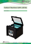
ChE 130, Spring 2015
Protocol 6: Analysis of double-stranded DNA with agarose gel electrophoresis
Background:
DNA is commonly analyzed by tracking its rate of migration through a semi-solid gel. The sizes
of DNA fragments in an experimental sample are estimated by comparing the rate of migration
of these fragments (or their relative locations on the gel) to that of fragments of DNA of known
size (commonly referred to as ladders). Double-stranded pieces of DNA greater than 50-100 bp
in length are typically analyzed by horizontal slab agarose gel electrophoresis. Agarose is a
linear polymer composed of alternating residues of D- and L-galactose joined by α-(1→3) and β(1→4) glycosidic linkages. Gelation of agarose results in a three-dimensional mesh of channels
whose diameters range from 50 nm to greater than 200 nm.
The rate of migration of DNA through agarose gels is determined by a number of factors
including:
molecular size of the DNA – Molecules of double-stranded DNA migrate through gel matrices at
a rate that is inversely proportional to the log of the number of base pairs. Larger molecules
migrate more slowly because of greater frictional drag and because they “worm” their way
through the pores of the gel less efficiently than smaller molecules.
agarose concentration– A DNA fragment of a given size will migrate at different rates through
gels containing different concentrations of agarose according to the following equation:
log10 µ = log10 µ0 – Kr t
where µ = electrophoretic mobility of DNA, t = gel concentration, µ0 = free electrophoretic
mobility of DNA, Kr = retardation coefficient.
DNA conformation – Superhelical circular (form I), nicked circular (form II), and linear (form
III) DNAs migrate through agarose gels at different rates. Under most circumstances form I will
migrate faster than form III.
applied voltage – At low voltages, the rate of migration of DNA fragments is proportional to the
applied voltage. Note that the effective range of separation decreases as voltage increases
because the mobility of high-molecular weight DNA increases differentially.
electrophoresis buffer – The composition and ionic strength of the electrophoresis buffer affects
the electrophoretic mobility of DNA. In the absence of ions, electrical conductivity is minimal
and DNA migrates slowly.
agarose – Different classes of agarose have different resolving powers and are used for different
applications. We will be using standard agarose for the separations performed in this laboratory.
We will also be using a Tris-acetate and EDTA (TAE) buffer for electrophoresis. TAE has a
relatively low buffering capacity and will become exhausted if electrophoresis is carried out for
long periods of time. Double-stranded DNA migrates ~10% faster in TAE than in other
commonly used buffers and TAE has better resolving power for larger DNA fragments. We will
1
ChE 130, Spring 2015
use a loading buffer composed of sucrose and two dyes, bromophenol blue and xylene cyanol FF.
The loading buffer serves to (i) increase the density of the DNA sample so that it sinks into the
well, (ii) add color to the sample to simplify the loading process, and (iii) add dyes to the sample
which will migrate toward the anode at predictable rates.
DNA is most easily visualized by a fluorescent nucleic acid stain that binds specifically to
nucleic acid molecules. The stain GelGreen (Phenix) will be included in the agarose slab used for
electrophoresis. This allows for instant visualization of DNA under blue light illumination.
Information in this background section was adapted from Sambrook J & Russell DW. Molecular cloning: a
laboratory manual, 3rd ed. Cold Spring Harbor Laboratory Press, Cold Spring Harbor, NY (2001).
Materials:
TAE (1X, fresh)
DNA
agarose
loading buffer (6X)
assorted pipetmen
100 ml graduated cylinder
gel box, tray, comb, and power supply
Gel Running Buffer (Used TAE)
FastGene GelPic LED BOX
weighing dish
Zymo spin columns
ADB Buffer
GelGreen (DNA stain, 100 000X)
double distilled autoclaved water (ddH2O)
benchtop microfuge
ready-to-load ladder stock
assorted tips
KimWipes
1.5 mL microfuge tube
microwave
125 ml Erlenmeyer flask
scale
2 ml collection tubes
DNA Wash Buffer
Method:
1. Prepare the gel tray for pouring of the gel solution by placing it into the tray apparatus and
placing black placeholders at the sides of the gel try. Make sure that the apparatus is level.
2. Based on the size of the DNA you will be separating on the gel, determine the appropriate
percentage gel to run. Full-length plasmids can be effectively separated using a 0.8% gel,
whereas smaller fragments (~ 100 bp) require a 1.5-2% gel. Medium-sized fragments can be
effectively separated using an intermediate agarose concentration. Note that these
percentages are in weight (gram) per volume (mL) units.
3. Make up the gel solution in a 250 ml Erlenmeyer flask. The total volume required for one gel
is 35 ml. Weigh out the appropriate amount of agarose using a metal spatula and weighing
dishes. Carefully pour the agarose into the flask. Measure out 50 ml of 1X TAE in a
graduated cylinder and pour the buffer into the flask over the agarose powder. Mix by
swirling. Note that the agarose will not go into solution.
4. Cap the flask loosely with a KimWipe and put it into the microwave. Heat the mixture to boil
in the microwave, being carefully not to allow the mixture to overflow. Typically heat on
high for 20 seconds, remove and swirl briefly, and then heat for another 10 seconds. Swirl
the mixture so that the agarose is melted and then cool briefly by allowing the solution to
cool slowly on the bench top or by running the flask under tap water. Once the mixture has
cooled for 1-2 minutes (the gel mixture should still be warm to the touch), add 0.5 µL of
GelGreen per 50 ml gel solution. Swirl to mix and pour ~35 ml of the gel solution into the
2
ChE 130, Spring 2015
gel tray. Place the appropriately sized comb into place and make sure that it is situated
correctly in the gel. Allow the gel to cool in the tray and solidify.
5. While the gel is solidifying (approximately 30 minutes), prepare the DNA solutions to be run.
For the small combs, solutions of 6-10 µl are typically run; for the large combs, solutions of
30 µl are run (these volumes are approximate). Add loading buffer to each sample such that
the final concentration is 1X loading buffer.
6. Select the appropriate DNA ladder (either 1 kb or 100 bp DNA ladder) depending on the
sizes of the DNA to be separated. The ladder comes pre-mixed with loading buffer.
7. Once the gel has solidified, carefully remove the black placeholders and comb. Orient the gel
such that the wells are closest to the cathode side of the gel box (black). Pour DNA Gel
Running Buffer (1X TAE) into the box such that the gel is completely, but just, covered.
8. Samples should be loaded using a L20 pipetman set to the appropriate volume. Mix DNA
samples prior to loading by pipetting up and down briefly. When loading a gel, it is easiest if
there are no air pockets in the pipet tip. Also, you will most likely have to stabilize your
loading arm either on the bench top or with your other arm. You’ll also have to position
yourself so that you can see the wells and place the pipet tip just over the well. Do not insert
the tip too deeply into the well as you will likely pierce through the well.
9. Once loaded run the gel at constant voltage at approximately 12 V/cm (typically between 80100 V). This setting can be modified depending on whether you want the separation to occur
faster or slower; however, we do not run gels of this size faster than 100 V. Better resolution
will be obtained at slower separation speeds.
10. Monitor the movement of the DNA through the gel by monitoring the location of the loading
dyes in the gel as it runs. The DNA will move towards the anode side of the gel box (red).
Stop the separation when the faster moving dye (purple color) is approximately 50-75%
across the length of the gel (adjusted as appropriate for the fragments you are separating).
Turn off the power supply and remove the gel tray from the running apparatus.
11. Lift the scanner lid and place it at the center of the sample tray.
12. The gel imager should be turned on and will likely be in standby mode unless it was recently
used. In order to image DNA gels, set the light switch to Blue (the White light setting can be
used for imaging plates). All relevant buttons and switches are labelled in the figure above;
you shouldn’t need to change any settings.
13. Flip the control switch to the right. Wait for an image of your gel to appear on the LCD
display.
3
ChE 130, Spring 2015
14. When an image appears, press Scan. Wait for the CAPTURING text on the LCD display to
appear and disappear; this indicates that your image has already be taken and saved to the SD
card.
15. Flip the control switch to the left (“SD”).
If using the gel imager connected to the computer:
16. Go to Windows Explorer, open up Removable Disk Drive and go into folder 200Media. The
most recent image (largest number) in the folder should be the image that was just taken.
Save it for your laboratory notebook. Please note that images taken with FastGene Gel
Imager cannot be quantified but can be used for image purposes.
If using the wireless gel imager:
16. You will need to have a smartphone with the PQI Air Card+ app installed. Go into your
phone settings and under WiFi networks, connect to the PQI Air Card+ network. Next, open
the PQI Air Card+ app and select the images icon. You will be able to browse through the
images stored on the SD card in the imager. Select the files you wish to save and either
download them to your phone or choose the ‘share’ option to email them to yourself.
Afterwards, disconnect your phone from the PQI Air Card+ WiFi network since only a
limited number of connections can be established at any one time.
17. Discard gel in wastebucket and clean sample tray dry with a kimwipe.
Please note that images taken with FastGene Gel Imager cannot be quantified but can be used
for image purposes.
Note: the FastGene Gel Imager User Manual is also available on the Desktop.
Gel Extraction:
1. Keep the control switch (on the top right hand side of the front panel) to the left. Put on
orange goggles (or use the orange plastic shield). The LED blue light is very bright and
will be uncomfortable if your eyes are exposed to it.
2. Use plastic gel cutters to excise your band of interest and place it into a labelled 1.5mL
microfuge tube. Gel cutters can be reused—please clean with a kimwipe.
3. Weigh the small piece of agarose on scale inside of 1.5 mL microfuge tube.
4. Add three volumes of ADB Buffer to 1 volume of agarose gel, assuming that 100 mg of
agarose gel is approximately 100 µl. If using a 2% agarose gel, use six volumes instead.
5. Incubate 1.5 mL microfuge tube at 50°C until gel slice has completely dissolved (~5-10
minutes). Vortex the tube every 2-3 minutes to aid in dissolving the agarose.
6. Optional: For DNA fragments larger than 8 kb, recovery can be improved by adding one
additional volume (volume of original gel slice) of water to the mixture.
7. Place Zymo-Spin column in a 2 mL collection tube.
8. Add the melted agarose mixture to the Zymo-Spin column and spin at 13,000 RPM for 30
seconds. Discard flow-through and repeat as necessary until all mixture has been bound
to the column.
4
ChE 130, Spring 2015
9. Wash DNA by adding 200 μl of DNA Wash Buffer to the Zymo-Spin column and spin at
13,000 RPM for 30 seconds.
10. Repeat the wash (repeat step 7).
11. Remove spin column from collection tube and place in a new 1.5 ml microfuge tube.
12. To elute DNA, add 6-25 µl of double distilled autoclaved water (ddH2O) to the spin
column. The choice in elution volume is a trade-off between DNA concentration and
DNA recovery. Lower volumes will give higher DNA concentrations but will sacrifice
total DNA eluted. Let the column stand for 1 minute before continuing. Make sure to
dispense the elution buffer directly on to the center of the spin column membrane,
taking care to avoid contact with the wall of the column or the membrane.
13. Spin at 13,000 RPM for 30 s to elute DNA from the spin column membrane.
14. Discard the spin column and save the 1.5 ml tube containing your sample. The excised
DNA fragment can be stored at -20°C in your personal freezer box or used immediately
in downstream applications.
5