1
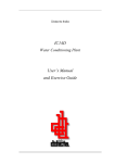
INSTALLATION AND ASSEMBLY INSTALLATION OF THE TONOMETER MODEL “TN-150B” ONTO THE SLIT LAMP. Remove the screw on the top of the slit lamp microscope head. NOTE: underneath the screw is a spring, use caution when removing screw. Attach the mounting block, with pin, on the microscope head. Use the flat head screw (fine threaded) that is supplied. Slide the tonometer onto the mounting block post. By rotating the instrument, the ball-bearing will be fixed into its 3 positions: in front to measure, and at both sides to operate the Slit Lamp. Install the tonometer prism into its holder. INSTALLATION OF THE TONOMETER MODEL “TN-150B” ONTO THE SLIT LAMP. Place the guide plate in the support hole of the Tonometer test bar on the slit lamp. Insert the pin of its base in one of two possible openings of the guide plate. These positions correspond with the biomicroscope so that ocular observation can be made through the right or left eye. Slide the tonometer on the support plate. Stability is ensured by the locking pins. WHEN NOT IN USE, RETIRE EL REMOVE THE TONOMETER FROM THE SLIT LAMP AND KEEP IT IN YOUR PACKAGE. INSTRUCTIONS FOR USE Preparing the patient 1. 2. 3. 4. Anesthetize both eyes. Both eyes must always be anesthetized to prevent movements of the lids during prolonged examination. Place a fluorescein paper strip near the external canthus in the lower conjunctival sac in the same manner as for the Shirmer test. After a few seconds in which the lachrymal fluid is sufficiently colored, the paper can be removed. The patient is asked to put his head on the chin and forehead-rest of the Slit Lamp. Regulate the microscope height for a comfortable observation. Page. 1 of 4 Preparing the Slit Lamp and the Applanation Tonometer 1. 2. 3. 4. 5. 6. Before measuring, make sure that the eyepieces are correctly focused. Bring the blue filter into the beam of the Slit Lamp and open the slit diaphragm completely. The angle between illumination and microscope should be about 60º so that the image appears bright and free of reflection. Place the tonometer in the forward click stop position. Observation is always monocular with the left eye. Clean the measuring prism with a disinfecting fluid that does not damage plastic. Afterwards dry with sterile cotton. Bring the pressure-arm into the notch position so that the axis of the measuring prism and the microscope coincide. Set the measuring drum at a pressure of about 1 g. If the prism touches the cornea without any force being applied, the pressure arm will vibrate and this may be disturbing to the patient. Instructions to the patient 1. 2. 3. The patient’s head should be pressed firmly against the chin and forehead rest. Instruct the patient to look straight ahead. The patient is repeatedly asked to keep his eyes wide open during the examination. It is also recommendable when necessary that the examiner hold open the lids of the examined eye with his fingers provided that no pressure is applied to the eyeball. Measurement 1. 2. 3. 4. 5. Immediately before measurement the patient should close his eyes for a moment so that the cornea is well moistened with lachrymal fluid and fluorescein. By moving the Slit Lamp forward, the measuring prism is brought into contact with the center of the cornea on the pupillary area. On contact, the limbus of the cornea shines with a bluish light. This is best observed by the naked eye from the side opposite to the illumination. As soon as the limbus lights up, the slit Lamp must stop moving. Only after contact with the cornea should then the microscope be used. The steady pulsation of the two fluorescein semi-circles, the size of which, at 1g, will depend on the intraocular tension, indicate that the instrument is in the correct position for measurement, Fig. 1. Any necessary correction has to be made by the joystick for height or lateral adjustment control of the Slit Lamp, until the flattened area is seen as 2 semicircles of equal size in the middle of the field of view, see Fig. 1. (Should this not be case, observe information under Sources of error.) The pressure on the eye is then increased by turning the measuring drum on the tonometer until the inner borders of the two fluorescein rings just touch each other, in other words, when they overlap with each pulsation of the eye, Fig.2. The width of the fluorescein band around the point of contact of the measuring prism should be about one tenth of the diameter of flattening. (0,3 mm). The reading is then made on the measuring drum of the amount of pressure applied. By multiplying this reading by ten, the intraocular pressure in mm Hg is found. Generalities concerning the measurement Measurements can be repeated frequently. The first measurements of excited or nervous patient produce higher readings than normal. When the patient is aware that the procedure is not all that uncomfortable, however, the tension falls to the true value in less than a minute. With the eyes wide open and a good anesthesia the patient does not feel anything at all. Trial measurements should therefore be made on both eyes, the results of which are to be ignored. Three further measurements should then be made and the values obtained are correct if they remain within a range of 0,5 mm Hg. When measuring an eye for a prolonged period of time desiccation of the corneal epithelium may appear on both eyes. In the examined eye a ring shaped lesion appears which takes the fluorescein coloration around the zone of contact. This small disturbance does not harm the patient. Whenever the other un-examined eye shows dried out patches in the epithelium which are stained fluorescein, it impedes an accurate measurement of the intraocular tension by means of Applanation tonometry. To avoid this condition, measurements should be made as rapidly as possible on each eye alternately. Although the above mentioned staining causes no harm or injury and disappears without any treatment, the visual acuity does in fact temporarily diminish by this medical intervention. A previous examination of the visual acuity and the measurement of visual fields should therefore be made before the Applanation tonometry measurements. Astigmatism correction If the cornea is spherical, measurements can be made on any meridian, but is most convenient to do it on the 0º meridian. If the cornea is not spherical, the flattened area is not circular but elliptic. When eyes with higher corneal astigmatism than 3 diopters are examined, a correction is necessary. This is achieved by rotating the prism until its graduation shows the same value as orientation of the higher radius of the cornea that must coincide with the red mark on the prism holder. Sources of error Errors can occur under the following conditions: 1. If the fluorescein ring is too wide or too narrow. Fig. 3: the fluorescein ring is too wide. Either the measuring prism was not dried after cleaning or the lids have come into contact with the prism during measurement. The pressure read from the drum will be higher than the real intraocular pressure. For correction the Slit Lamp has to be drawn back and the prism must be cleaned and dried with a piece of sterile cotton. Page. 2 of 4 Fig. 4: the fluorescein ring is too narrow. The lachrymal fluid has dried out during a prolonged measurement. The reading on the drum is lower than the actual intraocular pressure. The patient has to open and closed his eyes several times, then repeats the measurement to correct this error. 2. If the measuring prism does not touch the cornea. If the patient retreats slightly there will be irregular pulsation, because the measuring prism only temporarily touches the eye. If the patient retreats still further, the fluorescein ring will disappear completely. Fig. 5: if the Slit Lamp is moved too far forward towards the patient or the patient moves, during measurement, towards the Slit Lamp, the pressure arm could push against the eye with a force up to 8 g. The flattened area will be too large. Turning the measurement drum scarcely changes the picture. Draw the Slit Lamp back until regular pulsation of a correspondingly smaller surface is seen in the correct measuring position and the change of pressure immediately alter the size of the flattened area. 3. If the two semi-circles are not in the middle of the field of view. Fig. 6: the rings are too low. Correct with height adjustment control. Fig. 7 and 8: the Slit Lamp has to be lowered and moved with the joystick to the left. Fig. 9: The rings are too far to the right. Move the Slit Lamp by means of the joystick to the right. 4. If the inner borders of the fluorescein rings do not touch each other. Fig. 10: Here the outer border of the fluorescein rings touch. However, the inner borders do not touch, when they should in fact do so. Increase the pressure by turning the measuring drum. Fig. 11: Here the inner border of one semi-circle touches the outer border of the other. Increase the pressure still more. Fig. 12: The pressure has been increased too much. By reducing the pressure the circles will come together so that they touch each other correctly as in the Fig. 13. Fig. 13: Correct final position (like fig. 2). MAINTENANCE, VERIFICATION AND CLEANING THERE IS NO PERIODIC OR ROUTINE USER MAINTENANCE REQUIRED. Verification: the confirmation that the tonometer measures the force exercised by the counterbalance appropriately should be carried out periodically by the user. For this verification the check weight that is given as accessory must be used (Fig. 14). The periodicity of verifications will depend on the frequency of use of the instruments, and its realization is recommended if one observes some anomaly in its operation or in the suitable reading, if the apparatus has received some blow or it presents indications of damage or deterioration or if one suspects that it can have them. In case of detecting that the instrument doesn't measure the suitable force by the verification bar, it is recommended to send the instrument to calibrate. To carry out the verification, use the bar as follows: - with the bar held from its center and the drum marking “0”, the tonometer prism should remain vertical. with the bar held from the center mark and the drum marking “2”, the tonometer prism should remain vertical. with the bar held from the end mark and the drum marking “6”, the tonometer prism should remain vertical. Fig. 14 Cleaning: it is very important for the security and health of the patient to disinfecting the prism before each using. Disinfect the prism with a product that doesn't damage the methyl polymethacrylate (PMMA) material with which the prism is manufactured. Dry with a sterile cloth. STORAGE AND TRANSPORTATION TN-150B Applanation Tonometer may only be exposed to the following environmental conditions for transportation and storage: Page. 3 of 4 Storage: Temperature Relative Humidity Air Pressure from –10 ºC to +70 ºC from 10% to 95% from 700 hPa to 1060 hPa Transportation: Dismount the tonometer from the Slit Lamp, pack in a proper box and secure with adequate packing material DISINFECTING THE PRIMS REMOVE THE MEASURING PRISM FROM HOLDER THOROUGH CLEANING UNDER RUNNING (COLD) WATER KEEP DURING 30 - 60 SECONDS IMMERSE THE PRISMS INTO DISINFECTANT. SEE INSTRUCTIONS ABOUT SPECIFIC DISINFECTANT. CLEAN THOROUGHLY UNDER RUNNING (COLD) WATER MIN. 10 – MAX. 30 MINUTES DRY WITH A STERILE CLOTH PLACE INTO STERILE STORAGE CONTAINER RECOMMENDED PRODUCTS FOR DISINFECTION 1. - Hydrogen Peroxide. Concentration: 3%. Method: Immerse 10 minutes at 20 ºC 2. - Sodium hypochlorite (NaOCl). Concentration: 0, 0525%. Method: Immerse 10 minutes at 20 ºC DO NOT USE: Both products are effective against: - bacteria tuberculosis fungi viral infections ACCESSORIES A verification bar with fixing piece to the tonometer axis. Measuring Prism - Alcohol content agents UV disinfecting Sterilization Temperature max. 60 ºC FEATURES * Measuring range: from 0 to 80 mmHg in 2 mmHg increments * Accuracy: ± 2 mmHg * Diameter of the pneumatic face: 3,06 mm SAFETY – WARNINGS AND CAUTIONS The instrument should ONLY be used by qualified and specially trained personnel. BEFORE each use, check whether the contact surface of the cone is damaged and discard it if any damage is noticed. BEFORE each use, check that the cone to be used has been properly disinfected. DISINFECT the cone according to the cleaning method indicated in this instruction manual. There is a risk that the patient's cornea could be damaged if the cleaning of the cones is not done properly after the disinfection. It is recommended NOT TO USE the cone if it is used OVER TWO YEARS, because after that time, it is possible that the fluids with the cone has come into contact have penetrated into its interior, leading to potential problems of contamination. Change the cone by other previously disinfected BEFORE EACH USE. The owner of the instrument is responsible for training personnel in its correct use. The estimated useful life for the Applanation Tonometer is 10 years. Page. 4 of 4