1
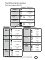
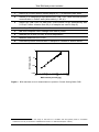
RNA and protein
purification
User manual
NucleoSpin® RNA / Protein
May 2014 / Rev. 09
Total RNA and protein isolation
Protocol-at-a-glance (Rev. 09)
NucleoSpin® RNA/Protein
1
Homogenization
of sample
2
Cell lysis
3
Filtration of
lysate
4
Adjust binding
conditions
5
Bind RNA
30 mg
350 μL RP1
3.5 μL ß-mercaptoethanol
11,000 x g,
1 min
350 μL ethanol (70 %)
11,000 x g,
30 s
RNA Purification
(RNA bound to the silica membrane)
Protein Purification
(protein in the column flow-through)
6
10
Desalt
silica
membrane
350 μL MDB
Precipitate
protein
1 vol PP
RT,
10 min
11,000 x g,
1 min
7
Digest
DNA
95 μL DNase
reaction mixture
RT,
15 min
8
Wash and
dry silica
membrane
1st wash
2nd wash
200 μL RA2
600 μL RA3
11,000 x g,
30 s
3rd wash
250 μL RA3
11,000 x g,
2 min
9
Elute
highly
pure RNA
10–700 μL
flow-through
11,000 x g,
5 min
11
Wash
protein
500 μL ethanol
(50 %)
11,000 x g,
1 min
12
Dry protein
pellet
13
Prepare
protein
sample
60 μL H2O
(RNase-free)
11,000 x g,
1 min
MACHEREY-NAGEL GmbH & Co. KG · Neumann-Neander-Str. 6–8 · 52355 Düren · Germany
Tel.: +49 24 21 969-270 · Fax: +49 24 21 969-199 · [email protected] · www.mn-net.com
RT,
5–10 min
20–100 μL
PSB-TCEP
95–98 °C,
3 min
11,000 x g,
1 min
Total RNA and protein isolation
Table of contents
1 Components 4
1.1 Kit contents
4
1.2 Reagents, consumables, and equipment to be supplied by user
5
1.3 About this user manual
5
2 Product description
6
2.1 The basic principle
6
2.2 Kit specifications
7
2.3 Handling, preparation, and storage of starting materials
11
2.4 Guideline for appropriate sample amount, precipitation, and
resolubilization volume for protein isolation
13
2.5 Elution procedures
14
3 Storage conditions and preparation of working solutions
15
4 Safety instructions
17
5Protocols
19
5.1 Total RNA and protein purification from cultured cells and tissue
19
5.2 Total RNA preparation from biological fluids
(e.g., serum, culture medium)
25
9
5.3 Total RNA preparation from up to 10 bacterial cells
7
26
5.4 Total RNA preparation from up to 5 x 10 yeast cells
27
5.5 Total RNA preparation from RNAlater® treated samples
28
5.6 rDNase digestion in solution
29
6Appendix
31
6.1 Protein quantification
31
6.2Troubleshooting
38
6.3References
42
6.4 Ordering information
43
6.5 Product use restriction / warranty
44
MACHEREY-NAGEL – 05 / 2014, Rev. 09
3
Total RNA and protein isolation
1
Components
1.1 Kit contents
NucleoSpin® RNA/Protein
10 preps
50 preps
250 preps
740933.10
740933.50
740933.250
9 mL
45 mL
225 mL
Protein Solving Buffer PSB
(without reducing agent)
2 x 1 mL
7.5 mL
40 mL
Reducing Agent TCEP
2 x 14 mg
107 mg
5 x 10⁷ mg
Lysis Buffer RP1
10 mL
25 mL
125 mL
Wash Buffer RA2
15 mL
15 mL
80 mL
Wash Buffer RA3
(Concentrate)*
6 mL
12 mL
3 x 25 mL
Membrane Desalting
Buffer MDB
10 mL
25 mL
125 mL
Reaction Buffer for rDNase
7 mL
7 mL
30 mL
1 vial (size C)
1 vial (size D)
5 vials (size D)
13 mL
13 mL
60 mL
NucleoSpin® Filters
(violet rings)
10
50
250
NucleoSpin® RNA / Protein
Columns (light blue rings,
plus Collection Tubes)
10
50
250
Collection Tubes (2 mL)
30
150
750
Collection Tubes (1.5 mL)
20
100
500
User manual
1
1
1
REF
Protein Precipitator PP
rDNase, RNase-free
(lyophilized)
RNase-free H2O
* For preparation of working solutions and storage conditions see section 3.
4
MACHEREY-NAGEL – 05 / 2014, Rev. 09
Total RNA and protein isolation
1.2 Reagents, consumables, and equipment to be supplied
by user
Reagents
•
•
•
•
96–100 % ethanol (to prepare Wash Buffer RA3)
70 % ethanol (to adjust RNA binding conditions)
50 % ethanol (to wash protein pellet)
Reducing agent (ß-mercaptoethanol, or DTT (dithiothreithol), or TCEP (Tris(2carboxyethyl) phosphine hydrochloride) to supplement lysis buffer
Consumables
•
•
1.5 mL microcentrifuge tubes for sample lysis
Disposable RNase-free tips
Equipment
•
Manual pipettors
•
Vortex mixer
•
•
•
•
Centrifuge for microcentrifuge tubes
Thermal heating block
Equipment for sample disruption and homogenization
Personal protection equipment (lab coat, gloves, goggles)
Additional material is furthermore needed for protein quantification, see section 6.1.
1.3 About this user manual
It is strongly recommended reading the detailed protocol sections of this user manual
if the NucleoSpin® RNA/Protein kit is used for the first time. Experienced users,
however, may refer to the Protocol-at-a-glance instead. The Protocol-at-a-glance is
designed to be used only as a supplemental tool for quick referencing while performing
the purification procedure.
All technical literature is available on the internet at www.mn-net.com.
Please contact Technical Service regarding information about changes of the current
user manual compared to previous revisions.
MACHEREY-NAGEL – 05 / 2014, Rev. 09
5
Total RNA and protein isolation
2
Product description
2.1 The basic principle
Introduction
Studies of gene expression at the level of transcription and translation by quantification
of RNA and protein are often hampered by the small sample size and the necessity of
different – often incompatible – techniques for RNA and protein isolation. Samples may
comprise biopsies, tumors, tissues, transgene organisms and others. The NucleoSpin®
RNA / Protein kit however enables isolation of RNA and protein from diverse sample
types. Protein and RNA are isolated without splitting the sample prior to protein / RNA
extraction. Thus, protein and RNA are obtained from one and the same sample and
not from two similar portions of one sample. This is especially valuable for unique,
small and precious samples. Isolated RNA is suitable for all common downstream
applications. RNA isolated with the NucleoSpin® RNA/Protein kit is of identical
quality as RNA isolated with the well proven NucleoSpin® RNA II kit. Isolated protein
is immediately suitable for SDS-PAGE, Western Blot analysis, and quantification with
recommended methods.
RNA and protein isolation
One of the most important aspects in the isolation of RNA and protein is to prevent
their degradation during the isolation procedure. With the NucleoSpin® RNA/Protein
method, cells are lysed by incubation in a solution containing large amounts of
chaotropic ions. This lysis buffer immediately inactivates virtually all enzymes (e.g.,
RNases and proteases) which are present in almost all biological materials. The
buffer dissolves even hardly soluble protein, creates appropriate binding conditions
which favor adsorption of RNA to the silica membrane and enables protein to pass the
specially treated NucleoSpin® RNA / Protein Column virtually quantitatively. Expensive
and harmful proteinase inhibitors or inhibitor cocktails are not necessary due to the
denaturing properties of the lysis buffer. Contaminating DNA, which is also bound to
the silica membrane, is removed by an rDNase solution which is directly applied onto
the silica membrane during the preparation (RNase-free rDNase is supplied with the
kit). Simple washing steps with two different buffers remove salts, metabolites and
macromolecular cellular components. Pure RNA is finally eluted under low ionic
strength conditions with RNase-free water (supplied).
Protein is isolated from the column flow-through. Protein is precipitated in denatured
form with a special buffer (Protein Precipitator PP) which effectively precipitates
protein. After a washing step the protein pellet is dissolved in Protein Solving Buffer
(PSB) containing the odourless reducing agent TCEP. The protein can thus readily be
applied to SDS-PAGE analysis. The kit is not recommended for isolation of native
proteins.
The RNA and protein preparation using NucleoSpin® RNA/Protein kits can be
performed at room temperature. The RNA eluate, however, should be treated with
care because RNA is very sensitive to trace contaminations of RNases, often found
on general lab ware, fingerprints and dust. To ensure RNA stability keep RNA frozen
6
MACHEREY-NAGEL – 05 / 2014, Rev. 09
Total RNA and protein isolation
at -20 °C for short-term or -70 °C for long-term storage. Recovered Protein dissolved in
Protein Solving Buffer is unproblematic concerning stability.
Simultaneous isolation of RNA, protein, and DNA (NucleoSpin® RNA/DNA Buffer
Set*)
The NucleoSpin® RNA/DNA Buffer Set (see ordering information) is a support set for
RNA and DNA isolation in conjunction with NucleoSpin® RNA II, NucleoSpin® RNA XS,
NucleoSpin® RNA Plant, or NucleoSpin® RNA / Protein.
This patented technology enables successive elution of DNA and RNA from one
NucleoSpin® Column with low salt buffer and water respectively. DNA and RNA are
immediately ready for downstream applications.
The combination of the NucleoSpin® RNA/DNA Buffer Set with NucleoSpin® RNA/
Protein allows parallel isolation of RNA, DNA, and protein from one undivided sample.
2.2 Kit specifications
•
NucleoSpin® RNA/Protein kits are recommended for the isolation of total RNA
and protein from cultured cells and tissue. The NucleoSpin® RNA/Protein kits
allow purification of pure RNA with an A260 / A280 ratio generally exceeding 1.9
(measured in TE buffer (pH 7.5)).
•
The isolated RNA is ready to use for applications like reverse transcriptasePCR** (RT-PCR), primer extension, or RNase protection assays.
•
The isolated protein is ready to use for SDS-PAGE, Western Blot analysis
and protein quantification with the Protein Quantification Assay (see ordering
information).
•
* DISTRIBUTION AND USE OF THE NUCLEOSPIN® RNA/DNA BUFFER SET IN THE USA IS PROHIBITED
FOR PATENT REASONS.
** PCR is patented by Roche Diagnostics.
MACHEREY-NAGEL – 05 / 2014, Rev. 09
7
Total RNA and protein isolation
Table 1: Kit specifications at a glance
Parameter
NucleoSpin® RNA/Protein
Technology
Silica-membrane technology
Format
Mini spin column
< 5 x 106 cells
< 30 mg human / animal tissue
< 100 mg plant tissue
Sample material
Total RNA
Total protein
Fragment size
> 200 nt
15–300 kDa
Typical yield
< 70 μg
< 1200 μg
A260/A280
1.9–2.1
–
>9
–
40–100 μL
10–100 μL
30 min/6 preps
35 min/6 preps
200 μg
–
Typical RIN (RNA integrity
number)
Elution volume /
Resolubilization volume protein
Preparation time (approx.)
Binding capacity
8
•
The standard protocol (section 5.1) allows purification of up to 70 μg of total
RNA per NucleoSpin®  RNA / Protein  Column from up to 5 x 106 cultured
cells, 30 mg of human / animal tissue, or 100 mg of plant tissue (see Table 1).
The isolated RNA can be used as template in a RT-PCR-reaction. Generally,
1–10 % of the eluate of total RNA prepared from 1 x 106 cells or 10 mg of tissue
is sufficient as template for RT-PCR. Intron-spanning primers for RT-PCR are
preferable if possible.
•
RNA prepared with NucleoSpin®  RNA / Protein is generally free of residual
DNA. However, minute traces of DNA may remain, if large amounts of material
rich in nucleic acids are used. If the isolated RNA will be used as template in
a RT-PCR-reaction, we recommend using lower quantities of sample material,
depending on cell or tissue type (in the range of 1 x 106 cells or 10 mg of tissue
resulting in about 20 μg of RNA).
MACHEREY-NAGEL – 05 / 2014, Rev. 09
Total RNA and protein isolation
•
The kit can be used for preparing RNA from different amounts of sample
material according to Table 2:
Table 2: Use of different amounts of sample material
Sample
•
Amount
Cultured animal cells
(e.g., HeLa cells)
Up to 5 x 10⁶
Animal tissue
Up to 30 mg
Bacteria
Up to 1 x 10⁹
Yeast
Up to 5 x 10⁷
Depending on sample type, the average yield is around 5–70 μg total RNA (see
Table 3). The A260 / A280 ratio, indicating purity of the RNA, generally exceeds
1.9.
Table 3: Overview on average yields of total RNA
isolation using NucleoSpin® RNA/Protein
Sample
Average yield
8 x 10⁴ HeLa cells
1.5 μg
5
4 μg
6
1 x 10 HeLa cells
14 μg
2 x 10⁶ HeLa cells
21 μg
4 x 10 HeLa cells
6
25 μg
6
50 μg
2.5 x 10 HeLa cells
5 x 10 HeLa cells
MACHEREY-NAGEL – 05 / 2014, Rev. 09
9
Total RNA and protein isolation
Protein yield
Protein yield depends on sample type, amount and quality as well as on homogenization
efficiency. Further, the utilized quantification method influences determined protein
yield. The following values were determined with the MACHEREY-NAGEL Protein
Quantification Assay and shall serve as a guideline for expected protein yield. It is
assumed that the complete sample amount is processed, i.e. the complete lysed
sample – after ethanol addition – is loaded onto the column and the complete 700 μL
flow through is subjected to protein precipitation. Note that in many cases precipitation
of only a portion of the column flow through (e.g.,100 μL) is recommended and will
yield enough protein in terms of absolute amount and concentration for SDS-PAGE
and Western Blot analysis.
As a guideline for appropriate precipitation volumes see section 2.4.
Table 4: Typical protein yield
10
Sample type and amount
Protein yield
Cultured human cells (e.g., HeLa, approx. 106 cells)
~ 50–150 μg
Plants (e.g., garden cress, approx. 100 mg)
~ 150–350 μg
Animal tissue (e.g., pig liver, approx. 30 mg)
~ 500–1200 μg
MACHEREY-NAGEL – 05 / 2014, Rev. 09
Total RNA and protein isolation
2.3 Handling, preparation, and storage of starting materials
RNA is not protected against digestion until the sample material is flash frozen or
disrupted in the presence of RNase inhibiting or denaturing agents. Therefore it is
important that samples are flash frozen in liquid N2 immediately and stored at -70 °C,
or processed as soon as possible. Samples can be stored in Lysis Buffer RP1 after
disruption at -70 °C for up to one year, at +4 °C for up to 24 hours or up to several hours
at room temperature. Frozen samples are stable up to 6 months. Frozen samples in
Buffer RP1 should be thawed slowly before starting with the isolation of total RNA.
Wear gloves at all times during the preparation. Change gloves frequently.
Cultured animal cells
are collected by centrifugation and directly lysed by adding Buffer RP1 according to
step 2 of the standard protocol (see section 5.1).
Cell lysis of adherent growing cells in a culture dish:
Completely aspirate cell-culture medium, and continue immediately with the addition of
Lysis Buffer RP1 to the cell-culture dish. Avoid incomplete removal of the cell-culture
medium in order to allow full lysis activity of the lysis buffer.
To trypsinize adherent growing cells:
Aspirate cell-culture medium and add an equal amount of PBS in order to wash the
cells. Aspirate PBS. Add 0.1–0.3 % trypsin in PBS and incubate for an appropriate time
to detach the cells from the dish surface. After cell detachment, add medium, transfer
cells to an appropriate tube (not supplied), and pellet by centrifugation for 5 min at
300 x g. Remove supernatant and continue with the addition of lysis buffer to the cell
pellet.
Human / animal and plant
tissues
are often solid and must therefore be broken up mechanically as well as lysed.
Depending on the disruption method, the viscosity of the lysed sample has to be
reduced further for optimal results. It is essential for efficient RNA preparation that all
the RNA contained in the sample is released from the cells by disruption and that the
viscosity of the sample is reduced by homogenization.
The most commonly used technique for disruption of animal tissues is grinding with a
pestle and mortar. Grind the sample to a fine powder in presence of liquid N2. Take care
that the sample does not thaw during or after grinding or weighing and add the frozen
powder to an appropriate aliquot of Buffer RP1 containing ß-mercaptoethanol and mix
immediately. The broken-up tissue must then be homogenized with a NucleoSpin®
Filter / Filter  L or by passing ≥ 5 through a 0.9 mm syringe needle.
Thawing of undisrupted animal tissue should exclusively be done in presence of
Buffer RP1 during simultaneous mechanical disruption, for example, with a rotorstator homogenizer. This ensures that the RNA is not degraded by RNases before the
preparation has started. The spinning rotor disrupts and simultaneously homogenizes
MACHEREY-NAGEL – 05 / 2014, Rev. 09
11
Total RNA and protein isolation
the sample by mechanical shearing within seconds up to minutes (homogenization
time depends on sample). Take care to keep the rotor tip submerged in order to avoid
excess foaming. To degenerate evolved foam, centrifuge 1 min at 400 x g. Select a
suitably sized homogenizer (5–7 mm diameter rotors can be used for homogenization
in microcentrifuge tubes).
Bacteria and yeasts
have to be incubated in lysozyme or lyticase / zymolase solutions, respectively
(see support protocols in section 5). By this treatment the robust cell walls of these
organisms are digested or at least weakened, which is essential for effective cell lysis
by Buffer RP1. For microorganisms with extremely resistant cell walls – like some
Gram-positive bacterial strains – it may be necessary to optimize the conditions of the
treatment with lytic enzymes or the cultivation conditions. After lysis, homogenization is
achieved by the use of a NucleoSpin® Filter or the syringe-needle method.
12
MACHEREY-NAGEL – 05 / 2014, Rev. 09
Total RNA and protein isolation
2.4 Guideline for appropriate sample amount, precipitation,
and resolubilization volume for protein isolation
The following Table 5 shall serve as a first guide for choosing appropriate amounts
of sample material, precipitation volume, and resolubilization volume. Depending on
sample type, and downstream application (e.g., Coomassie or silver stain, sensitivity
of antibody, detection system) appropriate volumes might deviate from the table below
and have to be determined experimentally.
Table 5: Guideline for appropriate sample amount
Amount of
Sample
Cultivated cells
(e.g., HeLa)
106
105
104
Animal tissue
(e.g., liver)
30 mg
Plant tissue
(e.g., garden
cress leaf)
3 mg 0.3 mg 100 mg 10 mg
Lysis Buffer RP1
incl. reducing
agent
350 μL
Ethanol
350 μL
1 mg
Column flowthrough to be
precipitated*
35 μL 350 μL 700 μL 35 μL 350 μL 700 μL 35 μL 350 μL 700 μL
Volume of Protein
Precipitator PP
35 μL 350 μL 700 μL 35 μL 350 μL 700 μL 35 μL 350 μL 700 μL
Buffer PSB used
for protein pellet
solubilization
100 μL 100 μL 20 μL 100 μL 100 μL 20 μL 100 μL 100 μL 20 μL
Protein sample to
be analysed on
SDS-PAGE with
Coomassie stain
10 μL
Protein sample to
be analyzed on
SDS-PAGE with
silver stain
1 μL
Protein sample
analyzed on
Western Blot
1–10 μL
*Protein pellets with a diameter of up to approximately 1–2 mm in size are ideally suited for subsequent
solubilization. Protein pellets exceeding volumes of approximately 10 μL should be avoided as large protein
pellets are harder to dissolve than small pellets. To obtain small protein pellets, adapt the volume of column flow
through in respect to the amount of sample material. Commonly small and even invisible protein pellets yield
sufficient protein for SDS PAGE and Western Blot analysis.
MACHEREY-NAGEL – 05 / 2014, Rev. 09
13
Total RNA and protein isolation
Solubilization of protein pellets and reduction of protein disulfide bonds
The NucleoSpin® RNA/Protein kit provides a protein sample buffer (Protein Solving
Buffer PSB) and the Reducing Agent TCEP.
The Protein Solving Buffer PSB is similar in composition and function to the buffer
commonly known as “Laemmli” buffer. For most applications, PSB may be substituted
by “Laemmli” buffer. However, for applications with large protein pellets (> approx.
1 mm, diameter) PSB is recommended.
TCEP is a powerful, multi-purpose and odourless reducing agent. It is non-volatile and
unlike commonly used reducing agents like DTT and ß-mercaptoehanol resistant to air
oxidation. TCEP reduces disulfide bonds as effectively as dithiothreitol (DTT). TCEP
reduces even most stable water-soluble alkyl disulfides selectively and completely over
a wide pH range.
Solubilization of TCEP in PSB according to the instruction, results in a PSB-TCEP
solution with a concentration of 50 mM TCEP (see section 6.1 for composition). This
provides sufficient molar excess to reduce peptide and protein disulfide bonds effectively
within a few minutes (in a range up to a protein concentration of approximately 1 μg/ μL).
2.5 Elution procedures
It is possible to adapt elution method and elution volume of water to the subsequent
application of interest. In addition to the standard method described in the individual
protocols (recovery rate about 70–90 %) there are several modifications possible.
•
High yield: Perform two elution steps with the volume indicated in the individual
protocol. About 90–100 % of bound nucleic acid will be eluted.
•
High yield and high concentration: Elute with the standard elution volume
and apply the eluate once more onto the column for reelution.
Eluted RNA should immediately be put on ice and always kept on ice for optimal stability
because almost omnipresent RNases (general lab ware, fingerprints, dust) will degrade
RNA. For short-term storage freeze at -20 °C, for long-term storage freeze at -70 °C.
14
MACHEREY-NAGEL – 05 / 2014, Rev. 09
Total RNA and protein isolation
3
Storage conditions and preparation of working
solutions
Attention:
Buffers RP1, RA2, and MDB contain chaotropic salt! Wear gloves and goggles!
CAUTION: Buffers RP1, RA2, and MDB contain guanidinium thiocyanate which can
form highly reactive compounds when combined with bleach (sodium hypochlorite).
DO NOT add bleach or acidic solutions directly to the sample-preparation waste.
•
Store lyophilized rDNase (RNase-free) at +4 °C on arrival (stable up to 1 year).
•
Store Reducing Agent TCEP at +4 °C on arrival.
•
All other kit components should be stored at room temperature (18–25 °C)
and are stable up to one year. Storage at lower temperatures may cause
precipitation of salts.
•
Check that 70 % ethanol is available as additional solution to adjust binding
conditions in the RP1-lysate.
•
Check that 50 % ethanol is available as additional solution to wash the protein
pellet.
Before starting any NucleoSpin® RNA/Protein protocol prepare the following:
•
rDNase (RNase-free): Add indicated volume of RNase-free H2O (see table
below) to the rDNase vial and incubate for 1 min at room temperature. Gently
swirl the vials to completely dissolve the rDNase. Be careful not to mix rDNase
vigorously as rDNase is sensitive to mechanical agitation. Dispense into
aliquots and store at -20 °C. The frozen working solution is stable for 6 months.
Do not freeze / thaw the aliquots more than three times.
•
Wash Buffer RA3: Add the indicated volume of 96–100 % ethanol (see table
below) to Buffer RA3 Concentrate. Mark the label of the bottle to indicate that
the ethanol is added. Store Wash Buffer RA3 at room temperature (18–25 °C)
for up to one year.
•
Protein Solving Buffer PSB and Reducing Agent TCEP: For SDS-PAGE
under reducing conditions (most common type of SDS-PAGE) transfer PSB
from one vial to one vial of the Reducing Agent TCEP. Mix gently to avoid
excessive foaming until the reducing agent is dissolved completely (this process
will require several minutes)*. Protein Solving Buffer containing Reducing Agent
TCEP (PSB-TCEP) is stable for several days at room temperature (18–25 °C)
and several months at 4 °C. For long term storage of PSB-TCEP keep at -20 °C.
* For 50 and 250 prep kits: For better handling, PSB-TCEP may be transferred into the original PSB vial (with
screw cap).
MACHEREY-NAGEL – 05 / 2014, Rev. 09
15
Total RNA and protein isolation
•
If SDS-PAGE under non-reducing conditions is intended consider the
following:
A: Omit addition the Reducing Agent TCEP to Buffer PSB.
B: Omit addition of ß-mercaptoethanol to Lysis Buffer RP1.
•
If other reducing agents than TCEP are preferred (e.g., DTT, ß-mercaptoethanol),
appropriate amounts should be added to PSB. Please consider limited stability
of DTT compared to TCEP.
•
If PSB-TCEP is turbid, warm up PSB-TCEP to > 25 °C before use until solution
is completely clear (i.e., all precipitate is dissolved completely). PSB-TCEP
has a half-life of approximately 5 months if stored at 4°C and approximately 7
months if stored at -20 °C.
NucleoSpin® RNA/Protein
10 preps
50 preps
250 preps
740933.10
740933.50
740933.250
6 mL
Add 24 mL ethanol
12 mL
Add 48 mL ethanol
3 x 25 mL
Add 100 mL ethanol
to each vial
rDNase,
RNase-free
(lyphilized)
1 vial (size C)
Add 230 μL
RNase-free H2O
1 vial (size D)
Add 540 μL
RNase-free H2O
5 vials (size D)
Add 540 μL
RNase-free H2O
to each vial
Reducing
Agent TCEP
2 x 14 mg
Add 1 mL PSB each
107 mg
Add 7.5 mL PSB
2 x 10⁷ mg
Add 7.5 mL PSB each
REF
Wash
Buffer RA3
(Concentrate)
16
MACHEREY-NAGEL – 05 / 2014, Rev. 09
Total RNA and protein isolation
4
Safety instructions
The following components of the NucleoSpin® RNA / Protein kits contain hazardous
contents.
Wear gloves and goggles and follow the safety instructions given in this section.
GHS classification
Only harmful features do not need to be labeled with H and P phrases up to 125 mL
or 125 g.
Mindergefährliche Eigenschaften müssen bis 125 mL oder 125 g nicht mit H- und P-Sätzen gekennzeichnet
werden.
Component Hazard contents
GHS symbol
Hazard Precaution
phrases phrases
Inhalt
Gefahrstoff
GHS Symbol
H-Sätze
P-Sätze
RP1
Guanidinium thiocyanate
30–60 %
Warning
Guanidiniumthiocyanat
30–60 %
Achtung
302, 412,
EUH031
260, 273,
301+312, 330
Guanidinium thiocyanate
30–60 % + ethanol 2035 %
Warning
226, 302,
412,
EUH031
210, 233, 260,
273, 301+312,
330, 403+235
315, 319
280, 302+352,
305+351+338,
332+313,
337+313
226
210, 233,
403+235
317, 334
261, 280,
302+352,
304+340,
333+313,
342+311, 363
RA2
TCEP
MDB
rDNase,
RNase-free
Guanidiniumthiocyanat
30–60 % + Ethanol 20–35 %
Achtung
Tris(2-carboxyethyl)
phosphine hydrochloride
70–100 %
Warning
Tris(2carboxy-ethyl)phoshine
Hydrochlorid 70–100 %
Achtung
Guanidinium thiocyanate
1–15 % + ethanol 5-20 %
Warning
Guanidiniumthiocyanat 1–15 %
+ Ethanol 5–20 %
Achtung
rDNase, lyophilized
Danger
rDNase, lyophilisiert
Gefahr
Hazard phrases
H 226
Flammable liquid and vapour.
H 302
Harmful if swallowed.
H 315
Causes skin irritation.
H 317
May cause an allergic skin reaction.
Flüssigkeit und Dampf entzündbar.
Gesundheitsschädlich bei Verschlucken.
Verursacht Hautreizungen.
Kann allergische Hautreaktionen verursachen.
MACHEREY-NAGEL – 05 / 2014, Rev. 09
17
Total RNA and protein isolation
H 319
Causes serious eye irritation.
H 334
May cause allergy or asthma symptoms or breathing difficulties if inhaled.
H 412
Harmful to aquatic life with long lasting effects.
EUH031
Contact with acids liberates toxic gas.
Verursacht schwere Augenreizung.
Kann bei Einatmen Allergie, asthmaartige Symptome oder Atembeschwerden verursachen.
Schädlich für Wasserorganismen, mit langfristiger Wirkung.
Entwickelt bei Berührung mit Säure giftige Gase.
Precaution phrases
P 210
Keep away from heat, hot surfaces, sparks, open flames and other ignition
sources. No smoking.
Von Hitze, heißen Oberflächen, Funken, offenen Flammen sowie anderen
Zündquellenarten fernhalten. Nicht rauchen.
P 233
Keep container tightly closed
P 260
Do not breathe vapours.
P 261
Avoid breathing dust.
P 273
Avoid release to the environment.
P 280
Wear protective gloves / eye protection.
P 301+312
IF SWALLOWED: Call a POISON CENTER/ doctor/…/if you feel unwell.
P 302+352
IF ON SKIN: Wash with plenty of water/…
P 304+340
IF INHALED: Remove victim to fresh air and keep at rest in a position comfortable for breathing.
Behälter dicht verschlossen halten.
Dampf nicht einatmen.
Einatmen von Staub vermeiden.
Freisetzung in die Umwelt vermeiden.
Schutzhandschuhe / Augenschutz tragen.
BEI VERSCHLUCKEN: Bei Unwohlsein GIFTINFORMATIONSZENTRUM / Arzt /…
anrufen.
BEI KONTAKT MIT DER HAUT: Mit viel Wasser/… waschen.
Bei Einatmen: An die frische Luft bringen und in einer Position ruhigstellen, die das
Atmen erleichtert.
P 305+351+338 IF IN EYES: Rinse continuously with water for several minutes. Remove
contact lenses if present and easy to do – continue rinsing.
BEI KONTAKT MIT DEN AUGEN: Einige Minuten lang behutsam mit Wasser spülen.
Vorhandene Kontaktlinsen nach Möglichkeit entfernen. Weiter spülen.
P 330
Rinse mouth.
P 332+313
If skin irritation occurs: Get medical advice / attention.
P 333+313
IF skin irritation or a rash occurs: Get medical advice / attention.
P 337+313
Get medical advice / attention.
P 342+311
If experiencing respiratory symptoms: Call a POISON CENTER/ doctor/…
P 403+235
Store in a well ventilated place. Keep cool.
P 363
Wash contaminated clothing before reuse.
Mund ausspülen.
Bei Hautreizung: Ärztlichen Rat einholen / ärztliche Hilfe hinzuziehen.
Bei Hautreizung oder -ausschlag: Ärztlichen Rat einholen / ärztliche Hilfe hinzuziehen.
Bei anhaltender Hautreizung: Ärztlichen Rat einholen / ärztliche Hilfe hinzuziehen.
Bei Symptomen der Atemwege: GIFTINFORMATIONSZENTRUM /Arzt/… anrufen.
Kühl an einem gut belüfteten Ort aufbewahren.
Kontaminierte Kleidung vor erneutem Tragen waschen.
For further information please see Material Safety Data Sheets (www.mn-net.com).
Weiterführende Informationen finden Sie in den Sicherheitsdatenblättern (www.mn-net.com).
18
MACHEREY-NAGEL – 05 / 2014, Rev. 09
NucleoSpin® RNA / Protein
5
Protocols
5.1 Total RNA and protein purification from cultured cells
and tissue
Joint protocol steps for RNA and protein purification.
Before starting the preparation:
•
1
Check if Wash Buffer RA3, rDNase, and Reducing Agent TCEP were prepared
according to section 3.
Homogenization of sample
Disrupt up to 30 mg of human / animal tissue or up to
100 mg of plant tissue (for homogenization methods
see section 2.3).
Disrupt
sample
Up to 5 x 106 eukaryotic cultured cells are collected by
centrifugation and lysed by addition of Buffer RP1 directly.
2
Cell lysis
Add 350 μL Buffer RP1 and 3.5 μL ß-mercaptoethanol
(ß-ME) to the cell pellet or to ground tissue and vortex
vigorously.
Note: As alternative to ß-ME the reducing agent DTT or TCEP
may be use instead of ß-ME. Use a final concentration of 10–
20 mM DTT or TCEP within the Lysis Buffer RP1.
3
+ 350 μL RP1
+ 3.5 μL ß-ME
Filtration of the lysate
Reduce viscosity and clear the lysate by filtration through
NucleoSpin® Filter: Place NucleoSpin®  Filter (violet ring)
in a Collection Tube, apply the mixture, and centrifuge for
1 min at 11,000 x g.
The lysate may be passed alternatively ≥ 5 times through a
0.9 mm needle (20 gauge) fitted to a syringe.
11,000 x g,
1 min
In case of visible pellet formation (depending on sample
amount and nature), transfer supernatant without any
formed pellet to a new 2 mL centrifuge tube (not included).
MACHEREY-NAGEL – 05 / 2014, Rev. 09
19
NucleoSpin® RNA / Protein
Important: To process higher amounts of cells (> 1 x 106)
or tissue (> 10 mg), the lysate should first be homogenized
using the 0.9 mm needle (20 gauge), followed by filtration
through NucleoSpin® Filter.
4
Adjust RNA binding conditions
Discard the NucleoSpin® Filter and add 350 μL ethanol
(70 %) to the homogenized lysate and mix by pipetting up
and down (approx. 5 times).
Alternatively, transfer flow-through into a new 1.5 mL
microcentrifuge tube (not provided), add 350 μL ethanol
(70 %), and mix by vortexing (2 x 5 s).
After addition of ethanol a stringy precipitate may become
visible which will not affect the RNA isolation. Be sure to
disaggregate any precipitate by mixing and to load all of
the disaggregated precipitate on the column as described
in step 5. Do not centrifuge at this stage in order to avoid
sedimentation of any precipitate.
5
+ 350 μL
70 % EtOH
Mix
Bind RNA
For each preparation, take one NucleoSpin® RNA/
Protein Column (light blue ring) placed in a Collection
Tube and load the lysate. Centrifuge for 30 s at
11,000 x g. Place the NucleoSpin® RNA / Protein Column
in a new Collection Tube (2 mL).
RNA and DNA are bound to the column membrane,
protein is contained in the flow-through.
Maximal loading capacity of NucleoSpin® RNA/Protein
Columns is 750 μL. Repeat the procedure if larger volumes
are to be processed.
Load sample
11,000 x g,
30 s
For RNA isolation continue with step 6.
It is recommended to continue the RNA isolation protocol first
and to perform the protein purification subsequently.
For protein isolation recover flow-through and continue
with step 10.
The protein containing flow-through is stable for several
hours at 4–8 °C.
Further steps for RNA purification (steps 6–9)
20
MACHEREY-NAGEL – 05 / 2014, Rev. 09
Recover
flow-through
for protein
isolation!
NucleoSpin® RNA / Protein
6
Desalt silica membrane
Add 350 μL MDB (Membrane Desalting Buffer) and
centrifuge at 11,000 x g for 1 min to dry the membrane.
Salt removal will enhance the effectivity of the following
rDNase digest much more effective. If the column outlet
has come into contact with the flow-through for any reason,
discard the flow-through and centrifuge again for 30 s at
11,000 x g.
7
Digest DNA
Prepare rDNase reaction mixture in a sterile
microcentrifuge tube (not provided): For each isolation,
add 10 μL reconstituted rDNase (also see section 3) to
90 μL Reaction Buffer for rDNase. Mix by flicking the
tube.
Apply 95 μL rDNase reaction mixture directly onto the
center of the silica membrane of the column. Incubate at
room temperature for 15 min.
8
+ 350 μL MDB
+ 95 μL
rDNase
reaction
mixture
RT,
15 min
Wash and dry silica membrane
+ 200 μL RA2
1st wash
Add 200 μL Buffer RA2 to the NucleoSpin® RNA/Protein
Column. Centrifuge for 30 s at 11,000 x g. Place the
NucleoSpin® RNA/Protein Column into a new Collection
Tube (2 mL).
11,000 x g,
30 s
Buffer RA2 will inactivate the rDNase.
+ 600 μL RA3
2nd wash
Add 600 μL Buffer RA3 to the NucleoSpin® RNA/Protein
Column. Centrifuge for 30 s at 11,000 x g. Discard flowthrough and place the NucleoSpin® RNA/Protein Column
back into the Collection Tube.
MACHEREY-NAGEL – 05 / 2014, Rev. 09
11,000 x g,
30 s
21
NucleoSpin® RNA / Protein
3rd wash
Add 250 μL Buffer RA3 to the NucleoSpin® RNA / Protein
Column. Centrifuge for 2 min at 11,000 x g to dry the
membrane completely. Place the NucleoSpin® RNA/
Protein Column into an RNase-free Collection Tube
(1.5 mL, supplied).
If for any reason, the liquid level in the Collection Tube
has reached the NucleoSpin® RNA/Protein Column after
centrifugation, discard flow-through, and centrifuge again.
9
+ 250 μL RA3
11,000 x g,
2 min
Elute highly pure RNA
Elute the RNA in 60 μL RNase-free H2O (supplied) and
centrifuge at 11,000 x g for 1 min.
If higher RNA concentrations are desired, elution can be
done with 40 μL for the NucleoSpin® RNA/Protein kit. Overall
yield, however, will decrease when using smaller volumes.
For further alternative elution procedures see section 2.5.
+ 60 μL
RNase-free
H 2O
11,000 x g,
1 min
Further steps for protein purification (steps 10–13)
Perform steps 1–5 of the NucleoSpin® RNA/Protein kit standard protocol
(homogenization, cell lysis, lysate filtration, adjusting of nucleic acid binding
condition, and binding of nucleic acids to the NucleoSpin® RNA/Protein
Column).
Use the NucleoSpin® RNA/Protein Column flow-through of step 5 (i.e., the
ethanolic lysate which has been passed throught the RNA binding column and
is as such deprived of nucleic acids) as starting point for protein precipitation
(step 10).
22
MACHEREY-NAGEL – 05 / 2014, Rev. 09
NucleoSpin® RNA / Protein
10
Precipitate protein
Transfer an appropriate amount (10–700 μL) of flow-though into a fresh
Collection Tube (1.5 mL, supplied).
See section 2.4 as guideline for choosing an appropriate amount.
Add one volume PP (Protein Precipitator). Mix vigorously.
Incubate mixture at room temperature for approximately 10 min.
Note: For samples of moderate to high protein content (e.g., 100 mg young plant
leaf, 30 mg liver) this incubation step may be omitted. For samples of low to medium
protein content (e.g., 15 mg young plant leaf) the 10 min incubation increases
protein yield relative to no incubation significantly. An incubation of longer than one
hour does not further increase protein yield.
Centrifuge for 5 min at 11,000 x g.
11
Wash protein
Remove supernatant by pipetting or decanting as complete as possible.
Add 500 μL of 50 % ethanol to the pellet (mixing or incubation at this step is
not necessary).
Centrifuge 1 min at 11,000 x g.
Remove supernatant by pipetting or decanting as completely as possible.
Note: Protein precipitate at this stage is quite different in appearance depending
on kind and amount of starting material. The precipitate might appear as no visible
pellet (e.g., for 10.000 cells, 0.3 mg liver and 1 mg leaf samples); a greenish tube
wall coating on one side of the tube (e.g., for leaf material); green or white pellet at
the bottom of the tube (e.g., for leaf and liver samples, respectively); green or white
crumbs at one side of the inner wall of the centrifuge tube (e.g., for leaf and liver
samples, respectively). If no precipitate is visible, mark the side of the tube where a
precipitate is expected in order to avoid touching this side of the inner tube wall with
the pipet tip during the washing step. See also section 2.4 how to avoid very large
protein pellets.
12
Dry protein pellet
Dry precipitate for 5–10 min at room temperature; keep lid open.
Note: Large pellets (e.g., complete precipitation of 700 μL column flow-through
form a 30 mg liver sample) need longer drying duration. Samples which are dried
incomplete may cause problems when loading the sample onto the gel due to
residual ethanol content. No problems with over-drying have been observed with
small-sized pellets. See also section 2.4 how to avoid very large protein pellets.
MACHEREY-NAGEL – 05 / 2014, Rev. 09
23
NucleoSpin® RNA / Protein
13
Prepare protein sample
Add 20–100 μL PSB-TCEP (Protein Solving Buffer containing reducing agent).
Assure that PSB-TCEP is clear (not turbid). If necessary, warm PSB-TCEP to > 25 °C
to dissolve turbidity.
See section 2.4 as guideline for choosing an appropriate amount of PSB-TCEP for
dissolving of protein pellets.
Disaggregate large and visible pellets with a pipet tip to facilitate subsequent
protein dissolution; this is not necessary for small and invisible pellets.
Incubate for 3 min at 95–98 °C for complete protein dissolving and denaturation.
Let sample cool down to room temperature.
Centrifuge for 1 min at 11,000 x g to pellet residual insolvable material.
Note: Depending on sample amount and nature there might be no visible pellet of
insolvable material up to large pellets of different size and structure. Do not disturb
residual precipitates at this stage. Protein will be in the supernatant. Do not centrifuge
samples at temperatures < 18 °C. SDS may precipitate at this temperature.
Recover supernatant for further analysis. See section 6.1 for suitable protein
quantification methods.
Note: At this stage samples can be stored at -20 °C for several months or at +4 °C for
several days. After storage, equilibrate sample to room temperature, mix, and then
centrifuge briefly before withdrawal of sample aliquots. Repeated sample denaturing
for 3 min at 95–98 °C is not necessary. Repetitive withdrawal, freezing, and thawing
for at least three times has shown constant sample quality.
Due to the strong denaturing purification method protein is precipitated in denatured
form with reduced solubility in water. Therefore resolubilization of the protein pellet
in PSB-TCEP or in traditional Laemmli buffer is recommended. The use of Protein
Solving Buffer PSB is not mandatory for dissolving protein. Alternatively to PSB,
PSB-TCEP, or Laemmli buffer, precipitated protein can be dissolved in 1 % SDS
or 8 M urea. Further, the protein pellet can be dissolved in urea / thiourea / CHAPS
buffers as used for 2-D electrophoresis. However, depending on the target protein,
the overall yield of solubilized protein may be reduced compared to PSB or PSBTCEP used as dissolving agent.
24
MACHEREY-NAGEL – 05 / 2014, Rev. 09
NucleoSpin® RNA / Protein
5.2 Total RNA preparation from biological fluids
(e.g., serum, culture medium)
Before starting the preparation:
•
1
Check if Wash Buffer RA3, rDNase, and Reducing Agent TCEP were prepared
according to section 3.
Homogenization of sample
Not necessary!
2
Cell lysis
Add 350 μL Buffer RP1 to 100 μL of sample and vortex vigorously.
3
Filtration of the lysate
Not necessary!
4
Adjust RNA binding conditions
Add 350 μL ethanol (70 %) to the lysate and mix by pipetting up and down
(approx. 5 times).
Proceed with step 5 of the NucleoSpin® RNA/Protein standard protocol
(section 5.1).
MACHEREY-NAGEL – 05 / 2014, Rev. 09
25
NucleoSpin® RNA / Protein
5.3 Total RNA preparation from up to 109 bacterial cells
Before starting the preparation:
•
1
Check if Wash Buffer RA3, rDNase, and Reducing Agent TCEP were prepared
according to section 3.
Homogenization of sample
Resuspend the bacterial cell pellet (Gram-negative strains) in 100 μL TE
buffer (10 mM Tris-HCl, 1 mM EDTA; pH 8) containing 0.2 mg/mL lysozyme by
vigorous vortexing. Incubate at 37 °C for 10 min.
For preparation of RNA from Gram-positive bacteria, resuspend cells in 100 μL
TE containing 2 mg / mL lysozyme. Depending on the bacterial strain, it may be
necessary to optimize incubation time and lysozyme concentration.
2
Cell lysis
Add 350 μL Buffer RP1 and 3.5 μL ß-mercaptoethanol (ß-ME) to the
suspension and vortex vigorously.
As alternative to ß-ME the reducing agent DTT or TCEP may be used. Use a final
concentration of 10–20 mM DTT or TCEP within the Lysis Buffer RP1.
3
Filtration of lysate
Reduce viscosity and turbidity of the solution by filtration through NucleoSpin®
Filters (violet rings). Place NucleoSpin® Filters in Collection Tubes, apply
mixture, and centrifuge for 1 min at 11,000 x g.
In case of visible pellet formation (depending on sample amount and nature)
transfer supernatant without any formed pellet to a new 2 mL centrifuge tube
(not included).
Alternatively, the lysate may be passed ≥ 5 times through a 0.9 mm needle (20 gauge)
fitted to a syringe.
4
Adjust RNA binding conditions
Add 350 μL ethanol (70 %) and mix by pipetting up and down (approx. 5 times).
Because of the much greater concentration of genome equivalents in a nucleic acid
preparation of bacteria compared with eukaryotic material, it may be necessary to
use a lower quantity of cells for the preparation.
Proceed with step 5 of the NucleoSpin® RNA/Protein standard protocol
(section 5.1).
26
MACHEREY-NAGEL – 05 / 2014, Rev. 09
NucleoSpin® RNA / Protein
5.4 Total RNA preparation from up to 5 x 10⁷ yeast cells
Before starting the preparation:
•
1
Check if Wash Buffer RA3, rDNase, and Reducing Agent TCEP were prepared
according to section 3.
Homogenization of sample
Harvest 2–5 mL YPD culture (5,000 x g; 10 min). Resuspend pellet
in sorbitol / lyticase buffer (50–100
U lyticase or zymolase in 1
M
sorbitol / 100 mM EDTA) and incubate at 30 °C for 30 min. Pellet the resulting
spheroplasts by centrifugation (1,000 x g; 10 min).
It may be necessary to optimize incubation time and lyticase / zymolase concentration,
depending on the yeast strain.
2
Cell lysis
Add 350 μL Buffer RP1 and 3.5 μL ß-mercaptoethanol (ß-ME) to the
suspension and vortex vigorously.
As alternative to ß-ME the reducing agent DTT or TCEP may be used. Use a final
concentration of 10–20 mM DTT or TCEP within the Lysis Buffer RP1.
3
Filtration of lysate
Reduce viscosity and turbidity of the solution by filtration through NucleoSpin®
Filters. Place NucleoSpin® Filters in Collection Tubes (2 mL), apply mixture,
and centrifuge for 1 min at 11,000 x g.
In case of visible pellet formation (depending on sample amount and nature),
transfer supernatant without any formed pellet to a new microcentrifuge tube
(not included).
Alternatively, the lysate may be passed ≥ 5 times through a 0.9 mm needle (20 gauge)
fitted to a syringe.
Due to the much higher concentration of genome equivalents in a nucleic acid
preparation of yeasts compared with cultured cells or tissue material, it may be
necessary to use a lower quantity of cells for the preparation.
Proceed with step 4 of the NucleoSpin® RNA/Protein standard protocol
(section 5.1).
MACHEREY-NAGEL – 05 / 2014, Rev. 09
27
NucleoSpin® RNA / Protein
5.5 Total RNA preparation from RNAlater® treated samples
Before starting the preparation:
•
1
Check if Wash Buffer RA3, rDNase, and Reducing Agent TCEP were prepared
according to section 3.
Sample preparation
Remove RNAlater® solution. Cut an appropriate amount of tissue.
2
Cell lysis
Add 350 μL Buffer RP1 and 3.5 μL ß-mercaptoethanol (ß-ME) to the sample.
Disrupt the sample material by using, for example, rotor-stated homogenizers
(for homogenization methods see section 2.3).
As alternative to ß-ME the reducing agent DTT or TCEP may be used. Use a
final concentration of 10–20 mM DTT or TCEP within the Lysis Buffer RP1.
Proceed with step 3 of the NucleoSpin® RNA/Protein standard protocol
(section 5.1).
28
MACHEREY-NAGEL – 05 / 2014, Rev. 09
NucleoSpin® RNA / Protein
5.6 rDNase digestion in solution
The on-column rDNase digestion in the standard protocol is very efficient and resulting
in minimal residual DNA content of the purified RNA. This DNA will not be detectable
in most downstream applications. Despite this, there are still certain applications which
require even lower contents of residual DNA. However, removal of DNA to a completely
undetectable level is challenging and the efficiency of an on-column DNA digestion is
sometimes not sufficient for downstream applications requiring lowest residual content
of DNA.
A typical example for such a demanding application is an RT-PCR reaction in which
the primer molecules do not differentiate between cDNA (derived from RNA) and
contaminating genomic DNA. Especially, if
•
•
•
high copy number targets are analyzed (e.g., multi gene family, mitochondrial,
plastidal or plasmid targets (from transfections))
the target gene is of a very low expression level
the amplicon is relatively small (< 200 bp).
DNA digestion in solution can efficiently destroy contaminating DNA. However, stringent
RNase control and subsequent repurification of the RNA (in order to remove buffer,
salts, DNase and digested DNA) are usually required.
The high quality, recombinant RNase-free DNase (rDNAse) in the NucleoSpin® RNA kits
facilitates such a digestion in solution in order to remove even traces of contaminating
DNA.
A
Digest DNA (reaction setup)
Add 6 μL Reaction Buffer for rDNase and 0.6 μL rDNase to 60 μL eluted
RNA.
Alternatively, premix 100 μL Reaction Buffer for rDNase and 10 μL rDNase and add
1 / 10 volume to one volume of RNA eluate.
B
Incubation
Incubate for 10 min at 37 °C.
MACHEREY-NAGEL – 05 / 2014, Rev. 09
29
NucleoSpin® RNA / Protein
C
Repurification of RNA
Repurify RNA with a suitable RNA clean-up procedure, for example, by use
of the NucleoSpin® RNA Clean-up or NucleoSpin® RNA Clean-up XS kit (see
ordering information) or by ethanol precipitation.
Ethanol precipitation, exemplary
Add 0.1 volume of 3 M sodium acetate, pH 5.2 and 2.5 volumes of 96–100 %
ethanol to one volume of sample. Mix thoroughly.
Incubate several minutes to several hours at -20 °C or +4 °C.
Note: Choose long incubation times if the sample contains low RNA concentration.
Short incubation times are sufficient if the sample contains high RNA concentration.
Centrifuge for 10 min at max. speed.
Wash RNA pellet with 70 % ethanol.
Dry RNA pellet and resuspend RNA in RNase-free H2O.
30
MACHEREY-NAGEL – 05 / 2014, Rev. 09
Total RNA and protein isolation
6
Appendix
6.1 Protein quantification
Quantification of protein dissolved in sample buffer such as PSB, PSB-TCEP, or
traditional Laemmli buffer is occasionally required prior to SDS-PAGE and Western
Blot analysis. However, major protein quantification assays are influenced by or are
incompatible with SDS and / or reducing agents commonly present in protein sample
buffers used for SDS-PAGE. A protein quantification procedure has to be chosen
carefully to ensure appropriate compatibility of the method with the protein dissolution
solution.
The NucleoSpin® RNA/Protein procedure allows several protein quantification methods
at different steps of the procedure:
•
•
•
Quantification of protein dissolved in PSB, PSB-TCEP or Laemmli buffer
(recommended)
Quantification of protein dissolved in alternative protein dissolution buffers
(1 % SDS or 8 M urea)
Quantification of protein within the column flow-through (i.e., prior to protein
precipitation, at step 5 of the standard protocol)
For most reliable results and convenience we recommend the MACHEREYNAGEL Protein Quantification Assay (for ordering information see section 6.4) to
quantify protein dissolved in PSB, PSB-TCEP, or Laemmli buffer.
Quantification of protein dissolved in PSB or PSB-TCEP
The concentation of protein dissolved in PSB, PSB-TCEP, or Laemmli buffer can be
determined with several methods.
Below you find a choice of quantification methods, which are compatible with PSB,
PSB-TCEP, and Laemmli buffer, but not all of the methods show the same sensitivity.
The following list compares the different sensitivities and gives assistance to find out
the most suitable protein quantification assay.
1: Protein Quantification Assay (highly recommended method!)
This is the most sensitive and convenient method for protein quantification in PSB or
PSB-TCEB. Highly recommended due to sensitivity and high compatibility. For ordering
information see section 6.4.
2: Method adapted from the publication Karlsson et al. 1994
For a detailed protocol of the Karlsson method, see page 35.
3: Pierce BCA Protein Assay Kit (reducing agent compatible)
Dilute the protein sample 1 : 5 with water to enable compatibility.
MACHEREY-NAGEL – 05 / 2014, Rev. 09
31
Total RNA and protein isolation
4: Bio-Rad DC Protein Assay
Dilute the protein sample 1 : 10 with water to enable compatibility.
5: Bio-Rad RC DC Protein Assay
Dilute the protein sample 1 : 5 with water to enable compatibility.
6: Serva ProtaQuant Assay Kit
According to manufacturer’s instructions, this assay should be compatible with PSB,
PSB-TCEP, and Laemmli buffer samples.
7: G-Biosciences SPNTM-Protein Assay
According to manufacturer’s instructions, this assay should be compatible with PSB,
PSB-TCEP, and Laemmli buffer samples.
8: Bio-Rad Protein Assay (Bradford)
This method has a very low tolerance towards SDS (0.1 % SDS for the Standard Assay
Procedure). Therefore, PSB, PSB-TCEP Laemmli buffer samples have to be diluted
considerably with water to reduce interference. After dilution of the sample 1 : 20 with
water protein can be quantified with the standard assay procedure. The microassay
procedure however, is not compatible with such samples, even after 1 : 50 dilution of
the sample with water.
Compatibility of protein quantification methods with PSB and PSB-TCEP
samples
#
Protein
quantification
assay
Input sample volume
(e.g., PSB-TCEP
sample)
Acceptable
protein
amount per
assay
Protein concentration
quantification range
(undiluted PSB-TCEP
sample)
1
Protein
Quantification
Assay* (highly
recommended,
REF 740967)
20 μL standard
(1–60 μL optional)
0.6–20 μg
0.03–1 μg/μL
(standard)
Karlsson protocol
(recommended,
see page 35 for
details)*
20 μL standard
(1–60 μL optional)
2
3
0.01–20 μg/μL
(optional)
0.6–20 μg
0.03–1 μg/μL
(standard)
0.01–20 μg/μL
(optional)
Pierce BCA Protein
Assay Kit (reducing
Agent compatible)*
25 μL of a 1 : 5
prediluted sample,
corresponding to 5 μL
original sample
3.125–50 μg
0.625–10 μg/μL
* Method tested in MN laboratory for compatibility with PSB, PSB-TCEP and Laemmli buffer protein samples.
32
MACHEREY-NAGEL – 05 / 2014, Rev. 09
Total RNA and protein isolation
#
4
5
Acceptable
protein
amount per
assay
Protein concentration
quantification range
(undiluted PSB-TCEP
sample)
100 μL of a 1:10
prediluted sample,
corresponding to 10 μL
original sample
(standard assay) or
20–150 μg
(standard)
2–15 μg/μL
5 μL of a 1:10
prediluted sample,
corresponding to 0.5 μL
original sample (micro
testtube assay)
1.0–7.5 μg
(micro)
100 μL of a 1:5
prediluted sample,
correspoding to 20 μL
original sample or
(standard assay)
20–150 μg
(standard)
25 μL of a 1:5
prediluted sample,
corresponding to 5 μL
original sample (micro
testtube assay)
5.0–37.5 μg
(micro)
20 μL
5–35 μg
0.25–1.75 μg/μL
1–10 μL
0.5–10 μg
0.05–10 μg/μL
100 μL of 1:20
prediluted sample,
corresponding to 5 μL
original sample
20–140 μg
4–28 μg/μL
Not recommended
_
_
Protein
quantification
assay
Input sample volume
(e.g. PSB-TCEP
sample)
Bio-Rad DC Protein
Assay*
Bio-Rad RC DC
Protein Assay*
6
Serva ProtaQuant
Assay Kit**
7
G-Bioscience
SPNTM-Protein
Assay**
8 a
Bio-Rad Protein
Assay – Standard
Assay Procedure*
(Bradford)
8 b
Bio-Rad
Protein Assay
– Microassay
Procedure*
(Bradford)
1–7.5 μg/μL
* Method tested in MN laboratory for compatibility with PSB, PSB-TCEP and Laemmli buffer protein samples.
**Method compatible with PLB-TCEP protein samples refering to manufacturer’s product information. Not tested
in MN laboratories.
MACHEREY-NAGEL – 05 / 2014, Rev. 09
33
Total RNA and protein isolation
Quantification of protein within the column flow-through
Alternative to quantification of protein dissolved in PSB-TCEP, protein can be quantified
within the ethanolic lysate column flow-through. Knowledge of protein concentration
in the column flow-through helps to choose an appropriate volume for subsequent
precipitation with Protein Precipatator PP. The following methods are suitable to
quantify protein in the column flow-through:
1: Bio-Rad Protein Assay (Bradford)
The standard assay procedure is compatibel with the column flow-through, however,
protein standards have to be prepared in an ethanolic lysate buffer (mix Buffer RP1 and
ethanol (70 %) in a ratio of 1 : 1). The microassay procedure is not compatible with the
column-flow-through!
2: Pierce BCA Protein Assay Kit (reducing agent compatible)
This method is compatible with the column flow-through, however, protein standards
have to be prepared in an ethanolic lysate buffer (mix Buffer RP1 and ethanol (70 %)
in a ratio of 1 : 1).
3: Bio-Rad DC Protein Assay
This method is compatible with the column flow-through, however, protein standards
have to be prepared in an ethanolic lysate buffer (mix Buffer RP1 and ethanol (70 %)
in a ratio of 1:1).
4: Bio-Rad RC DC Protein Assay
This method is compatible with the column flow-through, however, protein standards
have to be prepared in an ethanolic lysate buffer (mix Buffer RP1 and ethanol (70 %)
in a ratio of 1 : 1).
5: Roti-Nanoquant Assay
This method is compatible with the column flow-through, however, protein standards
have to be prepared in an ethanolic lysate buffer (mix Buffer RP1 and ethanol (70 %)
in a ratio of 1 : 1).
Protein quantification in alternative protein pellet dissolution buffers
The use of the PSB or PSB-TCEP buffer is not mandatory for solving proteins.
Precipitated protein (protein pellets) may be dissolved in alternative solutions, such
as 1 % SDS, 8 M urea, or in urea / thiourea / CHAPS buffers as used for 2-D
electrophoresis. However, depending on the target protein, the yield of solubilized
protein may be reduced compared to PSB or PSB-TCEP. Check manufacturers
product information to ensure compatibility of your protein quantification assay with
your alternative protein dissolving solution.
34
MACHEREY-NAGEL – 05 / 2014, Rev. 09
Total RNA and protein isolation
Quantification of protein dissolved in sample buffer
Protein Quantification Assay (MACHEREY-NAGEL)
For most reliable results and convenience we recommend the MACHEREY-NAGEL
Protein Quantification Assay to quantify protein dissolved in PSB, PSB-TCEP, or
Laemmli buffer (for ordering information see section 6.4). Alternatively, the Karlsson
protein quantification can be performed.
Protein quantification by Karlsson
The procedure presented below (based on the publication of Karlsson et al. 1994), is
also suitable for quantification of protein dissolved in Protein Solving Buffer PSB-TCEP
and may be followed alternatively.
Nucleic acids disturb protein quantification as described by Karlsson et al. 1994.
Protein samples obtained with the NucleoSpin® RNA / Protein kit are virtually free of
nucleic acids, thus, protein quantification is not affected.
Upon addition of TCA (Trichloracetic acid) to the sample, protein precipitates and causes
turbidity. The degree of turbidity is used for quantification relative to a sample with
known protein concentration. This test enables determination of protein concentration
in the range 5 ng/μL–20 μg/μL by using variable sample volumes of 1–60 μL.
Recommended sample volume
(protein dissolved in PSB-TCEP)
For protein
concentration in the
range of
Protein amount
per well
60 μL
0.01–0.33 μg/μL
0.6–20 μg
20 μL
0.03–1.00 μg/μL
0.6–20 μg
1 μL
0.6–20 μg/μL
0.6–20 μg
For the primary determination of protein concentration of the sample measure different
amounts, for example, 2 μL, 10 μL, and 50 μL. This assures that at least one value of
the three amounts tested falls within the range of the calibration curve. Further, for a
first rough estimation of expected protein yield also consider Table 4 in section 2.2 and
Table 5 in section 2.4.
Materials
•
TCA 60 % (Trichloracetic acid, not supplied)
•
Protein Solving Buffer with reducing agent (PSB-TCEP, see note below)
•
Multititer plate (not supplied)
•
BSA (Bovine Serum Albumin, not supplied)
Note: The volume of PSB-TCEP, provided with the kit, might not be sufficient to quantify
all isolated protein samples. Additional PSB-TCEP can either be ordered separately
(see ordering information) or easily be prepared (see composition of PSB-TCEP below).
MACHEREY-NAGEL – 05 / 2014, Rev. 09
35
Total RNA and protein isolation
Composition of PSB-TCEP
•
125 mM BisTris (Bis-(2-hydroxyethyl)-imino-tris(hydroxymethyl)-methane)
•
50 mM TCEP (Tris(2-carboxyethyl)phosphine Hydrochloride)
•
•
•
•
2 % SDS (sodium dodecyl sulphate)
20 % glycerol
0.01 % bromophenol blue
pH 6.8
Note: The composition of the previously used Protein Loading Buffer (PLB) has been
improved and is now called Protein Solving Buffer PSB (reduced concentration of SDS
and bromophenol blue).
The change in composition has increased the compatibility with protein quantification
methods (see above). For details on the composition of previous Protein Loading Buffer
(PLB) contact our technical service.
Method
Prepare a BSA stock solution with 40 mg/mL BSA in H2O.
Prepare a BSA dilution series:
Tube
Add PSB
to tube
Add BSA solution
to tube
Resulting BSA
concentration
Resulting BSA
in 20 μL
# 1
97.5 μL
2.5 μL BSA stock
solution (40 mg / mL)
1 μg/μL
20 μg
# 2
50 μL
50 μL from tube # 1
0.5 μg/μL
10 μg
# 3
50 μL
50 μL from tube # 2
0.25 μg/μL
5 μg
# 4
50 μL
50 μL from tube # 3
0.125 μg/μL
2.5 μg
# 5
50 μL
50 μL from tube # 4
0.063 μg/μL
1.25 μg
# 6
50 μL
50 μL from tube # 5
0.031 μg/μL
0.625 μg
# 7
50 μL
–
0 μg/μL
0 μg
Make sure that the protein concentration of your sample lies within the range of the
largest (# 1) and smallest (# 6) value of the calibration curve in order to obtain valid
measurements. Do not extrapolate beyond this range!
The prepared BSA dilution series is sufficient for subsequent determination of two
calibration curves.
36
MACHEREY-NAGEL – 05 / 2014, Rev. 09
Total RNA and protein isolation
1
Add 20 μL of each dilution series sample # 1–7 in microtiter plate wells.
2
Add 20 μL of samples (protein dissolved in PSB-TCEP) with unknown protein
concentration to further wells (alternatively 1–60 μL).
3
Add 40 μL PSB-TCEP to each well. Final volume: 60 μL (alternatively add
0–59 μL if other volumes than 20 μL of sample are used in step 2).
4
Add 40 μL TCA (60 %) to each well.
5
Mix until complete colour change from blue to yellow.
6
Incubate for 30 min (±3 min) at room temperature.
7
Measure extinction at 570 nm*.
8
Determine protein concentration of samples in relation to dilution series.
E570 (3 mm path)
1
0.1
0.01
E570
0.001
0.1
1
10
100
BSA amount per well [µg]
Figure 1: BSA standard curve for determination of protein in Protein Solving Buffer PSB.
* Measurement of extinction in the range of 530–700 nm is suitable and will typically result in correlation
coefficients of 0.99 (concentration of BSA dilution series vs. obtained absorption values).
MACHEREY-NAGEL – 05 / 2014, Rev. 09
37
Total RNA and protein isolation
6.2 Troubleshooting
Problem
Possible cause and suggestions
RNase contamination
RNA is
degraded / no RNA
obtained
•
Create an RNase-free working environment. Wear
gloves during all steps of the procedure. Change gloves
frequently. Use of sterile, disposable polypropylene tubes is
recommended. Keep tubes closed whenever possible during
the preparation. Glassware should be oven-baked for at least
2 hours at 250 °C before use.
Reagents not applied or restored properly
•
Reagents not properly restored. Add the indicated volume
of RNase-free water to rDNase vial and 96 % ethanol to
Buffer RA3 Concentrate and mix. Reconstitute and store
lyophilized rDNase according to instructions given in section
3.
•
Sample and reagents have not been mixed completely.
Always vortex vigorously after each reagent has been added.
•
No ethanol has been added after lysis. Binding of RNA to the
silica membrane is only effective in the presence of ethanol.
Kit storage
Poor RNA
quality or yield
•
Reconstitute and store lyophilized rDNase according to
instructions given in section 3.
•
Store other kit components at room temperature. Storage at
low temperatures may cause salt precipitation.
•
Keep bottles tightly closed in order to prevent evaporation or
contamination.
Ionic strength and pH influence A260 absorption as well as ratio
A260 / A280
•
For adsorption measurement, use 5 mM Tris pH 8.5 as
diluent. Please see also:
- Manchester, K L. 1995. Value of A260 / A280 ratios for
measurement of purity of nucleic acids. Biotechniques 19,
208–209.
- Wilfinger, W W, Mackey, K and Chomczyski, P. 1997. Effect of
pH and ionic strength on the spectrophotometric assessment
of nucleic acid purity. Biotechniques 22, 474–481.
38
MACHEREY-NAGEL – 05 / 2014, Rev. 09
Total RNA and protein isolation
Problem
Possible cause and suggestions
Sample material
Poor RNA
quality or yield
(continued)
•
Sample material was not stored properly. Whenever possible,
use fresh material. If this is not possible, flash freeze the
samples in liquid N2. Samples should always be kept at -70 °C.
Never allow tissues to thaw before addition of Buffer RP1.
Perform disruption of samples in liquid N2.
•
Insufficient disruption and / or homogenization of starting
material: Ensure thorough sample disruption and use
NucleoSpin® Filter s for easy homogenization of disrupted
starting material.
Sample material
Clogged
NucleoSpin®
Column/Poor
RNA quality
or yield
•
Too much starting material used: Overloading may lead to
decreased overall yield. Reduce amount of sample material
or use larger volume of Buffer RP1.
•
Insufficient disruption and / or homogenization of starting
material: Ensure thorough sample disruption and use
NucleoSpin® Filters / Filters L for easy homogenization of
disrupted starting material.
rDNase not active
•
Contamination
of RNA with
genomic DNA
Reconstitute and store lyophilized rDNase according to
instructions given in section 3.
rDNase solution not properly applied
•
Pipette rDNase solution directly onto the center of the silica
membrane.
Too much cell material used
•
Reduce quantity of cells or tissue used.
MACHEREY-NAGEL – 05 / 2014, Rev. 09
39
Total RNA and protein isolation
Problem
Possible cause and suggestions
DNA detection system too sensitive
The amount of DNA contamination is effectively reduced
during the on-column digestion with rDNase. Anyhow, it
can not be guaranteed that the purified RNA is 100 % free
of DNA, therefore in very sensitive applications it might still
be possible to detect DNA. The NucleoSpin® RNA II / Plant
system is checked by the following procedure: One million
HeLa cells are subjected to RNA isolation according to the
protocol. RNA eluate is used as template for PCR detection
of a 1 kb fragment in a 30 cycle reaction. Generally, no PCR
product is obtained while skipping the DNase digest usually
leads to positive PCR results.
Contamination
of RNA with
genomic DNA
(continued)
The probability of DNA detection with PCR increases with:
- the number of DNA copies per preparation: single copy
target < plastidial / mitochondrial target < plasmid transfected
into cells
- decreasing of PCR amplicon size.
•
Use larger PCR targets (e.g., > 500 bp) or intron spanning
primers if possible.
•
Use support protocol 5.6 for subsequent rDNase
digestion in solution.
Carry-over of ethanol or salt
Suboptimal
performance
of RNA in
downstream
experiments
•
Do not let the flow-through touch the column outlet after the
second RA3 wash. Be sure to centrifuge at the corresponding
speed for the respective time in order to remove ethanolic
Buffer RA3 completely.
•
Check if Buffer  RA3 has been equilibrated to room temperature
before use. Washing at lower temperatures lowers efficiency
of salt removal by Buffer RA3.
Store isolated RNA properly
•
40
Eluted RNA should always be kept on ice for optimal stability
since trace contaminations of omnipresent RNases (general
lab ware, fingerprints, dust) will degrade the isolated RNA.
For short term storage freeze at -20 °C, for long term storage
freeze at -70 °C.
MACHEREY-NAGEL – 05 / 2014, Rev. 09
Total RNA and protein isolation
Problem
Trouble with
resolubilization
of precipitated
protein in
PSB-TCEP
Protein
dissolved in
PSB-TCEP
escapes SDSPAGE gel slot
immediately
after loading
Unclear results
with commonly
used protein
quantification
systems
No protein
precipitate
(pellet) visible
PSB-TCEP
turbid or
partially
solidified
No / low
protein yield
Possible cause and suggestions
Protein pellets exceeding several millimeters in size are hard to
dissolve.
•
Use smaller volumes of column flow-through for protein
precipitation in order to obtain small sized pellets. Even
invisible protein pellets commonly yield enough protein for
SDS-PAGE and Western Blot analysis.
Protein pellet has not been dried sufficiently and contains residual
ethanol.
•
Increase drying time or decrease pellet size by precipitating a
smaller volume of column flow-through.
Most commonly used protein quantification systems are
incompatible with concentrations of SDS and / or reducing agents
present in Protein Loading Buffer.
•
Use a suitable quantification method as described in section
6.1.
•
If an other protein dissolution buffer than PSB or PSB-TCEP
has used for dissolving the protein pellet, ensure compatibility
of your buffer and quantification method of choice.
A small sample amount was used and/or a small volume of
column flow-through was used for precipitation.
•
Formation of a visible protein pellet is not required for
sufficient protein recovery. Even invisible protein pellets
commonly yield enough protein for SDS-PAGE and Western
Blot analysis.
PSB-TCEP may form a precipitate at temperatures below 18 °C.
•
Warm up ≥ 25 °C to dissolve turbidity completely.
Protein was resolubilized in water
•
Due to the strongly denatured form of the protein, the solubility
in water is significantly reduced. Use PSB-TCEP for protein
solubilization.
MACHEREY-NAGEL – 05 / 2014, Rev. 09
41
Total RNA and protein isolation
6.3 References
The following publications cite the use of the NucleoSpin® RNA/Protein kit:
Rodríguez-Jiménez FJ, Moreno-Manzano V, Lucas-Dominguez R, and SánchezPuelles JM (2008): Hypoxia Causes Down-Regulation of Mismatch Repair System and
Genomic Instability in Stem Cells. Stem Cells, May 2008; 10.1634/stemcells. 20071016.
Bahn A, Hagos Y, Reuter S, Balen D, Brzica H, Krick W, Burckhardt BC, Sabolic I,
and Burckhardt G (2008): Identification of a new urate and high affinity nicotinate
transporter – human organic anion transporter 10 (hOAT10, SLC22A13). J. Biol.
Chem. published 14 April 2008, 10.1074/jbc.M800737200
Weiske J, Albring KF, and Huber O (2007): The tumor suppressor Fhit acts as a
repressor of ß-catenin transcriptional activity. PNAS, Dec 2007; 104: 20344–20349.
The following publications show the general usefulness of the parallel extraction
of DNA, RNA, and protein from small and precious samples:
Coombs LM, Pigott D, Proctor A, Eydmann M, Denner J, and Knowles MA (1990):
Simultaneous isolation of DNA, RNA, and antigenic protein exhibiting kinase activity
from small tumor samples using guanidine isothiodyanate. Analytical Biochemistry 188,
338–343.
Banerjee S, Smallwood A, Chambers AE, and Nicolaides K (2003): Quantitative
recovery of immunoreactive proteins from clinical samples following RNA and DNA
isolation. BioTechniques 35 (3), 450-456.
Hoemann CD, Sun J, Chrzanowski V, and Buschmann MD (2002): A multivalent assay
to detect glycosaminoglycan, protein, collagen, RNA, and DNA content in milligam
samples of cartilage or hydrogel-based repair cartilage. Analytical Biochemistry 300,
1–10.
The following publication decribes the Reducing Agent TCEP:
Getz EB, Xiao M, Chakrabarty T, Cooke R and Selvin PR (1999): A comparison
between the sulfhydryl reductants Tris(2-carboxyethyl)phosphine and Dithiothreitol for
use in protein biochemistry. Analytical Biochemistry 273, 73-80.
The following publication describes a method for quantification of protein
dissolved in sampel buffer such as PSB:
Karlsson JO, Ostwald K, Kabjörn C, and Andersson M (1994): A method for protein
assay in Laemmli buffer. Analytical Biochemistry 219, 144–146.
42
MACHEREY-NAGEL – 05 / 2014, Rev. 09
Total RNA and protein isolation
6.4 Ordering information
Product
REF
Pack of
740933.10 / .50 / .250
10 / 50 / 250 preps
74096 7.50 / .250
50 / 250 preps
NucleoSpin® RNA
740955.10 / .50 / .250
10 / 50 / 250 preps
NucleoSpin® RNA XS
740902.10 / .50 / .250
10 / 50 / 250 preps
NucleoSpin® RNA Midi
740962.20
20 preps
NucleoSpin® RNA Clean-up
740948.10 / .50 / .250
10 / 50 / 250 preps
NucleoSpin® RNA Clean-up XS
740903.10 / .50 / .250
10 / 50 / 250 preps
NucleoSpin® RNA / DNA Buffer
Set*
740944
100 preps
740200.10 / .50
10 / 50 preps
Buffer RP1
740934.50
50 mL
Buffer RP1
740934.500
500 mL
740941
1
740395.107
107 mg
rDNase Set
740963
1
NucleoSpin® Filters
740606
50
Collection Tubes (2 mL)
740600
1000
NucleoSpin® RNA/Protein
Protein Quantification Assay
NucleoSpin® RNA Blood
Protein Solving Buffer Set
PSB / TCEP
TCEP
Porablot
See price list
Blotting Paper
See price list
* DISTRIBUTION AND USE OF THE NUCLEOSPIN® RNA / DNA BUFFER SET IN THE USA IS PROHIBITED
FOR PATENT REASONS.
MACHEREY-NAGEL – 05 / 2014, Rev. 09
43
Total RNA and protein isolation
6.5 Product use restriction / warranty
NucleoSpin® RNA/Protein kit components are intended, developed, designed, and
sold FOR RESEARCH PURPOSES ONLY, except, however, any other function of the
product being expressly described in original MACHEREY-NAGEL product leaflets.
MACHEREY-NAGEL products are intended for GENERAL LABORATORY USE
ONLY! MACHEREY-NAGEL products are suited for QUALIFIED PERSONNEL ONLY!
MACHEREY-NAGEL products shall in any event only be used wearing adequate
PROTECTIVE CLOTHING. For detailed information please refer to the respective
Material Safety Data Sheet of the product! MACHEREY-NAGEL products shall
exclusively be used in an ADEQUATE TEST ENVIRONMENT. MACHEREY-NAGEL
does not assume any responsibility for damages due to improper application of our
products in other fields of application. Application on the human body is STRICTLY
FORBIDDEN. The respective user is liable for any and all damages resulting from such
application.
DNA/RNA/PROTEIN purification products of MACHEREY-NAGEL are suitable for INVITRO-USES ONLY!
ONLY MACHEREY-NAGEL products specially labeled as IVD are also suitable for INVITRO-diagnostic use. Please pay attention to the package of the product. IN-VITROdiagnostic products are expressly marked as IVD on the packaging.
IF THERE IS NO IVD SIGN, THE PRODUCT SHALL NOT BE SUITABLE FOR INVITRO-DIAGNOSTIC USE!
ALL OTHER PRODUCTS NOT LABELED AS IVD ARE NOT SUITED FOR ANY
CLINICAL USE (INCLUDING, BUT NOT LIMITED TO DIAGNOSTIC, THERAPEUTIC
AND/OR PROGNOSTIC USE).
No claim or representations is intended for its use to identify any specific organism
or for clinical use (included, but not limited to diagnostic, prognostic, therapeutic, or
blood banking). It is rather in the responsibility of the user or - in any case of resale of
the products - in the responsibility of the reseller to inspect and assure the use of the
DNA/RNA/protein purification products of MACHEREY-NAGEL for a well-defined and
specific application.
MACHEREY-NAGEL shall only be responsible for the product specifications and the
performance range of MN products according to the specifications of in-house quality
control, product documentation and marketing material.
This MACHEREY-NAGEL product is shipped with documentation stating specifications
and other technical information. MACHEREY-NAGEL warrants to meet the stated
specifications. MACHEREY-NAGEL´s sole obligation and the customer´s sole remedy
is limited to replacement of products free of charge in the event products fail to perform
as warranted. Supplementary reference is made to the general business terms and
conditions of MACHEREY-NAGEL, which are printed on the price list. Please contact
us if you wish to get an extra copy.
There is no warranty for and MACHEREY-NAGEL is not liable for damages or defects
arising in shipping and handling (transport insurance for customers excluded), or
out of accident or improper or abnormal use of this product; defects in products or
44
MACHEREY-NAGEL – 05 / 2014, Rev. 09
Total RNA and protein isolation
components not manufactured by MACHEREY-NAGEL, or damages resulting from
such non-MACHEREY-NAGEL components or products.
MACHEREY-NAGEL makes no other warranty of any kind whatsoever, and
SPECIFICALLY DISCLAIMS AND EXCLUDES ALL OTHER WARRANTIES OF
ANY KIND OR NATURE WHATSOEVER, DIRECTLY OR INDIRECTLY, EXPRESS
OR IMPLIED, INCLUDING, WITHOUT LIMITATION, AS TO THE SUITABILITY,
REPRODUCTIVITY, DURABILITY, FITNESS FOR A PARTICULAR PURPOSE OR
USE, MERCHANTABILITY, CONDITION, OR ANY OTHER MATTER WITH RESPECT
TO MACHEREY-NAGEL PRODUCTS.
In no event shall MACHEREY-NAGEL be liable for claims for any other damages,
whether direct, indirect, incidental, compensatory, foreseeable, consequential, or
special (including but not limited to loss of use, revenue or profit), whether based upon
warranty, contract, tort (including negligence) or strict liability arising in connection with
the sale or the failure of MACHEREY-NAGEL products to perform in accordance with
the stated specifications. This warranty is exclusive and MACHEREY-NAGEL makes
no other warranty expressed or implied.
The warranty provided herein and the data, specifications and descriptions of this
MACHEREY-NAGEL product appearing in MACHEREY-NAGEL published catalogues
and product literature are MACHEREY-NAGEL´s sole representations concerning
the product and warranty. No other statements or representations, written or oral, by
MACHEREY-NAGEL´s employees, agent or representatives, except written statements
signed by a duly authorized officer of MACHEREY-NAGEL are authorized; they should
not be relied upon by the customer and are not a part of the contract of sale or of this
warranty.
Product claims are subject to change. Therefore please contact our Technical Service
Team for the most up-to-date information on MACHEREY-NAGEL products. You
may also contact your local distributor for general scientific information. Applications
mentioned in MACHEREY-NAGEL literature are provided for informational purposes
only. MACHEREY-NAGEL does not warrant that all applications have been tested in
MACHEREY-NAGEL laboratories using MACHEREY-NAGEL products. MACHEREYNAGEL does not warrant the correctness of any of those applications.
Last updated: 07 / 2010, Rev. 03
Please contact:
MACHEREY-NAGEL GmbH & Co. KG
Tel.: +49 24 21 969-270
[email protected]
Trademarks:
NucleoSpin® is a registered trademark of MACHEREY-NAGEL GmbH & Co. KG
RNAlater® is a registered trademark of AMBION, Inc.
SPN® is a registered trademark of G-Biosciences
All used names and denotations can be brands, trademarks, or registered labels of their respective
owner – also if they are not special denotation. To mention products and brands is only a kind of
information (i.e., it does not offend against trademarks and brands and can not be seen as a kind
of recommendation or assessment). Regarding these products or services we can not grant any
guarantees regarding selection, efficiency, or operation.
MACHEREY-NAGEL – 05 / 2014, Rev. 09
45