1
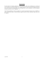
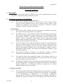
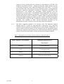
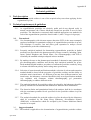
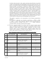
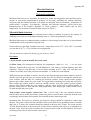
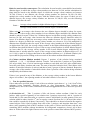
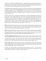
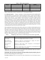
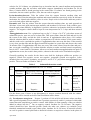
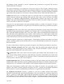
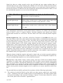
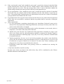
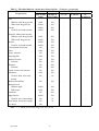
【Appendices】
!
(Translation from Original Chinese Version)
Product safety documents Technical guidelines
Introduction
For the details of technical guidelines for various product safety tests (including heavy metals and
toxic element test, pesticide residues test, microbial limit test, acute toxicity test, long term toxicity
test, local toxicity test, mutagenicity test, carcinogenicity test and reproductive and development
toxicity test), please refer to Appendix 1 - 9.
The technical guidelines in these appendices are general requirements for these tests. In addition to
these specified procedures, other procedures approved by the Chinese Medicines Board may also
be adopted.
April 2004
ii
Appendix I
Test for heavy metals and toxic element
Technical guidelines
1. Description
1.1 The test for heavy metals and toxic element is one of the required safety tests for the
registration of proprietary Chinese medicines (pCms).
2. Technical requirements & guidelines
2.1
Heavy metals & toxic element specified for tests
2.1.1 The Chinese Medicines Board has specified three heavy metals (namely
mercury, lead & cadmium) and one toxic element (namely arsenic) as the
required test parameters. The laboratory concerned is required to provide
quantitative methods for their determination.
2.2. Test methods
2.2.1 The laboratory shall establish precise and practical test methods to determine
the contents of the 4 heavy metals or toxic element.
2.2.2 By making reference to the relevant literature and information, the laboratory
may develop instrumental detection method and its corresponding sample
pre-treatment procedure. Applicable instrumental detection methods may
include Atomic Absorption Spectrometry (AAS), Inductively-Coupled
Plasma-Atomic Emission Spectrometry (ICP-AES)and Inductively-Coupled
Plasma- Mass Spectrometry (ICP-MS), etc.
2.2.3 The main purpose of the sample pre-treatment procedure is to breakdown the
matrices so that the heavy metals and toxic element are fully released for
determination. The laboratory may use microwave digestion method or other
effective pre-treatment methods such as acid digestion or ashing to obtain a
homogenous sample solution. The solution, after appropriate dilution, is used
for the determination.
2.2.4 The analytical methods developed by the laboratory should be properly
validated to ascertain that the methods are fitted for their intended use.
2.2.5 The detection limits and quantitation limits of the analytes shall be in
accordance with the maximum permitted levels specified for the heavy metals
and toxic element in pCms (see Table 1).
2.2.6 The analytical methods for heavy metals & toxic element in pCms developed by
the laboratory shall be accredited by the Hong Kong Laboratory Accreditation
Scheme (HOKLAS), or laboratories which are accepted by the Chinese
Medicines Board can also be accepted.
2.3 Suggested method
2.3.1 April 2004
This method employ inductively coupled plasma-mass spectrometry(ICP-MS)
to determine heavy metals and toxic element in the test sample. It is applicable
to the determination of contaminants, viz. Arsenic(As), Cadium(Cd),
Mercury(Hg) & Lead(Pb) in Chinese herbs and pCm samples. The sample is
digested by microwave-assisted nitric acid digestion and after dilution, the
iii
content of heavy metals and toxic element are determined by ICP-MS. The
method is described as follows: Weigh accurately the sample (about 0.5g) into
a microwave digestion vessel, add concentrated nitric acid (about 7.5 ml) and
mix well. Place the vessel into a microwave digestion system, set an appropriate
microwave digestion programme according to instructions described in the
Manufacturer’s user manual (the programme should gradually raise the
temperature to at least 180℃ and maintain at that temperature for at least 9.5
minutes) and start the digestion programme. After digestion, cool the digested
solution and transfer it into a volumetric container. Add an appropriate quantity
of internal standard Indium (In), make up to the volume with water and conduct
the determination using ICP-MS. As different instruments may have different
operation, user manuals or other relevant documents provided by the
manufacturer must be referred to when carrying out the determination.
2.3.2 The above suggested method is for reference only. The analytical method
adopted must be verified by inter-laboratory comparison or by analyzing
certified reference materials of similar matrices as the samples. Furthermore, in
order to ensure sufficient control over the analytical process, appropriate quality
control (QC) measures such as analysis of certified reference material, duplicate
sample and spiked sample shall be incorporated into the adopted methods.
Table 1: Permitted level for heavy metals and toxic element in pCms
Heavy Metals or Toxic element
Maximum permitted level
(Total intake)
April 2004
Mercury
36 micrograms/day
Lead
179 micrograms/day
Arsenic
1,500 micrograms/day
Cadmium
3,500 micrograms/dosage
iv
Test for pesticide residues
Appendix II
Technical guidelines
1. T
ask description
1.1 The test for pesticide residues is one of the required safety tests when applying for the
registration of pCms.
2. Technical requirements & guidelines
2.1. Pesticides specified for tests
2.1.1 As organochlorine pesticides are relatively stable and do not degrade easily in
natural environment, the Chinese Medicines Board has specified to test for such
pesticides. The laboratories concerned shall establish appropriate test methods for
each of the organochlorine pesticides listed in table 1 (total : 20 types in 9 groups).
2.2. Test methods
2.2.1 Gas chromatography with electron capture detection (ECD) is the most commonly
employed technique for quantitative analysis of organochlorine pesticides in pCms.
This technique is sensitive, fast and offers good separation to analyze several
organochlorine pesticides simultaneously.
2.2.2 Presently, analytical methods for determining organochlorine pesticides in herbal
medicines are described in the pharmacopoeias of many countries, including China,
Japan, United Kingdom and United States. Gas Chromatography with ECD is
essentially the standard technique adopted in these methods.
2.2.3 By making reference to the pharmacopoeia methods. Laboratories may optimize the
gas chromatographic conditions and develop their analytical methods for pCms.
Such methods should include a procedure to confirm the pesticides results by using
a column with different polarity and/or using mass spectrometry.
2.2.4 Selection of extraction and purification methods: selection of solvent and extraction
method should be based on the properties of pesticides residues and the ability to
minimize matrix interference. As different pCms may have different matrices and
interference, the laboratory concerned should develop several purification methods
(e.g. sulphuric acid treatment, column chromatography, gel permeation
chromatography) to address different needs.
2.2.5 The analytical methods for pesticide residues developed by the laboratory should be
properly validated to ascertain that the methods are fitted for their intended use.
2.2.6 The detection limits and quantitation limits of the analytes shall be in accordance
with the maximum permitted levels specified for the pesticide residues in pCms
(see Table 1).
2.2.7 The analytical methods for pesticide residues in pCms developed by the laboratory
shall be accredited by the Hong Kong Laboratory Accreditation Scheme
(HOKLAS), or laboratories which are accepted by the Chinese Medicines Board
can also be accepted.
2.3. Suggested method
2.3.1 This method is applicable to the determination of organochlorine pesticide residues
April 2004
v
in Chinese herbs and pCms. After sample extraction and purification, the pesticide
residues are determined by gas chromatography with electron capture detection. Any
positive results shall be further confirmed by gas chromatography-mass spectrometry
whenever possible. The method is described as follows: weight accurately the
sample (about 10 g) and add an appropriate quantity of ethyl acetate. After ultrasonic
extraction, evaporate the solvent using a rotary vacuum evaporator at elevated
temperature and under reduced pressure. Purify the residue using gel permeation
column and florisil column. Add an appropriate amount of internal standard (e.g.
1-bromo-2 nitrobenzene) and determine the content of each pesticide by gas
chromatography-electron capture detection. When carrying out the above operations,
instrument manufacturer’s user manuals should be referred to. For those samples
suspected to contain organochlorine pesticide, another chromatographic column with
different polarity may be used to confirm the results. Whenever possible, positive
results should be confirmed by gas chromatography-mass spectrometry.
This method is applicable to the determination of the following organochlorine
pesticides:
Aldrin, dieldrin, cis-chlordane, trans- chlordane, oxychlordane, p,p-DDE, p,p-DDD,
o,p-DDT, p,p-DDT, endrin, heptachlor, heptachlor epoxide, hexachlorobenzene,
hexachlorocyclohexane (including isomers like α ,β, δ), lindane, quintozene,
pentachloroaniline and methyl pentachlorophenysulphide.
2.3.2 The above suggested method is for reference only. The analytical method adopted
must be verified by inter-laboratory comparison or by analyzing certified reference
materials of similar matrices as the samples. Furthermore, in order to ensure
sufficient control over the analytical process, appropriate quality control (QC)
measures such as analysis of certified reference material, duplicate sample and
spiked sample shall be incorporated into the adopted methods.
Table 1: Maximum permitted levels of organochlorine pesticide residues
Names of pesticides
Test Parameters
Maximum permitted
level (mg/kg)
0.05
1
Aldrin and dieldrin
Sum of aldrin and dieldrin
2
Chlordane
Sum of cis-chlordane,
trans-chlordane and oxychlordane
0.05
3
DDT
Sum of p,p’-DDT, o,p-DDT,
p,p’-DDE and p,p’-TDE
1.0
4
5
Endrin
Heptachlor
Endrin
Sum of heptachlor and heptachlor
epoxide
0.05
0.05
6
Hexachlorobenzene
Hexachlorobenzene
0.1
7
Hexachlorocyclohexane
Sum of α-, β- and δ-isomers.
0.3
8
Lindane
(γ-BHC)
Lindane
(γ-BHC)
0.6
9
Quintozene
Sum of quintozene,
pentachloroaniline and methyl
pentachlorophenyl sulphide.
1.0
April 2004
vi
Appendix III
Microbial limit test
Technical guidelines
Microbial limit tests are to determine the number of viable microorganisms and specified species
present in non-sterile pharmaceutical products of all kinds, including the starting materials,
excipients and the finished products. For details on microbial limit test methods, ‘preparation of
culture media’, ‘reagents’, ‘test solutions’, ‘diluents’ and ‘indicator solutions’, please refer to the
Pharmacopoeia of the People’s Republic of China (volume 1, 2000 edition) and its 2002
Supplement. For criterion of microbial limit, please see Table 5.
Microbial limit detection
Sampling of the substances being examined must be taken at random. In general, the quantity of
sampling (more than two minimum packaging units) is 3 times the quantity for testing.
Microbial limit tests are conducted under conditions of strict aseptic procedures to avoid accidental
contamination of the preparation during the test.
Unless otherwise specified, incubate bacteria at a temperature of 30~35℃, at 25~28℃ for moulds
or yeast and at 36℃±1℃for reference microorganisms.
The test results are reported in unit per g, per ml or per 10cm2.
Procedures
1. Total aerobic count & moulds and yeast count.
(1) Plate count: Use homogenized solution for examination1, dilute to 1: 102 、1:103 etc serial
dilutions. Transfer three successive 10-fold dilutions of 1 ml each to a 90 mm diameter petri dish
separately. Then to each dish add 15 ml of culture medium at 45℃. Mix well, and allow the
contents to solidify. Invert the petri dish and incubate, 2~3 dishes for each dilution.
While nutrient agar medium is used for bacteria count, Rose bengal agar culture media is used for
moulds count. In special cases, the former media may also be used for counting the moulds and
yeast colonies, and the latter for counting the bacteria colonies. Yeast extract powder peptone
glucose agar culture media for yeast count. For preparations containing bee honey and royal jelly,
Rose bengal agar culture media is used for moulds colonies and yeast extract powder peptone
glucose agar culture media for yeast colonies, and the count results will be added together to
provide a total sum. For fluid and semi fluid preparations, Rose bengal agar culture media is used
for both moulds and yeast count.
Total aerobic count negative control test: Take 1 ml of each of the test substance diluents,
transfer them into 4 sterile petri dishes, prepare the plate for total aerobic count and moulds count
accordingly, then incubate and examine, there should not have bacteria growth.
Incubation time for bacteria is 48 hours, the bacteria colonies should be counted at 24-hour and
48-hour. The count result at 48-hour should be taken as the final result. Incubation time for moulds
and yeast is 72 hours, the colonies should be counted at 48-hour and 72-hour. The count result at
72-hour should be taken as the final result. If the bacteria colonies form a layer, it is unsuitable for
bacteria count. After the count, calculate the average bacteria count of each dilution, and report
according to the rules for total aerobic count report.
April 2004
vii
Rules for total aerobic count report: The calculation for total aerobic count shall be based on the
dilution degree in which the average colony numbers are between 30~300, and the calculation for
fungi shall be based on the dilution degree in which the average colony numbers are between
30~100. If there is one dilution, the average colony numbers are between 30~300 (30~100),
multiply the colony number in this degree with the dilution times for report. If there are two
dilution degrees, the average colony numbers are between 30~300(30~100), use the following
formula to calculate the ratio
Ratio =
Average colony number in higher dilution degree×dilution times
Average colony number in lower dilution degree×dilution times
When ratio<2, an average value between the two dilution degrees should be taken for report;
When ratio>2, the average colony number in lower dilution degree multiplied by dilution times
should be taken for report. If there are 3 dilution degrees in which the average colony numbers are
between 30~300, an average value between the latter two dilution degrees should be taken for
report. If no dilution degree has an average colony number between 30~300, the average colony
number in a degree (in which the average colony number are) closest to 30 or 300 multiplied by
dilution times should be taken for report. If the average colony numbers in all the dilution degrees
are higher than 300 (100), the average colony number in the highest dilution degree multiplied by
its dilution times shall be taken for report. If the average colony numbers in all the dilution degrees
are lower than 30, the average colony number in the lowest dilution degree multiplied by its
dilution times is usually taken for report. If the average colony number in dilution degree of 1:10
(or 1:100) is equal to or bigger than that in the undiluted solution (or 1:10 diluted solution), the
report shall be prepared according to the test result of the culture medium dilution method.
(2) Culture medium dilution method: Prepare 3 portions of the solution being examined
(undiluted 、1:10 、1:100 diluted solution). Transfer 1 ml each of the 3 portions to 5 petri dishes
(0.2 ml per dish). Pour 15 ml of nutrient agar medium to each dish, mix well, after solidification,
invert the petri dishes, incubate and count. The sum of microbial colonies from 5 Petri dishes is the
number of microbial colonies of each ml and that of total 3 groups of data are obtained. Report the
average value of the number of microbial colonies from 3 portions of the solution being examined
multiplied by the dilution times.
If there is no growth in any of the dilutions, or the average colony number in the lowest dilution
degree is less than 1, the reporting number of microbial colonies is less than 10.
2. Test for specified bacteria
Unless otherwise specified, using 10 ml of the test solution (equivalent to 1 g, 1 ml, 10cm2 of the
substance being examined), inoculate directly or after pretreatment. After isolation and
multiplication, use Gram’s stain, biochemical test and autoagglutination test to detect the specified
bacteria.
(1) Escherichia coli
Take 3 portions of bile salt lactose culture medium, 100ml for each
portion. Add a specified quantity of test solution into 2 portions respectively, and in 1 portion of
which add 50~100 control bacteria as a positive control. Add diluent (in same quantity of the test
solution added in others) into the 3rd one as negative control. Incubate for 18~24 hours (the culture
time could be extended to 48 hours if necessary). There should be no bacteria found in the negative
control subject. Take 0.2ml of culture from each of the 3 portions, and inoculate them into 3 test
tubes (each) containing 5ml of MUG medium. At the point of 5 hours and 24 hours, take the one
containing MUG medium without inoculation for background comparison, observe all of these
tubes in 365nm ultraviolet light. Fluorescence in the positive control tube indicates MUG positive;
April 2004
viii
fluorescence in MUG tube containing solution for testing also indicates MUG positive. If there is
no fluorescence, it indicates MUG negative. Then add some Indole solution into the MUG tubes to
observe the colors on the surface of the solution: rose color indicates positive result, if the color of
the test solution remains unchanged, then the result indicates negative.
If the negative control subject appears negative, the positive control subject shows normal growth,
and the bile salt lactose culture medium containing test solution appears clean, which show there is
no bacteria growth, it can be concluded that no Escherichia coli has been detected. If the test
solution suggests MUG positive, and the Indole test is also positive, it can be concluded that
Escherichia coli has been detected. With MUG negative and Indole test is negative, no Escherichia
coli has been detected.
If MUG is found positive and Indole test found negative, or if MUG found negative and Indole test
found positive, test solution (cultured in bile salt lactose culture medium) shall be inoculated
streakily on a eosin methylene blue agar plate or on a MacConkey agar plate, and culture for
18~24 hours to observe the results. If there is no colony found growing in the split plate, it can be
concluded that no Escherichia coli has been found. If there are some colonies growing, 2~3
suspect colonies should be chosen for Indole test (I), methyl red test (M), acetomethylmethanol
growing test (V-P), citrate usage test (C), and Gram's stain, as well as microscopic detection, the
results can be assessed according to the specification in Table 1 below.
Indole test (I): Take the suspect colonies or slant culture, inoculate them into peptone water
medium, incubate for 24 hours, and then add several drops of Indole solution along the wall of the
test tubes to observe the color change on the surface of the liquid: rose color indicates positive
result, if the color remains unchanged, this indicates negative result.
Methyl red test (M): Take the suspect colonies or slant culture, inoculate them into phosphate
dextrose peptone water medium, incubate for 48±2 hours, and then add several drops of methyl
red indicator liquid to observe the immediate color change: bright red or tangerine colour change
indicates positive result, while yellow color change indicates negative result.
Acetomethylmethanol growing test(V-P): Take the suspect colonies or slant culture, inoculate
them into phosphate dextrose peptone water medium, incubate for 48±2 hours, then add 1ml of
α-naphthol into every 2ml of the culture fluid, mix well. Add 0.4ml of 40% potassium hydroxide
solution, shake thoroughly, and observe the color change: red color appears within 4 hours
indicates positive result, while absent of this red-color-reaction indicates negative result.
Citrate usage test (C): Take the suspect colonies or slant culture, inoculate them into citrate
medium, and incubate for 48~72 hours. If there are colonies growing on the medium slant and the
culture medium changes its color from green to blue, the result is considered positive. If the color
remains unchanged, the result is considered negative.
See table 1 for result assessment. For the suspect bacteria colonies (see note c、d) with reaction
results different from what are specified in column MUG-I, then isolate and multiply again, and
biochemical tests should be carried out for further confirmation.
April 2004
ix
Table 1: MUG result assessment
MUG-I
Eosin methylene blue agar
IMVic
Results
Escherichia coli detected
+ +
No Escherichia coli detected
- -
no bacterial growth
No Escherichia coli detected
+ -
bacterial growth
+ -
- + - -c Escherichia coli detected e
bacterial growth
- +
+ + - - d Escherichia coli detected e
Note: c, d for c with result as + + - - or d with result as - + - -, the colonies
shall be isolated again for MUG-I and IMVic test. e Gram-negative Bacillus.
(2) Salmonella species:
Prepare 3 portions of 100 ml nutrient broth culture medium, add a
specified quantity of test solution into 2 portions respectively, and in 1 portion of which adds in
control bacterial suspension as positive control. Add diluent (in same quantity of the test solution
added in others) into the 3rd one as negative control. Incubate for 18-24 hours. Negative control
should not have bacterial growth. Transfer 1 ml of the other 2 portions of culture fluid and
inoculate separately to 10 ml of sodium tetrathionate brilliant green culture media, incubate for
18-24 hours. Inoculate streakily to bile salt sulfur milk agar (or agar media for salmonella, shigella
genus) culture medium and MacConkey agar medium (or eosin-methylene blue agar) plates
separately. Incubate for 18-24 hours or extended up to 40-48 hours. When bacterial colonies are
seen in the plate of positive control test, no bacterial colony is seen in the plate of the test solution,
or none of the colony conforms the morphological characteristics described in the table 2, it can be
concluded that the salmonella species has not been found.
If the morphologic characteristics of the colonies conform or are similar to the description given in
table 2. Select 2~3 colonies and inoculate to triple sugar iron agar culture medium slant. Inoculate
the positive control at the same time. Incubate for 18-24 hours, red colour appears on the surface
and yellow at the bottom, hydrogen sulfide positive for the slant of the positive control. If no
evidence of red colour on the surface and yellow colour at the bottom for the suspected slant of
substance being examined, it can be concluded that the Salmonella species has not been found.
Otherwise, following test should be carried out for further detection.
Table 2
Morphologic characteristics of salmonella species on selective agar media
Culture medium
Bile salt, sulphur
lactose agar
Colonial characteristics
Colourless to light orange colour, semi-transparent, the center of
colonies can be slightly black, all black or no black
Agar medium for
salmonella, shigella
Colourless to light red, semi-transparent or opaque, the center of
colonies can be black in colour sometimes.
Eosin-methylene
blue agar
Colourless to light orange colour, transparent or semi-transparent,
smooth moist circular colonies.
MacConkey agar
medium
Colourless to light orange colour, transparent or semi-transparent, dark
in the center of the colonies occasionally.
Indole test: The test shall be carried out according to the specification under the section of
“Escherichia coli” and assess the results accordingly.
Urease test: Take the culture from the suspect bacteria medium slant and inoculate it into urea agar medium slant. After 24 hours incubation, if the slant becomes red, the test result is positive; if the color remains unchanged, the result is negative. Potassium cyanide test: Take the nutrient broth culture liquid that has been incubating the suspect April 2004
x
colonies for 20~24 hours, use platinum loop to inoculate into the control medium and potassium
cyanide medium, plug the test tubes with rubber stoppers immediately and incubate for 24~48
hours. Colonies shall be found growing in the control tube. If colonies are found growing in the
tested tube, the test result is positive; if no, the test result is negative.
Lysine decarboxylase test: Take the culture from the suspect bacteria medium slant and
inoculate it into lysine decarboxylase medium and control medium respectively. After 24~48 hours
incubation, the control tube shall be yellow in color. Purple color in tested tube indicates positive
test result, and yellow indicates negative.
Dynamics test: Take the culture from the suspect bacteria medium slant, use stab approach to
inoculate it into semi-solid nutrient agar medium, and incubate for 24 hours. If the colonies grow
from the stab points diffusely, its dynamics is positive, otherwise its dynamics shall be recorded as
negative. The negative culture shall be kept in room temperature for 2~3 days and then assessed
again.
Autoagglutination test: Use a platinum loop to dip 2~3 loops of A~F”0” polyvalent serum of
salmonella species, onto one end of a clean slide. Take some culture from the slant and mix it with
the serum on the slide, and tilt the slide in and out. If agglutination takes place, 0.9% sodium
chloride solution shall be used to conduct a comparison test with the culture (of the same strain),
and the result can be concluded as positive if agglutination does not recur this time. If the reaction
is very slow, put the slide and the dipped cotton ball together in a petri dish, and observe the result
20 minutes later. If agglutination still does not occur, take some culture from the slant and put it
into a test tube containing 0.9% sodium chloride solution to make enriched bacteria suspension,
heat in 100℃ water bath for 30 minutes, allow to cool and conduct autoagglutination test again. If
agglutination occurs, the result is then positive, otherwise the result can be concluded as negative.
Generally speaking, the results for the above tests shall be: hydrogen sulfide test positive (or
negative), Indole test negative, urease test negative, potassium cyanide test negative, lysine
decarboxylase test positive, dynamics test positive, and A~F”0” polyvalent autoagglutination test
positive. The test results are listed in Table 3.
Table 3
Serial
No.
Salmonella detection results
Autoagglutination test (A~F “0” serum)
Agglutination Agglutination reaction at 0.9% sodium chloride
reaction
100℃ for 30 minutes solution comparison
1
Positive
2
Negative
3
Negative
Positive
Negative
Biochemical
Test
Conclusion
Negative
Tally
Negative
Tally
Salmonella
detected
Not tally
Salmonella
not detected
Salmonella
detected
For the above tests, if there is any discrepancy or suspect reaction observed with the culture,
further tests will be carried out before final conclusion can be drawn.
(3) Pseudomonas aeruginosa: Prepare 3 portions of 100 ml bile salt lactose culture medium,
add a specified quantity of test solution into 2 portions respectively, and in 1 portion of which adds
control bacterial suspension as positive control. Add diluent (in same quantity of the test solution
added in others) into the 3rd one as negative control. Incubate for 18~24 hours. The negative
control should not have bacterial growth. Streak the other two cultures on cetrimide (cetyl
trimethyl ammonium bromide) agar culture plate, incubate for 18~24 hours. If bacterial colonies
are seen in positive control plate and no bacterial colonies or suspicious colonies in the plates of
April 2004
xi
the substance being examined, it can be concluded that pseudomonas aeruginosa has not been
found within the test solution.
The typical morphology of pseudomonas aerugionosa colony is flat, irregular, diffusing margin,
moist surface, greyish white with bluish green pigment diffused in the surrounding. If the growth
colonies have the above characteristics, select 2~3 suspect colonies, inoculate separately to nutrient
agar culture slant, incubate for 18-24 hours. Take the culture for the Gram stain and oxidase test.
Oxidase test: Put a piece of clean filter paper in a petri dish, and use a sterile glass rod to apply
culture from the nutrient agar medium slant onto the filter paper, add a few drops of freshly
prepared 1% dimethyl-p-phenylenediamine dihydrochloride solution and observe the colour
change. If the color turns gradually from pink to violet red within 30 seconds, oxidase test positive
can be concluded, or otherwise it is negative.
If proven it is not gram-negative non spore-forming bacillus or oxidase test negative, it can be
concluded that no pseudomonas aeruginosa has been detected. Otherwise pyocyanin test shall be
carried out.
Pyocyanin test: Take some of the above agar culture and inoculate it onto the slant of the medium
specially for pyocyanin detection, incubate it for 24 hours, add 3~5ml of chloroform into the test
tube, crush the medium and shake well. Leave for a while, remove the chloroform into another test
tube, add about 1ml of hydrochloric acid (1mol/L), shake and leave for a while again. If pink color
is observed in the hydrochloric acid layer, positive pyocyanin test can then be concluded. Negative
control shall also be carried out at the same time for confirmation.
When the negative control test is found negative, Gram-negative bacillus and oxidase test found
positive, and pyocyainin test also found positive, it can be concluded that pseudomonas aeruginosa
has been detected.
For those culture that are found as pyocyanin test negative, the following tests shall be carried out.
Nitrate reduction aerogenic test: Take some culture from the nutrient agar medium slant and
inoculate it into nitrate peptone water medium. After 24 hours incubation, if there is any gas
generated in the Durham tube, the result is positive.
42℃ growth test: Take some culture from the nutrient agar medium slant and inoculate it into
0.9% sterile sodium chloride solution to make bacterial suspension. Immediately put it into water
bath at 41℃ ± 1℃ to incubate for 24~48 hours. If there is bacterium and moss growth, the test
result is positive, otherwise the result is negative.
Gelatin liquidation test: Use the inoculating needle to dip some culture from the nutrient agar
medium slant, stab to inoculate it into the gelatin medium and culture for 24 hours, and put it into a
refrigerator for 10~30 minutes. If the medium is still in liquid condition, the test result is positive.
When the Gram-negative bacillus and oxidase test results are positive, the pyocyanin test is
negative, the results of nitrate reduction aerogenic test, 42℃ growth test and gelatin liquidation
test are positive, it can be concluded that pseudomonas aeruginosa has been detected.
(4) Staphylococcus aureus: Prepare 3 portions of 100 ml of sodium tellurite broth culture
medium (or nutrient broth), add a specified quantity of the test solution into 2 portions respectively
and in 1 portion of which adds control bacterial suspension as positive control. Add diluent (in
same quantity of the test solution added in others) into the 3rd portion as negative control. Incubate
for 18-24 hours (or up to 48 hours, if necessary). Negative control shall not have bacterial growth.
April 2004
xii
Streak the other two culture growth on the egg yolk high salt agar culture medium plate or
mannitol high salt agar culture medium plate. Incubate for 24-72 hours. When bacterial colonies
are seen in positive control plate, and no bacterial colony is seen in the plate of the test solution, or
the bacterial colonies are different from the morphologic characteristics described in table 4, it can
be concluded that staphylococcus aureus has not being found.
Table 4 Morphologic characteristics of staphylococcus aureus on selected agar media
Medium
Characteristic colonial morphology
Egg yolk high salt agar
Golden yellow, circular convex, regular margin, opaque outer circle
due to degradation of lecithin, colony diameter 1~2 mm
Mannitol high salt agar
Golden yellow, circular convex, yellow outer circle, colony diameter
0.7~1 mm
If the morphologic characteristics of the growth colonies conform or are similar to the description
given in table 4, select 2~3 suspect colonies, inoculate separately onto nutrient agar culture
medium slant. Incubate for 18-24 hours. Take the cultures for Gram’s stain and plasma coagulase
test.
Plasma coagulase test: Take 3 test tubes, add 0.5ml of mixture of plasman-sterile water (1:1)
into each of them. Add 0.5ml of the nutrient broth culture fluid (or enriched bacterial suspension)
of the tested strain into one of these 3 tubes, and add 0.5ml of nutrient broth culture fluid (or
bacterial suspension) containing staphylococcus aureus into another tube as positive control, add
0.5ml of nutrient broth or 0.9% sterile sodium chloride into the last tube as negative control.
Incubate these 3 tubes at the same time. Determine the results 3 hours later, then observe them at a
suitable time interval within the 24-hour culture period. Plasma in the negative control tube should
still be in liquid condition, while the plasma in the positive control tube should have been
coagulated. If the plasma in the tube containing the test strain is now coagulated, the test result is
positive. Should the plasma in either the positive control tube or the negative control tube not in
the condition as they are supposed to be, re-test is necessary.
When the plasma in both the negative and positive control tube are in their expected conditions,
and if the test strain is found positive in both Gram-positive coccus test and plasma coagulase test,
it can be concluded that staphylococcus aureus has been detected.
n Preparation of the plasma: Using a sterile syringe, suck 1ml of sterile 0.9% sodium chloride
containing 5% sodium citrate, and aseptically take 9ml of rabbit (or goat, human) blood, gently
shake for a few minutes. Wait until the blood does not coagulate, put it in a sterile centrifugation
tube, centrifugate it to separate the plasma. Suck the separated plasma with a sterile pipette and
transfer it into a sterile test tube, store the tube in refrigerator. In order to verify the usability of
plasma, known positive plasma coagulase test for staphylococcus aureus should be used for the
test.
The microbial limit test shall be performed in accordance with the following requirements:
(1) The criterion of microbial limit for pCms shall cover three aspects, namely the “total aerobic
count”, “moulds & yeast count” and the presence of specified bacteria. The subject product shall
be assessed according to the criterion of all these 3 aspects. For the limit criterion for various
dose forms, please refer to the table 1 appended below.
April 2004
xiii
(2)
If the “total aerobic count” and “moulds & yeast count” exceeds the criterion of microbial limit
of that particular dose form in the first test, samples shall be taken randomly from the same
batch to repeat the test twice. Take the average value of the 3 tests to determine whether the
product being examined meet the specified criterion for that particular test.
(3)
For eye preparations, if the “moulds & yeast count” exceeds the specific criterion of microbial
limit in the first test, samples shall be taken randomly from the same batch to repeat the test
twice. The product being examined passes the requirements only when no evidence of moulds &
yeast growth has been found in the last 2 tests.
(4)
If specified bacteria for any dose form is detected in the first test, the result of the first detection will be final, no re-test shall be conducted, and the test product shall be assessed as failing the
requirement. Other requirements:
i. For oral preparations containing animal-origins raw ingredients, Salmonella must not be
detected. For oral preparations containing animal horn, royal jelly, bee honey and Colla Corii
Asini, test for Salmonella is exempted.
ii. For preparations containing crude drug powder used for haemostatis or on wounds, ulcers or
deep tissue, clostridium tetani must not be detected.
iii. Moulds and yeast detection: the liquid/semi-solid preparations containing no sugar, royal
jelly or bee honey and the solid preparations should be examined for moulds; liquid or
semi-solid preparations containing sugar, royal jelly or bee honey should be examined both
for moulds and yeast.
iv. Vaginal preparations: In every 1g or 1ml of the preparation, the aerobic count should not
exceed 100, the moulds & yeast count should not exceed 10, and Staphylococcus aureus and
Pseudomonas aeruginosa should not be detected. For those dose forms containing crude
drug powder, Clostridium tetani should not be detected.
(5)
v. Preparations found with mildew and growing acarid are considered not meeting the
requirements.
vi. Injections should be tested for sterility.
For those dose forms not listed in the table below, they will be considered by the Chinese
Medicines Board on individual basis.
April 2004
xiv
Table 5:Microbial limit for various dose form of pCms
Item 1
Item 2
Preparations
Total aerobic Moulds &
count
yeast Count
Pills
Without crude drug powder
With crude drug powder
Powder
Used for oral and external
Granules, tablets and capsules
Without crude drug powder
With crude drug powder
Troches
Used for oral and external
Concentrated decoctions
Glues
Syrup, mixtures
Dripping pills
Medicinal wines
External
Tinctures
External
Liquid extracts and extracts
Ointments
Used for burn, ulcer and
wound
Medicinal distillates
Medicinal teas
Without sugar
With sugar
Liniments
Suppositories
Used for ulcer, hemorrhage
Nasal drops, aerosols or sprays
Eye drops
Note:
1000
30000
30000
30000
100
100
100
100
-
1000
10000
10000
10000
100
1000
100
1000
500
500
100
100
100
1000
100
100
100
100
100
100
100
100
100
100
100
100
100
100
100
10
-
100
100
-
10000
1000
100
10000
100
100
100
100
100
100
100
10
10
-
-
“ -“ stands for not detecting in per g or per ml.
April 2004
(Unit: per g or per ml)
Item 3 (Specified bacteria)
E. coli
S. aureus
Ps.
aeruginosa
xv
-
-
-
-
-
-
-
-
-
-
-
-
-
-
Appendix IV
Acute toxicity test
Technical guidelines
(A) Test method
The acute toxicity of a drug is generally represented by its median lethal dose (LD50). If the
value of median lethal dose is not obtainable with practical measures because of the limits on
concentration or volume of the medicine, the acute toxicity can be represented by its
maximum tolerable dose (MTD).
(i) Median lethal dose (LD50)
- This refers to the amount which can cause death to half of the test animals in a
single administration.
- The route of administration shall be same as for clinical trial.
- Observe for at least 7 days after administration, record the toxic reactions, weight
changes and the temporal distribution of death of the test animals. Inspection should
be carried out immediately after the death of animals. If necessary, microscopic
examination should be conducted on organ or tissue which shown pathological
changes at autopsy.
(ii) Maximum tolerable dose (MTD)
- This refers to the maximum dose that the test animals can tolerate without dying.
The medicine may be administered to the animals once or several times in a day.
- The route of administration shall be same as for clinical trial.
- Administer the drug to the test animals once or 2~3 times in a day, observe for 7
consecutive days, record the reaction of the animals in detail, calculate the total
quantity of the administered drug, and deduce the equivalent amount in clinical
administration.
April 2004
xvi
Long-term toxicity test
Appendix V
Technical guidelines
(A)
Test method
Animal species:The test animals shall include two species of animals (including both
rodent and non-rodent)*, three groups in each species. Each group is
used for the test of one dose level; the male and female animals are in
equal proportion in each group. White rats are the most commonly used
animals in rodent species, at least 20 to be used in a group. Dogs and
monkeys are most commonly used animals in non-rodent species, at
least 6 in a group.
Dose Levels: 3 different dose levels should be used, in principle, the lowest dose level
should be slightly higher than the effective dose level obtained in the
pharmacodynamics study, in which no toxicity effect should be observed
in any of the test animals. With the highest dose level, apparent toxicity
effect can be observed in some of the test animals.
Route of administration: The route of administration shall be same as for clinical trial.
Administration period:If in clinical practice, the medicine is for single administration
or repetitive administration spanning less than a week, the test
administration period should be 2 weeks to a month; If the medicine is
for repetitive administration spanning over a week, the test
administration term should be 3~4 times of the period of clinical
treatment (usually, not over 6 months in maximum for rodents, and not
over 9 months for non-rodents).
If the test administration period is not within the aforementioned range,
the applicant should provide supporting explanation.
Observation: This includes observation with the naked eye and pathological diagnosis
on physical signs, weights, appearance, blood test, and functionality of
livers, kidneys, and other important organs. For all the animals that have
died during administration period, autopsy shall be carried out to
determine the cause of the death. All survivors should be autopsied at
the end of the administration period.
* The following pCms must use two species of animals (including both rodent and non-rodent)
in conducting long-term toxicity test:
1. new medicine that its prescription comprises of a newly discovered Chinese herb, a new
medicinal part of a Chinese herb, an active group extracted from Chinese herb or a set of
active groups extracted from a compound prescription;
2. Chinese medicine injection;
3. new medicine that its formulation is a new Chinese medicine prescription, with altered
route of administration or with altered dose form, and its prescription contains Chinese
herb medicine(s) listed in Schedule 1 of the Chinese Medicine Ordinance or contains
incompatibility of drugs, such as “incompatibility among eighteen medicaments” or
“mutual antagonism among nineteen medicaments” .
For pCm that does not fall into any of the categories mentioned above, if the results of
long-term toxicity test in rats do not show any toxic effects, the applicant is not required to
conduct long-term toxicity test in non-rodent at this stage. However, if necessary, the Chinese
Medicines Board can require the applicant to conduct long-term toxicity test in non-rodent.
April 2004
xvii
Appendix VI
Local toxicity test
Technical guidelines
(A)
Test method
(I) Skin irritation test
i. Experimental animals and grouping:
a) White rabbits or white guinea-pigs can be used as the test animals.
b) Young animals should be chosen when conducting tests for infant skin
preparations.
c) The test animals are divided into 2 groups: the group with intact skin,
and the group with wounded skin. For each test animal, the comparison
is made with the skin on the two sides of its body.
d) Numbers in each group: For rabbits and mini pigs, 3~4 in each group;
for guinea-pigs, 5~6 in each group. Each group should contain same
number of male and female.
ii. Preparations to be tested
In the test, the preparations should be in the same dose form as in clinical
practice.
iii. Pre-test treatment
a) 24 hours before application, remove the hair on both sides (spine as the
centerline) of the back of the test animal. The unhaired area shall be
about 10% of the whole surface area of its body.
b) After removing hair, check to see whether the skin has been wounded.
Wounded skin shall not be used for tests that with intact skin.
c) P
reparation of wounded skin:
Sterilize the skin (after removing hair), and use appropriate method to
cut the skin (e.g. use sterile scalpel to cut open the skin with several
cross lines, or use abrasive paper to rub skin to cause wound) until the
skin is slightly oozing blood. Both sides of the skin should have similar
wound level.
iv. Experimental procedure
a) Apply the preparations to be tested onto the unhaired area on one side of
the back of the test animal, and apply placebo onto another side.
b) Cover the unhaired area with pledget and fasten it properly with
adhesive tape.
c) Every animal should be kept in individual cage.
d) After 24 hours of application (or after any other appropriate period of
time), use warm water or non-irritating solvent to wash away the
remaining test substance or placebo.
e) At the 1st, 24th, 48th and 72nd hour after the test substance is removed,
observes with the naked eye and carries out necessary histopathologic
examinations, and record whether there is any erythema and edema
observed in application area.
v. Test results and evaluation
a) Record all skin reactions during the observation period. Refer to the
following table to assess the score of the observed skin conditions.
Compare the score with set values to evaluate the irritation intensity of
the test preparations.
b) Record the recovery process of the irritation effect.
April 2004
xviii
Irritation scoring system
Erythema and eschar formation
No erythema
Very slight erythema (barely perceptible)
Well-defined erythema
Moderate to severe erythema
Severe erythema (beet-redness) to slight, eschar formation
The highest score for erythema is 4
Edema formation
No edema
Very slight edema (barely perceptible)
Slight edema (edges of area well defined by definite raising)
Moderate edema (raised approximately 1 mm)
Severe edema (raised more than 1 mm and extending
beyond the area of exposure)
The highest score for edema is 4 Total Maximum Score (erythema score + edema score)
Intensity Evaluation
Average Score
0 – 0.49
0.5 – 2.99
3.0 – 5.99
6.0 – 8.0
c)
Score
0
1
2
3
4
0
1
2
3
4
8
Evaluation
Non-irritating
Slightly irritating
Moderately irritating
Intensely irritating
The test results shall be processed statistically.
(II) Skin Sensitization Test
i. Test animal
Use white guinea-pigs as the test animals. 10 guinea-pigs in each group, with
same number of male and female.
ii. Control groups
In addition to the treatment group, two control groups shall be set up for
comparison:
- Negative control group (administer with placebo)
- Positive control group (administer with positive sensitizer)
iii. Preparations to be tested
In the test, the preparations shall be in the same dose form as in clinical practice.
iv. Experimental procedure
a) 24 hours before application, remove hair on both sides (spine as the
centerline) of the back of the test animal (guinea-pigs). The unhaired area is
about 3x3 cm2 in each side.
b) Sensitization contacts:
(1) Apply the preparations onto the unhaired area of the left side of the
animal.
(2) Cover the unhaired area with pledget and fasten it properly with
adhesive tape.
(3) Every test animal should be kept in individual cage.
(4) The preparation should be allowed to come into contact with the skin
for 6 hours each time.
April 2004
xix
(5) Apply the preparation 3 times; 2nd time on the 7th day and 3rd time on
the 14th day.
c) Provocation contacts:
14 days after the last application, apply the preparation onto the unhaired
area of the right side of the animal. After removing the preparation,
immediate, at the 24th, 48th and 72nd hour respectively, observe and record
whether there is any anaphylactic reaction on the skin.
v. Test results and evaluation
a) Pay attention to see whether the animals have serious general anaphylactic
reactions like asthma, titubation or shock.
b) Compare the condition with that in the control groups, and give scores to
animals’ local anaphylactic reactions to evaluate the level of the
anaphylactic reactions.
Sensitization Scoring system
Erythema and eschar formation
No erythema
Very slight erythema
Well-defined erythema
Moderate to severe erythema
Severe erythema (beet-redness) to slight, eschar formation
Edema formation
No edema
Very slight edema (barely perceptible)
Slight edema (edges of area well defined by definite raising)
Moderate edema (raised approximately 1 mm)
Severe edema (raised more than 1 mm and extending
beyond the area of exposure)
Score
0
1
2
3
4
0
1
2
3
4
c) Calculate the sensitization ratio of each group, and compare the test results
of the test group with the control group.
Sensitization =
ratio
the number of animals with erythema, edema or
general anaphylactic reactions
X 100%
The total
number of the
test animals
Skin sensitization evaluation:
Sensitization ratio(%)
0-10
11-30
31-60
61-80
81-100
Evaluation
No sensitization
Slight sensitization
Moderate sensitization
High sensitization
Extreme sensitization
d) The test results shall be processed statistically.
(III) Mucous membrane irritation test
April 2004
xx
1. Ocular irritation test
a) Test animal:
White rabbits (at least 4) shall be chosen as test animal.
b) Control comparison method:
The two eyes of the animal can be compared with each other. Apply the
preparation into the conjunctival sac in one of the eyes of the test animal,
and use a placebo for the other eye for comparison.
c) Experimental procedure:
In the test, the preparations to be tested shall be in the same dose form as in
clinical practice. Apply the preparation into the conjunctival sac in one of
the eyes of the test animal, and placebo into the other eye, then make the
animal close its eyes for about 10 seconds.
d) Observation and evaluation of results:
At the 6th, 24th, 48th, 72nd hour and on the 7th day (respectively) after
application of the preparation, observe the local reactions of cornea, iris and
conjunctiva. Histopathologic examinations may also be carried out when
necessary.
Compare the test results (reaction of the eye with preparation applied) with
the control results (reaction of the eye with placebo applied), and evaluate
the ocular irritation of the preparation. The test results shall be processed
statistically.
2. Irritation tests with other mucous membrane areas
The following methods are applicable to nasal drops, inhalants, buccal
applications, auristillas, rectal and vaginal suppositories, etc.
a)
Selection of proper test animals:
(1) For nasal drops, inhalants, buccal applications, auristillas:
guinea-pigs or white rats shall be used as test animals.
(2) For rectal and vaginal suppositories: Rabbits or white rats shall be
used as test animals. b) Grouping: (1) Set up two different doses groups and one negative control group.
(2) Use placebo for negative control group.
c) Experimental procedure:
In the test, the preparation being tested shall be in the same dose form as
in clinical practice. Follow the clinical route of administration, keep the
preparation in contact with the local mucous membrane for 4 hours. If it is
too much for a single application, the preparation can also be divided into
several applications and applied evenly within the 4 hours.
d) Observation and evaluation of results:
(1) At the 24th, 48th hour and on the 7th day (respectively) after
application of the preparation, observe any irritation reaction of the
test animals and its recovery process. In addition, general symptoms
shall also be observed and recorded.
(2) At various observation time, kill some of the test animals to conduct
histopathologic examinations with targeted local mucous
membranes.
(3) During the test, if any test animals are found with toxic reaction or
died, the major viscera of the dead animals (both died or killed) shall
be observed carefully, and histopathologic examinations shall be
carried out.
(4) Compare the test results with the control group, and evaluate the
irritation intensity of the preparation. The test results shall be
processed statistically.
April 2004
xxi
Appendix VII
Mutagenicity test
Technical guidelines
(A) Test methods
(I) Reverse mutation test in bacteria (1)
Strains:
Mutated strain of Salmonella typhimurium with histidine defect TA
1535, TA 1537, TA 98, TA 100 and Escherichia coli WP2 uvr A.
Dose Levels: At least 5 different dose levels should be chosen for test.
Control Groups: Negative control, positive control and S9(2) control.
S9 control means: With and without metabolic activation, allow the
bacteria to come into contact with the test substances, and observe its
reverse mutation.
Metabolic activation means: Use the mammalian liver microsome
enzyme (i.e. S9) (which has been processed with exutory mixture) to
transform the test substances into highly reactive metabolites.
Test method: Either a plate incorporation or a preincubation method should be
used, observe the result 48 hours later to determine the number of
revertant colonies that have been induced. For each concentration
level, at least 3 plates shall be prepared to calculate the mean value
of revertant colonies.
Assessment: Record the number of revertant colonies that have been induced by
the test substances. Process the numbers statistically. If the number is
over 2 times greater than in the negative control group, and is
dependence to the dose level, the test result is positive.
Notes:
(1) The principle of bacterial r everse mutation test is: The mutated strain of Salmonella
typhimurium with histidine defect (the strain that cannot grow in media containing no
histidine) can be reverted to original strains (the strain that can grow in media containing no
histidine) under the effect of exutories. In this test, add the test substances and check the
number of revertant colonies growing in the medium that contains no histidine to evaluate the
mutagenicity of the test substances.
(2) S9 - mammalian liver microsome enzyme
Preparation of S9: Inject some exutory enzyme (For example: Aroclor 1254) into adult rat,
kill the rat 5 days after the injection, take out its liver aseptically and undergo further process,
then put it into 9000xg centrifugal machine to centrifugate for 10 minutes, and collect the
supernatant.
(3) Number of revertant colonies – the number of colonies of original strains that are reverted
from the mutated strains under the effect of exutory.
(II) Chromosomal aberration test with mammalian cells in culture.
Cells:
Primary or established cell lines of mammalian cells in culture
should be used.
Dose levels:
At least 3 different dose levels should be employed.
Control groups: Negative control, positive control and S9(2) control.
Solvent or normal saline group should serve as a negative control; A substance known to cause chromosomal aberrations should be employed as a positive control. S9 control means: With and without metabolic activation conditions,
April 2004
xxii
allow the cells to come into contact with the test substances, and
observe the induced aberration.
Metabolic activation means: Use the mammalian liver microsome
enzyme (i.e. S9) (which has been processed with exutory mixture) to
transform the preparation to be tested into highly reactive
metabolites.
Medicinal activity and specimen preparation time: The test substances shall be put in
contact with the cells for a period of time before observation. The
cells can be collected after the test substances has been in contact
with the cells for 24 and 48 hours, and the specimen shall then be
prepared for observation. Cells in metabolic activation group shall be
in contact with the preparation for at least 6 hours.
Microscopic examination: At least 100 metaphase cells shall be observed for each
concentration level. Use oil-microscope (usually 2500-4000 times)
to observe the number and type of chromosomal aberrations.
According to different concentration, comparison and different
contact time, present the observations in a table.
Assessment: If the test result meets any of the following two conditions and is
statistically meaningful, it is assessed as positive:
1. The increase of chromosomal aberrations that induced by the
preparation is dependence to the dose levels.
2. Repeatable increase can be observed at a certain test point.
(III) Micronucleus test with rodents
Animals: Mice should normally be used. Generally 10 in each group with
same number of male and female (or at least 6 adult male mice in
each group).
Route and method of administration : Normally, the expected clinical route of
administration should be used. Single administration is the most
common method, while multiple administrations are also applicable.
Dose levels: At least three dose groups should be employed. Negative and
positive control groups should also be set up for comparison
purpose.
Control groups: Solvents or normal saline group can serve as a negative control. A
positive control group should receive a substance known to induce
micronuclei.
Microscopy: Animals should be sacrified at an appropriate time after
administration of the test substance, and bone marrow smears
prepared. Normally, observation should be made of the incidence of
micronuclei in 1000 polychromatic erythrocytes per animal. The
relative frequency of polychromatic erythrocytes and total
erythrocytes should also be calculated (in %).
Assessment: From statistical point of view, if the test result meets any of the
following two conditions, it can be assessed as positive:
(1) The increase in percentage of micronucleus erythrocytes
(induced by the preparation) is dependence to the dose levels.
(2) Repeatable increase can be observed at a certain test point.
April 2004
xxiii
Carcinogenicity test
Appendix
VIII
Technical guidelines
(A) Experimental method
The test can be divided into two stages: the preliminary and the full-scale carcinogenicity
study. The purpose of preliminary carcinogenicity study is to determine the highest dose that
can be used in the full-scale carcinogenicity study. If the relevant information (for highest
dose) is available, the preliminary study can be omitted. When selecting the test animals,
factors which should be considered include: the animal’s resistance against infectious disease,
its life span, incidence of spontaneous tumor formation, as well as its sensitivity to known
carcinogens.
(I)
Preliminary carcinogenicity study
Animals: The animal should be the same as in full-scale study. It is desirable
to use two species.
Dose levels & Grouping: At least 3 different dose levels should be chosen for test.
In addition, rodents should be used to set up control groups for
comparison. 10 males and 10 females in each group.
Administration period: Usually, the medicine should be administered continuously
for 90 days. However, this period may be extended if necessary.
The substance being examined and the route of administration: both
should be the same as in the full-scale test.
of
them
Observation & examination: For all animals in each group, the general signs should
be observed daily and body weight measured once a week. Autopsy
and gross observations on organs and tissues should be performed on
dead animals on each occasion and on surviving animals at the end
of the administration period. Organs or tissues with gross changes
should be examined histopathologically.
Evaluation of results: The dose in the preliminary carcinogenicity study that
inhibits body weight gain by less than 10% in comparison with the
control and causes neither death due to toxic effects nor remarkable
changes in the general signs and laboratory examination findings of
the animals. This is the highest dose to be used in the full-scale
carcinogenicity study.
(II)
Full-scale carcinogenicity study
Animals: At least two species of both sexes should be used as test animals,
such as rats and mice.
Dose levels & Grouping: At least 3 dose groups and a control group should be
employed. A blank control group should also be employed. There
should be at least 100 animals in each group, males and females in
equal numbers.
Route of administration: Normally, the expected route of clinical application
should be used.
Administration period: Over 24 months for rats, over 18 months for mice.
Experimental procedure: - All animals in each group should be observed daily for
April 2004
xxiv
general signs. At the beginning of the administration period,
measure their weights and food-intakes once a week, 13 weeks later,
measure weights and food-intake at least once every four weeks.
Minimize incidence of death caused other than tumors.
- Animals that appear to be moribund during the experimental
period should be isolated or sacrificed and autopsied immediately
and organs and tissues should be examined macroscopically and
histopathologically. At the time of sacrifice, blood samples should
be taken to measure red and white blood cells as well as to prepare
smear specimens.
- If tumorous pathologic changes (or suspect tumorous pathologic
changes) are found with animals in the test and control groups,
histopathologic examinations should be carried out with the
following organs: skin, mammary glands, lymph nodes, salivary
glands, sternums, spine, femurs, thymuses, tracheas, lungs and
bronchi, heart, thyroids and parathyroid glands, tongues, esophagus,
stomach, duodenums, large intestines, small intestines, livers,
pancreases, spleens, kidneys, adrenal glands, testicles, ovaries,
gonads and their accessory organs, eyeballs, pituitary gland, spinal
cord etc…
- If no tumorous pathologic changes are visible with the naked-eye
on animals in both the test and control groups, histopathologic
examinations should be carried out to the above mentioned organs of
animals in the highest dose group. If there is any tumorous
pathologic changes found in the examination, histopathologic
examinations should then be carried out on all the test animals.
Evaluation of results: A test substance is considered to be positive for carcinogenicity
when any of the following types of response has been observed in
the carcinogenicity study(in accordance with statistical calculation):
i) Development of tumours of a type seen in the test group and not
seen in the control group
ii) Development of tumours seen with greater frequency in the test
group, compared with the control group.
iii) Greater varieties of organs and tissues involved in tumour
development in the test group, compared with the control group.
iv) Earlier development of tumours in the test group, though in the
absence of any significant difference in the incidence of tumours
between the test group and the control group.
April 2004
xxv
Appendix IX
Reproductive and development toxicity test
Technical guidelines
(A) Experimental methods
The test should be carried out in 3 segments:
Segment I. General reproductive toxicity test (the test conducted prior to
and in the early stages of pregnancy)
Animals: Rats and mice should be used as test animals. At least 20 males and
20 females should be employed.
Dose levels & Grouping: 2~3 different dose groups and 1 negative control group should be
included. Positive control group may be included if necessary.
Route of administration: Normally, the expected route of clinical application should be used.
Administration periods: Male – Starting from 60 days before mating to the day of successful
copulation. Female – Starting from 14 days before mating, during mating and after successful copulation until the beginning of organogenesis. Experimental procedure: A male and a female test animal should be housed together for two
weeks. According to the requirements of each individual case, the
substance may be administered only to the male animal or only to
the female animal (e.g. the pCms relate to pregnancy may be
administered only to the female test animals). Check daily to see if
the animals have successfully copulated. During the administration
period, the animals’ general signs, changes in body weights and
food-intakes should be carefully recorded.
After successful copulation, females should be autopsied at term,
and calculate the rate of successful pregnancy, number and
percentage of dead foetus, number and percentage of alive foetus, as
well as the weights of foetus. The foetus’s appearances and the
development condition of their viscera and bones should be
carefully checked. Histological examinations may be carried out
when necessary.
Males used for mating and females without successful copulation
should be autopsied at an appropriate time, and gross observation on
organs and tissues should be made.
Segment II. Teratogenic test (the test conducted during the organogenesis
period)
Animals:
Females of at least one species of rodent (at least 30) and one species of non-rodent (at least 12) should be used. Dose levels & Grouping: 3 different dose groups and 1 negative control group should be employed. A positive control group is generally desirable.
Route of administration: Normally, the expected clinical route of administration should
be used.
Observation & examination: During the experimental period, general signs, body
weights and food intake should be measured for all dams until the
final phase of pregnancy. All dams should be autopsied at term,
body weight of each dam, number of corpus luteums, total number
of foetus, number and percentage of dead foetus, number and
April 2004
xxvi
percentage of live foetus, and the weights, appearance and sexes of
the live foetus.
Segment III. Perinatal toxicity test (the test conducted during the
perinatal and lactation periods)
Animals: Choose at least one of the animal species that had been used in the
2nd stage of the test, e.g. rats, mice (15~20 in each group) or rabbits
(8~12 in each group).
Dose levels & Grouping: 3 different dose groups and 1 negative control group should be
used.
Route of administration: Normally, the expected clinical route of administration should
be used.
Observation & Examination: - During the experimental period, carefully observe and
record the general signs, weights and food-intakes of the dams.
- Observe and record the average litter size, mortality of the foetus,
weights of live foetus, genders of foetus, and whether there is any
abnormality with their appearance. After the partus of the dams,
house them with their own offspring together and allowed them to
nurse their offspring. Observe and record the survival, growth and
development of their offspring.
- Keep observing the effect of the test substance on the offspring of
the test animal. Breed some of their offspring and observe their
survival, growth and development, including their behaviors,
reproductive performance and any other possible abnormalities.
Reproductive performance of offspring should be examined on the
basis of establishment of copulation and pregnancy. The learning
capacity of their offspring may be measured and assessed if
necessary.
April 2004
xxvii