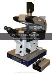
Download NTEGRA Spectra Probe NanoLaboratory
Transcript
NTEGRA Probe NanoLaboratory NTEGRA Spectra Upright configuration Instruction Manual ` NTEGRA Spectra Probe NanoLaboratory (Upright Configuration with Solar TII Spectrometer) Instruction Manual 1 February 2011 Copyright © NT-MDT Web Page: http://www.ntmdt.com General Information: [email protected] Technical Support: [email protected] NT-MDT Co., building 100, Zelenograd, 124482, Moscow, Russia Tel.: +7-499-735-03-05 Fax: +7-499-735-64-10 Read me First! Observe safety measures for operation with devices containing sources of laser radiation. Do not stare into the beam. A label warning about the presence of laser radiation is attached to the measuring head (Fig. 1) as well as to the laser sources. Fig. 1 Before you start working with the instrument, get acquainted with the basic safety measures and the operation conditions for the instrument! If you are a beginner in scanning probe microscopy, we recommend you to familiarize with basic SPM techniques. “Fundamentals of Scanning Probe Microscopy” by V.L. Mironov gives a good introduction to the subject. This book is available on the Internet, http://www.ntmdt.com/support. Feedback Should you have any questions, which are not explained in the manuals, please contact the Service Department of the company ([email protected]) and our engineers will give you comprehensive answers. Alternatively, you can contact our staff on-line using the ask-online service (http://www.ntmdt.com/online). User’s documentation set The following manuals are included into the user’s documentation set: − Instruction Manual – is the guidance on preparation of the instrument and other equipment for operation on various techniques of Scanning Probe Microscopy. The contents of the user’s documentation set may differ depending on the delivery set of the instrument. − SPM Software Reference Manual – is the description of the control program interface functions, all commands and functions of the menu and, also a description of the Image Analysis module and the Macro Language “Nova PowerScript”. − Control Electronics. Reference Manual – is the guide to SPM controller, Thermocontroller and Signal Access module. Some equipment, which is described in the manuals, may not be included into your delivery set. Read the specification of your contract for more information. The manuals are updated regularly. Their latest versions can be found in the site of the company, in the section “Techsupport” (http://www.ntmdt.com/support). Table of Contents NTEGRA Spectra PNL (Upright Configuration). Instruction Manual Table of Contents 1. OVERVIEW.............................................................................................................................................. 3 2. DESIGN 2.1. 2.2. 2.3. 2.4. 2.5. 2.6. 2.7. ............................................................................................................................................... 4 BASE UNIT ........................................................................................................................................ 6 OPTICAL MEASURING HEAD ............................................................................................................. 8 2.2.1. AFM Probe Holder ............................................................................................................. 11 2.2.2. STM Probe Holder .............................................................................................................. 13 2.2.3. Side Illumination Module .................................................................................................... 13 RADIATION DELIVERY AND RECORDING SYSTEM ............................................................................ 15 SPECTROMETER .............................................................................................................................. 17 DETECTORS .................................................................................................................................... 18 LASERS ........................................................................................................................................... 21 OPTICAL FIBER TRANSPORT SYSTEM.............................................................................................. 23 3. PRINCIPLE OF OPERATION OF THE OPTICAL SPECTRAL SYSTEM .................................. 24 4. BASIC SAFETY MEASURES .............................................................................................................. 27 5. OPERATING CONDITIONS ............................................................................................................... 30 6. STORAGE AND TRANSPORT REGULATION ............................................................................... 31 7. GENERAL REQUIREMENTS ON INSTALLATION ...................................................................... 32 8. SETUP AND INSTALLATION ............................................................................................................ 33 8.1. 8.2. 9. PREPARING THE SPECTROMETER .................................................................................................... 33 8.1.1. Installing and Aligning the Optical Fiber Transport System .............................................. 33 8.1.2. Installing the Spectrometer and the Optical Microscope.................................................... 35 CONNECTING THE ELECTROMECHANICAL UNITS ............................................................................ 36 POWERING SEQUENCE..................................................................................................................... 38 10. PREPARING FOR AFM MEASUREMENTS .................................................................................... 41 10.1. 10.2. 10.3. 10.4. 10.5. 10.6. 10.7. 10.8. 10.9. PREPARING AND INSTALLING THE SAMPLE ..................................................................................... 41 INSTALLING THE SIDE ILLUMINATION MODULE.............................................................................. 47 INSTALLING THE AFM PROBE HOLDER ........................................................................................... 48 INSTALLING THE AFM PROBE ........................................................................................................ 49 INSTALLING THE MEASURING HEAD ............................................................................................... 51 INSTALLING THE OPTICAL FIBER TRANSPORT SYSTEM ................................................................... 52 LAUNCHING THE CONTROL PROGRAM ............................................................................................ 54 ADJUSTING THE SPECTRAL UNIT .................................................................................................... 55 ADJUSTING THE OPTICAL CANTILEVER DEFLECTION DETECTION SYSTEM .................................... 59 11. PREPARING FOR STM MEASUREMENTS..................................................................................... 63 11.1. 11.2. 11.3. 11.4. 11.5. 11.6. 11.7. 11.8. 11.9. PREPARING AND INSTALLING THE SAMPLE ..................................................................................... 63 INSTALLING THE SIDE ILLUMINATION MODULE.............................................................................. 65 INSTALLING THE STM TIP HOLDER................................................................................................. 65 MANUFACTURING THE STM TIP ..................................................................................................... 66 INSTALLING THE STM TIP .............................................................................................................. 67 INSTALLING THE MEASURING HEAD ............................................................................................... 68 INSTALLING THE OPTICAL FIBER TRANSPORT SYSTEM ................................................................... 68 LAUNCHING THE CONTROL PROGRAM ............................................................................................ 69 SPECTRAL UNIT ADJUSTMENT ........................................................................................................ 69 1 NTEGRA Spectra PNL (Upright Configuration). Instruction Manual 12. PERFORMING MEASUREMENTS ................................................................................................... 70 12.1. APPROACHING THE SAMPLE AND FOCUSING THE LASER BEAM ...................................................... 73 12.1.1. Approaching and Focusing for Modes with the AFM Probe.......................................... 73 12.1.2. Approaching and Focusing for Modes with the STM tip................................................ 75 12.1.3. Approaching and Focusing for Modes without the Probe.............................................. 78 12.2. ADJUSTING CHANNELS OF EXCITING AND DETECTION ................................................................... 79 12.2.1. Adjusting Detection Parameters of the CCD-camera .................................................... 80 12.2.2. Adjusting Detection Parameters of the PMT Module..................................................... 84 12.2.3. Adjusting Detection Parameters of the APD Module..................................................... 87 12.3. ADJUSTING SCAN PARAMETERS ..................................................................................................... 89 12.4. SCANNING ...................................................................................................................................... 91 12.4.1. Scanning with Recording by the CCD-Camera.............................................................. 92 12.4.2. Scanning with Recording by the PMT Module ............................................................... 94 12.4.3. Scanning with Recording by the APD Module ............................................................... 95 12.5. SAVING DATA................................................................................................................................. 96 12.6. FINISHING THE WORK ..................................................................................................................... 97 APPENDIX 1. OPTICAL VIEWING SYSTEM ........................................................................................................... 99 1.1. 1.2. 2 ............................................................................................................................................ 99 CCD07 OPTICAL VIEWING SYSTEM ............................................................................................... 99 CCD27 OPTICAL VIEWING SYSTEM ............................................................................................. 100 1.2.1. Basic Units ........................................................................................................................ 100 1.2.2. Assembling Videomicroscope for Optical Measuring Head ............................................. 103 1.2.3. Assembling Videomicroscope for Universal Measuring Head.......................................... 105 Chapter 1. Overview 1. Overview NTEGRA Spectra PNL instrument is equipped with high performance optics that provides scanning of the sample area under the tip with the resolution of 0.4 μm. Its optical measuring head serves for illuminating the surface with visible light (minimum spot size 0.4 μm) and for collecting the scattered radiation including radiation from immediate vicinity of the tip. Additional capability is confocal Raman microscopy that uses the spectrometer of the instrument. A large value of the numerical aperture of the objective lens (NA=0.7) allows illuminating the surface at wide angle with high intensity of the normal electric component in the light spot under the probe. This enables measuring tip-enhanced optical effects, including the surface-enhanced Raman scattering. NTEGRA Spectra PNL provides the following options: − performing spectral measurements at a certain point and acquiring spectral characteristics of various materials when the instrument operates as a regular spectrometer; − measuring secondary signal intensity in the selected wavelength range in the mode of layer-specific volumetric scanning of the area 100x100x30 μm; − acquiring optical images of the object when the instrument operates as a regular laser confocal microscope; − detecting the object landscape with atomic resolution as well as collecting its electrical, magnetic and nanomechanical properties with force microscopy techniques; − performing nanoscale measurements of different optical properties relevant to techniques of nonaperturate near-field scanning optical microscopy. The particular feature of the instrument is its capability to detect optical properties with a number of nonlinear effects, surface-enhanced Raman scattering, and the so-called Tip-Enhanced Raman spectroscopy. 3 NTEGRA Spectra PNL (Upright Configuration). Instruction Manual 2. Design Fig. 2-1 shows general view of the NTEGRA Spectra PNL instrument. Fig. 2-1. NTEGRA Spectra PNL NTEGRA Spectra PNL consists of the following main parts: 1. Base unit (see sec. 2.1 on p. 6). It holds the measuring head, the scanner, and the sample holder; 2. Optical measuring head (see sec. 2.2 on p. 8). It provides acquisition of the surface image of the investigated object. Installed on the base unit; 3. Laser radiation delivery and recording system (see sec. 2.3 on p. 15). It serves for linking the spectral unit with the measuring head. Mounted on the holder of the base unit; 4. Spectral unit, including: - Spectrometer (see sec. 2.4 on p. 17). It serves to carry out spectrometry measurements and to form the image; - Lasers (see 2.6 on p. 21). Generate radiation that is delivered to the spectrometer or to the measuring head through the optical fiber transport system; - Optical fiber transport system (see 2.7 on p. 23). It serves for delivery of laser radiation to the spectrometer or to the measuring head; 5. Detectors (see sec. 2.5 on p. 18). Serve for detecting signals coming from the investigated object. The spectrometer can be supplied with the following detectors: - CCD camera; - APD module; - PMT module; 4 Chapter 2. Design 6. Optical viewing system (see Appexdix 1 on p. 99). Provides monitoring the sample both at normal and at small angle to the sample surface for following purposes: - aiming the laser beam at the cantilever; - selecting the scan range on the sample surface; - visual inspection of the probe in processes of approaching and scanning. 7. Control system: - Main SPM controller; - Slave SPM controller; - Computer; - Interface boards. The main units of the NTEGRA Spectra PNL are shown in the block diagram of the Fig. 2-2. Fig. 2-2. NTEGRA Spectra PNL block diagram 5 NTEGRA Spectra PNL (Upright Configuration). Instruction Manual 2.1. Base Unit The base unit is shown in Fig. 2-3. Fig. 2-3. Main components of the base unit 1 – exchangeable mount; 2 – stepper motor; 3 – temperature and humidity sensor; 4 – LC-display The base unit is contains a stepper motor with an approach mechanism that serves to place a sample under the probe. Data on ambient temperature and humidity are measured with the sensor 3 and then displayed by the LC-display 4. Base unit electrical slots Slots for connecting the devices installed on the base unit: HEAD – two identical connectors for connecting the measuring head; SCANNER – scanner connector, used for connecting either the exchangeable scanner or the measuring head scanner; SCAN+SENSOR – connector for the scanner or for the scanner with built-in sensors, used for connecting either the exchangeable scanner or the measuring head scanner; Т° –connector of a heating element, used for connecting a heating stage, heating liquid cells etc.; SM – connector of the stepper motor; AFAM – connector of the ultrasonic piezoelectric transducer; BV – bias voltage jack, used for applying bias voltage to the sample. 6 Chapter 2. Design Slots for connecting the bias unit to the control system: CONTROLLER 1 – connector for connecting to the HEAD connector of the SPM controller; CONTROLLER 2 – connector for connecting to the CONTROLLER 2 connector of the SPM controller; CONTROLLER T° – connector for connecting to the thermocontroller. Exchangeable mount The exchangeable mount is installed into the base unit of the device and is fixed with three screws. Design of the PNL allows for several types of the exchangeable mount. Here, a mount of SCC01NTF model is used. Basic parts of the exchangeable mount are shown in Fig. 2-4. Fig. 2-4. Exchangeable mount 1, 2 – measuring head seats; 3 – mirror mounting frame; 4 – positioning device; 5 – measuring head cables holder; 6 – approach mechanism cylinder; 7 – grounding jack; 8 – screws for fixing to the base unit. There are two types of sockets on the exchangeable mount (see pos. 1, 2 in Fig. 2-4). Depending on its type, a measuring head can be installed either into the slots 1 or into the slots 2. The frame 3 is used as a support of subsidiary mirrors of the optical viewing system. The positioning device 4 (Fig. 2-5) holds a device that carries the sample (sample holder, scanner etc.). 7 NTEGRA Spectra PNL (Upright Configuration). Instruction Manual Fig. 2-5. Scanner with movement sensors mounted on the positioning device The positioning device is placed on the movable cylinder of the approach mechanism 6. This enables to move the sample up and down. The microscrews 1 and the spring stops 2 (Fig. 2-6) provide adjusting in the XY plane. Fig. 2-6. Positioning device 1 – microscrews; 2 – spring stops 2.2. Optical Measuring Head The optical measuring head is a dedicated version of the scanning probe microscope. Its probe is located under a lens of large numerical aperture and of high spatial resolution. The optical measuring head provides an option of side illumination of the sample with laser radiation. This option needs the side illumination module to mount on the measuring head. 8 Chapter 2. Design Optical measuring head (Fig. 2-7) (hereinafter – measuring head) consists of the following components: − Optical cantilever deflection detection system; − Universal lens; − Universal lens holder; − Probe holder. The probe holder is exchangeable. Type of the probe depends on the measurement mode. The following holders are used for different measurement modes: − AFM probe holder; − STM probe holder. The optical cantilever deflection detection system 1 is mounted on the assembly of the lens holder 2 (Fig. 2-7). The lens hovers with a system of elastically deformed arms that provides high accuracy of beam focusing on the object as well as good stability of the assembly. The unit mount 3 contains microscrews for positioning the probe. Fig. 2-7. Optical AFM measuring head 1 – optical cantilever deflection detection system; 2 – lens holder; 3 – mount with the probe holder and the positioning device Main components of the AFM optical measuring head are shown in Fig. 2-8. 9 NTEGRA Spectra PNL (Upright Configuration). Instruction Manual Fig. 2-8. Measuring head 1, 2 – screws for XY probe adjusting; 3, 4 – photodiode mirror adjusting screws; 5 –screw for fine focusing of the lens; 6, 7 –screws for XY lens adjusting; 8 – lens positioning device spring stop; 9 – entrance aperture; 10, 11, 12 – screw legs; 13 – lens; 14 –spring stop of the probe holder positioning device The measuring head serves for measurements with confocal microscopy. Besides, contact and semi-contact modes of AFM as well as STM techiques are available. The main functional component of the measuring head is the Mitutoyo M Plan Apo 100 lens (pos. 13 in Fig. 2-8). The numerical aperture of the lens is 0.7 and the focal length is 2 mm while the working distance is 6 mm. The lens provides the the following options along with conventional AFM scan techniqiues: − Viewing the sample with resolution limited by 0.4 μm; − Delivery and focusing the exciting laser radiation; − Aiming the laser beam at the probe’s tip; − Detecting the probe deflection (needs an additional semiconductor laser diode to generate radiation of 650 nm or 830 nm wavelength). The screw for fine focusing of the lens 5 is located in front of the lens. The microscrews 1 and 2 move the probe holder relative to the lens. Moving the probe holder provides directing the laser beam of the optical cantilever deflection detection system (hereinafter referred to as detection system) to the cantilever reflective surface. The photodiode mirror is adjusted with the screws 3 and 4 so that the beam reflected from the probe could come directly to the centre of the photodiode. For adjusting the measuring head in angle and height, three screw legs 10, 11, and 12 are used. The microscrews 6 and 7 provide positioning of the lens against the probe and external optical devices. * 10 ATTENTION! Height adjustment of the legs 10, 11, and 12 is not recommended as this can misalign the optical system. Chapter 2. Design Cantilever deflection in AFM modes is detected with the detection system operating a semiconductor laser (with λ =830 nm or 650 nm) that supplies radiation through the lens. The system dichroic mirror selectively reflects the radiation of the semiconductor laser. It provides a wide spectral range (400 <λ < 800 nm for the laser with λ =830 nm) thus allowing for observing the sample through the optical microscope and for optical exciting the sample surface. High sensitivity of the detection system is due to a special design of the laser formation system. Confocal microscopy uses a special probe (Fig. 2-9) with the tip being outside the edge of the cantilever. Fig. 2-9. Confocal microscopy probe Fig. 2-10. AFM probe Table 2-1. Optical measuring head specification Parameter Value Sample size Up to ∅40×10 mm Scanning type Sample Scanning range 100×100×7 μm Lens numerical aperture 0.7 Optical resolution 0.4 μm Lens focal distance 2 mm Visual field ~80 μm Laser wavelength − 830 nm in 400÷800 nm; spectral range − 650 nm in working spectral 400÷600 nm, 700÷900 nm range Dimensions (L×W×H) 150×130×130 nm Weight 1.5 kg 2.2.1. working AFM Probe Holder AFM probe holder (see Fig. 2-11) is placed on the measuring head. It can be replaced with another holder if necessary. It serves for measurements in AFM modes. 11 NTEGRA Spectra PNL (Upright Configuration). Instruction Manual Fig. 2-11. AFM probe holder Design of the AFM probe holder is shown in Fig. 2-12. The probe is placed on the sapphire bench-shaped support and then fixed with the clamping spring 2. The spring moves up and down with the handle 3. Fig. 2-12 Design of the AFM probe holder 1 – sapphire support; 2 – clamping spring; 3 – handle The AFM probe holder is placed in the mount of the measuring head and then secured with the spring stop 1 (see Fig. 2-13). The screws 2 and 3 serve to move the probe holder for adjusting the optical cantilever deflection detection system. The AFM probe holder is connected electrically to the slot 4 in the measuring head (see Fig. 2-13). Fig. 2-13. AFM probe holder installed on the measuring head 1 – spring stop; 2, 3 – screws for X and Y movement of the probe; 4 – probe holder connector 12 Chapter 2. Design 2.2.2. STM Probe Holder STM probe holder (see Fig. 2-14) is placed on the measuring head. It can be replaced with another holder if necessary. It serves for measurements in the STM mode. Fig. 2-14. STM probe holder Design of the STM probe holder is shown in Fig. 2-15. The probe 1 is inserted into slots of the holder 2 and then fixed with the spring 3. Protrusion of the sharpened end of the probe outside the holder should not exceed 3÷4 mm. Fig. 2-15 Design of the STM probe holder 1 – probe; 2 – holder; 3 – clamping spring The STM probe holder is mounted on the measuring head quite similarly to the AFM probe holder (see Fig. 2-13). The STM probe holder is connected electrically to the HEAD socket in the base unit of the instrument. With the STM probe holder, the socket of the measuring head is left open. 2.2.3. Side Illumination Module To operate with side illumination of the sample, the measuring head is equipped with the exchangeable module for side illumination (see Fig. 2-16). The side illumination module provides delivery of the exciting laser radiation at angle of 60º to normal to the sample surface as well as aiming the laser beam at the probe tip. Positioning of the laser beam on the sample is controlled with the micrometric screws 1 and 2, while focusing uses the screw 3. 13 NTEGRA Spectra PNL (Upright Configuration). Instruction Manual Fig. 2-16. Side illumination module 1, 2 – screws for X and Y movements of the laser spot; 3 – screw for fine focusing of the laser beam; 4 – positioning device; 5 – entrance aperture The side illumination module is secured with four screws on the front side of the lens holder (Fig. 2-17). Fig. 2-17. Side illumination module installed on the measuring head Laser radiation comes into the side illumination module through the port 5. To focus the laser beam on the cantilever, the focusing module is used (Fig. 2-18). This module is screwed into a hole that is located inside the side illumination module. Focusing modules for different radiation wavelengths are available. 14 Chapter 2. Design Fig. 2-18. Focusing module 2.3. Radiation delivery and recording system The radiation delivery and recording system (Fig. 2-19) provides linking the spectral unit with the measuring head. The module is mounted to the back of the base unit and to the Optical viewing system holder (see Appendix 1 on p. 99). Fig. 2-19. Radiation delivery and recording system 1, 2 – adjusting screws of the steering mirror (M2); 3 – laser beam focusing screw (L1); 4 –scanner; 5, 6 – adjusting screws of the steering mirror M1; 7 – a turret with exchangeable mirrors (M3); 8 – entrance aperture; 9 – stopping screw; 10 – adjusting ring Schematics of delivery and detection of laser radiation is presented in Fig. 2-20 where the input beam is colored with green and the reflected one (used for detection) is red. With the side illumination of the sample, the radiation delivery and recording system operates only with the reflected beam. In this case, the laser beam is delivered through the side illumination module. * ATTENTION! For adjusting the laser beam, use the screws 1, 2, and 3. Do not turn the screws 5 and 6 and the screws 9 and 10 located on the radiation feedthrough as this can misalign the optical system. 15 NTEGRA Spectra PNL (Upright Configuration). Instruction Manual The exciting laser radiation comes through the entrance aperture 8 of the radiation feedthrough. For fine positioning of the radiation on the entrance aperture 8, the radiation feedthrough can move vertically (with the adjusting ring 10) for ±10 mm and rotate around its axis. The stopping screw 9 fixes the feedthrough in the desired position. Fig. 2-20. Schematics of delivery and detection of laser radiation M1 – mirror; L1, L2 – lenses; M2 – steering mirror; M3 – exchangeable mirror; 1, 2 – adjusting screws of the steering mirror; 3 – laser beam focusing screw; 4 –scanner; 7 – turret with exchangeable mirrors Radiation delivery and recording system contains a mirror M1 (Fig. 2-20) at its entrance. With the screws 5 and 6, the mirror M1 is aligned so that the laser beam is directed along the lens L1 axis. The screw 3 is used to move vertically the lens L1 that focuses the laser beam on a point located in the front focal plane of the lens L2. The lens L2 patterns the spot (taken from the mirror M2) in the entrance pupil of the measuring head lens. Thereby, a laser beam comes from the steering mirror M2 and goes to the lens pupil under various angles, but it invariably falls into the center of the pupil. The steering mirror M2 can be controlled either manually with the screws 1, 2 or instrumentally by the piezoelectric scanner 4. The mirror M3 is located on the turret and can be changed. The turret has four states that correspond to four types of mirrors: M0 – totally reflecting mirror. With this option, no portion of the laser beam transmits the mirror and so image on the display is unavailable. M1 – partially transparent mirror (reflection to transmission ratio is 10/90). The display shows an image of the sample and of the laser spot. 16 Chapter 2. Design M2 – mirror is absent and the laser radiation does not fall onto the sample. The display shows only an image of the sample. M3 – custom mirror. In particular, this can be a mirror with a high reflection in the range of 600÷800 nm and a high transmission outside it for the laser with the wavelength of 633 nm. 2.4. Spectrometer Raman spectrometer is used to carry out spectroscopy and to form the image. A detailed description is given in the manual Raman Spectrometer. User’s Manual. Fig. 2-21. Raman spectrometer Modularity of the spectrometer structure, a wide variety of lasers and microscopes, high-precision elements for operation with three laser, automatic adjustment of optical components after switching between the lasers – all this factors make the system flexible thus enabling a wide range of applications to perform. 17 NTEGRA Spectra PNL (Upright Configuration). Instruction Manual 2.5. Detectors CCD-camera CCD-camera (Fig. 2-22) provides a wide measurement range and has brilliant spectral and time characteristics. It contains a built-in thermoelectrical cooling system (Peltier element), which allows to cool the CCD-matrices down to –90 °С. Fig. 2-22. CCD-camera Table 2-2. CCD-camera technical characteristics Parameter Value Quantity of sensors (pixels) 1024×255 Sensing element size 26 μm2 Active area size 26.6×6.7 mm2 Vertical scanning rate 16 ms # NOTE. Model of the CCD-camera can change without prior notice. APD module Avalanche photodiode (APD module) (Fig. 2-23) serves for counting photons during a predefined time intervals. It contains a built-in cooling system which provides cooling the device thus lowering the thermal noise. 18 Chapter 2. Design Fig. 2-23. ADP module Fig. 2-24. Spectral range Table 2-3. APD module technical characteristics Parameter Value Ambient temperature +5÷40ºC Working range at temperature 22ºC 400÷1060 nm Active area diameter 175 μm Maximum light intensity 10 000 photons/sec # NOTE. In some configurations of NTEGRA Spectra PNL, the ADP module is missed or replaced by a cooled PMT. 19 NTEGRA Spectra PNL (Upright Configuration). Instruction Manual PMT module Photomultiplier module (PMT module) (Fig. 2-25) serves for converting optical radiation acquired from the sample into electrical signals. Fig. 2-25. PMT module Fig. 2-26. Photomultiplier sensitivity 20 Fig. 2-27. Effective spectral range Chapter 2. Design Table 2-4. PMT module technical characteristics Parameter Value Sensitivity (at 420 nm) 3.0×10¹⁰ V/W Operating spectral range 185÷850 μm Output voltage shift ±3 mV Current-to-voltage conversion gain 1×10⁶ V/A Frequency range from 0 to 20 kHz 2.6. Lasers Depending on configuration, the delivery set of the NTEGRA Spectra PNL may contain from one to three lasers. The standard configuration contains He-Ne, diode and solid-state lasers. Besides, equipping with some additional lasers of a wide wavelength range (from 350 to 850 nm) is possible. * ATTENTION! Direct and scattered radiation is hazardous to eyesight. Avoid eye contact with laser radiation. LM473 solid-state laser Table 2-5. Specifications of the solid-state laser Parameter Value Transverse mode TEM00 Wavelength 473 nm Polarization linear Degree of polarization 100:1 Output power max 50 mW Beam divergence (1/e2) < 1.2 mrad Long-Term Drift <3% Warm-up Period < 15 minutes Operating temperature 10 ÷ 40 ºС 21 NTEGRA Spectra PNL (Upright Configuration). Instruction Manual LM532 diode laser Table 2-6. Specifications of the diode laser Parameter Value Transverse mode TEM00 Wavelength 532 nm Polarization linear Degree of polarization 100:1 Output power up to 22±2 mW Beam divergence (1/e2) 0.6±0.1 mrad Long-Term Drift <2% Warm-up Period < 10 minutes Operating temperature 15 ÷ 50 ºС LM633 Helium-Neon laser Table 2-7. Specifications of the Helium-Neon laser Parameter Value Transverse mode TEM00 Wavelength 633 nm Polarization linear Degree of polarization > 500:1 Output power Up to 35 mW Pointing stability < 0.03 mrad after 30 minutes Beam divergence (1/e2) 0.66 mrad Long-Term Drift ±2 % Warm-up Period < 15 minutes Operating temperature –20 to 40 ºС 22 Chapter 2. Design 2.7. Optical Fiber Transport System Optical fiber transport system (hereinafter – OTS) and radiation feedthrough (see Fig. 2-28) are designed for easy, safe and secure transfer the laser radiation to the NTEGRA Spectra PNL entrance port or immediately to the sample when the side illumination module is used. a) Radiation feedthrough: 1 – mounting flange; 2 – housing; 3 – guide bush; 4 – adjusting screws; 5 – central backing-up screw b) OTS: 1 – «Input» socket; 2 – polarization key, 3 – «Output» socket; 4 – cover plugs Fig. 2-28. Radiation feedthrough and OTS Polarization plane of the radiation is defined by the line mark on the Output socket of OTS (see Fig. 2-29). Rotate the socket to the desired position when installing OTS. Fig. 2-29. Output socket. Line mark indicates the polarization plane Table 2-8. OTS technical characteristics Parameter Value Fiber type single-mode Pigtail length 2÷3 m Beam diameter at entrance 0.65 mm Transmission efficiency > 65 % Output beam divergence diffraction limit Maximum input power ≤ 100 mW 23 NTEGRA Spectra PNL (Upright Configuration). Instruction Manual 3. Principle of Operation of the Optical Spectral System Optical layout of the NTEGRA Spectra PNL is shown in Fig. 3-1. Fig. 3-1. Simplified optical diagram of the NTEGRA Spectra PNL 1 – triple input unit; 2, 3, 4, 6, 7 – mirrors; 5 - 97% mirror; 8 – beam expander; 9 – turret with three sets of changeable edge or notch filters; 10, 11 – lenses; 12, 13 – adjustable pinhole; 14 – diffraction gratings; 15, 16 – adjustable density neutral filter; 17, 18 – polarizer; 19 – filter cartridge; PMT – photomultiplier tube; CCD – camera; APD – avalanche photodiode The laser beam goes from the triple input unit 1, passes the mirrors 2 and 3, the adjustable density neutral filter 15 and a polarizer 17, and comes to the beam expander 8. The laser beam expander 8 automatically adjusts the diameter and divergency of the beam regarding the laser in operation. The mirrors 4 and 5 direct the beam to the notch filter 9 that reflects the beam to the radiation delivery and recording system. The measuring head lens focuses the beam on the sample. Fig. 3-2 explains the NTEGRA Spectra PNL layout with the side illumination module employed. 24 Chapter 3. Principle of Operation of the Optical Spectral System Fig. 3-2. Simplified optical functional diagram of the NTEGRA Spectra PNL with the side illumination module employed 1 – triple input unit; 2, 3, 4, 6, 7 – mirrors; 5÷97% mirror; 8 – beam expander; 9 – turret with three sets of changeable edge or notch filters; 10, 11 – lenses; 12, 13 – adjustable pinhole; 14 – diffraction gratings; 15, 16 – adjustable density neutral filter; 17, 18 – polarizer; 19 – filter cartridge; 20 – OTS; 21 – focusing module; PMT – photomultiplier tube; CCD – camera; APD – avalanche photodiode The laser beam goes from the triple input unit 1, passes the mirrors 2 and 3, and comes into the optical fiber transport system 20. Further, the beam passes the focusing module 21 and impinges the sample at angle of 60 º to the vertical direction being focused with the lenses. Radiation scattered by the sample is detected with one of the schemes below for both layouts explained above. Laser optical scheme Laser optical scheme is designed to form the confocal image of the sample by the elastically scattered radiation. Besides, this scheme is useful for juxtaposing the probe tip and the focus of the laser beam. The scattered radiation passes through the measuring head lens. Then, after reflection by the notch filter 9 and by the mirror 4, it is collimated and transferred back into the spectrometer. A portion of the scattered light (~3 %) passes through the mirror 5 and comes to the Reflection unit. The lens 11 focuses the beam on the adjustable pinhole 12. If necessary, the laser beam can be attenuated with the adjustable circular density neutral filter 16 before directing it to the PMT. A confocal 2D or 3D image is formed by simultaneous scanning the sample and recording the scattered intensity with the PMT. 25 NTEGRA Spectra PNL (Upright Configuration). Instruction Manual Spectral optical scheme Spectral optical scheme is designed to form the confocal image of the sample by the secondary radiation that is composed of spectral components absent in the illuminating light. Luminescent radiation emitted by the sample is collimated by the measuring head lens. Then, it passes the notch filter 9, the polarizer 18 and the filter cartridge 19. After focusing with the lens 10, the secondary radiation passes the adjustable pinhole 13 at the monochromator entrance. Analysis of the secondary radiation is performed either by collecting its spectral composition the CCD-camera or by detecting intensity of a particular spectrum line (selected by positioning the diffraction grating 14 and the mirror 7) with APD or PMT (incl. photons calculation mode). If the monochromator is equipped with a mirror instead of the diffraction grating, the desired wavelength is selected by the notch filter of the filter cartridge 19. 26 Chapter 4. Basic Safety Measures 4. Basic Safety Measures General Safety Measures − Ground the instrument before operation! − Do not disassemble any part of the instrument. Disassembling of the product is permitted only to persons certified by NT-MDT Co. − Do not connect additional devices to the instrument without prior advice from NT-MDT Co. − The instrument contains precision electro-mechanical parts. Therefore, protect it from mechanical shocks. − Protect the instrument against the influence of extreme temperature and moisture. − For transport, provide proper packaging of the instrument to avoid its damage. Electronics − Before operation, set the voltage selector of the SPM controller to the position corresponding to local electrical power line (this is only done with the controller being off!). − Switch the instrument off before any operation on connecting/disconnecting its cable connectors. Disconnecting or connecting the cable connectors during operations may cause damage to the electronic circuit and disable the instrument. A warning label is attached to the SPM controller of the instrument (Fig. 4-1). Fig. 4-1 27 NTEGRA Spectra PNL (Upright Configuration). Instruction Manual Laser − Observe safety measures for operation with devices containing sources of laser radiation. See the warning label on the back panel of the instrument (Fig. 4-2). Fig. 4-2 Optical components Optical components include the following units and elements: − optical elements in the spectrometer unit; − Optical Fiber Transport System (OTS); − laser. To avoid contamination of the optical elements with dust or moisture, keep the room clean and never disperse sprays or other substances near the instrument. Do not touch the optical elements. For cleaning the optical components, use special cleaning sets. * ATTENTION! Never uncover the spectrometer without extreme necessity. Only an authorized person is permitted to clean elements of the spectrometer. In the case of contamination, request for help to the manufacturer. When handling the OTS, do not curve a fiber-optic cable and never exceed the limit radiation power. For storing or transporting, cover the slots with the special caps. If the entrance or the exit aperture is contaminated, do not use the instrument and contact the manufacturer. You must be extremely careful when using a laser! * ATTENTION! Do not look at the exit aperture or at the OTS. Remember that laser radiation can damage your eyesight even after reflection. Adjust optical elements with minimum power of the laser. Use eye protectors to reduce the laser’s power. 28 Chapter 4. Basic Safety Measures Radiation detectors Radiation detectors include CCD-camera and APD (PMT). When storing or handling the CCD-camera, avoid ingress of contamination into the camera. Before connecting the water cooling system, turn off the computer and disconnect the camera from the computer. Prevent ingress of moisture into the camera case. Keep the vent holes on the case open when using the camera. Prevent too intensive illumination of the camera. Make sure that the camera is at the room temperature when finishing the work. All operations with the disconnected APD must be carried out in a dark room. Control the intensity of the light falling onto the APD. Too intensive light can damage the photo detector. 29 NTEGRA Spectra PNL (Upright Configuration). Instruction Manual 5. Operating Conditions For normal operation of the instrument, the following operating conditions are recommended: − Ambient temperature: +18÷23º˚С; − Temperature stability: better 1˚С per hour; − Relative humidity less 60 %; − Atmospheric pressure: 760 ±30 mmHg; − Supply voltage 220 V (+10%/-15%), 50/60 Hz with grounding; − The working room must be protected from mechanical vibrations, airflows and acoustic noise, both internal and external; − Use a massive vibration-isolating optical bench for mounting the PNL; − RMS vibration measured in the 1/3 octave frequency band must not exceed the values given in the graph; − The device must be protected from the direct sunbeams; − The base unit must be located on a separate table, away from the computer and from monitors, to reduce electromechanical noise. Heat flow and draughts badly influence the instrument. Do not operate the PNL if temperature or humidity is outside the recommended limits. 30 Chapter 6. Storage and Transport Regulation 6. Storage and Transport Regulation Storing The units must be stored in package in a clean room with a stable temperature and humidity: − storage temperature (20±10) °С; − Storage humidity less 80 %. Transportation − Transport the device in a package, excluding possible damage. 31 NTEGRA Spectra PNL (Upright Configuration). Instruction Manual 7. General Requirements on Installation NTEGRA Spectra PNL is a precise optical device. Special requirements on the work room, on the power supply system, and on the optical table should be met. NTEGRA Spectra PNL should be installed in a dark room of area of 12 m2 (3×4 m2). The room must be equipped both with ceiling lighting and with a separate source of weak scattered light. A system of thermal stabilization or, at least, of good ventilation should be provided. Remember that an operating laser can disperse up to 4 kW of heating power. Keep dust concentration as low as possible. The electric wiring of the room must meet the following requirements: − at least 8 standard voltage sockets for mains 220 V/50 Hz with the total power of 1 kW. These are used to supply the microscope, the spectrometer, the controllers, computers and monitors; − at least 8 standard voltage sockets for mains 220 V/50 Hz with the power of 4.5 kW (current up to 20 A). These are used to connect the laser. Other supply voltage lines are defined by the user. The CCD-camera needs water cooling for operation. Thus, the corresponding infrastructure must be provided in the room. The PNL is mounted on an optical table of dimensions 2x1 m2 or bigger with a standard net of threaded connections М5 (grid pitch 50 mm). 32 Chapter 8. Setup and Installation 8. Setup and Installation The NTEGRA Spectra PNL is put into operation by an authorized person after installation. Restarting the instrument is needed only if the exciting laser has been changed. 8.1. Preparing the Spectrometer 8.1.1. * Installing and Aligning the Optical Fiber Transport System ATTENTION! Do not look through the laser exit aperture. Direct or scattered laser radiation is hazardous and may cause eye injuries. To install and align the optical fiber transport system, perform the following steps: 1. Put the laser radiation feedthrough onto the laser source of the optical fiber transport system. 2. Put an opaque screen (a sheet of paper) in front of the laser exit aperture. Choose the minimal laser power and turn it on. Close the exit aperture with a shutter. 3. Insert the alignment auxiliary tube 1 into the bush and then, not applying excessive effort, and fasten it with the central backing-up screw 2 (Fig. 8-1). Fig. 8-1. Laser radiation feedthrough 1 – auxiliary tube; 2 – central backing-up screw; 3 – vertical adjusting screws; 4 – horizontal adjusting screws 4. Open the laser shutter. 5. The first stage of the adjustment is aimed at finding the proper position of the shutter in the XY plane. Carefully rotate the adjusting screws in pairs, vertical and horizontal (Fig. 8-2). Try to change the depth of twisting-in the screws in pair for the same value. Follow the spot on the display. The brighter is it, the more precise is the adjustment. 33 NTEGRA Spectra PNL (Upright Configuration). Instruction Manual Fig. 8-2. Positioning the OTS input slot 6. Now, the input slot butt is to be positioned in the XY plane with the α and β angles reduced to zero. Rotate the adjusting screws by turn and follow the spot on the display to the effects. With proper adjustment, you will see a bright spot in the center and several symmetric rings of scattered light (you may probably see a slight violet plasma line). 7. Carefully close the laser gate. Remove the auxiliary tube not touching the screws. 8. Install the auxiliary tube to the shutter with the other end. 9. Repeat steps 4÷7. With proper adjustment, the laser beam optimally passes through the diaphragm of the auxiliary tube in its both positions. 10. Insert the INPUT end of the transport optical fiber into the bush firmly. Make sure that the polarization key enters the pocket on the radiation feedthrough shutter. * ATTENTION! The OUTPUT end of the optical fiber is tightly fixed in the spectral unit. Removal of the optical fiber end from the spectral unit will totally misalign the system. 11. In the Control program, choose the Beam Splitter and Beam expander parameters corresponding to the wavelength of the laser. 12. Set the ND filter to the “0” position. 13. Open the exit shutter of the laser. 14. Put a white opaque screen (a sheet of paper) at the exit of the spectral unit. A light spot should be seen on it. Adjust the spot by inspecting brightness of the image on the display. The brighter is the image, the better is adjustment. After optimizing the spot, carefully direct the laser beam from exit of the spectral unit to the photometric unit. 15. Next, adjust the laser beam with indications of the photometer. Now, you have to find the proper position of the input slot butt in the XY plane and reduce α and β to zero. Rotate all the adjusting screws and inspect indications of the photometer. The system is adjusted when the OTS passes more than 60% of the laser power and the spectral unit passes more than 40% of the power. 34 Chapter 8. Setup and Installation 16. Finish the adjusting procedure by fixing the screws with the fixing nuts. This completes adjusting the optical fiber transport system. 8.1.2. Installing the Spectrometer and the Optical Microscope The spectrometer unit and the inverted optical microscope are the main parts of the NTEGRA Spectra PNL. The PNL scheme requires that these parts are installed in the same plane and hardly fixed on the optical table (Fig. 8-4). Installing and adjusting the spectrometer is performed by a service engineer. * ATTENTION! Do not move the spectral unit manually. Any displacement of the spectral unit or the base unit of the INTEGRA instrument will misalign the system. Fig. 8-3 and Fig. 8-4 show how to connect the spectrometer cables and to locate the spectrometer relatively to the microscope. Fig. 8-3. Spectrometer cable connections 35 NTEGRA Spectra PNL (Upright Configuration). Instruction Manual The base unit is placed to the left of the spectrometer (Fig. 8-4) so that the laser beam coming from the spectrometer would fall into the laser radiation feedthrough located in the base unit. Fig. 8-4. Location of the base unit, the spectrometer, and the lasers (top view) 8.2. Connecting the Electromechanical Units * ATTENTION! Before connecting or disconnecting any parts of the instrument, power off the controller. Any connections during the instrument operation may result in damage of the electronic circuitry. The NTEGRA Spectra PNL instrument operates under control of two controllers, main and slave. The main controller model depends on the size of the exchangeable mount BL222NNTF (BL222RNTF) for the 50-μm or BL227NNTF (BL227RNTF) for 100-μm exchangeable mount. The slave controller model is BL222SS. Commutation schematic of NTEGRA Spectra PNL is shown in Fig. 8-5. Below, the procedure on connection of electromechanical units is explained. 36 Chapter 8. Setup and Installation Fig. 8-5. Commutation schematics of the NTEGRA Spectra electromechanical units To connect electromechanical units, perform the following steps: 1. Release the spring clips (Fig. 8-6) and install the scanner into the positioning device as it is shown in the Fig. 8-7. Fig. 8-6. Spring clips Fig. 8-7. Scanner installed in the positioning device 37 NTEGRA Spectra PNL (Upright Configuration). Instruction Manual 2. When using a scanner with sensors, connect the scanner slot to the SCAN+SENSOR located on the base unit. When using a scanner without sensors you may connect the scanner connectors to the SCANNER slot or to the SCAN+SENSOR slot. 3. Connect the measuring head to the HEAD slot at the base unit. 4. Connect the base unit to the main controller by connecting the CONTROLLER1 socket of the base unit to the HEAD socket of the main controller with the first cable and the CONTROLLER2 socket of the base unit to the corresponding socket of the main controller with the second cable. 5. Connect the spectral unit to the COM port of the computer. 6. Connect the PMT module to the EXTENSION socket of the main controller. 7. Connect the radiation delivery and recording system to the CONTROLLER2 socket of the slave controller. 8. Connect the CCD-camera, the APD module, the main and the slave controllers to the interface boards of the computer. 9. Before operation, set the power switch of the SPM controllers to the position corresponding to value of the local electrical power line (this is only done with the controller being off!). 10. Connect the controllers to the power point. * ATTENTION! Controllers can be connected to an electrical power supply line of 110/220 V (60÷50 Hz) after setting the voltage selection switch to the position corresponding to this power supply line. Not following this instruction may cause damage to the electronic components. 9. Powering Sequence Switching on * ATTENTION! Tighten up all connectors before turning the controllers on. Disconnection of connectors during operation may cause damage to the electronic components. 1. Switch on the spectrometer pushing the button at the power box. 2. Switch on the main and the slave controllers using the toggles at the front panel. 3. Switch on the computer. * 38 ATTENTION! Launch the Control program only with the controllers and the spectrometer switched on. Chapter 9. Powering Sequence 4. Launch the Nova program with one of the following methods: - Using a shortcut located on the Desktop; - Using the Nova.exe file located in the program directory. A Nano30PRE-starting (Fig. 9-1) dialog box will open to indicate the controllers initialization process. Fig. 9-1. Nano30 Pre-starting dialog 5. Select the desired options. 6. Click the button. This will initialize the selected movable elements. Main window of the Nanospectrum control program will be shown on the display. 7. Switch on the lasers. In the left bottom of the Main window, a device status indicator is displayed. The indicator may be in one of three states: – initialization was successful. The device is ready to work; – the controller is switched off; – the interface board is not installed, or the controller is not valid. # NOTE. It is recommended to start the instrument 40 minutes before starting measurements for proper warm-up. The device is now ready for operation. 39 NTEGRA Spectra PNL (Upright Configuration). Instruction Manual Switching off 1. Retract the probe from the sample. 2. Disable the feedback. 3. Warm the CCD matrix to the normal conditions. 4. Disable the laser. 5. Disable the controllers. 6. Switch off the spectrometer. 7. Close the Control program. 40 Chapter 10. Preparing for AFM Measurements 10. Preparing for AFM Measurements Preparation for AFM measurements includes the following operations: 1. Preparing and Installing the Sample (i. 10.1 on p. 41). 2. Installing the Side Illumination Module (i. 10.2 on p. 47) 3. Installing the AFM Probe holder (i. 10.3 on p. 48). 4. Installing the AFM Probe (i. 11.4 on p. 49). 5. Installing the Measuring Head (i. 10.5 on p. 51). 6. Installing the Optical Fiber Transport System (i. 10.6 on p. 52). 7. Launching the Control Program (i. 10.7 on p. 54). 8. Adjusting the Spectral Unit To adjust the spectral unit to the laser wavelength, perform the following steps: (i. 10.8 on p. 55). 9. Adjusting the Optical Cantilever Deflection Detection System (i. 10.9 on p. 59). These operations will be explained in details below. 10.1. Preparing and Installing the Sample The instrument allows for measuring samples of 40÷50 mm in diameter and up to 10 mm in thickness. The samples are fixed on a substrate that is installed onto the scanner sample stage. Two types of measuring scanners are available. So, the procedure on preparing and installing the sample depends on the scanner type and should be done in one of two following ways: − Preparing and installing the sample on the scanner with sensors:; − Installing the sample on the scanner without sensors. These operations will be explained in details below. Preparing and installing the sample on the scanner with sensors: The scanner substrate with capacitive sensors is a ferromagnetic disc (Fig. 10-1), which is installed onto a magnetic holder located in the scanner. The sample is fixed to the substrate using a double-sided adhesive tape or glue. 41 NTEGRA Spectra PNL (Upright Configuration). Instruction Manual Fig. 10-1. Metal substrate Fig. 10-2. Sample stage A sample can also be mounted on a sapphire substrate of dimensions 24×19×0.5 mm. This option needs the special sample stage (see Fig. 10-2) to be used. To prepare and install the sample, perform the following steps: 1. Prepare a clean metal or sapphire substrate. Cut off a strip of a double-sided adhesive tape, slightly wider in size than the sample. 2. Stick the adhesive tape to the substrate, smooth its surface out with the back of the tweezers to remove air bubbles between the substrate and the adhesive tape. 3. Put the sample on the adhesive tape and carefully press it with tweezers in several places (not touching the area intended for the investigation). # NOTE. After the sample is fixed on the substrate, a noticeable vertical drift of the sample can occur within one hour (due to the slow relaxation of sticky tape). This drift should be taken into account. If the task requires the minimum drift, prepare the sample beforehand (at least, an hour before the investigation). 4. Place the sapphire substrate with the sample on the sample stage. Insert the substrate from the side of two support balls under the spring clips so that the clipses secure the substrate while the substrate bottom is supported by the three support balls (see Fig. 10-3). If a metal substrate is used, skip this step and go to i. 5. Fig. 10-3. Sapphire substrate on the sample stage * ATTENTION! When working with a thick sample, you need to account for its height in the program. The default value of the sample is set to 0.5 mm (For more details how to set up the sample height see p. 3). 5. Place the metal substrate on the magnetic fastener of the scanner or on the sample stage with a sapphire substrate (see Fig. 10-4). The sample stage should come to the center of a magnetic holder and lay on the three support balls. 42 Chapter 10. Preparing for AFM Measurements Fig. 10-4. Loading the sample into the scanner with sensors Installing the sample on the scanner without sensors Samples are fixed to special polycrystalline sapphire substrates, which are available in the hardware set of the instrument. Dimensions of these substrates are 24×19×0.5 mm. The sample fixes to the substrate with double-sided adhesive tape or glue. # NOTE. Any plate of 0.5 mm thickness can be used as an adapter substrate. This plate should be wide enough to allow the spring clips of the sample holder (or the sample stage) to retain it firmly. The substrate SU001 is recommended for use with small size samples (less than 10÷12 mm diameter). The substrate SU002 is recommended for use with bigger size samples (greater than 10÷15 mm diameter). # NOTE. For studying a transparent sample, the SU002 substrate is recommended because the SU001 substrate may emit some spectral lines to the acquired spectrum thus worsening experimental results. Fig. 10-5. Substrate SU001 Fig. 10-6. Substrate SU002 43 NTEGRA Spectra PNL (Upright Configuration). Instruction Manual # NOTE. To mount a small sample, use the substrate SU001 with an about 2mm thick adapter plate glued to it. Thickness of the plate should be large enough to raise the sample above the spring clips of the sample stage, while its width should be less than the distance between the clips. The sample is fixed to the adapter plate. To prepare and install the sample, perform the following steps: 1. Prepare a clean sapphire substrate. Cut off a strip of a double-sided adhesive tape, slightly wider than the sample. 2. Stick the adhesive tape to the substrate, smooth its surface out with the back of the tweezers to remove air bubbles between the substrate and the adhesive tape. 3. Put the sample on the adhesive tape and carefully press it with tweezers in several places (not touching the area intended for the investigation) # NOTE. After the sample is fixed on the substrate, a noticeable vertical drift of the sample can occur within one hour (due to the slow relaxation of sticky tape). This drift should be taken into account. If the task requires the minimum drift, prepare the sample beforehand (at least, an hour before the investigation). 4. Place the sapphire substrate with the sample on the sample stage. Insert the substrate from the side of two support balls under the spring clips so that the clipses secure the substrate while the substrate bottom is supported by the three support balls (see Fig. 10-7). * ATTENTION! When working with a thick sample, you need to account for its height in the program. The default value of the sample is set to 0.5 mm (For more details how to set up the sample height see p. 3). Fig. 10-7. Installing the sample on the scanner without sensors 44 Chapter 10. Preparing for AFM Measurements For operation in modes requiring electrical connection between the sample and the instrument, substrates equipped with a spring contact are recommended. This spring contact is used for constant electrical bias of the sample (Fig. 10-8). Fig. 10-8. Substrate with the spring contact When you use special substrates an electrical connection between the sample and the device is provided by a spring contact, which is connected with a plug end to the corresponding slot located on the base unit. There are two jacks on the base (Fig. 10-9), the jack 1 to supply the desired bias voltage (BV), and the grounding jack 2. * ATTENTION! Do not use the BV slot for grounding as the BiasVoltage is applied both to the probe holder and to the BV. Fig. 10-9 1 – bias voltage jack; 2 – grounding jack Setting the sample height The sample thickness influences the geometrical dimensions of the acquired image. With the same elongation of the scanner tube, a thicker sample will move to a larger distance from the probe in the XY plane (Fig. 10-10) thus increasing size of the image. To account for this effect, the height of the sample above the scanner should be taken correctly. 45 NTEGRA Spectra PNL (Upright Configuration). Instruction Manual Fig. 10-10. Increase of the image size with height of the sample When defining the sample thickness, be careful not to miss thickness of the sample used for XY calibration of the scanner. To define the sample height, perform the following steps: 1. Open the Scanner Calibration Setup dialog (Fig. 10-11) by selecting the Main menu command Settings Æ Calibrations Æ Change Calibrations. 2. In the Sample Height input field of any of the tabs, enter the sample height (Fig. 10-11). - If the substrate is of the same thickness as that of calibration, enter thickness of the sample itself. - If the substrate is thicker than that used for calibration, enter the sample thickness increased by the height difference of the substrates. For the sample stage (Fig. 10-2), the difference is 4.8 mm. Fig. 10-11. Scanner Calibration Setup dialog 46 Chapter 10. Preparing for AFM Measurements 3. Press OK to save the changes and close the window. For another sample, value of the Sample Height field of the Scanner Calibration Setup dialog can change. 10.2. Installing the Side Illumination Module If upside illumination of the sample is used, skip this section and go to instructions of i. 10.3 «Installing the AFM Probe holder» on p. 48. To apply side illumination of the sample, install the side illumination module on the measuring head. To install the side illumination module, perform the following steps: 1. Place the measuring head on a plane surface. 2. Unscrew four screws for fixing the side illumination module on the measuring head. These screws are at the front side of the lens holder (see Fig. 10-12). Fig. 10-12. Measuring head. Arrows indicate screws for fixing the side illumination module 3. Secure the side illumination module with the four screws using a 3 mm Allen key (see Fig. 10-13). 47 NTEGRA Spectra PNL (Upright Configuration). Instruction Manual Fig. 10-13. Side illumination module installed on the measuring head 4. Turn the measuring head upside down. Screw the focusing module into the hole inside the side illumination module (see Fig. 10-14). Fig. 10-14. Focusing module installed on the measuring head This completes installation of the side illumination module on the measuring head. 10.3. Installing the AFM Probe holder Ti install the AFM probe holder, perform the following steps: 1. Place the measuring head with its bottom up on a plane surface. 2. Pull the clamp (Fig. 10-15). 3. Place the AFM probe holder so that support balls of the holder stand on sapphire seats on the base of the measuring head (Fig. 10-15). Screws of the measuring head must come to the corresponding slots with their caps. 4. Release the clamp. 48 Chapter 10. Preparing for AFM Measurements Fig. 10-15. AFM probe holder installed. Arrow indicates handle of the clamp (here the measuring head is shown without the side illumination module) 5. Connect the AFM probe holder electrically to the measuring head (see Fig. 10-15). This completes installation of the AFM probe holder on the measuring head. 10.4. Installing the AFM Probe To install or to replace the AFM probe, perform the following steps: 1. Take the measuring head, turn it over, and place it on a plane surface. 2. Take the probe from the container with the tweezers (Fig. 10-16). Make sure that the working side of the chip with cantilevers is directed to you during the installation. Do not turn the chip over because probes in the container are packed with their tips pointing upwards. Fig. 10-16. Container with probes 49 NTEGRA Spectra PNL (Upright Configuration). Instruction Manual 3. Press the handle to raise the pressing spring (see Fig. 10-17). Handle of the clamping spring Fig. 10-17. Probe holder 4. Place the probe on the forepart of the holder and move it under the spring (see Fig. 10-18). Fig. 10-18. Installing the probe 5. Correct position of the probe with the tweezers so that it seats firmly on the forepart. Release the spring. # NOTE. The probe chips of some models may be of trapezium shape. When installing such a probe, make sure that it is not inclined to the forepart of the holder. Fig. 10-19. Probe installed into the holder Now, the probe is installed into the holder. 50 Chapter 10. Preparing for AFM Measurements 10.5. Installing the Measuring Head To install the measuring head, perform the following steps: 1. Rotate the manual approach knob clockwise until the scanner comes to its lowest position (pos. 1 on Fig. 10-20). This will secure the probe and the sample during installation of the measuring head. Fig. 10-20 1 – manual approach; 2 – measuring head seats 2. Place the screw legs of the measuring head down to the seats 2 of the exchangeable mount so that the front legs (these are fixed with check-nuts) come into the seat with a hole and into the seat with a groove (see Fig. 10-21). Fig. 10-21. Measuring head installed into the base unit (here the measuring head is equipped with the side illumination module) * ATTENTION! Length of the measuring head legs is agjusted during the installation procedure. Do not re-adjust it as this will result in inclination of the lens and thus in misalignment of the optical detection system. 51 NTEGRA Spectra PNL (Upright Configuration). Instruction Manual 3. Tuck in the head cable into the cable holder. Connect the measuring head to the HEAD socket in the base unit of the instrument. 4. See from one side and rotate the manual approach knob anticlockwise to approach the sample to the probe to distance of ~2 mm. This completes installation of the measuring head. 10.6. Installing the Optical Fiber Transport System Hereafter, the spectrometer is assumed to be ready for operation that is, the main OTS is installed and adjusted (see sec. 8.1 «Preparing the Spectrometer» on p. 33). The main OTS links the laser with the spectral unit to deliver the laser radiation to the input port of the spectrometer. For operation with the side illumination module, another OTS is required to link the laser with the optical measuring head. This addition is necessary to deliver the laser radiation to the sample with the side illumination module installed in the instrument. This section explains the procedure on installing the second OTS. If the instrument is not equipped with the side illumination module, skip this section and go to i. 10.7 «Launching the Control Program». * ATTENTION! Never look through the output aperture of the laser! Direct and scattered radiation is hazardous to eyesight. To install and to align the optical fiber transport system, perform the following steps: 1. Press and hold the central backing-up screw of the radiation feedthrough of the laser source (Fig. 10-22). Pull the Input socket of the OTS to remove the socket from the radiation feedthrough. The Input socket was inserted into the assembly in the procedure on preparing the spectrometer for operation. Fig. 10-22. Radiation feedthrough 52 Chapter 10. Preparing for AFM Measurements * ATTENTION! The Output end of the OTS was secured inside the spectral unit when the spectrometer was prepared for operation. Do not remove the Output end from the spectral unit as that will result in misalignment of optical components. 2. Take the Input socket of the second OTS with your right hand while, with your left hand, press and hold the central backing-up screw of the radiation feedthrough on the laser source. Insert the Input socket into the slot against the stop. Make sure that the polarization line marker of the socket comes into the cavity in the radiation feedthrough (see Fig. 10-22). 3. Insert the Output socket of the second OTS into the hole of the side illumination module against the stop (see Fig. 10-23). Fig. 10-23. Output socket installed 4. Secure the Output socket by screwing two fixing screws with 1.5 mm Allen key (see Fig. 10-24). The key is supplied with the delivery set of the instrument. a) b) Fig. 10-24. Securing the Output socket 53 NTEGRA Spectra PNL (Upright Configuration). Instruction Manual This completes installation of the second optical fiber transportation system. 10.7. Launching the Control Program * ATTENTION! Tighten up all connectors before turning the controllers on. Disconnection of connectors during operation may cause damage to the electronic components. 1. Switch on the lasers. 2. Switch the spectrometer on with the button on the power supply. 3. Switch the controllers on with the toggle switches on the front panel. 4. Switch on the computer. * ATTENTION! The controllers must be on before starting the control program. 5. Start the Nova program using one of the options below: - by clicking the program icon available in the computer desktop; - by opening the file Nova.exe available in the program folder. Nano30PRE-starting (Fig. 10-25) dialog will open. It controls initialization of the spectrometer. Fig. 10-25. Nano30 Pre-starting dialog 54 Chapter 10. Preparing for AFM Measurements 6. In the Type of pre-starting procedure group, select one of the following options: - Full – for the initial start of the program in the beginning of the working day. With this option, positions of all movable components of the system are initiated - Fast – for restart of the program during the working day. With this option, only selected components are initiated. Components recommended for re-initialization: - Objective XYZ (two fields corresponding to the lens in front of the monochromator and to the lens in front of the photomultiplier in the light detection system); - Pinhole (two fields). 7. Click the button to initialize the selected movable elements. Control program Main window will appear on the screen. 10.8. Adjusting the Spectral Unit To adjust the spectral unit to the laser wavelength, perform the following steps: button in the Instrument Parameters Adjustment panel to 1. Click the open the Nano 30 dialog (see Fig. 10-26). 2. Switch to the 1. Entrance tab by clicking the corresponding title (see Fig. 10-26). Fig. 10-26. 1. Entrance tab of the Nano 30 dialog 3. Close the shutters of all lasers. Fields Entrance 1, Entrance 2, and Entrance 3 must indicate . 4. Switch to the 2. Excitation tab (see Fig. 10-27) by clicking the corresponding title. 55 NTEGRA Spectra PNL (Upright Configuration). Instruction Manual Fig. 10-27. 2. Excitation tab of the Nano 30 dialog 5. Define the following parameters in the 2. Excitation tab: - In the ND-filter input field, enter attenuation N of the exciting laser power (intensity will be reduced by 10N factor). With zero in this field, no attenuation will be applied while maximum attenuation is 3. The desired value can be adjusted with the buttons at step given in the Step field. - In the Beam Expander field, select the desired wavelength of the exciting laser to set position of the beam expander lens. - In the Beam Splitter and Half Wave Plate fields, select the desired filter turret position according to wavelength of the exciting laser. 6. Switch to the 1. Entrance tab (Fig. 10-28) by clicking the corresponding title. 56 Chapter 10. Preparing for AFM Measurements Fig. 10-28. Entrance tab of the Nano 30 dialog 7. In the tab 1. Entrance, select a laser for operation by clicking the corresponding button. This will change the button caption to and open the corresponding laser shutter in the spectrometer thus supplying the optical system with exciting radiation. * ATTENTION! The wavelength of the selected laser must fit the filter chosen in the Beam Splitter option in the 2.Excitation tab. Otherwise, the laser beam may come through the spectrometer slit and overload the CCD-matrix or the PMT thus damaging them. 8. Switch to the Spectrometer tab by clicking the corresponding title (see Fig. 10-29). 57 NTEGRA Spectra PNL (Upright Configuration). Instruction Manual Fig. 10-29. Spectrometer tab of the Nano 30 dialog a. In the Exit Port field, select position of the deflecting mirror at exit of the spectrometer (see pos. 7 in Fig. 3-1). b. In the Exit Slit field, define the exit slit size: - With CCD selected, the Exit Slit value must be zero. - With PMT selected, enter the desired slit size in the Exit Port field. c. Select the diffraction grating in the Grating drop-down list. d. In the input field of the Wave Length panel, enter position of the diffraction grating according to the required spectrum range and press the <Enter> key. Then click the ResGo or GoTo button to set the grating to the defined position. After clicking the ResGo button, the grating will first take zero position, and then will move to the position corresponding to the selected wavelength. (This option allows to detect the actual position of the grating, and it is recommended when the grating position was not changed for a long time). If the GoTo button is used, the grating will immediately move to the defined position. e. Click the button to apply the settings. f. Click the button in the Shutter field to open the shutter at the spectrometer entrance. After opening, the button will change its appearance to . * 58 ATTENTION! The entrance shutter is automatically closed when the grating is replaced. So, you will have to open the shutter after every replacement of the grating to receive the signal. Chapter 10. Preparing for AFM Measurements 10.9. Adjusting the Optical Cantilever Deflection Detection System Adjusting the optical cantilever deflection detection system can be performed in one of two ways: − by the spotlight from the illuminator; − by laser beam coming from the spectrometer. In both cases, the adjustment procedure is the same. So, only adjustment with the illuminator is described below. 1. Set the Optical viewing system (see Appendix 1 on p. 99) lens above the hole for radiation delivery to the measuring head. 2. Switch on the illuminator, CCD-camera, and the viewing system monitor if they are off. 3. Watch the probe and move it with the screws 1 and 2 (see Fig. 10-30) to the position where the light spot appears on the probe chip (pos. 1 in Fig. 10-31). a) b) Fig. 10-30. Adjusting elements of the detection system 1, 2 – screws for X and Y positioning of the probe;3 – screw for fine focusing of the lens; 4, 5 – screws for positioning of the photodiode; 6, 7 – screws for X and Y movement of the lens (here the measuring head is shown with the side illumination module) Fig. 10-31. Aiming the laser spot at the cantilever 59 NTEGRA Spectra PNL (Upright Configuration). Instruction Manual 2. Move the laser spot until it comes in front of the probe chip by displacing the probe with the screw 1 forward to the moment the spot appears on the sample (pos. 2 in Fig. 10-31). 3. Move the probe with the screw 2 along the laser spot until the spot comes to the cantilever (pos. 3 in Fig. 10-31). This results in displaying a distinct bright spot on the optical viewing system monitor. 4. Focus the objective on the end of the cantilever by turning the screw 3 (see Fig. 10-32). Monitor the focus on the image of the optical viewing system. Fig. 10-32. Objective focused on the end of the cantilever 5. Turn the screw for fine focusing of the lens counterclockwise until the specific image of the tip apex (bright point) appears. This shifts the objective focus from the cantilever to the tip apex (see Fig. 10-33). Fig. 10-33. Objective focused on the tip apex bright point displays the tip apex # 60 NOTE. Focusing the objective on the tip apex is available only in configurations with special probes for confocal scanning (see Fig. 2-9). Chapter 10. Preparing for AFM Measurements 3. Further adjustments are performed in the Aiming tab of the Control program (available through the button in the Main operations panel). The Aiming tab (Fig. 10-34) contains a graphical panel to view the laser spot on the photodiode and a table of the current photodiode signal values. The signals refer to: DFL the differential signal between the top and the bottom photodiode halves. LF the differential signal between the left and the right photodiode halves. Laser the total signal coming from all four sections of the photodiode. It is proportional to the laser radiation intensity, reflected from the cantilever. Fig. 10-34. Aiming tab 6. Adjust the Laser signal (Fig. 10-35) in the Aiming tab to its maximum by moving the probe with the screws 1 and 2. The Laser signal should be in the range 5÷30 nA. 61 NTEGRA Spectra PNL (Upright Configuration). Instruction Manual Fig. 10-35. Laser signal achieved its maximum 7. Adjust the laser spot at the center of the photodiode (Fig. 10-36) by moving the photodiode with the screws 4 and 5. For higher precision, find a position where the signals DFL and LF are close to zero, with the total signal Laser being sufficiently high. Fig. 10-36. Laser spot adjusted at the center of the photodiode This completes preparation of the instrument for measurements with the AFM probe. 62 Chapter 11. Preparing for STM Measurements 11. Preparing for STM Measurements Preparation for STM measurements includes the following operations: 1. Preparing and Installing the Sample (i. 11.1 on p. 63). 2. Installing the Side Illumination Module (i. 11.2 on p. 65). 3. Installing the STM Tip holder (i. 11.3 on p. 65). 4. Manufacturing the STM Tip (i. 11.4 on p. 66). 5. Installing the STM Tip (i. 11.5 on p. 67). 6. Installing the Measuring Head (i. 11.6 on p. 68). 7. Installing the Optical Fiber Transport System (i. 11.7 on p. 68). 8. Launching the Control Program (i. 11.8 on p. 69). 9. Spectral Unit Adjustment (i. 11.9 on p. 69). These operations will be explained in details below. 11.1. Preparing and Installing the Sample To fix samples during the STM investigations, special substrates with an electric contact to apply bias voltage to the sample are available (see Fig. 11-1). Fig. 11-1. Substrate with a spring contact To prepare and install the sample, perform the following steps: 1. Prepare a clean substrate. Cut off a strip of a double-sided adhesive tape, slightly wider than the sample. 2. Stick the adhesive tape to the substrate, smooth its surface out with the back of the tweezers to remove air bubbles between the substrate and the adhesive tape. 3. Put the sample on the adhesive tape and carefully press it with tweezers in several places (Fig. 11-2). Do not touch the area intended for the investigation. 63 NTEGRA Spectra PNL (Upright Configuration). Instruction Manual # NOTE. After the sample is fixed on the substrate, a noticeable vertical drift of the sample can occur within one hour (due to the slow relaxation of sticky tape). This drift should be taken into account. If the task requires the minimum drift, prepare the sample beforehand (at least, an hour before the investigation). Fig. 11-2. Prepared substrate with a sample installed 4. Turn the contact spring with the tweezers to apply the tunneling voltage to the sample and fix it somewhere close to the boundary of the sample. The central part of the sample should be freely accessible (Fig. 11-2). * ATTENTION! When working with a thick sample, you need to account for its height in the program. The default value of the sample is set to 0.5 mm (For more details how to set up the sample height see p. 3). 5. Install the substrate with the sample on the sample stage, sliding it in sideways under the fixing clips. The spring contact should be seen during insertion (Fig. 11-3). Make sure that the spring contact does not touch any metal parts of the tip holder. 6. Insert the voltage terminal into BV connector on the base unit (see Fig. 11-3). Fig. 11-3. Installing a sample 64 Chapter 11. Preparing for STM Measurements 11.2. Installing the Side Illumination Module Confocal microscopy with the STM probe can be performed either with or without use of the side illumination module. If the side illumination module will not be used, skip this section and go to i. 11.3 «Installing the STM Tip holder» on p. 65. Otherwise, install the module on the measuring head. For detail on installation, refer to i. 10.2 «Installing the Side Illumination Module» on p. 47. 11.3. Installing the STM Tip holder To install the STM tip holder, perform the following steps: 1. Place the measuring head with its bottom up on a plane surface. 2. Pull the clamp (see Fig. 11-4). Fig. 11-4. Measuring head. Arrow indicates handle of the clamp (here the measuring head is shown without the side illumination module) 3. Place the STM tip holder so that support balls of the holder stand on sapphire seats on the base of the measuring head (Fig. 11-5). Screws of the measuring head must come to the corresponding slots with their caps. 4. Release the clamp (see Fig. 11-5). 65 NTEGRA Spectra PNL (Upright Configuration). Instruction Manual Fig. 11-5. STM tip holder installed This completes installation of the STM tip holder on the measuring head. 11.4. Manufacturing the STM Tip The tip is the sharpened end of a platinum-iridium (PtIr), or platinum-rhodium (PtRo) (with platinum content of about 80 %), or tungsten (W) wire, 8÷10 mm long with a diameter of 0.25÷0.5 mm. The sharpness of the tip can be evaluated by imaging a reference sample with known surface characteristics, for example Highly Oriented Pyrolytic Graphite (HOPG). There are two techniques of manufacturing an STM tip: 1. By cutting the wire apex with scissors (PtIr, PtRo) (see below); 2. By electrochemical etching (W, Pt, PtIr, PtRo). The simplest technique of STM tip manufacturing is cutting the wire apex with the scissors. It provides the apex radius of curvature less 10 nm. Sharp-edged scissors and tweezers with kinks on the interior surface (available in the toolkit supplied with the microscope) are used to cut the wire. * 66 ATTENTION! Do not use the wire cutting scissors for other purposes. Chapter 11. Preparing for STM Measurements Fig. 11-6 To prepare the apex, perform the following steps: 1. Clamp the wire with the tweezers so that it projects beyond its edge for 2÷3 mm (Fig. 11-6). 2. Cut the wire at an angle of 10÷15 degrees as close to its apex as possible and simultaneously pull the scissors along the wire axis to separate the part being cut off. This is done to avoid the contact between the cutting edges of the scissors and the tip apex. This procedure implies rather a tearing off the wire in the last moment than truncating it. This provides a sharp apex, formed at the wire’s end (Fig. 11-7). Fig. 11-7. Typical shape of a wire cutoff (apex of the tip) 3. Check the cutoff shape using the optical microscope (magnification 200x is recommended). Repeat the cutting process, if necessary. * ATTENTION! Avoid any contact with the tip apex in order not to damage it. The overall length of the tip should not exceed 10 mm. After cutting the wire, annealing the apex in the flame of an alcohol lamp for 1÷2 seconds is recommended to remove organic substances. Check the apex of the tip using the optical microscope. The cutoff section should be bright, with no traces of black or dust. 11.5. Installing the STM Tip To install or to replace the STM probe, perform the following steps: 1. Turn the measuring head over and place it on a plane surface. 2. Take the wide tweezers in one hand. By the other hand, take the tip with the narrow tweezers. 67 NTEGRA Spectra PNL (Upright Configuration). Instruction Manual * ATTENTION! Avoid contacting a sharpened apex with any surfaces during installation. 3. Squeeze the clamping spring using the wide tweezers 4. Insert the probe into slots of the holder with the blunt end down. Protrusion of the sharpened end of the probe outside the holder should not exceed 3÷4 mm (see Fig. 11-8). Fig. 11-8. Tip installed into the holder 5. Release the spring. The tip should be fixed firmly in the holder. # NOTE. The quality of the tip sharpness and firmness of the tip fixation in the clamp are the primary factors that determine the quality of results obtained with STM. 11.6. Installing the Measuring Head For details on installation of the measuring head on the base unit refer to i. 10.5 «Installing the Measuring Head » on p. 51. The socket of the measuring head is free for STM measurements because the STM probe holder is connected to the HEAD socket of the base unit. 11.7. Installing the Optical Fiber Transport System Hereafter, the spectrometer is assumed to be ready for operation that is, the main OTS is installed and adjusted (see sec. 8.1 «Preparing the Spectrometer» on p. 33). The main OTS links the laser with the spectral unit to deliver the laser radiation to the input port of the spectrometer. For operation with the side illumination module, another OTS is required to link the laser with the optical measuring head. This addition is necessary to deliver the laser radiation to 68 Chapter 11. Preparing for STM Measurements the sample with the side illumination module installed in the instrument. For details on installation of the second OTS, refer to i. 10.6 «Installing the Optical Fiber Transport System» on p. 52. This section explains the procedure on installing OTS for operation without the side illumination module. * ATTENTION! Never look through the output aperture of the laser! Direct and scattered radiation is hazardous to eyesight. To install and align the optical fiber transport system, do the following: 1. Take the Input socket of the OTS. The Output socket of the OTS is secured inside the spectral unit. 2. Press and hold the central backing-up screw of the radiation feedthrough of the laser source Insert the Input socket into the slot against the stop. Make sure that the polarization line marker of the socket comes into the cavity in the radiation feedthrough (see Fig. 10-8). Fig. 11-9. Radiation feedthrough This completes installation of the second optical fiber transportation system. 11.8. Launching the Control Program For details on launching the Control program, refer to i. 10.7 «Launching the Control Program» on p. 54. 11.9. Spectral Unit Adjustment For details on adjusting the spectral unit, refer to i. 10.8 «Adjusting the Spectral Unit » on p. 55. 69 NTEGRA Spectra PNL (Upright Configuration). Instruction Manual 12. Performing Measurements * ATTENTION! Before any measurements, make sure that switches of the block diagram are set properly for the selected configuration of the instrument. Fig. 12-1 shows switches layout for measurements in contact mode with the Spreading Resistance technique. Notice that in this case the lower switch position corresponds to grounding the sample though the sample is connected electrically to the BV socket and, so, it is applied to bias voltage. The Ipr button should be pressed in. Fig. 12-1. Switches layout for measurements with the Spreading Resistance technique For semi-contact modes (EFM, Kelvin Probe, or Capacitance Contrast technique), the switches layout of the block-diagram should correspond to that shown in Fig. 12-2. Notice that in this case the Ipr button is now released and the lower switch connects directly to the ground. 70 Chapter 12. Performing Measurements Fig. 12-2. Switches layout for measurements in a semicontact mode (EFM, Kelvin Probe, or Capacitance Contrast) Electric lithography in the contact mode with the Kelvin probe technique needs the switches layout shown in Fig. 12-3. Notice that in this case the lower switch position corresponds to grounding the sample though the sample is connected electrically to the BV socket and, so, it is applied to bias voltage. The Ipr button should be released. Fig. 12-3. Switches layout for electrical lithography 71 NTEGRA Spectra PNL (Upright Configuration). Instruction Manual AFM measurements For details on performing measurements in contact and semi-contact AFM modes, refer to the manual PNL NTEGRA. Performing Measurements, Appendix. STM measurements For details on performing STM measurements, refer to the manual PNL NTEGRA. Performing Measurements, Appendix. Measuring with the Confocal Microscopy Mode Confocal microscopy can be performed both with AFM and STM probes. Preparatory procedures for confocal microscopy depend on the type of the probe used. Before measuring with the AFM probe prepare for operation according to instructions of i. 10 «Preparing for AFM Measurements» on p. 41. Before measuring with the STM tip prepare for operation according to instructions of i. 11 «Preparing for STM Measurements» on p. 63. For performing measurements with confocal microscopy, the user should be aware of principles of contact and semi-contact AFM modes as well as of STM modes. The confocal scanning includes the following main operations: 1. Approaching the Sample and Focusing the Laser Beam : - case of the AFM probe (i. 12.1.1 on p. 73); - case of the STM tip (i. 12.1.2 on p. 75); - case of operation without probe sensor (i. 12.1.3 on p. 78). 2. Adjusting Channels of Exciting and Detection (i. 12.2 on p. 79). 3. Adjusting Scan Parameters (i. 12.3 on p. 89). 4. Scanning (i. 12.4 on p. 91). 5. Saving Data (i. 12.5 on p. 96). 6. Finishing the Work (i. 12.6 on p. 97). These operations will be explained in details below. 72 Chapter 12. Performing Measurements 12.1. Approaching the Sample and Focusing the Laser Beam Confocal microscopy requires that the sample is located in the lens focal plane. Depending on the measuring technique, the probe is located in one of the following ways: − close to the sample surface; − far from the sample surface or even absent in the measurement layout. The latter means that we have three preparatory procedures: − Approaching and Focusing for Modes with the AFM Probe (i. 12.1.1 on p. 73); − Approaching and Focusing for Modes with the STM tip (i. 12.1.2 on p. 75); − Approaching and Focusing for Modes without the Probe (i. 12.1.3 on p. 78). 12.1.1. Approaching and Focusing for Modes with the AFM Probe Approaching the sample to the probe This procedure serves to approach the sample to the probe. It depends on the selected mode (contact, semi-contact). For detailed description, refer to the manual Performing Measurements, Part 3. Here, only procedural sequences are presented. Contact AFM: − Selecting the controller configuration; − Setting initial level of the DFL signal; − Approaching the sample to the probe; − Adjusting the working level of the feedback amplification ratio. Semi-contact AFM: − Selecting the controller configuration; − Adjusting the piezodriver working frequency; − Setting initial level of the Mag signal; − Approaching the sample to the probe; − Adjusting the working level of the feedback amplification ratio. 73 NTEGRA Spectra PNL (Upright Configuration). Instruction Manual Fine focusing With proper adjustment of the optical detection system, the sample surface should be at the lens focus. If the focus appears shifted from the surface, fine focusing is required. To obtain fine focusing, perform the following steps: 1. In the Auxiliary Operations Area (accessible through the button at the right top of the Program window), select the Scheme tab to open the corresponding window. 2. Click the secondary mouse button on the button of the scheme window. In the popped-up list (Fig. 12-4), select Save to lock the position of the Z-section of the scanner. Fig. 12-4. Selecting the Save item # a) NOTE. Be aware that leaving the probe in the Save state for long time is risky because the probe can be damaged due to the scanner drift. b) Fig. 12-5. Measuring head installed (here the measuring head is shown with the side illumination module) 1, 2 – screws for X and Y positioning of the probe;3 – screw for fine focusing of the lens; 4, 5 – screws for positioning of the photodiode; 6, 7 – screws for X and Y movement of the lens 8 – manual approach knob; 9 – screw for fine focusing of the laser; 10,11 – screws for X and Y movement of the laser beam 3. 74 Focus the lens on the sample surface by rotating the screw 3 (Fig. 12-5). Chapter 12. Performing Measurements Adjusting the Photodiode Position To adjust the photodiode position with the fine focus, perform the following steps: 1. Press button to open the feedback circuit (when the circuit is opened, the button is released). 2. Switch to the Aiming tab by pressing the button in the Main Operations Panel. 3. Rotate the photodiode positioning screws 4, 5 (Fig. 12-5) to move the laser spot in the position indicator to the center of the photodiode. Fig. 12-6. Laser spot adjusted at the center of the photodiode 4. Close the feedback circuit by pressing the button. 12.1.2. Approaching and Focusing for Modes with the STM tip Approaching the sample to the probe Before approaching the sample to the probe, make sure that the probe is in the viewing field of the optical viewing system. If necessary, move the probe under the lens of the optical viewing system (Fig. 12-7) with the screws 1 and 2 (Fig. 12-5). Watch movement of the probe either immediately or on the screen of the optical viewing system. 75 NTEGRA Spectra PNL (Upright Configuration). Instruction Manual Fig. 12-7. Probe in the viewing field of the optical viewing system Now, approach the sample to the probe. It depends on the selected mode (contact, semicontact). For detailed description, refer to the manual Performing Measurements, Part 3. Here, only the procedural sequence is presented. 1. Selecting the controller configuration; 2. Approaching the sample to the probe; 3. Adjusting the working level of the feedback amplification ratio. As the approach completes, the sample surface appears out of the lens focus (see Fig. 12-8). Fig. 12-8. Sample surface is out of the lens focus Focus the lens on the sample surface by rotating the screw 1 (see Fig. 12-9). 76 Chapter 12. Performing Measurements Fig. 12-9. Sample surface is in the objective focus Focusing the lens on the tip apex 1. Switch off the illuminator that is located on the positioning holder of the optical viewing system (see Appendix 1 on p. 99). 2. Open the feedback loop by clicking the button in the Main Parameters panel (this button is released when the feedback loop is open). 3. Watch the probe from a side and on the screen of the optical viewing system and aim the laser beam at the tip apex (Fig. 12-10). Move the laser beam by rotating the screws 10 and 11 (see (Fig. 12-5). Fig. 12-10. Laser beam aimed at the tip apex 4. Rotate the screw 9 (Fig. 12-5) to focus the laser beam on the tip apex (Fig. 12-11). 77 NTEGRA Spectra PNL (Upright Configuration). Instruction Manual Fig. 12-11. Laser beam focused on the tip apex 5. Switch on the illuminator. This will produce the screen image that shows the sample surface with the laser spot focused on the tip apex (see Fig. 12-12). Fig. 12-12. Both sample surface and laser beam in the objective focus 12.1.3. Approaching and Focusing for Modes without the Probe For operation without the probe, it is either removed from the instrument or retracted far away from the sample. In both cases, approaching should provide focusing the sample in the lens focal plane. If the probe remains installed, the lens is preliminarily moved away from the probe so that the probe locates at a secure distance from the sample surface, that is, 1÷2 mm above the lens focal plane. If the probe is removed, go to i. 4 of the procedure below. To approach and to focus without the probe, perform the following steps: 1. Rotate the screw 3 for fine focusing of the lens (Fig. 12-5) to focus the lens on the cantilever. 78 Chapter 12. Performing Measurements 2. Rotate the screw 3 for fine focusing counterclockwise until the typical image of the cantilever tip (bright point) appears. The cantilever will be out of the focus, but the lens will be focused on the probe tip (see Fig. 2-9). With the STM probe, the probe will be out of the focus while the objective will be focused on the tip apex. 3. Do 3÷4 revolutions of the screw 3 to move the lens down. This will place the probe to the safe distance (1÷2 mm above the lens focal plane). # NOTE. To remove the cantilever from the lens viewing field, use the screws 1, 2 (Fig. 12-5). 4. Use the spectrometer to attenuate the laser beam to its minimum (see the manual on the spectrometer). 5. Approach the sample to the lens slowly and very carefully by rotating the manual approach knob and observing the sample on the display of the viewing system. Control the lens focus by contrast of the sample image or by focusing the laser beam on the sample (if the spectrometer laser is on). * ATTENTION! Be careful when approaching the sample. If the sample-probe distance is less than the one, the probe can contact the sample surface thus being damaged. 12.2. Adjusting Channels of Exciting and Detection The spectrometer is assumed to be enabled and adjusted according to i. 8.1 on p. 33. For recording the signal from the sample surface, the following detectors are employed: − PMT module – for measuring optical characteristics of the sample surface; − APD module – for detecting a signal at a predefined wavelength; − CCD-camera – for detecting signals in the desired spectral ranges, for analyzing the acquired data and/or for recording the complete spectrum at every scan point (socalled acquisition of the complete spectrum map). A specific procedure on adjusting channels of exciting and detection depends on the module used to detect the signal. It is performed with one of the following procedures: 1. Adjusting Detection Parameters of the CCD-camera (i. 12.2.1 on p. 80); 2. Adjusting Detection Parameters of the PMT Module (i. 12.2.2 on p. 84); 3. Adjusting Detection Parameters of the 77HAPD Module (i. 12.2.3 on p. 87). These operations will be explained in details below. 79 NTEGRA Spectra PNL (Upright Configuration). Instruction Manual 12.2.1. Adjusting Detection Parameters of the CCD-camera 1. Switch to the Spectra tab by clicking the Panel. button in the Main Operations 2. Go to the Andor CCD tab by clicking the corresponding title in the left top of the Spectra tab. 3. Define cooling temperature of the CCD-matrix (in the range of -40÷-50°C) in the Set T(°C) input field available in the Control panel of the Andor CCD tab. button placed to the right 4. Start cooling the CCD-matrix by pressing the of the input field. The cooling process will begin and the text caption of the pressed-in button will indicate the current temperature of the matrix. 5. Open the Nano 30 dialog with the Adjustment panel. button in the Instrument Parameters a. Go to the 4. Registration tab (Fig. 12-13) by clicking the corresponding title. Fig. 12-13. Registration tab of the Nano 30 dialog b. Enter 100 μm in the Pinhole input field. c. Click the button to activate and to save the modifications made. 6. Adjust detection parameters in the Control Program Main window through the following steps: a. Enter the exposure time in the range of 0.2÷1 s in the Exposure time input field. b. Define the horizontal speed of spectrum recording by selecting the appropriate item in the H list of the Shift Speed field (Fig. 12-14): 1 – corresponds to the maximum scanning speed; 32 – corresponds to the minimum scanning speed that provides better signal/noise ratio. 80 Chapter 12. Performing Measurements Fig. 12-14. Instrument Parameters Adjustment panel c. To acquire a spectrum from the whole CCD-matrix, click the d. Collect the spectrum by clicking the button. button (Fig. 12-15). Fig. 12-15. Spectrum collected from the whole CCD-matrix # NOTE. To activate the option of scan-time updating the spectrum image, click the button. # NOTE. A spectrum to be saved should be appended to the current data file as a separate frame using the Toolbar button. 7. Adjust positioning of the entrance lens: a. Go to the 4. Registration tab of the Nano 30 window by clicking the corresponding title. b. Click the button in the Objective XYZ field. This opens an additional window (Fig. 12-16). 81 NTEGRA Spectra PNL (Upright Configuration). Instruction Manual Fig. 12-16. Registration tab with the Objective XYZ window opened c. In the step input fields of the groups Reg.ObjXYZ-X drive and Reg.ObjXYZY drive, enter the value four times less Pinhole. d. Adjust the signal to its maximum by moving the entrance objective (pos. 10 on Fig. 3-1) over the X and Y directions with the buttons in the groups Reg.ObjXYZ-X drive and Reg.ObjXYZ-Y drive. If the spectrum is still unavailable, position of the entrance lens (pos. 10 on Fig. 3-1) should be tuned. 1. In the Pinhole input field of the 4. Registration tab, enter the value of 1000 μm that corresponds to complete opening of the monochromator slit. * ATTENTION! Observe maximum intensity of the signal received by the CCD camera. If the signal exceeds 40 000, reduce level of Exposure time. 2. Narrow the slit opening gradually entering decreasing values in the Pinhole field (e.g., using the sequence 1000 Æ 500 Æ 300 Æ 100) and observe the signal from the sample on the CCD-matrix. If the signal drops significantly in the course of narrowing the slit opening, move the entrance lens (pos. 10 on Fig. 3-1) in the X and Y directions 82 Chapter 12. Performing Measurements with the buttons in the groups Reg.ObjXYZ-X drive and Reg.ObjXYZ-Y drive (Fig. 12-16) to obtain reversal of the signal to the level already achieved. # NOTE. When varying the Pinhole value, the step fields of the groups Reg.ObjXYZX drive and Reg.ObjXYZ-Y drive are recommended to change so that their values take 20÷25 % of the current Pinhole value. With the lens positioned properly, ratio of the signals acquired from the sample at slit openings of 100 and of 300 μm should not exceed 1.5. To improve the signal/noise ratio at low levels of the signal, the spectrum is collected only from a limited region of the CCD-matrix that receives the major portion of the radiation: 1. To receive the image from the CCD-matrix, press the button. 2. Acquire the scan by clicking the button. The acquired image will display where the signal is concentrated (Fig. 12-17). Fig. 12-17. Image collected with the CCD-matrix 3. Using the collected image, select a band of the CCD-matrix that receives the spectral signal from the sample. This band will bound an area of the CCD-matrix in measuring the desired signal. To define the band, perform the following steps: a. Click the button in the Control panel of the Andor CCD tab. b. Place the mouse pointer in the line of the image where the signal is maximum and click the primary mouse button. This results in highlighting the selected line (Fig. 12-18) and in displaying its number in the Center input field in the Control panel of the Andor CCD tab. c. In the Width input field on the Control panel of the Andor CCD tab, define width of the desired band so that it covers the major portion of the image (Fig. 12-19). 83 NTEGRA Spectra PNL (Upright Configuration). Instruction Manual Fig. 12-18. Central line defined Fig. 12-19. Band width defined 4. To acquire a spectrum with the defined band of the CCD-matrix, press the button. button. The spectrum 5. Start collecting the spectrum by clicking the (Fig. 12-20) collected over the limited band of the CCD-matrix is of better signal/noise ratio than the spectrum collected over the whole CCD-matrix. Fig. 12-20. Spectrum collected over a limited band of the CCD-matrix 12.2.2. Adjusting Detection Parameters of the PMT Module 1. Switch to the Spectra tab by clicking the Panel. button in the Main Operations 2. Go to the Andor CCD tab by clicking the corresponding title in the left top of the Spectra tab. 3. Open the Nano 30 dialog box with the Parameters Adjustment panel. button in the Instrument a. Go to the 3. Reflection tab (Fig. 12-21) by clicking the corresponding title. 84 Chapter 12. Performing Measurements Fig. 12-21. Reflection tab of the Nano 30 dialog b. Enter 100 μm in the Pinhole input field. c. Click the button to activate and to save the modifications made. 4. Click the button at the right top of the Program Main window to open the Auxiliary Operations Area. a. Choose Ext2 in the Signal section of the oscilloscope at the bottom of the Auxiliary Area (Fig. 12-22). Fig. 12-22. Ext2 signal selected in the oscilloscope b. Activate the oscilloscope with the button. 5. Choose the PMT1 signal in the drop-down list of the Main Parameters panel (Fig. 12-23). Fig. 12-23. Selecting the PMT1 signal 85 NTEGRA Spectra PNL (Upright Configuration). Instruction Manual 6. Double-click in the field to the right from the list (Fig. 12-24) to open a slider that adjusts voltage applied to the PMT. Move the slider and observe the Ext2 signal in the oscilloscope. The working level of the Ext2 signal is in the range 0.1÷1 V (level of 10 V corresponds to saturation of the PMT). Click anywhere inside of the Program window to activate the desired voltage. Fig. 12-24. Adjusting voltage applied to the PMT # NOTE. PMT1 voltage higher 1000 V is not recommended. 7. Adjust positioning of the entrance lens: a. Go to the 3. Reflection, tab of the Nano 30 window by clicking the corresponding title. b. Click the button in the Objective XYZ field. This opens an additional window (Fig. 12-25). Fig. 12-25. Reflection tab with the Objective XYZ window opened c. In the step input fields of the groups Reg.ObjXYZ-X drive and Reg.ObjXYZY drive, enter the value four times less Pinhole. d. Narrow the slit opening gradually entering decreasing values in the Pinhole field (e.g., using the sequence 1000 Æ 500 Æ 300 Æ 100) and observe the signal from the sample on the CCD-matrix. If the signal drops significantly in the course of narrowing the slit opening, move the entrance lens (pos. 10 on Fig. 3-1) in the X 86 Chapter 12. Performing Measurements and Y directions with the buttons in the groups Reflected.Obj X drive and Reflected.Obj Y drive to obtain reversal of the signal to the level already achieved. With the lens positioned properly, ratio of the signals acquired from the sample at slit openings of 100 and of 300 m should not exceed 1.5. # NOTE. When varying the Pinhole value the step fields of the groups Reflected.ObjXYZ-X drive and Reflected.ObjXYZ-Y drive are recommended to change so that their values take 20÷25 % of the current Pinhole value. 12.2.3. Adjusting Detection Parameters of the APD Module Adjusting the APD module needs the signal spectrum to be collected (the procedure on collecting the spectrum is explained in sec. 12.2.1 on p. 80). To adjust the APD module, perform the following steps: 1. Go to the Photon Counter tab by clicking the corresponding title in the left top of the Spectra tab. 2. Adjust the following parameters in the Control panel of the Photon Counter tab (Fig. 12-26): a. In the input fields WL Min and WL Max, define the wavelength range to cover the peak of the collected spectrum. Fig. 12-26. Control panel of the Photon Counter tab b. In the Point Count input field, define the total of points to perform measurement. c. In the Integration Time input field, define duration of collecting the signal at every point. 3. Open the Nano 30 dialog box with the Parameters Adjustment panel. button in the Instrument a. Go to the Spectrometer tab (Fig. 12-27) by clicking the corresponding title. 87 NTEGRA Spectra PNL (Upright Configuration). Instruction Manual Fig. 12-27. Spectrometer tab of the Nano 30 dialog b. Enter 50÷100 μm in the Exit Slit input field to define opening of the exit slit. c. Click the button to activate and to save the modifications made. d. Open the shutter on exit of the spectrometer by clicking the button in the Shutter group. The button changes its appearance to . The photons number field in the Control panel of the Photon Counter tab (Fig. 12-28) displays a nonzero number. Fig. 12-28. Photons number field 4. Launch the measurement procedure with the button in the Control panel of the Photon Counter tab (see Fig. 12-26). The viewer of the signal distribution starts drawing the plot of signal against the wavelength. On completion of measurements, the viewer displays the plot of the signal distribution over the defined range. 5. Find the wavelength of the signal maximum in the plot. Activate the marker with the button (see Fig. 12-26). Drag it with the mouse to the maximum of the plot. The marker will display the abscissa (wavelength) and the ordinate (signal) at the marked point. 6. Go to the Spectrometer tab of the Nano 30 dialog (see Fig. 12-27) by clicking the corresponding title. 88 Chapter 12. Performing Measurements a. Enter position of the diffraction grating according to the desired spectral range in the input field of the Wave Length panel (see Fig. 12-27). b. Click the ResGo button or the GoTo button to move the diffraction grating at the desired position. With clicking the ResGo button, the grating first moves to its zero position and then proceeds to the position corresponding to the selected wavelength. (This sequence provides more precise positioning of the grating. It is recommended if the grating was not repositioned for a long time). With clicking the GoTo button, the grating immediately moves to the position corresponding to the selected wavelength. 12.3. Adjusting Scan Parameters Scanning can be performed by the sample (with the scanner of the exchangeable mount) or by the laser (with the steering mirror of the Laser Radiation delivery and recording system) In both modes, scanning starts with the button in a corresponding tab. For scanning-by-sample, it is the Scan(Master) tab while for scanning-by-laser it is the Scan(Slave). Operations in both modes are similar. They are executed in the corresponding tab that will be for brevity referred to as Scan below. To adjust the scan parameters, perform the following steps: 1. Go to the Scan tab by clicking the corresponding title at the left top of the Spectra tab (Fig. 12-29). Fig. 12-29 2. Select between 2D and 3D scanning with the button. State “released” of the button corresponds to 2D scanning while state “pressed-in” means 3D scan (Fig. 12-30). a) 2D scanning b) 3D scanning Fig. 12-30. Selecting dimensionality and direction of scanning 3. Select direction of scanning in the drop-down list (Fig. 12-30). 89 NTEGRA Spectra PNL (Upright Configuration). Instruction Manual 4. Define numbers of scan points at every direction: - for 3D scanning – enter numbers of points in the input fields for every direction (Fig. 12-31). - for 2D scanning – enter numbers of points in the input fields for selected directions. The input field for the third direction will be inaccessible. X Y Z Fig. 12-31. Defining numbers of scan points for every direction 5. Select the scan area. for 2D scanning: a. Enable the function of selecting the scan area by pressing the (Fig. 12-32) in the Toolbar. button b. Change size and position of the scan area with the mouse. Fig. 12-32. Scan area in XY plane for 3D scanning: a. Enable the function of selecting the scan area in the XY plane by pressing the button (Fig. 12-33) in the Toolbar. b. Change size and position of the scan area in the XY plane with the mouse. c. Define the upper and lower limits of scanning in the Z-direction by moving with the mouse the sliders and at the right side of the scan viewer. 90 Chapter 12. Performing Measurements Fig. 12-33. Area of 3D scanning 6. Define the measurement time for all scan points. Regarding the detector in use, the time is given by the following parameters: for CCD-camera – Exposure Time in the Andor CCD tab; for PMT module – Time/Point in the Scan tab; for APD module – Integration Time in the Photon Counter tab. # NOTE. The Time/Point parameter provides also the speed of returning the probe to the beginning of a scan line. 12.4. Scanning For recording signals from the sample surface, the following detectors are employed: − PMT module – for measuring optical characteristics of the sample surface; − APD module – for detecting a signal at a predefined wavelength; − CCD-camera – for detecting signals in the desired spectral ranges, for analyzing the acquired data and/or for recording the complete spectrum at every scan point (socalled acquisition of the complete spectrum map). So, depending on the detector in use, scanning is performed with one of the following procedures: 1. Scanning with Recording by the CCD-Camera (i. 12.4.1 on p. 92); 2. Scanning with Recording by the PMT Module (i. 12.4.2 on p. 94); 3. Scanning with Recording by the APD Module (i. 12.4.3 on p. 95). These operations will be explained in details below. 91 NTEGRA Spectra PNL (Upright Configuration). Instruction Manual 12.4.1. Scanning with Recording by the CCD-Camera The measurements are carried out according to the “spectral” optical scheme (see. “Principle of Operation of the Optical Spectral System” on p. 24). It is supposed that the CCD-camera have been adjusted for recording the signal (see sec. 12.2.1 on p. 80) and for scanning (see sec. 12.3 on p. 89). Controls of scanning-by-sample are in the Scan(Master) tab while controls of scanning-bylaser – in the Scan(Slave) tab. Further the selected tab will be called Scan for convenience. To scan with recording the signal by the CCD-camera, perform the following steps: 1. Go to the Andor CCD tab by clicking the corresponding title at the left top of the Spectra tab. 2. Click the spectrum region selection button tab. in the Control panel of the Andor CCD 3. Press the <Ctrl> key. Holding the key pressed, highlight with the mouse the desired regions around peaks in the collected spectrum (Fig. 12-34) (the procedure on collecting the spectrum is explained in sec. 12.2.1 on p. 80). Fig. 12-34. Peaked regions are selected in the spectrum The highlighted regions will be displayed in the Regions table (Fig. 12-35). Fig. 12-35. List of highlighted regions 4. For every selected region, define functions to apply for processing spectral data in the course of scanning. To this purpose, select the desired options in the Functions panel (Fig. 12-36). 92 Chapter 12. Performing Measurements Fig. 12-36. Functions panel 5. To record the full spectrum at every scan point, press the Full Spectrum Recording button. 6. Go to the Scan tab (Fig. 12-37) by clicking the corresponding title at the left top of the Spectra tab. Fig. 12-37. Control panel of the Scan tab 7. Open the Select Signal window (Fig. 12-38) with the Control panel of the Scan tab. button in the Fig. 12-38. Selecting the CCD signal in the Select Signal window 8. Select the CCD option in the Select Signal window and click the enable the modifications and to close the window. 9. Launch scanning with the * button to button (Fig. 12-37. ). ATTENTION! Do not open any other tab of the program until scanning completes because they may interfere with the running process. 93 NTEGRA Spectra PNL (Upright Configuration). Instruction Manual Clicking the button results in analyzing the selected spectrum regions at every scan point with the functions selected in the Function panel of the Andor CCD tab. Outputs of the functions will be displayed in separate 2D viewers. If the full spectrum is recorded at every scan point (the Full Spectrum Recording button is pressed in), the data are saved in a separate frame of the Data tab. Analysis of that frame can be performed in the Data tab. 12.4.2. Scanning with Recording by the PMT Module The measurements are carried out according to the “laser” optical scheme (see “Principle of Operation of the Optical Spectral System” on p. 24). The intensity of the scattered laser radiation is recorded by the PMT of the reflected radiation detection unit. It is supposed that the PMT module have been adjusted for recording the signal (see sec. 12.2.2 on p. 84) and for scanning (see sec. 12.3 on p. 89). Controls of scanning-by-sample are in the Scan(Master) tab while controls of scanning-bylaser – in the Scan(Slave) tab. Further, the selected tab will be called Scan for convenience. To scan with recording the signal by the PMT module, perform the following steps: 1. Go to the Scan tab (Fig. 12-39) by clicking the corresponding title at the left top of the Spectra tab. Fig. 12-39. Control panel of the Scan tab 2. Open the Select Signal window (Fig. 12-40) with the Control panel of the Scan tab. Fig. 12-40. Selecting the PMT signal in the Select Signal window 94 button in the Chapter 12. Performing Measurements 3. Select the PMT option in the Select Signal window and click the enable the modifications and to close the window. 4. Launch scanning with the * button to button (see Fig. 12-39). ATTENTION! Do not open any other tab of the program until scanning completes because they may interfere with the running process. Clicking the the sample surface. button results in displaying the optical characteristics distribution of 12.4.3. Scanning with Recording by the APD Module Measurements are performed according to the “spectral” optical scheme (see 3 «Principle of Operation of the Optical Spectral System» on p. 24). It is supposed that the APD module have been adjusted for recording the signal (see sec. 12.2.3 on p. 87) and for scanning (see sec. 12.3 on p. 89). Controls of scanning-by-sample are in the Scan(Master) tab while controls of scanning-bylaser – in the Scan(Slave) tab. Further, the selected tab will be called Scan for convenience. To scan with recording the signal by the APD module, perform the following steps: 1. Go to the Scan tab (Fig. 12-41) by clicking the corresponding title at the left top of the Spectra tab. Fig. 12-41. Control panel of the Scan tab 2. Open the Select Signal window (Fig. 12-38) with the Control panel of the Scan tab. button in the 95 NTEGRA Spectra PNL (Upright Configuration). Instruction Manual Fig. 12-42. Selecting the PMS signal in the Select Signal window 3. Select the PMT option in the Select Signal window and click the enable the modifications and to close the window. 4. Launch scanning with the * button to button (Fig. 12-39). ATTENTION! Do not open any other tab of the program until scanning completes because they may interfere with the running process. Clicking the wavelength. button results in displaying the signal distribution for the selected 12.5. Saving Data Data collected during the scanning procedure are stored in the RAM. They can be viewed and analyzed in the Data tab (accessible through the button in the Main Operations panel). To save the data on the hard disk, perform the following steps: 1. Select the File Æ Save Main menu command. 2. A dialog box will appear. Define the folder to store the data (by default, it is C:\Program Files\NT-MDT\Nova). 3. Type in a filename and save it with the extension *.mdt. # 96 NOTE. By default, the images obtained are stored in files “NoNameXX.mdt”, where XX is the file index in the folder Nova. Chapter 12. Performing Measurements 12.6. Finishing the Work 1. Regarding the detector in use, perform the following steps: CCD-camera Enter 20°C for heating temperature of the CCD-matrix in the Set T(°C) input field in the Control panel of the Andor CCD tab. Wait until the CCD-matrix is heated to room temperature. * ATTENTION! Do not exit the Control program until temperature of the CCD-matrix is lower 0° С to prevent damage of the CCD-matrix. PMT module Enter zero voltage in the PMT1 input field of the Main Parameters panel. APD module Close the APD shutter on the spectrometer exit by clicking the button in the Shutter group on the Spectrometer tab of the Nano 30 window. This closes the shutter and changes appearance of the button to . 2. If the AFM probe sensor was in use, open the feedback loop by releasing the button. 3. Retract the sample from the probe as follows: a. Go to the Approach tab with the (Fig. 12-43). button in the Main Operations panel Fig. 12-43. Control panel of the Approach tab b. Double-click in the Moving input field of the Backward group (Fig. 12-44) on the Control panel of the Approach tab to open a slider. Position the slider at the value of 2÷3 mm. Fig. 12-44 97 NTEGRA Spectra PNL (Upright Configuration). Instruction Manual c. To retract the sample from the probe, click the group (Fig. 12-45). button of the Backward Fig. 12-45 4. Close the laser shutter at the entrance of the spectrometer by clicking the button in the group of the employed laser on the 1. Entrance tab of the Nano 30 window. This closes the shutter and changes appearance of the button to . 5. Switch off the laser. 6. Switch off the SPM controllers. 7. Switch off the spectrometer. 8. Close the Control program. 98 Appendix 1. Optical Viewing System Appendix 1. Optical Viewing System The Optical Viewing System of NTEGRA Spectra Probe NanoLaboratory (Upright configuration) provides focusing at different distances from the sample surface thus enabling the following functions: − pointing a laser beam onto the cantilever; − selection of the sample surface research area; − approaching and scanning control. PNL NTEGRA Spectra (Upright configuration) is delivered with the optical viewing system of one of the following models: − CCD07 model (see i. 1.1 on p. 99); − CCD27 model (see i. 1.2 on p. 100). These models are described in details below. 1.1. CCD07 Optical Viewing System CCD07 Optical Viewing System serves to operate with an optical measuring head. Assembly and adjustment of the optical viewing system are performed by personnel of NT-MDT Co. when installing the equipment. Fig. 1-1 shows a general view of the NTEGRA Spectra system with the optical viewing system in place. Basic parts of the optical viewing system (Fig. 1-1): − positioning stand 4; − videomicroscope holder 5; − videomicroscope positioner 6; − Videomicroscope: - CCD camera 7; - Tube 8; - Beam splitter 9; - Illuminator 10. 99 NTEGRA Spectra PNL (Upright Configuration). Instruction Manual Fig. 1-1. General view of NTEGRA Spectra with the CCD07 optical viewing system in place 1 – base unit; 2 – optical measuring head; 3 – laser radiation delivery and detection unit; 4– positioning stand; 5 – videomicroscope holder; 6 – videomicroscope positioner; 7 – CCD camera; 8 – tube; 9 – beam splitter; 10 – illuminator 1.2. CCD27 Optical Viewing System 1.2.1. Basic Units Fig. 1-2 shows a general view of the NTEGRA Spectra system with the CCD27 optical viewing system in place. Basic parts of the optical viewing system (Fig. 1-2): − Positioning stand 4; − Stand extender 5; − Videomicroscope holder 6; − Videomicroscope positioner 7; − Videomicroscope 8. 100 Appendix 1. Optical Viewing System Fig. 1-2. General view of NTEGRA Spectra with the CCD27 optical viewing system in place 1 – base unit; 2 –measuring head; 3 – laser radiation delivery and detection unit; 4– positioning stand; 5 – stand extender; 6 – videomicroscope holder; 7 – videomicroscope positioner; 8– videomicroscope The CCD27 optical viewing system can operate both with the optical measuring head and with the universal one. Assembly and adjustment of the optical viewing system are performed by personnel of NT-MDT Co. when installing the equipment. Assembly of the videomicroscope depends on the type of the measuring head to be used. On exchanging the measuring head, the customer reassemble the videomicroscope on his own. Fig. 1-3 shows design of the videomicroscope (pos. 8 in Fig. 1-2) for different types of the measuring head. 101 NTEGRA Spectra PNL (Upright Configuration). Instruction Manual a) Videomicroscope for optical measuring head a) Videomicroscope for universal measuring head Fig. 1-3. Videomicroscope 1 – CCD camera; 2 – tube; 3 – beam splitter; 4 – illuminator; 5 – illuminator start/stop button; 6 – telescope; 7 – lens; 8 – adapter Procedures on assembling the videomicroscope are the following: − Assembling Videomicroscope for Optical Measuring Head (see i. 1.2.2 on p. 103); − Assembling Videomicroscope for Universal Measuring Head (see i. 1.2.3 on p. 105). 102 Appendix 1. Optical Viewing System 1.2.2. Assembling Videomicroscope for Optical Measuring Head NOTE. If the instrument uses the CCD27 optical viewing system and it has been adjusted to operate with the universal measuring head, disassemble the videomicroscope of the optical viewing system first through steps given in i. 1.2.3 on p. 105 in reverse order. Then proceed to assembling the videomicroscope. Assembling videomicroscope 1. Insert the adapter of the videomicroscope (see Fig. 1-3) into the lower end of the tube and secure it with three screws (Fig. 1-4). Fig. 1-4. Mounting the adapter. Arrow indicates the adapter 2. Insert the beam splitter with the illuminator into the lower end of the adapter and secure it with three screws (Fig. 1-5). Fig. 1-5. Mounting the beam splitter. Arrow indicates the beam splitter This completes assembling the videomicroscope. 103 NTEGRA Spectra PNL (Upright Configuration). Instruction Manual Preparing optical viewing system for operation With the videomicroscope assembled, prepare the optical viewing system for operation by performing the following steps: 1. Turn on the illuminator of the videomicroscope with the button 7 (Fig. 1-6). Fig. 1-6. General view of NTEGRA Spectra with the CCD27 optical viewing system in place 1 – latch; 2 – coarse adjustment knob; 3 – centering screw; 4 – stand angular position fixing screw; 5 – positioning screws of the videomicroscope; 6 – fixing screws of the videomicroscope; 7 – illuminator start/stop button 2. Inspect the image in the monitor of the optical viewing system to verify that radiation from the illuminator of the videomicroscope comes to the center of the lens of the optical measuring head (the object under study should be irradiated uniformly). Achieve uniform irradiation of the sample by repositioning the videomicroscope with the positioning screws 5. Now, the optical viewing system is ready for operation. 104 Appendix 1. Optical Viewing System 1.2.3. Assembling Videomicroscope for Universal Measuring Head NOTE. If the instrument uses the CCD27 optical viewing system and it has been adjusted to operate with the universal measuring head, disassemble the videomicroscope of the optical viewing system first through steps given in i. 1.2.2 on p. 103 in reverse order. Then proceed to assembling the videomicroscope. Assembling videomicroscope 1. Insert the telescope (see Fig. 1-3) into the lower end of the tube and secure it with three screws (Fig. 1-7). Fig. 1-7. Mounting the telescope. Arrow indicates the telescope 2. Insert the beam splitter with the illuminator into the lower end of the telescope and secure it with three screws (Fig. 1-8). 105 NTEGRA Spectra PNL (Upright Configuration). Instruction Manual Fig. 1-8. Mounting the beam splitter. Arrow indicates the beam splitter 3. Screw the lens supplied with the optical viewing system into the lower end of the beam splitter (Fig. 1-9). Fig. 1-9. Mounting the lens. Arrow indicates the lens This completes assembling the videomicroscope. 106 Appendix 1. Optical Viewing System Preparing optical viewing system for operation With the videomicroscope assembled, prepare the optical viewing system for operation by performing the following steps: 1. Remove the mirror from the beam path by turning the turret of the laser radiation delivery and detection unit with the knob 8 to position М2 (see i. 2.3 “Radiation delivery and recording system” on p. 15). Fig. 1-10. General view of NTEGRA Spectra with the CCD27 optical viewing system in place 1 – latch; 2 – coarse adjustment knob; 3 – centering screw; 4 – stand angular position fixing screw; 5 – positioning screws of the videomicroscope; 6 – fixing screws of the videomicroscope; 7 – illuminator start/stop button; 8 – turret rotation knob 2. Using the coarse adjustment knob 2, move the videomicroscope holder to its midposition. 3. Turn on the illuminator of the with the button 3. 4. Loosen the stand angular position fixing screw 4. 5. Using the centering screw 3, adjust angular position of the videomicroscope to view the target object. 6. Fix the stand with the screw 4. 107 NTEGRA Spectra PNL (Upright Configuration). Instruction Manual 7. Verify correct positioning of the videomicroscope by viewing the cantilever (or sample) on the monitor of the optical viewing system (the image should be free of distortions). If necessary, loosen the fixing screws 6 and reposition the videomicroscope. Adjust position of the videomicroscope observing the cantilever (or sample) on the monitor of the optical viewing system. Fix the videomicroscope with the screws 6. 8. Using the coarse focusing knob 2, focus the videomicroscope on the cantilever (sample). Adjust the videomicroscope with the adjustment screws 5 to place the center of the field of view on the object to observe. Now, the optical viewing system is ready for operation. 108 109


