1
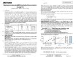
BioVision For research use only rev. 02/13 MMP-3 Activity Fluorometric Assay Kit (Catalog #K783-100; 100 assays; Store kit at -20oC) II. MMP-3 Assay Buffer MMP-3 Substrate Mca Standard (1mM) MMP-3 Positive Control (lyophilized) IV. K783-100 25 ml 200 µl 20 µl 1 vial Cap Code WM Red Yellow Green Part Number K783-100-1 K783-100-2 K783-100-3 K783-100-4 Storage and Handling: Store the kit at -20°C, protect from light. Allow Assay Buffer to warm to room temperature before use. Briefly centrifuge vials before opening. Read the entire protocol before performing the assay. Reagent preparation: MMP-3 Positive Control: Reconstitute with 100 µl assay buffer. Aliquot and store at -20oC. Avoid repeated freeze/thaw cycles. Use within one week. V. Add 50 µl of the Reaction Mix to each well containing the samples and positive controls. Mix well. 4. Measurement: Read Ex/Em = 325/393 nm R1 at T1. Read R2 again at T2 after incubating the reaction at room temperature for 60 min (or incubate longer time if the sample activity is low), protect from light. The RFU of fluorescence generated by hydrolyzes of the substrate is ∆RFU = R2 – R1. It is recommended to read kinetically to choose the R1 and R2 values that fall within the linear range of the Standard Curve. 5. Calculation: Subtract the 0 Standard from the Standard readings. Plot the Standard Curve and apply the ∆RFU to the standard curve to get B nmol of Mca (amount of unquenched Mca generated between T1 and T2). Kit Contents: Components III. 3. Reaction Mix: Mix enough reagents for the number of assays to be performed. For each well, prepare a total 50 µl Reaction Mix: 48 µl MMP-3 Assay Buffer 2 µl MMP-3 substrate MMP-3 Assay Protocol: 1. Standard Curve Preparation: Mix 5 µl 1mM MMP Mca Standard with 495 µl MMP-3 Assay Buffer to generate a 10 µM standard solution. Add 0, 10, 20, 30, 40, 50 µl to each well individually. Adjust to a final volume of 100 µl/well with Assay Buffer to generate 0, 0.1, 0.2, 0.3, 0.4, 0.5 nmol/well of Mca Standard. Read fluorometrically at Ex/Em=325/393 nm. 2. Sample Preparations: Tissues (50 mg) or cells (1×10⁶) can be homogenized in ~ 200 µl ice-cold MMP-3 Assay Buffer then centrifuged to remove insoluble material at 13,000 g, 10 minutes. Serum sample can be directly diluted in the MMP-3 Assay Buffer. Prepare test samples of up to 50 µl/well with MMP3 Assay Buffer in a 96-well plate. We suggest testing several doses of your sample to make sure the readings are within the Standard Curve range. . For Positive Control use 5-10 µland adjust well volume to 50 µl with Assay Buffer. BioVision Incorporated 155 S. Milpitas Boulevard, Milpitas, CA 95035 USA MMP-3 Activity = ۰ ሺ܂ି܂ሻ× ܄ ×Sample Dilution Factor = nmol/min/ml = mU/ml Where: B is the Mca amount from MMP Mca Standard Curve (in nmol). T1 is the time of the first reading (R1) (in min). T2 is the time of the second reading (R2) (in min). V is the pretreated sample volume added into the reaction well (in ml). Unit Definition: One unit is defined as the amount of enzyme that will generate 1.0 µmol of unquenched Mca per minute at room temperature. MMP-3 Mca Standard MMP-3 Positive Control 4000 2000 y = 8.964x - 73 R² = 0.9974 0 RFU Ex/Em=325/393nm Introduction: The matrix metalloproteinase-3 (MMP-3, stromelysin-1) exhibits a number of activities that would make it a particularly good tumor promoter. Like several other MMPs, MMP-3 was first cloned and later recloned as a cancer-specific gene. In addition to degrading numerous extracellular matrix components, MMP-3 can activate gelatinase B, the collagenases and several serpin-type serine proteinase inhibitors. Moreover, it can release a number of cell surface molecules, including E-cadherin, a known contributor to cancer development. In BioVision’s MMP-3 Assay Kit, MMP-3 hydrolyzes a specific FRET substrate to release the quenched fluorescent group Mca, which can be detected fluorometrically at Ex/Em = 325/393 nm. The kit provides a rapid, simple, sensitive and reliable test which can also be used as a high throughput assay of MMP-3 activity. The assay sensitivity is < 50 µU. This kit can be used with our MMP-3 inhibitor, GM6001 (Biovision #1799) as a control. In addition, we also offer a human recombinant MMP-3 enzyme (Biovision #7783) and a MMP-3 inhibitor Screening Kit (Biovision #K793-100), separately. RFU (Ex/Em=325/393nm) I. Positive Control 5000 4000 3000 Back Ground 2000 0 100 200 300 400 500 0 Mca Standard(pmol) 20 40 Time (min) 60 RELATED PRODUCTS: Human recombinant proteins: MMP-1, -2, -3, -8, -9, -11, -12, -13 MMP antibodies to: MMP-1, -2, -3, -8, -9, -11, -12, -13, -17, -19 MMP blocking peptides to: MMP-3, -8, -9, -11, -12 MMP-3 Inhibitor GM6001 MMP-3 Inhibitor Screening Kit FOR RESEARCH USE ONLY! Not to be used on humans. Tel: 408-493-1800 | Fax: 408-493-1801 www.biovision.com | [email protected] Page 1 of 2 BioVision rev. 02/13 For research use only GENERAL TROUBLESHOOTING GUIDE: Problems Cause Solution Assay not working • Use of ice-cold assay buffer • Assay buffer must be at room temperature • Omission of a step in the protocol • Refer and follow the data sheet precisely • Plate read at incorrect wavelength • Check the wavelength in the data sheet and the filter settings of the instrument • Fluorescence: Black plates (clear bottoms) ; Luminescence: White plates ; Colorimeters: Clear plates • Use of a different 96-well plate Samples with erratic readings Lower/ Higher readings in Samples and Standards Readings do not follow a linear pattern for Standard curve Unanticipated results • Use of an incompatible sample type • Refer data sheet for details about incompatible samples • Samples prepared in a different buffer • Use the assay buffer provided in the kit or refer data sheet for instructions • Cell/ tissue samples were not completely homogenized • Use Dounce homogenizer (increase the number of strokes); observe for lysis under microscope • Samples used after multiple free-thaw cycles • Aliquot and freeze samples if needed to use multiple times • Presence of interfering substance in the sample • Troubleshoot if needed • Use of old or inappropriately stored samples • Use fresh samples or store at correct temperatures until use • Improperly thawed components • Thaw all components completely and mix gently before use • Use of expired kit or improperly stored reagents • Always check the expiry date and store the components appropriately • Allowing the reagents to sit for extended times on ice • Always thaw and prepare fresh reaction mix before use • Incorrect incubation times or temperatures • Refer datasheet & verify correct incubation times and temperatures • Incorrect volumes used • Use calibrated pipettes and aliquot correctly • Use of partially thawed components • Thaw and resuspend all components before preparing the reaction mix • Pipetting errors in the standard • Avoid pipetting small volumes • Pipetting errors in the reaction mix • Prepare a master reaction mix whenever possible • Air bubbles formed in well • Pipette gently against the wall of the tubes • Standard stock is at an incorrect concentration • Always refer the dilutions in the data sheet • Calculation errors • Recheck calculations after referring the data sheet • Substituting reagents from older kits/ lots • Use fresh components from the same kit • Measured at incorrect wavelength • Check the equipment and the filter setting • Samples contain interfering substances • Troubleshoot if it interferes with the kit • Use of incompatible sample type • Refer data sheet to check if sample is compatible with the kit or optimization is needed • Sample readings above/below the linear range • Concentrate/ Dilute sample so as to be in the linear range Note: The most probable list of causes is under each problem section. Causes/ Solutions may overlap with other problems. BioVision Incorporated 155 S. Milpitas Boulevard, Milpitas, CA 95035 USA Tel: 408-493-1800 | Fax: 408-493-1801 www.biovision.com | [email protected] Page 2 of 2