1
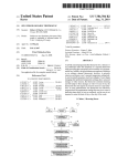
US008289378B2
(12) United States Patent
Soto-Thompson et a].
(54)
(75)
HIGH RESOLUTION DIGITAL VIDEO
COLPOSCOPE WITH BUILT-IN POLARIZED
LED ILLUMINATION AND COMPUTERIZED
CLINICAL DATA MANAGEMENT SYSTEM
Inventors: Marcelo Esteban Soto-Thompson,
Honolulu, HI (US); Andrew Beaumont
Oct. 16, 2012
OTHER PUBLICATIONS
D. G. Ferris, et al., “Modern ColposcopyiTextbook and Atlas,” pp.
250-662, American Society for Colposcopy and Cervical Pathology
Kendall/ Hunt Publishing Co., Dubuque, Iowa.
B.S. Apgar et al., “Colposcopy: Principles and Practice,” pp. 115
132, W.B. Saunders Company, Philadelphia, PA 2002.
Subject to any disclaimer, the term of this
patent is extended or adjusted under 35
U.S.C. 154(b) by 947 days.
multispectral digital colposcope,” Gynecologic Oncology 107, S21
Gustafsson, Honolulu, HI (US)
(73) Assignee: STI Medical Systems, LLC, La Jolla,
CA (US)
Notice:
US 8,289,378 B2
K. T. Schomacker et al., Novel optical detection system for in vivo
identi?cation and localization of cervical intra-epithelial neoplasia, J.
Biomed Optics 11(3), pp. 034009-1 to 034009-02, 2006.
J .E. Kendrick et al., “LUMATM Cervical Imaging System,” Expert
Rev. Med. Devices 4(2), pp. 121-129, 2007.
E. Hecht., “Optics,” pp. 270-332, Addison-Wesley, 2nd edition 1987.
Welch Allyn, “Video Colposcope,” Directions for Use, 2007.
S. Nakappan et al., “Methodology of real time quality control for the
Whitesell, Honolulu, HI (US); Ulf Peter
(*)
(10) Patent N0.:
(45) Date of Patent:
S222, 2008.
(21) Appl. N0.: 12/291,s90
(22)
Filed:
Acgih, “Electromagnetic Radiation and Fields,” pp. 13-43, 2007,
Cincinati, OH.
Nov. 14, 2008
* cited by examiner
(65)
Prior Publication Data
US 2010/0026785 A1
Feb. 4, 2010
Related US. Application Data
(60)
Provisional application No. 61/137,684, ?led on Aug.
1, 2008.
(51)
Int. Cl.
Primary Examiner * Tonia L Dollinger
Assistant Examiner * Adam Cooney
(74) Attorney, Agent, or Firm * Martin E. Hsia
(57)
H04N132/02
(52)
(58)
This invention uses LEDs and cross-polarization to produce
(2006.01)
US. Cl. ............................. .. 348/47; 348/57; 348/58
Field of Classi?cation Search ................... .. 348/47
See application ?le for complete search history.
(56)
ABSTRACT
References Cited
bright, high-resolution digital images, both With and Without
glint (Which adversely affects the clarity of standard colpo
scopic images), as Well as streaming video at loWer resolu
tion. The invention alloWs for deeper layers of the tissue to be
more e?iciently visualized at multiple magni?cations,
thereby enhancing the invention’s diagnostic capabilities,
U.S. PATENT DOCUMENTS
5,929,443 A
6,766,184 B2
2006/0184040 A1*
2006/0215406 A1*
7/1999 Alfano et al.
and it includes a focusing subsystem and a computerized data
management system to archive and annotate still image data.
7/ 2004 Utzinger et al.
8/2006
Keller et al.
9/2006
Thrailkill .................... .. 362/252
................ .. 600/476
9 Claims, 9 Drawing Sheets
US. Patent
0a. 16, 2012
Sheet 1 019
FIG. 1
US 8,289,378 B2
US. Patent
0a. 16, 2012
Sheet 2 of9
US 8,289,378 B2
34
49
36
FIG. 2A
34
49
36
FIG. 2B
34
49
36
FIG. 2C
US. Patent
0a. 16, 2012
Sheet 3 of9
US 8,289,378 B2
FIG. 3A
34
> 49
36
FIG. 3B
34
49
36
FIG. 3C
US. Patent
0a. 16, 2012
US 8,289,378 B2
Sheet 4 0f 9
49
FIG. 4A
LA 4
49
FIG. 4B
4
49
36
FIG. 4C
US. Patent
0a. 16, 2012
Sheet 5 of9
FIG. 5A
FIG. 5B
US 8,289,378 B2
US. Patent
Oct. 16, 2012
Sheet 6 0f 9
US 8,289,378 B2
86
85
83
FIG. 6A
84
FIG. 6B
US. Patent
0a. 16, 2012
Sheet 7 of9
US 8,289,378 B2
2O
4/’
34
I‘
24
4/’
FIG. 7A
34
22
FIG. 7B
US. Patent
0a. 16, 2012
Sheet 8 of9
US 8,289,378 B2
20, 24
FIG. 8A
20, 24
IG. 8B
IG. 8C
US. Patent
0a. 16, 2012
Sheet 9 of9
3
6
US 8,289,378 B2
‘_-"-’
10
¢—-——!
11
¢——>
12
<———&
13
I """ ".
*"--":
FIG. 9
14
|
US 8,289,378 B2
1
2
HIGH RESOLUTION DIGITAL VIDEO
COLPOSCOPE WITH BUILT-IN POLARIZED
LED ILLUMINATION AND COMPUTERIZED
CLINICAL DATA MANAGEMENT SYSTEM
addition, different colored ?lters are often used to accentuate
blood vessel patterns that cannot be easily seen by using
regular White light.
Although the standard colposcopic exam and regular
screening have led to dramatic decreases in the overall inci
dence of cervical cancer, neW technologies can further
This application claims priority to US. provisional patent
application No. 61/137,684 for “CervicalMD C30 Imaging
Subsystem”, ?led on Aug. 1, 2008.
enhance the sensitivity and speci?city of currently accepted
colposcopic practices. Digital imaging is one such technol
ogy that can revolutioniZe medical imaging and enables
sophisticated computer programs to assist the physician With
CAD (Computer-Aided-Detection or Computer-Aided-Di
agnosis). The combination of digital imaging and CAD could
have a direct impact on improving Women’s health, and
TECHNICAL FIELD
This invention relates to medical imaging and, more spe
ci?cally, to a device and process that suppresses specular
decrease the associated cost, by automatically identifying
CIN in real-time With high sensitivity and speci?city. This
re?ection (glint) through cross-polarization, producing bright
cross-polarized and parallel-polarized images at multiple
magni?cations in real-time, thereby enhancing the visualiZa
Would mean feWer false-positive biopsies, or ultimately,
elimination of biopsies. A CAD system operating as an
tion of diagnostically relevant features Within a subject (such
adjunct to colposcopy could minimize the high variability
among colposcopists and enable consistent, higher standards
as organs or tissue). The device also preferably includes a
focusing subsystem, and a computeriZed data management
20
system for archival purposes and for the annotation of digital
data.
for accuracy. A product realiZation Where a CAD system is
incorporated into a loW-cost device, creating in effect a
machine expert colposcopist, Would have the potential of
increasing the availability and cost-effectiveness of screening
in developing countries.
BACKGROUND ART
25
Although this invention is being disclosed in connection
Digital imaging provides a means for implementing a com
puteriZed clinical data management system. This data man
With cervical cancer, it is applicable to many other areas of
agement system could provide management, display, and
medicine. Uterine cervical cancer is the second most common
cancer in Women WorldWide, With nearly 500,000 neW cases
annotations of the acquired digital data, as Well as automation
of the Work?oW associated With colposcopy. The system
and over 270,000 deaths annually (IARC, “Globocan 2002
database,” International agency for research in cancer, 2002,
30
alloW the use of electronic patient data records, and interface
and integrate With standard systems for handling, storing,
printing and transmitting information in medical imaging,
incorporated herein by reference). Because invasive disease
is preceded by pre-malignant Cervical Intraepithelial Neopla
sia (CIN), if detected early and treated adequately, cervical
cancer can be universally prevented (D. G. Ferris, J. T. Cox,
D. M. O’Connor, V. C. Wright, and J. Foerster, Modern Col
could simplify the administration of patient data and history,
such as DICOM (Digital Imaging and Communication in
35
Medicine). DICOM is a standard for handling, storing, print
ing, and transmitting information in medical imaging. It
poscopy. TexlbookandAllas, pp. 1-699,American Society for
includes a ?le format de?nition and a network communica
Colposcopy and Cervical Pathology, 2004, incorporated
herein by reference). Colposcopy is the primary diagnostic
tions protocol. The communication protocol is an application
protocol that uses TCP/IP (the standard intemet protocol) to
method in the United States to detect CIN and cancer folloW
ing an abnormal cytological screen (Papanicolaou smear or
40
communicate betWeen systems. DICOM ?les can be
exchanged betWeen tWo entities that are capable of receiving
pap smear). The purpose of a colposcopic examination is to
image and patient data in DICOM format. Digital imaging
identify and rank the severity of lesions, so that biopsies
representing the highest-grade abnormality can be taken, if
necessary. The biopsies are then microscopically evaluated
alone is also a pre-requisite for telemedicine applications,
further increasing the availability of screening and detection
45
For the colposcopic exam, an optical colposcope is typi
cally used, and has been used for such purposes for almost 80
years. A colposcope is a binocular microscope With a built in
White light source and objective lens attached to a support
ery upon Which a CAD system operates must be of high visual
quality. One factor contributing to poor cervical imagery is
specular re?ection (glint), Which is perfect, mirror-like
50
mechanism (B. S. Apgar, BrotZman, G. L. and SpitZer, M.,
Colposcopy: Principles and Practice, W.B. Saunders Com
pany: Philadelphia, 2002, incorporated herein by reference).
At loW levels of magni?cation, (comparable to a circular ?eld
of vieW of approximately 50 to 100 mm) the entire vagina and
cervix can be visualiZed and this setting is typically used to
obtain a general impression of the surface structure and archi
tecture. Medium magni?cations (comparable to a circular
55
60
mirror itself. Because this color information may be impor
tant in detecting cancer precursors, reducing the amount of
glint in an image is helpful in producing high-quality images
vagina and the cervix. These higher magni?cations are often
necessary to detect and identify certain vascular patterns
for diagnostic purposes. HoWever, it is not alWays desirable to
eliminate all the glint from an image because an image of a
tissue or organ that contains glint may look more natural and
indicative of the presence of more advanced pre-cancerous or
cancerous lesions. During the colposcopic exam, acetic acid
and iodine solutions are usually applied to the surface of the
cervix to improve the visualiZation of abnormal areas. In
re?ection of light from a surface, in Which light from a single
incoming direction (i.e., a ray) is re?ected into a single out
going direction. Glint is undesirable because it effectively
eliminates color information in an image, and also results in
the introduction of artifacts (misrepresentations of tissue
structures) in the image. Glint eliminates color information
because its mirror-like re?ection shoWs the color of the light
source, and not of the underlying tissue, much as a mirror
shoWs the color of a re?ected light, and not the color of the
?eld of vieW of approximately 15-30 mm) and high magni?
cations (comparable to a circular ?eld of vieW of approxi
mately 5-15 mm) are utiliZed for detailed analysis of the
in rural areas and developing countries.
In order to reliably assess colposcopic features, the imag
by a pathologist based on the morphology of the tissue.
65
three-dimensional. In addition, colposcopists analyZe the
glint patterns on the cervix to assess the surface contour of
lesions, an important feature used to evaluate lesion severity.
US 8,289,378 B2
4
3
as vascular patterns, bright light sources are helpful in pre
The prior art describes a number of Ways to reduce the
serving the clarity of an image.
in?uence of or eliminate glint. Physicians using optical col
HoWever, these bright light sources must not exceed
poscopes can change their ?eld-of-vieW and/or the lightning
conditions to either move the glint to different parts of the
cervix and maintain the region of interest glint-free, or to a
acceptable thresholds for patient exposure to ultraviolet (UV)
and infrared (IR) radiation (as described by the American
Conference on Governmental Industrial Hygienists (ACGIH)
in, Threshold Limit Values (TLVs) for Chemical Substances
large extent eliminate the glint completely. Another method
involves using multiple light sources directed at different
angles toWards an object (see for example K. T. Schomacker,
T. M. Meese, C. Jiang, C. C. Abele, K. Dickson, S. T. Sum,
and R. F. FleWelling, Novel optical detection system for in
vivo identi?cation and localization of cervical intra-epithelial
neoplasia, J. Biomed Optics 11(3), 034009-1-12, 2006, and J.
and Physical Agents and Biological Exposure Indices (BEIs),
Signature Publications, 2008, incorporated herein by refer
ence). Exposure to UV radiation has the potential of acute
adverse health effects such as erythema and photokeratitis,
and can cause DNA damage in the cells. Presently, the expo
sure to UV radiation is minimized, if not completely elimi
nated, by employing a UV blocking ?lter in the light source
E. Kendrick, W. K. Huh, and R. D. Alvarez, LUMATM Cer
vical Imaging System, Expert Rev. Med. Devices 4(2), 121
beam path prior to the light being available to human vieWing
129, 2007, incorporated herein by reference). By illuminating
and exposure (such as described in Welch Allyn Video Col
the cervix at different angles and acquiring several images,
the position of the glint on the surface of the cervix is different
betWeen the different images, and the images can be com
bined to create a glint-free combined image.
The use of polarization ?lters, each of Which functions in
the same Way as a pair of polarized sunglasses, is another glint
20
ogy 107, S21-S222, 2008, incorporated herein by reference).
reducing or eliminating technique Well knoWn in the art (see
for example, E. Hecht., Optics, Addison-Wesley, 1st edition
1972, 2nd edition 1987, 3rd edition 1997, 4th edition 2001).
25
The polarization ?lter method utilizes one polarization ?lter
placed at the light source and another ?lter rotated to approxi
blocking ?lter, the heat generated by the IR radiation puts
this cross polarization scheme, the re?ections from the sur
glint-free object or image. Cross-polarization is employed,
for example, in commercially available colposcopes (Welch
Allyn Video Colposcope, User’s manual, 2007, incorporated
herein by reference), and research colposcope systems (such
Many bright light sources also contain a large amount of IR
Which is essentially excess heat. Similar to UV radiation, the
exposure to IR radiation is minimized or eliminated by the
utilization of an IR blocking ?lter. Although the heat exposure
of the user or patient is minimized or eliminated by an IR
mately 900 positioned in front of the detector. By applying
face of the object under study are substantially minimized, if
not completely eliminated, and the end result is an essentially
poscope, User’s manual, 2007, incorporated herein by refer
ence) and S. Nakappan, S-Y. Park, D. Serachitopol, R. Price,
M. Cardeno, S. Au, N. Mackinnin, C. MacAulay, M. Follen,
and B. M. Pikkula, Methodology of real time quality control
for the multispectral digital colposcope, Gynecologic Oncol
30
stress on the optical and mechanical components of the light
source assembly and may signi?cantly decrease the lifetime
of the components, as Well as of the light source itself.
Further, some bright light sources require a long start-up
time before the output intensity is stable. This means there is
a Wait time before the device can be utilized in a clinical
35
examination, possibly decreasing the cost-effectiveness of
as described in S. Nakappan, S-Y. Park, D. Serachitopol, R.
such a device.
Price, M. Cardeno, S. Au, N. Mackinnin, C. MacAulay, M.
Follen, and B. M. Pikkula, Methodology of real time quality
A third factor contributing to poor cervical imagery is
non-uniform lighting, Which can lead to non-uniformity of
control for the multispectral digital colposcope, Gynecologic
Oncology 107, S21-S222, 2008, incorporated herein by ref
40
diagnosis based on the resulting image.
erence). In these systems, the polarization ?lters are typically
actuated by computer controlled rotating ?lter Wheels or
manually operated rotating ?lter holders, either on the light
source side or detection side, or both. Being able to remove or
rotate the polarization ?lters, alloWs for the acquisition of
45
both cross-polarized imagery Without glint and regular imag
ery With glint. A drawback of using manually operated or
computer controlled mechanical assemblies to sWitch or
rotate the polarization ?lters is the inevitable Wear and tear
and ultimate failure of these units over time. In addition,
mechanical sWitch or rotational devices Will introduce a delay
50
betWeen the image vieWing or capture of cross polarized and
regular imagery. During this delay, signi?cant movement of
the colposcope and/ or the patient can occur. This movement
can make it extremely dif?cult to register (align) images and
track diagnostically important features, such as blood vessels
of varying sizes. This is especially true for a fully automated
55
body surface Which includes recording at a ?rst time a ?rst
multispectral digital image of the surface including the
spectral digital image of the surface including the region, and
60
comparing the ?rst and the subsequent images. Also, such a
method in Which the ?rst and subsequent images are high
magni?cation images, and further including recording loW
magni?cation images that include the high magni?cation
respect to imaging systems incorporating polarizing compo
nents, is that polarization inherently results in a loss of light.
Because brightness (or intensity) is an integral part of the
achievable contrast (i.e. the difference in visual properties
that make an object distinguishable from other objects and the
background) in the captured images, and because the contrast
of an image is important in detecting cancer precursors such
The folloWing patents may be considered relevant to the
?eld of the present invention:
US. Pat. No. 4,979,498 to Oneda et al., incorporated herein
by reference, discloses a video cervicoscope system for the
examination of the cervix comprising: a rigid, elongated
tubular member having a light guide; imaging means at the
distal end of said tubular member, a disposable, light-trans
mitting, sleeve disposed about the distal end of said tubular
member; and transmitting means to transmit an image vieWed
by said imaging means proximally to a control box Wherein
said image is received and stored.
US. Pat. No. 5,836,872 to Kenet et al., incorporated herein
by reference, discloses a method for monitoring a region of a
region, recording at a subsequent time a subsequent multi
CAD system that does not rely on human direction or inter
vention.
Another factor contributing to poor cervical imagery, With
the brightness (or intensity) in the resulting image. Non
uniforrnity of brightness impairs the potential accuracy of any
images. Also disclosed is a method for forming a diagnosti
cally useful classi?cation of pigmented skin lesions, using
65
such a method to construct a database containing quantita
tively extracted selected features from images recorded from
a plurality of skin lesions, and correlating the features from
US 8,289,378 B2
5
6
each such lesion in the database With the medical history of
the skin lesion from Which the image Was recorded. Further,
a method for diagnosis of a premelanomatous or early mela
nomatous condition includes using the method for character
a desired object. Re?ected light from the object entering the
housing through the glass cover is passed through a second
polarizer, Which is adjustably mounted in the barrel portion of
the housing and Which is preferably oriented to pass depolar
izing a surface region including the lesion and comparing the
ized light emitted from an illuminated object, and is then
imaged by optics onto a black and White CCD (charged
features of the lesion so obtained With the features in a data
base obtained from a number of skin lesions including lesions
knoWn to be premelanomatous or early melanomatous, or
coupled device) detector (camera). The optics may include a
lens that is disposed Within the barrel portion and is adjustably
spaced relative to the CCD detector. The detector is coupled
classifying the features of the lesion according to the diag
nostically useful classi?cation of pigmented skin lesions.
U.S. Pat. No. 5,929,443 to Alfano et al., incorporated
herein by reference, discloses a method and apparatus for the
imaging of objects based on the polarization and depolariza
to a Wireless transmitter mounted in the housing, the trans
mitter transmitting the output from the detector to a remotely
located Wireless receiver. The Wireless receiver is coupled to
a computer, Which then processes the output from the detec
tor. The processed output is then displayed on a display. The
tion of light. In one embodiment, a surface of a turbid medium
is imaged by illuminating the surface of the turbid medium
With light, Whereby light is backscattered from the illumi
nated surface of the turbid medium, detecting a pair of
complementary polarization components of the backscat
tered light, and forming an image of the illuminated surface
using the pair of complementary polarization components.
The illuminating light is preferably polarized (e.g., linearly
display may be remotely situated for remote expert diagnosis.
U.S. Pat. No. 6,766,184 to Utzinger et al., incorporated
herein by reference, discloses methods and apparatus for
generating multispectral images of tissue. The multispectral
images may be used as a diagnostic tool for conditions such as
20
polarized, circularly polarized, elliptically polarized), Where,
for example, the illuminating light is linearly polarized, the
pair of complementary polarization components are prefer
ably the parallel and perpendicular components to the polar
ized illuminating light, and the image is formed by subtract
ing the perpendicular component from the parallel
cervical cancer detection and diagnosis. Primary radiation is
produced With an illumination source. The primary radiation
is ?ltered to select a ?rst Wavelength and a ?rst polarization.
Tissue is illuminated With the ?ltered primary radiation to
generate secondary radiation, Which is ?ltered to select a
25
second Wavelength and a second polarization. The ?ltered
secondary radiation is collected With a detector, and a plural
ity of multispectral images of the tissue is generated accord
component, by taking a ratio of the parallel and perpendicular
ing to different combinations of ?rst and second Wavelengths
and ?rst and second polarizations With an analysis unit in
components or by using some combination of a ratio and
operable relation With the detector. Apparatus utilizing the
invention include endoscopes and colposcopes.
difference of the parallel and perpendicular components.
U.S. Pat. No. 5,989,184 to Blair, incorporated herein by
reference, discloses an apparatus for digital colposcopy and
videography Which comprises a digital imaging camera that
is operably coupled to the optical path of the digital colpo
30
scope by means of a beam splitter so that a digital image of the
35
U.S. Patent Application Publication No. 2006/ 0141633 to
Balas, incorporated herein by reference, discloses a method
and an apparatus for the in vivo, non-invasive, early detection
of alterations and mapping of the grade of these alterations,
caused by the biochemical and/or the functional characteris
tics of epithelial tissues during the development of tissue
atypias, dysplasias, neoplasias and cancers. The method is
40
poral and spectral alterations in the characteristics of the light
cervico-vaginal tissue can be captured. The digital imaging
camera and digital colposcope are mounted to one end of an
articulating arm of the apparatus. Digital processing means is
operably connected to the digital imaging camera to create a
digital image. The digital processing means is housed in a
stand of the assembly.
U.S. Pat. No. 6,277,067 to Blair, incorporated herein by
reference, discloses a method and portable apparatus for the
based on the simultaneous measurement of the spatial, tem
that is re-emitted from the tissue under examination, as a
result of a combined tissue excitation With light and special
chemical agents. The topical or systematic administration of
visual examination and grading of cervical epithelium by
means of a hand-held colposcopy assembly capable of pro
45
these agents results in an evanescent contrast enhancement
betWeen normal and abnormal areas of tissue. The apparatus
ducing a digital image of the cervix. The apparatus enables
real-time imaging and archiving of images of the entire cervix
enables the capturing of temporally successive imaging in
for the purpose of detecting cancerous and pre-cancerous
measured data, the characteristic curves that express the
agent-tissue interaction kinetics, as Well as numerical param
eters derived from these data, are determined in any spatial
tissue, and by virtue of computerized image processing, sug
gests an objective diagnosis of the cervical epithelium by
one or more spectral bands simultaneously. Based on the
50
point of the examined area. Mapping and characterization of
means of a loW cost, portable, hand-held digital colposcope.
U.S. Pat. No. 6,587,711 to Alfano et al., incorporated
herein by reference, discloses an apparatus for examining an
object, such as skin, mucosa and cervical tissues, for the
the lesion are based on these parameters.
U.S. Patent Publication No. 2006/ 0184043 to Tromberg et
al., incorporated herein by reference, discloses an improve
55
ment in a method for quantitative modulated imaging to per
form depth sectioned re?ectance or transmission imaging in a
turbid medium, such as human or animal tissue. The method
is directed to the steps of encoding a periodic pattern of
illumination, preferably With a ?uorescent excitation Wave
60
portion of the housing. A manually-operable sWitch for con
length When exposing a turbid medium to the periodic pat
tern, to provide depth-resolved discrimination of structures
Within the turbid medium; and reconstructing a non-contact
trolling actuation of each of the four LED’s is accessible on
three dimensional image of the structure Within a turbid
purpose of detecting cancer and precancerous conditions. In
one embodiment, the apparatus includes a gun-shaped hous
ing having a handle portion and a barrel portion. The front end
of the barrel portion is open, and a glass cover is mounted
therein. Red, green, blue, and White LED’s are disposed
Within the handle portion of the housing, and are electrically
connected to a battery and are also disposed Within the handle
the handle portion of the housing. An optical ?ber is disposed
inside the housing and is used to transmit light from the four
LED’s through a ?rst polarizer disposed in the barrel portion
of the housing and then through the glass cover to illuminate
medium. As a result, Wide ?eld imaging, separation of the
65
average background optical properties from the heterogene
ity components for a single image, separation of super?cial
features from deep features based on selection of spatial
US 8,289,378 B2
8
7
frequency of illumination, or qualitative and quantitative
structure, function and composition information, is extracted
from spatially encoded data.
The present invention is a device that preferably creates
pairs of clear images, a glint-free image (cross-polarized
image) and an image With glint (parallel-polarized image), at
multiple magni?cations. The term cross-polarized (XP)
US. Patent Application No. 2006/0215406 to Thrailkill,
refers to When a ?rst polarization orientation is perpendicular
to a second polarization orientation, Whereas the term paral
lel-polarized (PP) refers to When a ?rst polarization orienta
tion is parallel to a second polarization orientation. In the
present invention, PP can also mean singly-polarized, Where
there is only one polarization orientation, or unpolarized. The
XP and PP images can be used in conjunction With each other
for diagnostic purposes. For example, XP and PP images can
be registered (aligned) and then faded into each other to aid a
clinician in cancer detection (as described in co-pending,
incorporated herein by reference, discloses a medical diag
nostic instrument, Which could be a colposcope for examin
ing cervical tissue, and includes a light source comprising an
annular array of high intensity light emitting diodes (LEDs).
The LED array includes a central access opening Which pro
vides vieWing access for the colposcope optical components
to the illumination site. The array includes a plurality of sets
of LEDs, With each set including a red, blue and green emit
ting LED. The intensities of the red, blue and green LEDs,
respectively, are controllable With a controller to continu
commonly assigned US. patent application Ser. No. 12/228,
ously vary or tune the spectral characteristics of the illumi
nation from the light source. Selected color mixes can be
stored in a memory for later retrieval.
US. Patent Publication No. 2007/0213590 to Squicci
298, entitled “A Method of Image Manipulation to Fade
BetWeen TWo Images” ?led Aug. 11, 2008, incorporated
herein by reference). Fading betWeen the tWo images alloWs
a clinician to detect important features that may be masked by
narini, incorporated herein by reference, discloses a portable
multi-functional endoscopic device and method for use in the
20
examination of tissue to permit diagnostic, therapeutic or
glint, While at the same time retaining to a desired extent the
natural and three-dimensional shape of tissue or an organ
(such as the cervix) in the image.
To suppress glint, the ?rst preferred embodiment prefer
anatomical assessment data to be transmitted, recorded, or
analyzed. The device includes a base unit sized and con?g
ably employs cross-polarization, Which alloWs for deeper
ured to be held in a human hand to permit functional and
directional control of the device, an interchangeable head
assembly sized and con?gured to be inserted into an ori?ce
being removably connectable to the base unit, and an in?at
able tissue stabilizer disposed external to a distal end of the
device. In preferred aspects, the endoscopic device has an
25
image sensor, light source, lens, air pump, and Working tools.
30
layers of tissue to be visualized at multiple magni?cations,
thereby further enhancing the invention’ s diagnostic capabili
ties. To produce a brighter and more uniform illuminated
surface With a denser light emission than typical ?uorescent
and incandescent light sources, the light source in this
embodiment preferably includes tWo sets of multiple light
emitting diodes (LEDs), Which are preferably turned on and
US. Patent Application No. 2008/ 0049997 to Chin, incor
porated herein by reference, discloses an image enhancement
off rapidly by electronically changing the operating current or
system that includes a data source Which provides image data
voltage, to directly and uniformly illuminate a ?eld of vieW.
The light from the LEDs is preferably polarized through an
illumination polarizer before reaching the ?eld of vieW.
of an object, enhancement data storage including image
enhancement information, an image enhancement unit con
35
The present invention’s use of LEDs offers several ben
e?ts. LEDs, unlike ?uorescent and incandescent lights, are
solid-state components, alloWing for faster sWitching on and
?gured to enhance the image data based on the image
enhancement information, and a color display con?gured to
display a monochrome image representing the enhanced
image data on a screen thereof. The enhanced image data may
include a gray level scale of at least 32 bits per pixel.
US. Patent Application Publication No. 2005/004365 to
40
off, so that sWitching betWeen parallel-polarized image
acquisition and cross-polarized image acquisition can be
achieved more easily and quickly. The invention preferably
Zelenchuk, incorporated herein by reference, discloses a sys
uses ?xed illumination polarizers (IP) in front of the tWo sets
tem and method for the in situ discrimination of healthy and
of LEDs, instead of mechanically moving polarizing ?lters.
Rapidly sWitching betWeen the LEDs (rapidly alternating
diseased tissue. A ?beroptic probe is employed to direct ultra
violet illumination onto a tissue specimen and to collect the
?uorescent response radiation. The response radiation is
observed at three selected Wavelengths, one of Which corre
sponds to an isosbestic point. In one example, the isosbestic
point occurs at about 431 nm. The intensities of the observed
signals are normalized using the 431 nm intensity. A score is
45
50
determined using the ratios in a discriminant analysis. The
tissue under examination is resected or not, based on the
diagnosis of disease or health, according to the outcome of the
discriminant analysis.
55
DISCLOSURE OF THE INVENTION
The present invention addresses the problem of glint inher
ent in performing a standard colposcopic exam and addresses
the limitations of using mechanical ?lter Wheels or rotating
?lter assemblies, the brightness of the light source, the health
aspects of exposure to UV and IR hazards, and the extended
start-up time of most bright light sources by employing a
60
non-mechanical, non-moving (?xed or stationary) polariza
tion assembly With an electronically sWitchable light source
each set of LEDs on and off) also reduces the in?uence from
patient movement betWeen successive images, alloWing for
better registration (alignment) betWeen successive images
and improving the possibility of detection of diagnostically
important features. An LED assembly is also less expensive
65
and more compact than the typical illumination sources using
either ?uorescent or incandescent lighting With bundles of
?ber optic cables, and LEDs are also more reliable than using
either ?uorescent or incandescent lights because they are
rated for approximately 50,000 hours of use and one million
on/off cycles. As a comparison, the rated lifetimes for ?uo
rescent or incandescent lights are typically less than 10,000
hours With many having a typical lifetime of only 1000 hours.
LEDs also generate less heat than ?uorescent and incandes
cent lights. Therefore, less cooling and less thermal insulation
are required for the device housing, alloWing for smaller
poWer suppliesie.g., smaller fans that produce less noise
and vibration for patient and operator comfort. The thermal
e?iciency of LEDs also reduces heat, Which generates ther
mal expansion of the device’s mounts and can degrade the
optical quality of the images. LEDs also require less poWer
system based on high-intensity light emitting diodes (LEDs),
and are more e?icient in converting electrical poWer con
Which do not to generate any UV or IR radiation.
sumption to visible light, ultimately reducing the poWer con
US 8,289,378 B2
9
10
sumption of the device, and enabling battery operation in
cervical tissue) in one direction (e.g., toWards one of the
remote areas, if necessary. Further, light emitted by an LED is
cameras), diffuse light refers to light that is scattered in all
also emitted over a broad ?at area unlike conventional incan
directions and is unpolarized. It is Well knoWn in the art that
descent and ?uorescent lights, thus providing for more uni
form illumination.
unpolarized light can be described as a combination of both
PP and XP light. Because a camera captures only that portion
of the diffuse light that is re?ected in the camera’s direction,
the diffuse re?ected light does not have the same visually
Moreover, as patient safety is of utmost importance in in
vivo examinations, the fact that the spectral emission of
LEDs, unlike ?uorescent lights, does not contain a signi?cant
amount of ultraviolet (UV) light is extremely bene?cial. This
fact eliminates the need for the present invention to utilize UV
?lters, detectors and hardWare, all of Which are used in con
impairing effect (i.e., glare) on the image as glint.
When the ?rst total re?ected light reaches the PBS, that
beam splitter both polarizes and splits the light into ?rst and
second beams of light Which contain substantially perpen
junction With UV emitting light sources to monitor, measure,
control, or minimize UV output to prevent UV damage to the
preferably directs the horizontally-polarized glint and any
examined region.
other diffuse re?ected light that has a horizontal orientation (a
dicular polarization orientations to one another. The PBS
The inventors are unaWare of any other imaging device that
?rst PP output) to a ?rst camera Which creates a ?rst PP
incorporates the advantages of rapidly sWitching betWeen
image. The secondbeam of light split by the PBS contains any
different sets of LEDs to instantly, directly, and uniformly
diffuse re?ected light With a polarization orientation that is
substantially perpendicular to the ?rst beam (a ?rst XP out
illuminate a ?eld of vieW for near-simultaneous imaging of
put) and is directed to a second camera Which creates a ?rst
tissue or an organ; and cross-polarization to suppress the
negative effects of glint Without compromising the brightness
of the image(s).
20
The ?rst presently preferred embodiment of the invention
preferably uses tWo sets of LEDs. Each LED in a set has a
separate non-moving (?xed or stationary) illumination polar
izer (IP) in front of it, With the ?xed IP in front of each of the
25
Where the second polarization orientation is substantially per
pendicular to the ?rst polarization orientation). This time, the
second total re?ected light Will contain vertically-polarized
30
polarization orientation Which is substantially perpendicular
output, and direct the second PP output (vertically-polarized
glint and any diffuse re?ected light that has a vertical orien
35
appropriate polarization.
a horizontal orientation) Will be directed by the PBS to the
tional separate polarizing element, preferably a polarizing
beam splitter (PBS), Which simultaneously polarizes and
ined region. Ideally, a polarizing beamsplitter splits the beam
into tWo beams that have orthogonal (perpendicular) polar
izations so that the horizontally-polarized light (or light hav
ing a ?rst polarization orientation) re?ects off the PBS and is
directed toWard one camera, and vertically-polarized light (or
light having a second polarization orientation) is transmitted
through the PBS toWard the second camera.
Thus, When the ?rst set of LEDs is turned on, the light
?rst camera to create a second XP image.
40
45
sWitching betWeen the tWo sets of LEDs (by rapidly sWitching
one set on and the other set off, and vice versa), the tWo
50
(or light having a ?rst polarization orientation) Which directly
illuminates a subject (such as tissue or an organ) in the ?eld of
vieW. Part of the horizontally-polarized light re?ects off the
55
While the other part is partially absorbed by the subject and
then re?ected as diffuse re?ected light (described beloW). The
re?ected glint maintains its horizontal polarization (or ?rst
polarization orientation) to become horizontally-polarized
glint (or glint having a ?rst polarization orientation). The light
that penetrates the tissue Will gradually become unpolarized
60
cameras in the device Will acquire four separate images of the
same examined region, tWo images at a ?rst magni?cation,
and tWo images at a second magni?cation. Of the tWo images
at a ?rst magni?cation, one image Will be parallel-polarized
and the other Will be cross-polarized. The same applies to the
tWo images at a second magni?cation: one Will be parallel
polarized and the other Will be cross-polarized.
Rapidly sWitching betWeen the tWo sets of LEDs by elec
tronically turning their operating current on and off, alloWs
for near-simultaneous acquisition of images that are co-reg
istered (co-aligned) by the tWo cameras and controls Whether
the imaging system captures parallel- or cross-polarized
images, and at What magni?cation the images are captured by
after a su?icient number of scattering events, and a portion
Will be re?ected from the tissue as diffuse re?ected light. The
the cameras. PP images Will resemble standard colposcopic
?rst total re?ected light re?ected by the subject thus contains
horizontally-polarized glint (or glint having a ?rst polariza
The tWo cameras are preferably CCD (charged coupled
device) or CMOS (complimentary metal oxide semiconduc
tor) cameras that are co-aligned (aligned With each other) but
have different magni?cations. The ?rst camera preferably has
a ?rst magni?cation (a full-vieW or a magni?cation that
alloWs the entire subject of interest to be imaged), and the
second camera preferably has a second magni?cation (a mag
ni?ed vieW or a magni?cation that alloWs for the vieWing of
the smallest diagnostically important features). By rapidly
passes through an IP to become horizontally-polarized light
subject as horizontally-polarized glint (described beloW),
tation) to the second camera Which creates a second PP
image. The second XP output (any total re?ected light that has
The present invention also preferably includes an addi
splits an incident beam of light into tWo beams and directs the
split beams toWards tWo cameras (detectors), so that each
camera receives a differently polarized image of the exam
glint (or glint having a second polarization orientation) and
diffuse re?ected light. The PBS Will noW split the second total
re?ected light into a second PP output and a second XP
does not have be a vertical orientation but can be any second
to the ?rst polarization orientation). Preferably, the lPs are
integrally formed into a single polarizing element, With each
LED in a set placed behind a corresponding region having the
Alternatively, When the second set of LEDs is turned on,
the light passes through an IP to become vertically-polarized
light (or any light having a second polarization orientation,
LEDs in the ?rst set having a horizontal orientation or being
horizontally-polarized (it does not have to be a horizontal
orientation but can be any ?rst polarization orientation), and
the ?xed IP in front of each of the LEDs in the second set
having a vertical orientation or being vertically-polarized (it
XP image. For all preferred embodiments of the invention, the
PP image Will alWays be created from the split beam of light
Which contains glint, Whereas the XP image Will be created
from the split beam of light that does not contain glint.
65
images, and although XP images Will be visually blurrier,
they Will lack the glint present in the PP images.
tion orientation) and diffuse re?ected light. While glint refers
A second preferred embodiment comprises tWo sets of
to light that is re?ected off one surface (e.g., the surface of the
LEDs and uses one camera at a single level of magni?cation
US 8,289,378 B2
11
12
or continuous magni?cation by the use of a zoom lens. In this
information to the operator and technicians; and (iii) include
comprehensive information, With respect to each image data
second preferred embodiment, by rapidly switching betWeen
set, about the state of the system When the images Were
acquired. It also preferably contains a fully integrated user
interface, in Which physical buttons are tied to the hardWare
the tWo sets of LEDs, after passing the unpolarized light
through an IP, the device Will acquire tWo near simultaneous
images With the same magni?cation of the same examined
region, one XP and one PP image. Although the use of a zoom
and softWare platforms, permitting the functionality of the
lens involves a mechanical movement of optical components
With an associated time delay, it Would alloW for the acquisi
management system With similar features is described in
device to be adapted, if necessary, to neW functions. A data
co-pending, commonly assigned US. patent application Ser.
tion of XP and PP image pairs With different magni?cations.
A third preferred embodiment is a simpler system Which is
No. 1 1/184,046 entitled “Uterine Cervical Cancer Computer
Aided Diagnosis”, ?led on Feb. 3, 2005, incorporated herein
similar to the ?rst embodiment, but includes only one set of
LEDs, and uses tWo cameras. In this preferred embodiment,
by reference.
light (or light having a ?rst polarization orientation) to
The present invention is also preferably a self-contained
system, meaning it is unnecessary to sWitch betWeen a col
poscope and a physically separate computer to revieW and
annotate images, and the system eliminates cable bundles that
directly and uniformly illuminate a subject in a ?eld of vieW.
Would otherWise be present betWeen a colposcope and a com
The horizontally-polarized light that interacts With the sub
ject is re?ected as total re?ected light that includes horizon
tally-polarized glint and diffuse re?ected light. When the total
re?ected light reaches the PBS, the PBS preferably directs the
horizontally-polarized glint and any diffuse re?ected light
puter.
unpolarized light from the single set of LEDs is polarized
When it passes through an IP to create horizontally-polarized
The presently preferred embodiment of the invention also
discloses a process of creating polarized light; illuminating a
20
having a horizontal orientation (PP output), to the ?rst camera
Which creates the PP image, and directs any diffuse re?ected
light into parallel-polarized and cross-polarized outputs; pro
ducing a parallel-polarized image from the parallel polarized
light having a vertical orientation @(P output) to the second
camera Which creates the XP image. By adding zoom control
to the cameras, PP and XP images at different magni?cations
can be acquired.
25
BRIEF DESCRIPTION OF DRAWINGS
30
emphasizes the collection of glint-free @(P) images only.
Again, the device polarizes unpolarized light from the single
set of LEDs by passing the unpolarized light through an IP to
create horizontally-polarized light (or light having a ?rst
polarization orientation) to illuminate a subject in a ?eld of
35
vieW. The horizontally-polarized light that interacts With the
subject is re?ected as total re?ected light that includes hori
zontally-polarized glint and diffuse re?ected light. When the
total re?ected light reaches the PBS, it directs any diffuse
re?ected light having a vertical orientation (XP output) to the
40
45
50
region, in order to achieve optimal focus. Preferably, the tWo
cameras are able to operate in video mode (loWer resolution
but faster image capture) and camera mode (maximum reso
lution at a loWer image capture rate for the acquisition high
resolution imagery With all of the diagnostically features
LEDs. FIG. 4A shoWs a con?guration containing a total of 8
LEDs, FIG. 4B shoWs a con?guration containing a total of 4
LEDs, and FIG. 4C shoWs a con?guration containing a total
of 2 LEDS.
FIGS. 5A and 5B are conceptual vieWs of the preferred
embodiment to illustrate the process of cross-polarization.
single camera system.
FIG. 7A is a side conceptual vieW of the detection system
The presently preferred embodiment of the invention also
60
for a tWo camera system, and FIG. 7B is a side conceptual
vieW of the detection system for a one camera system.
65
FIGS. 8A, 8B, and 8C are schematics of the focusing
subsystem Which shoW the camera object plane, the camera
and the position of the focused light relative to the cervix, for
three different focus states. In FIG. 8A, the cervix is in focus,
in FIG. 8B, the cervix is too close, and in FIG. 8C, the cervix
pletely digital data ?oW, simplifying data transfer and storage,
and minimizing the risk of human error. The data manage
ment system alloWs for (i) the use of image-enhancement
algorithms; (ii) the use of image-processing algorithms; (iii)
system diagnostics; (ii) present more comprehensive system
tion) LEDs. FIG. 3A shoWs a con?guration containing a total
of 8 LEDs; FIG. 3B shoWs a con?guration containing a total
of 4 LEDs; and FIG. 3C shoWs a con?guration containing a
total of 2 LEDS.
FIGS. 4A, 4B, and 4C are conceptual front vieWs of the
illumination system illustrating the con?guration for one set
FIG. 6A is a front vieW of one of the camera systems, and
FIG. 6B is a side vieW thereof.
video mode to achieve satisfactory focus before the invention
collects the XP and PP images in camera mode.
the use of annotation programs; (iv) digital archiving; and (v)
remote vieWing. This also alloWs the invention to (i) perform
of horizontally-polarized (or having a ?rst polarization direc
FIG. 5A shoWs a tWo camera system and FIG. 5B shoWs a
55
present). The focusing subsystem is preferably utilized in
includes a data management system that alloWs for a com
of LEDs. FIG. 2A shoWs a con?guration containing a total of
8 LEDs, FIG. 2B shoWs a con?guration containing a total of
4 LEDs, and FIG. 2C shoWs a con?guration containing a total
of 2 LEDS.
FIGS. 3A, 3B, and 3C are conceptual front vieWs of the
of vertically-polarized (or second polarization direction)
position of a focused beam of light (from eg a laser beam) to
assess the distance betWeen the device and the examined
FIG. 1 is a conceptual side vieW of the main components of
the illumination system.
FIGS. 2A, 2B and 2C are conceptual front vieWs of the
illumination system illustrating the con?guration for tWo sets
illumination system illustrating the con?guration for one set
camera Which creates the XP image. By adding zoom control
to the camera system, XP images at different magni?cations
can be acquired.
The presently preferred embodiment of the invention also
includes a focusing subsystem (as described in co-pending,
commonly assigned US. patent application Ser. No. 12/221,
267, entitled “Single Spot Focus Control” ?led Jul. 31, 2008,
incorporated herein by reference) that utilizes the vertical
output and a cross-polarized image from the cross-polarized
output; and using the parallel polarized image and cross
polarized image to enhance the visualization of tissue.
A fourth preferred embodiment is simpler than all of the
previously described embodiments. It uses only one set of
LEDs and one camera. The fourth preferred embodiment
?eld of vieW With the polarized light; re?ecting the polarized
light off a subject in the ?eld of vieW; splitting the re?ected
is too far.
FIG. 9 is a ?oW chart of the data management system.
US 8,289,378 B2
13
14
BEST MODE FOR CARRYING OUT THE
INVENTION
tion. Again, a person of ordinary skill in the art could easily
design the LED con?guration for a number of LEDs other
than 2, 4, and 8 (for example, 6, l0, l2, 14, etc.).
FIG. 5A and FIG. 5B illustrate the process of cross-polar
ization in the preferred embodiment of the invention. The
presence of light With a polarization orientation perpendicu
lar to the plane of the paper (horizontally-polarized light) is
indicated by an “O” (eg 70), and the presence of light With a
polarization orientation parallel to the plane of the paper
(vertically-polarized light) is indicated by an up and doWn
Initially, a subject 72 (such as an organ or tissue) in a ?eld
of vieW is directly illuminated by the presently preferred
embodiment’s illumination subsystem, as shown in FIG. 1.
The purpose of the illumination subsystem is to provide even
and direct illumination of the subject in the ?eld of vieW With
suf?cient intensity (brightness) for the invention to acquire
images quickly enough to avoid or minimize blur due to
motion (for eg patient movement). The illumination sub
system preferably comprises the folloWing main compo
nents: (a) front lens 34, (b) illumination polarizer (IP) 36, (c)
doWn arroW denotes unpolarized light (eg 66).
LED lenses 42, (d) one or more sets of LEDs 49, (e) illumi
by illuminating a ?eld of vieW With unpolarized light 66 from
arroW (eg 82). The presence of both an “O” and an up and
As illustrated in FIG. 5A, XP and PP images are obtained
nation board 50 With control electronics and (f) poWer supply
15. The laser 51 of the focusing system is also shoWn, as is the
at least one set of LEDs 49, Which is ?ltered through an IP 36
to produce horizontally-polarized light (or any light having a
?rst polarization orientation) 70. When the horizontally-po
subject 72 being illuminated, and the light beam direction 70
of the illumination system. The LEDs 49 are preferably
mounted onto the illumination board 50, Which also provides
control electronics to set the operating current and voltage,
and to rapidly turn the LEDs 49 on and off. The LED lenses 42
focus the divergent light from the unpolarized LED 49 into
light output beams 66. An IP 36 is placed in front of the LED
lenses 42. The front lens 34 directs the polarized LED light
output 70 toWards the subject 72 to illuminate the entire ?eld
of vieW that contains the subject 72.
Front vieWs of the illumination system shoWing the LEDs
49, IP 36, and front lens 34 are displayed in FIG. 2A-2C, FIG.
3A-3C, and FIG. 4A-4C. Con?gurations for tWo sets of LEDs
are illustrated in FIG. 2A-2C Whereas con?gurations for one
set of LEDs are illustrated in FIG. 3A-3C and FIG. 4A-4C.
larized light 70 interacts With a subject 72 (such as cervical
20
tissue) in the ?eld of vieW, it partially penetrates the tissue,
and is also partially re?ected as horizontally-polarized glint
(or glint having a ?rst polarization orientation). A portion of
the light that penetrates the tissue Will gradually become
unpolarized, and re?ect from the tissue as diffuse re?ected
light. The total re?ected light 74 Will be a combination of both
25
(i) the horizontally-polarized glint, and (ii) the diffuse
re?ected light. When the total re?ected light 74 reaches an
additional polarizing element designed to split the light into
perpendicular orientations (such as a polarizing beam splitter
or PBS 22), it splits the total re?ected light into a PP output 76
30
and a XP output 82 that are received by separate cameras 20
and 24. The PP output creates the PP image, and contains
The polarizing direction of the light is indicated by the
horizontally-polarized glint and any diffuse re?ected light
arroWs.
having the same polarization (horizontally-polarized diffuse
re?ected light). The PP image Will look substantially similar
to standard colposcopic data. The other output, the XP output,
FIGS. 2A-2C illustrate the preferred embodiment using
tWo sets of LEDs 49, the solid arroWs designating the ?rst set
of LEDs 49 and the outlined arroWs designating the second
set of LEDs 49. The direction of the arroWs designates the
35
is vertically-polarized (vertically-polarized does not mean
the light has to be vertically-polarized but can be any light
polarization orientation of the LED light 70 after being trans
having a second polarization orientation that is at a substantial
mitted by the IP 36. The outlined arroWs have a horizontal
polarization orientation (or a ?rst polarization orientation),
40
angle to the ?rst polarization described above, preferably
substantially perpendicular), and contains only the diffuse
and the solid black arroWs have a vertical orientation (or a
re?ected light having the same vertical-polarization, but no
second polarization orientation). For tWo sets of LEDs 49, the
IP 36 is preferably designed to polarize the light from each set
of LEDs 49 such that the second polarization orientation is
glint. Thus, a ?rst camera 20 creates a PP image and a second
camera 24 creates the XP image.
FIG. 5B illustrates the concept of using one camera 24 only
substantially perpendicular to the ?rst polarization orienta
45
of one of the cameras and FIG. 6B is a side vieW thereof. As
49 With perpendicular polarization orientations (i.e. tWo
polarization orientations), the con?guration Will preferably
50
be designed With the total number of LEDs as a multiple of 2
to ensure similar intensity (brightness) and uniform illumi
nation for both polarization orientations. Although not shoWn
55
such as 6, l0, l2, 14, etc. The circular design and alternating
illumination for both polarization orientations.
possible image resolution is preferably achieved, at the
60
tion of the arroWs again designating the polarization orienta
tion of the light from the LED. Here, the IP 36 is designed to
polarize the light from the LEDs 49 in a single polarization
orientation only. FIG. 3A, FIG. 3B and FIG. 3C, and FIG. 4 A,
FIG. 4B and FIG. 4C illustrate the use of 8, 4, and 2 LEDs,
respectively. Here any number of LEDs can be used; hoWever,
a circular con?guration Will again ensure uniform illumina
a regular video camera for positioning and manual focus. In
this mode, a loW resolution, high frame rate image stream is
produced. In camera mode, the present invention Will tempo
rarily change the imaging sensor settings so that the highest
polarization orientation of the LEDs 49 Will ensure uniform
FIGS. 3A-3C and FIGS. 4A-4C, illustrate an alternative
embodiment Which uses only one set of LEDs 49, the direc
seen in FIGS. 6A and 6B, each camera preferably comprises
the folloWing components: an imaging sensor 83, a lens
mount 84, an output cable connector housing 85, and an
output cable connector 86. The sensor 83 is preferably able to
run in tWo modes of operation: video mode and camera mode.
In video mode, the operator manipulates the device much like
in FIG. 2A-2C, a person of ordinary skill in the art could
easily design the LED con?guration for additional LEDs,
and, thus, acquiring an XP image only.
FIG. 6A and FIG. 6B illustrate the camera con?guration of
the presently preferred embodiment. FIG. 6A is a front vieW
tion. FIG. 2A, FIG. 2B, and FIG. 2C illustrate the use of 8, 4,
and 2 LEDs, respectively, to achieve illumination With light of
perpendicular polarization orientations. For tWo sets of LEDs
expense of a loWer frame rate. The ?nal XP and PP images of
the present invention are preferably acquired in camera mode,
and are stored for later revieW, image annotation, and pro
cessing. XP and/ or PP images can also be registered (aligned)
65
and then faded into each other to aid a clinician in arriving at
a diagnosis
FIG. 7A shoWs a side vieW of a presently preferred embodi
ment of the detection system Which contains tWo cameras,
US 8,289,378 B2
15
and FIG. 7B shows a side vieW of another embodiment of the
detection system Which contains only one camera. The place
ment of the front lens 34, polarizing element (such as a
polarizing beam splitter) 22, and the cameras 20 and 24 are
shoWn therein.
For a tWo camera system, as seen in FIG. 7A, the optics of
the ?rst camera 20 are preferably designed to capture a full
16
The presently preferred embodiment of the invention also
includes a data management system. FIG. 9 shoWs a ?owchart
5
of the data management system. The data management sys
tem 3, includes a database of image and patient data, and
interacts With the data acquisition system 7 and external
sources, such as PACS (Picture Archiving and Communica
tion System) 10, pathology reports 11, patient data records
?eld of vieW of the subject, and the secondary optics of the
12, telemedicine applications 13, and optional other sources
second camera 24 are preferably designed to capture a mag
de?ned by the user 14. PACS refer to computers or netWorks
ni?ed vieW of a smaller region-of-interest (typically being
able to resolve the smallest diagnostically relevant feature). In
dedicated to the storage, retrieval, distribution and presenta
tion of images. The medical images are preferably stored in a
format that is independent of this invention and Widely used.
The most common format for medical image storage is pres
the preferred embodiment Which uses tWo sets of LEDs, the
optical paths of the tWo sets of LEDs are polarized perpen
dicular to one another after being split by the PBS 22, and
ently DICOM (Digital Imaging and Communications in
depending on Which set of LEDs is turned on, the image
outputs received and displayed by both cameras Will be either
Medicine).
(i) a full-vieW XP image; (ii) a magni?ed region-of-interest
vieW PP image; (iii) a full-vieW PP image; or (iv) a magni?ed
region-of-interest XP image. The PP and XP images are sub
digital data ?oW, simplifying data transfer and storage, and
stantially co-registered because of the rapid sWitching
This data management system alloWs for a completely
minimizing the risk of human error. The data management
20
betWeen (or sWitching on and off) of the illumination source
(LEDs), and can be used in conjunction With each other for
system alloWs for (i) the use of image-enhancement algo
rithms; (ii) the use of image-processing algorithms; (iii) the
use of annotation programs; (iv) digital archiving; and (v)
remote vieWing. This also alloWs the invention to (i) perform
diagnostic purposes, for example, fading betWeen them. In
system diagnostics; (ii) present more comprehensive system
the embodiment Which uses only one set of LEDs, the optics
of the tWo camera systems 20 and 24 are preferably designed
to capture the same ?eld of vieW of the subject, this vieW
being either the full-vieW or a magni?ed region-of-interest
information to the operator and technicians; and (iii) include
comprehensive information, With respect to each image data
25
set, about the state of the system When the images Were
acquired. It also preferably contains a fully integrated user
interface, in Which physical buttons are tied to the hardWare
vieW. Here, the image outputs received and displayed Will be
and softWare platforms, permitting the functionality of the
either (i) a full-vieW (or magni?ed) XP image or (ii) a full
vieW (or magni?ed) PP image.
30
device to adapt, if necessary, to neW functions.
For a single (one) camera system, as seen in FIG. 7B, the
optics of the camera 24 are preferably either a ?xed focal
INDUSTRIAL APPLICABILITY
length lens con?gured to capture the full ?eld of vieW of the
subject, or a zoom lens con?gured to capture the full ?eld of
vieW of the subject as Well as a magni?ed vieW of the subject.
This invention uses LEDs and cross-polarization to pro
35
The presently preferred embodiment of the invention also
includes a focusing subsystem Which helps achieve optimal
focus of the images. This focusing subsystem analyzes
images taken in the video mode to assess if the invention is in
focus before images are acquired in camera mode. As dis
played in FIGS. 8A, 8B and 8C, a focus light source (eg a
focused LED or a laser) 51 produces a focus beam 53 that is
focused toWard the optical axis 60 through a lens 34, the
displacement of the focus beam (Ay) in the ?eld of vieW of the
camera uniquely determines the distance (Az) needed to align
the examined region (region or object of interest) 72 With the
camera object plane 62, Where the focus beam 53 meets the
optical axis 60. When the examined region is aligned With the
camera object plane, as seen in FIG. 8A, the examined region
is ‘in focus’ . When the focus beam is in the loWer pixels of the
images) and parallel-polarized images (images With glint),
preferably at multiple magni?cations. The invention is appli
40
of vieW by using at least one image With glint and a glint-free
image.
45
degrees. Preferably the angle 0t should be approximately in
the range of 4 to 15 degrees. Optimally, the angle should be
approximately in the range of 5 to 8 degrees.
What is claimed is:
1 . A device for enhancing visualization of tissue in a ?eld of
vieW comprising:
50
a ?rst set of LEDs to emit light onto said ?eld of vieW,
Whereby When said ?rst set of LEDs is turned on, said
?rst set of LEDs emits light onto said ?eld of vieW
through a ?rst illumination polarizer placed betWeen
said ?rst set of LEDs and said ?eld of vieW, to create
55
polarized light in said ?eld of vieW having a ?rst polar
ization orientation, and Wherein said polarized light hav
ing said ?rst polarization orientation is re?ected by said
tissue as ?rst total re?ected light;
a second set of LEDs to emit light onto said ?eld of vieW,
Whereby When said second set of LEDs is turned on, said
second set of LEDs emits light onto said ?eld of vieW
direction the operator should move the device to achieve
optimal focus. Although the focus light source 51 is shoWn to
be located beloW the optical axis 60 in FIG. 8A, FIG. 8B and
FIG. 8C, it can be positioned anyWhere off the optical axis.
The system is operable as long as the focus beam and the
optical axis are Within a certain angle (B) of each other, and
the focus beam is not obstructed by any object other than the
region of interest. The focus system is operable When the
angle 0t is approximately betWeen the ranges of 2 to 60
cable to any imaging device in Which it is desirable to enhance
visualization of a subject (such as tissue or an organ) in a ?eld
image, the examined region is too close, as shoWn in FIG. 8B,
and the subsystem is ‘out of focus’. When the focus beam is
in the upper pixels of the image, the examined region is too
far, as seen in FIG. 8C, and the subsystem is also ‘out of
focus’. The output of the focusing subsystem suggests Which
duce bright, high-resolution digital images, both With and
Without glint, that preserves image clarity While suppressing
glint, and creates both cross-polarized images (glint-free
60
through a second illumination polarizer placed betWeen
said second set of LEDs and said ?eld of vieW, to create
polarized light in said ?eld of vieW having a second
polarization orientation, and Wherein said polarized
light having said second polarization orientation is
65
re?ected by said tissue as second total re?ected light;
Wherein said ?rst set of LEDs and said second set of LEDs
all emit visible light of the same color;
US 8,289,378 B2
17
wherein said second polarization orientation is substan
tially perpendicular to said ?rst polarization orientation;
a polarizing element to receive said ?rst total re?ected light
and said second total re?ected light and split said total
re?ected light into a ?rst parallel-polarized output and a
?rst cross-polarized output When said ?rst set of LEDs is
illuminated, and a second parallel-polarized output and
a second cross-polarized output When said second set of
LEDs is illuminated;
a ?rst camera to receive said ?rst parallel-polarized output 10
to create a ?rst parallel-polarized image When said ?rst
18
3 . A device for enhancing visualization of tissue in a ?eld of
vieW comprising:
a ?rst set of LEDs to emit light onto said ?eld of vieW,
Wherein all LEDs in said ?rst set of LEDs emit visible
light of the same color;
an illumination polarizer placed betWeen said ?rst set of
LEDs and said ?eld of vieW to cause said light emitted
onto said ?eld of vieW; to become horizontally-polar
ized light, Wherein said horizontally-polarized light is
re?ected by said tissue as total re?ected light;
a polarizing element to receive said total re?ected light that
set of LEDs is illuminated, and to receive said second
cross-polarized output to create a second cross-polarized image When said second set of LEDs is illuminated;
both polarizes and splits said total re?ected light into
?rst and second beams of light Which contain substan
tially perpendicular polarization orientations to one
a second camera to receive said ?rst cross-polarized output 15
another, Wherein one of said polarization orientations is
to create a ?rst cross-polarized image When said ?rst set
of LEDs is illuminated, and to receive said second parallel-polarized output to create a second parallel-polarized image When said second set of LEDs is illuminated;
electronic sWitching means to rapidly sWitch betWeen illu- 20
minating said ?rst set of LEDs and illuminating said
second set of LEDs to reduce in?uence from patient
movement betWeen successive images, alloWing forbetter registration betWeen said successive images; and
Whereby said parallel-polarized images and said cross- 25
substantially parallel to polarization of said horizon
tally-polarized light and the other of said polarization
orientations is substantially perpendicular to polariza
tion of said horizontally-polarized light, Whereby said
polarizing element receives said total re?ected light and
splits said total re?ected light into a parallel-polarized
output and a cross-polarized output;
a ?rst camera to receive said cross-polarized output to
polarized images enhance visualization of said tissue.
create a cross-polarized image;
asecond camera to receive saidparallel-polarizedoutputto
create a parallel-polarized image; and
2. A device for enhancing visualization of tissue in a ?eld of
Whereby said cross-polarized image and said parallel-po
vieW comprising:
larized image enhance visualization of said tissue.
4. A device according to any one of claim 1 or 2, Wherein
a ?rst set of LEDs to emit light onto said ?eld of vieW,
Whereby When said ?rst set of LEDs is turned on, said 30 said ?rst illumination polarizer and said second illumination
?rst set of LEDs emits light onto said ?eld of vieW
polarizer are preferably integrally formed.
through a ?rst illumination polarizer placed betWeen
5. A device according to any one of claim 1 or 3, Wherein
said ?rst camera is at a ?rst magni?cation and said second
said ?rst set of LEDs and said ?eld of vieW, to create
camera is at a second magni?cation, Wherein said second
polarized light in said ?eld of vieW having a ?rst polar
ization orientation, and Wherein said polarized light hav- 35 magni?cation is greater than said ?rst magni?cation.
ing said ?rst polarization orientation is re?ected by said
6. A device according to any one of claim 1, 2, or 3, further
tissue as ?rst total re?ected light;
comprising a focusing subsystem to achieve optimal focus.
a second set of LEDs to emit light onto said ?eld of vieW,
7. A device according to any one of claim 1, 2, or 3, further
Whereby When said second set of LEDs is turned on, said
comprising a computerized data management system for
second set of LEDs emits light onto said ?eld of vieW 40 archival purposes and for the annotation of digital data.
through a second illumination polarizer placed betWeen
said second set of LEDs and said ?eld of vieW, to create
8. A process to suppress glint and preserve image clarity
comprising:
polarization orientation, and Wherein said polarized
polarizing unpolarized light from LEDs to produce polar
ized light having a ?rst polarization orientation;
light having said second polarization orientation is 45
re?ected by said tissue as second total re?ected light;
Wherein said second polarization orientation is substan-
illuminating a ?eld of vieW containing tissue With said
polarized light having a ?rst polarization orientation;
re?ecting said polarized light off said tissue as total
polarized light in said ?eld of vieW having a second
tially perpendicular to said ?rst polarization orientation;
re?ected light;
apolarizing element to receive said ?rst total re?ected light
and said second total re?ected light and split said total 50
re?ected light into a ?rst parallel-polarized output and a
?rst cross-polarized output When said ?rst set of LEDs is
illuminated, and a second parallel-polarized output and
a second cross-polarized output When said second set of
LEDs is illuminated;
55
a camera to receive one of said cross-polarized outputs to
create a cross-polarized image When one of said set of
polarizing and splitting said total re?ected light into a
parallel-polarized output and a cross-polarized output;
rapidly electronically sWitching betWeen said parallel-po
larized output and said cross-polarized output to reduce
in?uence from patient movement betWeen successive
images, alloWing for better registration betWeen succes
sive images;
collecting said rapidly electronically sWitched parallel-po
larized outputs and said cross-polarized outputs;
LEDs is illuminated, and to receive one of said parallel-
producing a set of cross-polarized images from said cross
polarized outputs to create a parallel-polarized image
When the other set of LEDs is illuminated;
60
electronic sWitching means to rapidly sWitch betWeen illu
minating said ?rst set of LEDs and illuminating said
polarized output and a set of parallel-polarized images
from said parallel polarized outputs; and
Whereby said set of cross-polarized image and said set of
parallel-polarized images can be registered and faded
second set of LEDs to reduce in?uence from patient
into each other to enhance visualization of said tissue.
movement betWeen successive images, alloWing for bet
9. A process to suppress glint and preserve image clarity
ter registration betWeen said successive images; and
65 comprising:
Whereby said cross-polarized image and said parallel-po
larized image enhance visualization of said tissue.
polarizing unpolarized light from LEDs to produce polar
ized light having a ?rst polarization orientation;
US 8,289,378 B2
19
illuminating a ?eld of vieW containing tissue With said
polarized light having a ?rst polarization orientation;
re?ecting said polarized light off said tissue as total
re?ected light;
polarizing and splitting said total re?ected light into a
parallel-polarized output and a cross-polarized output;
rapidly electronically sWitching betWeen said parallel-po
larized output and said cross-polarized output to reduce
in?uence from patient movement betWeen successive
images, alloWing for better registration betWeen succes
sive images;
20
collecting said rapidly electronically sWitched cross-polar
ized outputs;
producing a set of cross-polarized images from said cross
polarized outputs; and
Whereby said set of cross-polarized images enhances visu
alization of said tissue by removing glint to alloW for
deeper layers of said tissue to be visualized at multiple
magni?cations.