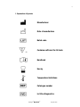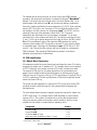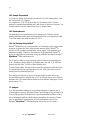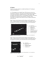
1
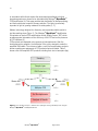
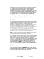
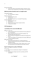
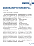
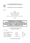
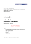
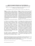
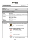
Mentype® MycoDermQS Manual For fast and reliable detection of 21 dermatomycosis causing pathogens In-Vitro-Diagnostics 50 45-21310-0050 CH1200105 Biotype Diagnostic GmbH Moritzburger Weg 67 D-01109 Dresden Germany Made in Germany 2 Biotype Diagnostic GmbH develops, produces and markets their PCR-based rapid Mentype® Detection Kits. Our products provide customers with fast and reliable testing methods for professional medical diagnostics. Our Mentype® Test Kits guarantee highest quality standards for clinical research and diagnostics. For information and enquiries about the Mentype® MycoDermQS PCR Amplification Kit, please do not hesitate to get in touch or visit www.biotype.de/en/home.html. Mentype® MycoDermQS August 2012 3 Trademarks and Disclaimer Mentype® and i-sep® are registered trademarks of Biotype Diagnostic GmbH. Ficoll® is a registered trademark of GE Healthcare Bio-Sciences AB. GelRedTM is a registered trademark of Biotium Inc. GeneAmp® is a registered trademark of Roche Molecular Systems. Mastercycler® is a registered trademark of Eppendorf AG. NucleoSpin® is a registered trademark of Machery Nagel GmbH & Co. KG. QIAamp® is a registered trademark of Qiagen GmbH. ZR Fungal/Bacterial DNA MiniPrep™ is a registered trademark of Zymo Research Corporation. © 2012, Biotype Diagnostic GmbH, all rights reserved. Mentype® MycoDermQS August 2012 4 Table of Contents 1. Explanation of Symbols .................................................................................. 5 2. Kit Content .................................................................................................... 6 3. Storage ......................................................................................................... 6 4. Additionally Required Materials and Devices .................................................... 6 5. General Precautions....................................................................................... 7 6. Safety Information ......................................................................................... 7 7. Quality Control............................................................................................... 8 8. Product Use Limitations ................................................................................. 8 9. Diagnostic Evaluation ..................................................................................... 8 10. Product Warranty......................................................................................... 8 11. Technical Assistance ................................................................................... 8 12. Introduction ................................................................................................. 9 13. Sampling .................................................................................................. 11 14. DNA Preparation ........................................................................................ 11 14.1 Sample Lysis and DNA Extraction Using Sampletype i-sep® SQ CE-IVD . 12 14.2 DNA Purification .................................................................................. 12 14.3 Nail Samples ....................................................................................... 12 15. PCR Amplification ...................................................................................... 13 15.1 Master Mixes Preparation .................................................................... 13 15.2 PCR Amplification Parameter ............................................................... 14 16. Agarose Gel Electrophoresis ....................................................................... 15 16.1 Electrophoresis Chamber Preparation ................................................... 15 16.2 Sample Preparation ............................................................................. 16 16.3 Electrophoresis.................................................................................... 16 16.4 Gel Staining Using GelRedTM................................................................. 16 17. Analysis .................................................................................................... 16 18. Results ..................................................................................................... 17 19. Troubleshooting ......................................................................................... 19 19.1 PCR: ................................................................................................... 19 19.2 Electrophoresis.................................................................................... 20 20. References................................................................................................ 22 21. Notes ........................................................................................................ 23 Mentype® MycoDermQS August 2012 5 1. Explanation of Symbols Manufacturer Date of manufacture Batch code Contains sufficient for 50 tests 50 Handbook Use by Temperature limitations Catalogue number In-Vitro-Diagnostics Mentype® MycoDermQS August 2012 6 2. Kit Content Mentype® MycoDermQS PCR Amplification Kit Catalogue number 45-21310-0050 Sufficient for 50 reactions Nuclease-free water Reaction Mix D (REM D) Taq2 (DNA Polymerase) 2 x 1.9 ml 1 x 500 µl 1 x 40 µl; 1 x 10 µl Primer mix PCR1 Control-DNA PCR1 (2 ng/µl) Quality Sensor PCR1 (25 fg/µl) Reference Ladder PCR1 50 µl 10 µl 50 µl 80 µl Primer mix PCR2 Control-DNA PCR2 (2 ng/µl) Quality Sensor PCR2 (25 fg/µl) Reference Ladder PCR2 50 µl 10 µl 50 µl 80 µl Buffer L (conc.)* Buffer N 3 x 0.6 ml 3 x 1.5 ml *Add 1.4 ml water before use. 3. Storage Store the PCR components (such as REM D, Taq2, Primer, H20, Control-DNA) at -20°C. Primer mixes and reference ladders must be stored protected from light. Repeated thawing and freezing (> 2 x) should be avoided, as this may reduce the sensitivity. If the reagents are to be used only intermittently, they should be frozen in aliquots. Storage at +4°C should not exceed a period of five hours. DNA samples as well as quality sensors and post-PCR reagents (reference ladders, or DNA size standards) should be stored separately from the PCR reagents. Buffer L and N can be stored at ambient temperature. The expiry date is indicated on the kit cover. 4. Additionally Required Materials and Devices Disposable powder-free gloves DNA isolation kit (Section 14) Pipettes (adjustable) Sterile pipette tips with filters Vortex mixer Desktop centrifuge with rotor for 2 ml reaction tubes Heating block or incubator (56ºC) PCR thermal cycler Agarose gel electrophoresis system with power supply Gel documentation system Mentype® MycoDermQS August 2012 7 To carry out the gel electrophoresis further reagents are needed in addition to the Biotype® PCR Amplification Kit: Reagent 6x Loading buffer containing bromphenole blue and Ficoll 400 GelRed (DNA-specific fluorescence dye) peqGold low melt agarose TBE-buffer 1x TE-Puffer Supplier Order number Applichem GmbH A3144 Biotium Inc. PeqLab Biotechnologie GmbH Applichem GmbH Applichem GmbH 41003 35-1030 A0972 A2575 5. General Precautions The user should always pay attention to the following: Use sterile pipette tips with filters. Store and extract positive material (specimens, controls and amplicons) separately from all other reagents and add it to the reaction mix in a spatially separated facility. Thaw all components thoroughly at room temperature before starting an assay. When thawed, mix the components and centrifuge briefly before pipetting. Work quickly on ice or in a cooling block. 6. Safety Information The PCR Amplification Kit contains the following potentially hazardous chemicals: Kit component Buffer L (conc.) Chemical 10 % potassium hydroxide Hazards GHS05 (Corrosive) H302 - Harmful if swallowed. H314 - Causes severe skin burns and eye damage. GHS07 (Attention!) H290 - May be corrosive to metals. P262: Do not get in eyes, on skin, or on clothing P280 - Wear protective gloves/protective clothing/eye protection/face protection. P301+330+331: IF SWALLOWED: Rinse mouth. Do NOT induce vomiting P303+361+353: IF ON SKIN (or hair): Remove/Take off immediately all contaminated clothing. Rinse skin with water/shower P305+351+338: IF IN EYES: Rinse cautiously with water for several minutes. Remove contact lenses if present and easy to do continue rinsing. P310 : Immediately call a POISON CENTER or doctor/physician Observe the Material Safety Data Sheets (MSDS) for all Biotype® products, which are available on request. Please contact the respective manufacturers for copies of the MSDS for any additionally needed reagents. Mentype® MycoDermQS August 2012 8 7. Quality Control All kit components undergo an intensive quality assurance process at Biotype Diagnostic GmbH. The Biotype Diagnostic GmbH is accredited under DIN EN ISO 9001:: 2008. In accordance with our ISO-certified Quality Management System, each lot of Mentype® MycoDermQS Amplification Kit is permanently monitored in order to ensure unrestricted usability and consistent product quality. Please contact us if you have any questions regarding quality assurance. 8. Product Use Limitations All reagents may exclusively be used in In-Vitro-Diagnostics (IVD). The product is to be used by personnel specially instructed and trained in the In-VitroDiagnostics procedures (EN375) and PCR only. Strict compliance with the user manual is required for optimal PCR results. Attention should be paid to expiration dates printed on the box and labels of all components. Do not use expired components. 9. Diagnostic Evaluation The Mentype® MycoDermQS Amplification Kit has undergone a series of evaluation studies including a clinical performance assessment fulfilling the requirements of In-Vitro-Diagnostics Directive 98/79/EC (IVDD). A declaration of conformity was prepared and the CE mark was granted by the local governing body for medical devices. 10. Product Warranty The Biotype Diagnostic GmbH guarantees the performance of all products in the manner described in our product literature. The purchaser must determine the suitability of the product for its particular use. Should any product fail to perform satisfactorily due to any reason other than misuse, the Biotype Diagnostic GmbH will replace it free of charge or refund the purchase price. We reserve the right to change, alter, or modify any product to enhance its performance and design. Please inquire for more information. A copy of our terms and conditions can be found on the back of your invoice. 11. Technical Assistance The Biotype Diagnostic GmbH provides a high quality technical support. Our support is staffed by experienced scientists with extensive practical and theoretical expertise in sample and assay technologies and the use of our products. If you have any questions or experience any difficulties regarding the Mentype® MycoDermQS Amplification Kit, please do not hesitate to contact us. Our customers are a major source of information regarding advanced or specialised uses of our products. This information is helpful to other scientists as well as to our team. We therefore encourage you to contact us if you have any suggestions about product performance or new applications and techniques. For technical assistance and more information, please see our website at www.biotype.de/en/home.html, send us an email to [email protected] or give us a call at +49 (0) 351 8838 400. Mentype® MycoDermQS August 2012 9 12. Introduction A definitive diagnosis of dermatophyte infection needs to be done before the initiation of antifungal therapy because of the long duration of the treatment and its high cost, and of the potential side effects of the drugs. Mentype® MycoDermQS is a polymerase chain reaction (PCR) based multiplex application for fast, reliable, easy to use and routine-fit diagnostics of dermatomycosis also known as dermatophytosis or ringworm. Mentype® MycoDermQS is best suited for skin-, nail-, and hair-specimens. From DNA isolation to diagnosis the Mentype® MycoDermQS exceeds traditional procedures by performing highest quality identification of dermatophytes within 24 hours, consequently, providing up to 4 weeks gain in time. The multiplex PCR application eliminates the eventuality of miss interpretation caused by the dominance of a particular species or an increased contamination risk of culture plates over the long analysis time. The assay detects 21 dermatomycosis causing pathogens either species- or genus-specific (Table 1; [1]). Table 1. Dermatomycosis causing pathogens detected using the Mentype® MycoDermQS Amplification Kit. Genus Epidermophyton Microsporum Microsporum Trichophyton Trichophyton Trichophyton Species floccosum canis (1) gypseum (2) rubrum interdigitale (3) spp. Candida spp. Trichosporon Scopulariopsis Aspergillus cutaneum brevicaulis spp. Genus-specifically detected species T. rubrum T. interdigitale (3) T. tonsurans T. violaceum T. verrucosum Arthroderma benhamiae C. albicans C. tropicalis C. glabrata C. krusei (Pichia kudriavzevii, Issatchenkia orientalis) C. guilliermondii (Meyerozyma guilliermondii) C. parapsilosis A. fumigatus A. flavus A. niger A. versicolor (1) teleomorph: Arthroderma otae, (2) teleomorph: Arthroderma gypseum, (3) teleomorph: Arthroderma vanbreuseghemii Mentype® MycoDermQS August 2012 10 In accordance with clinical experts the most important pathogens eliciting dermatomycosis were chosen to be detectable with Mentype® MycoDermQS PCR Amplification Kit. This assay enables fast clarification of disease aetiology and thus contributes to specific therapy selection. The latter is particularly important in light of growing resistance to antimycotics [2, 3]. Modern technology designed for discerning users provides reliable results in just few working steps (Figure 1). The Mentype® MycoDermQS Amplification Kit contains an internal PCR amplification control (Quality Sensor “QS”) which provides precise information on the efficiency of the PCR and on the presence of PCR inhibitors [4]. PCR products are separated using agarose gel electrophoresis. After the electrophoresis is complete, the molecules in the gel are stained to make the amplified DNA visible. The reference ladder, a mix of all amplification products, allows unambiguous assignment of PCR products fast and reliable. Table 2 shows a list of all amplified PCR products including their size in base pairs (bp). Sampling Sample Lysis ≥12 h DNA Extraction 2-2.5 h Master Mix PCR1 Master Mix PCR2 Multiplex-PCR 2-2.5 h Gel Electrophoresis 1h Analysis of Results 0.5 h Figure 1. From sample to analysis - detection of 21 pathogens causing dermatomycosis using the Mentype® MycoDermQS PCR Amplification Kit. Mentype® MycoDermQS August 2012 11 A validation was carried out using a Mastercycler® ep gradient S (Eppendorf AG, Hamburg) and the electrophoresis system iMupid Mini Agarose Gel Electrophoresis System (Helixx Technologies Inc., Toronto, Canada) in combination with the agarose gel documentation system BioVision 3000 (Vilber Lourmat Deutschland GmbH, Eberhardzell, Germany) equipped with a 312 nm UV light source and a 590 nm emission filter. The camera was set diaphragm 2.8 and shutter speed 1-3 s. For DNA visualisation the intercalating dye GelRed (Biotium Inc.) was used in a freshly prepared solution according to manufacturer's protocol. The complementary analysis software package Bio1D advanced (Vilber Lourmat Deutschland GmbH) was used for the interpretation and analysis of all agarose gels. 13. Sampling The diagnosis of dermatophytosis requires samples that are correctly collected. A simple and fast extraction method of the DNA directly from patient samples is necessary for routine application of a molecular based detection methodology of dermatophyte infections. The following materials can be used for isolation of dermatophytes: skin, nail, hair and swabs. Using sterile instruments, a sufficient sample should be removed taken from the edge of the infected area, which corresponds to the active zone of the lesion. The procedure should be performed by an experienced staff under aseptic conditions. To reduce a risk of sample contamination, the area, from where the sample will be taken, should be first disinfected with 70 % ethanol. Note! To improve the efficiency of mycological examination, samples should be obtained before any local or systemic antifungal treatment. Due to electrostatic attraction, plastic boxes such as Petri dishes are unsuitable shipping samples. They can be used to collect the sample. For a good location of skin scales, nails and dark or light hairs, dishes may be placed over coloured paper. The sample is collected using a regular nylon flocked dry swab (3520CS01; Copan Diagnostics Inc., US) moisturised with sterile water. The collected sample should be immediately transferred into a sterile vial. We recommend Sampletype i-sep® SQ CE-IVD (Catalogue number 63-002010050). The vial should be sealed and packed for transport following guidelines of shipping clinical samples. 14. DNA Preparation Since performance of the Mentype® MycoDermQS assay is mostly depending on quality and quantity of applied pathogen DNA, we recommend standardised and already validated methods for sampling and DNA isolation. Mentype® MycoDermQS August 2012 12 14.1 Sample Lysis and DNA Extraction Using Sampletype i-sep® SQ CE-IVD The sample lysis is carried out prior to the use of the actual DNA isolation procedure to further purify the DNA. To prepare the lysis buffer calculate the volumes of components that are needed based on the number of reactions. Include up to 5 % excess volume to compensate for pipetting losses. Gently mix 180 µl lysis buffer per sample + 20 µl proteinase K. Sample Lysis: All samples should be centrifuged for about 10 s Bring the shaking incubator to 56°C Sample provided in a Sampletype i-sep® SQ filter column placed in a 2 ml collection tube Open the tube, add 200 µl lysis buffer (~56°C) and close the lid Place the assembly into a thermo shaker at 850 rpm at 56°C and incubate overnight (recommended), alternatively use an incubator Centrifuge the Sampletype i-sep® SQ at 13.400 rpm for 1 min Discard the filter column Close the lid of the collection tube and store the lysate for further processing; tube contains 200 µl lysate containing the pathogenic DNA Collected lysate is directly processed in a subsequent DNA purification 14.2 DNA Purification A selection of DNA isolation kits, based on silica membrane technology, is listed below. Please carry out the DNA isolation according to the manufacturer’s instructions. An overnight sample treatment using proteinase K, yet at least 12 hours, is recommended. The following isolation kits are recommended: DNA-Isolation Kit QIAamp® DNA Mini Kit NucleoSpin Tissue ZR Fungal/Bacterial DNA MiniPrep™ Manufacturer Qiagen Macherey & Nagel Order No. 51304 740952.250 ZymoResearch D600 14.3 Nail Samples To ensure an effective DNA isolation we recommend the utilisation of buffer L and buffer N in the case of nail containing samples. Both buffers are part of the Mentype® MycoDermQS DNA Amplification Kit. Mentype® MycoDermQS August 2012 13 The sample lysis is carried out prior to the use of the actual DNA isolation procedure. Any kit specific lysis buffer is replaced by Mentype® MycoDermQS buffer L. Prior to the first use, buffer L (conc.) has to be diluted using 1.4 ml distilled water. Both buffers, L and N, can be stored at ambient temperature. Ensure the sample equilibrates to room temperature (15-25°C). Heat a heating block to 90°C for further use. A volume of 100 µl of buffer L is added to the nail sample in the filter column of a Sampletype i-sep® SQ CE-IVD, the lid is closed and the single tube assembly is kept at 90°C for 15 minutes. Alternatively the nail sample can be processed in a standard 1.5 ml micro centrifuge vial. Let the sample cool below 56°C and briefly centrifuge the tube (1 min, >1000 x g) to remove drops from the inside of the lid. Add 80 µl buffer N and 20 µl proteinase K (part of the respective DNA isolation kit), mix by vortexing, and incubate at 56°C overnight or at least 12 hours until the tissue is completely lysed. Centrifuge the Sampletype i-sep® SQ CE-IVD at 13.400 rpm for 1 min. Discard the filter column and use the lysate for a subsequent DNA purification. This remaining DNA isolation procedure is carried out following the manufacturer’s instructions. 15. PCR Amplification 15.1 Master Mixes Preparation All components should be mixed (vortex) and centrifuged for about 10 s before preparing the master mix. A maximum of 7.0 µl sample volume can be applied in PCR1. Adjust the final reaction volume to 25 µl with nuclease-free water. The amount of DNA used for the assay depends on the concentration and quality of prior isolated material. Best performance is achieved using fungal DNA in a range of 50 pg up to 250 pg in PCR1 and between 25 pg and 100 pg in PCR2, respectively. The total concentration of DNA, both fungal and human, should not exceed 200 ng. The primer mix was optimised in a way that 40 PCR cycles gave enough PCR products to obtain a sufficient signal in the analysis based on agarose gel electrophoresis. The table below shows volumes of applied reagents to prepare the master mix of PCR1 with using 7.0 µl sample volume (DNA-template) in a total reaction volume of 25 µl. The number of reactions to be set up shall be determined taking into account positive and negative control reactions. Add one or two reactions to this number to compensate for pipetting errors. Components Nuclease-free Water Reaction Mix D* Primer Mix PCR1 QS Sensor PCR1 (25 fg/µl) Multi Taq2 DNA Polymerase (hot start, 2.5 U/µL) Volume Master Mix Volume 10.5 µl 5.0 µl 1.0 µl 1.0 µl 0.5 µl 18.0 µl * contains Mg2+, dNTPs, BSA Mentype® MycoDermQS August 2012 14 In PCR2 the recommended maximum sample volume is 4.0 µl in a total reaction volume of 25 µl. The table below shows the volumes of all required reagents to prepare the master mix of PCR2 using 4 µl sample. If less sample volume is used, adjust the final reaction volume to 25 µl using nuclease-free water. Note! The QS Sensor has to be diluted fresh each time for PCR2. QS (25 fg/µl) has to be diluted 1:200 to a final concentration of 125 ag/µl using an appropriate volume of nuclease-free water (e.g. 1 µl QS in 199 µl nucleasefree water). Again, the number of reactions to be set up shall be determined taking into account positive and negative control reactions. Add one or two reactions to this number to compensate for pipetting errors. Components Nuclease-free Water Reaction Mix D* Primer Mix PCR2 QS Sensor PCR2 (125 ag/µl) Multi Taq2 DNA Polymerase (hot start, 2.5 U/µL) Volume Master Mix Volume 13.5 µl 5.0 µl 1.0 µl 1.0 µl 0.5 µl 21.0 µl * contains Mg2+, dNTPs, BSA Positive Control A low concentrated solution of Control-DNA is used to verify the PCR amplification. Control-DNA PCR1 and PCR2 (both 2 ng/µl) has to be diluted 1:200 to a final concentration of 10 pg/µl using an appropriate volume of nuclease-free water (e.g. 1 µl Control-DNA in 199 µl nuclease-free water). Instead of template DNA pipette 1 µl diluted Control-DNA into reaction-tubes containing the PCR master mix. The criterion of a successful PCR amplification is fulfilled in the case of a clearly detectable positive control. Negative Control Nuclease-free water serves as negative control. Pipette respective volume instead of the DNA-template into reaction tubes containing the PCR master mix of PCR1 or PCR2, respectively. 15.2 PCR Amplification Parameter Perform a “hot start” PCR in order to activate the Multi Taq2 DNA Polymerase and to prevent the formation of non-specific amplification products. Mentype® MycoDermQS August 2012 15 Standard program of PCR1 and PCR2, respectively, recommended for all DNA samples. Temperature 94°C 94°C 62°C 72°C 94°C 60°C 72°C 10°C Time 4 min (hot start to activate Multi Taq2 DNA Polymerase) 30 s 60 s 5 cycles 90 s 30 s 60 s 35 cycles 90 s ∞ hold 16. Agarose Gel Electrophoresis For general instructions on instrument setup and analysis software packages please refer to the manufacturer’s user manual. Electrophoresis and gel documentation (gel doc) are carried out using standard equipment. Quality and concentration of agarose and the separation length are the main criteria to form distinct bands on the gel. Sensitivity is related to the thickness of the gel, the intercalating dye and the optical quality of the gel doc system. The reference ladder and/or a PCR using the Control-DNA, both part of the kit, can be used to calibrate the gel doc system. Minimal specifications: 2 % agarose e.g. peqGOLD Universal-Agarose (PeqLab Biotechnologie GmbH) or similar in 1x TBE-running buffer 6x Loading buffer containing bromphenole blue and Ficoll 400 Separation length ≥ 7 cm Gel thickness ≤ 5 mm Loaded sample volume ≤ 12 µl Electrophoresis conditions: max. 6 Volt / cm (electrode-to-electrode clearance) at ambient temperature (≤ 26°C) After the electrophoresis is complete, the molecules in the gel are stained to make them visible. The intercalating dye GelRed (Biotium Inc.) is recommended and should be used always freshly prepared. 16.1 Electrophoresis Chamber Preparation Only powder free gloves should be worn to prevent any background fluorescence. A fundamental overview of gelelectrophoresis can be found in Sambrook et al. 1989 [5]. Furthermore, please refer to manufacturer’s instructions. Mentype® MycoDermQS August 2012 16 16.2 Sample Preparation 5 µl reference ladder are thoroughly mixed with 5 µl of 6x loading buffer; load the first well with 10 µl marker. For samples mix 10 µl PCR product with 2 µl 6x loading buffer. Continue loading the samples quantitatively and finish off with a final lane of marker. It is recommended to use one marker lane every 10 sample lanes. 16.3 Electrophoresis Corresponding to the manufacturer use a maximum of 6 Volt/cm until the distance between starting point and bromphenole blue front measures at least 7 cm. Best results are achieved using 90-120 V. 16.4 Gel Staining Using GelRedTM GelRedTM (Biotium Inc.) is a new generation of fluorescent nucleic acid gel stain designed to replace the highly toxic ethidium bromide (EtBr). GelRedTM is superior to EtBr by having a combination of low toxicity, high sensitivity and exceptional stability. Therefore, visualization of DNA bands with GelRedTM is highly recommended and was used during the validation process of Mentype® MycoDermQS DNA Amplification Kit. For 50 ml of a ready-to-use gel staining mixture (3 times concentrated) add 5 ml 1 M sodium chloride with 45 ml distilled water and add 15 µl GelRedTM Nucleic Acid Gel Stain 10.000x in Water (Biotium Inc.). To ensure best quality, the stain mixture should be prepared freshly each time. The typical staining time used was about 50 min. During this time, keep the staining mixture protected from light. Once staining is achieved, a picture of the gel should be taken with a gel documentation system equipped with a 312 nm UV light source and a 590 nm emission filter. The camera specs should be set as follows; diaphragm 2.8 and shutter speed 1-3 s. 17. Analysis A gel documentation software for an automated analysis of agarose gels is recommended in general. Windows Bitmap-Format (bmp) or Tagged Image File Format (Tiff) are saved and loaded into a gel documentation software to carry out an automated amplicon identification and amplicon size analysis. Please refer to manufacturer’s instructions. A list of the amplicon sizes for the Mentype® MycoDermQS DNA Amplification Kit can be found in Table 2. Mentype® MycoDermQS August 2012 17 18. Results Amplicon size analysis allows for a reliable identification of 21 pathogens causing dermatomycosis. A 1.231 bp amplicon of the Quality Sensor (QS) is the main criterion for a successful PCR experiment. The band can be partially depressed in presence of very high concentration of pathogen-specific or human genomic DNA. An amplicon of the positive control, 100 pg template concentration, guarantees the sensitivity of the experiment (see also page 10 Positive Control). Furthermore, the negative control should not result in additional bands other than the QS one. Figure 2 shows a typical agarose gel with pathogen-specific amplicons of PCR1 in relative comparison to the reference ladder, an equimolar mixture of all amplified PCR products and the QS band visible in all PCR preparations. RL1 A B C D E F G (-) RL1 QS QS: RL1: (-): A: B: C: D: E: F: G: Quality Sensor Reference ladder PCR1 Negative control Aspergillus spp. Microsporum canis Candida spp. Epidermophyton floccosum Trichosporon cutaneum Scopulariopsis brevicaulis Microsporum gypseum Figure 2. Electrophoresis PCR1: 2 % agarose gel; separating length: 7.0 cm; electrophoresis conditions: 6 V/cm; gel doc: GelRedTM. Figure 3 shows a typical agarose gel with pathogen-specific amplicons of PCR2 in relative comparison to the reference ladder, an equimolar mixture of all amplified PCR products and the QS band visible in all PCR preparations. RL2 H I J (-) RL2 QS QS: RL2: (-): H: I: J: Quality Sensor Reference ladder PCR2 Negative control Trichophyton interdigitale Trichophyton rubrum Trichophyton spp. Figure 3. Electrophoresis PCR2: 2 % agarose gel; separating length: 7.0 cm; electrophoresis conditions: 6 V/cm; gel doc: GelRedTM. Mentype® MycoDermQS August 2012 18 Table 2. Fragment size of the amplified PCR templates detected using the Mentype® MycoDermQS Amplification Kit. Parameter Aspergillus spp. Microsporum canis Candida spp. Epidermophyton floccosum Trichosporon cutaneum Scopulariopsis brevicaulis Microsporum gypseum Trichophyton interdigitale Trichophyton rubrum Trichophyton spp. Quality Sensor (QS) PCR1 Size [bp]# 226 383 421 497 557 649 761 PCR2 Size [bp]# 298* 373* 1036 1231 1231 # ( ) Due to possible differences in separation during electrophoresis, 5 % drift between PCR amplicon and corresponding fragment of the reference ladder can be tolerated. (*) For T. interdigitale and T. rubrum a second template (Trichophyton spp.) is amplified. Mentype® MycoDermQS August 2012 19 19. Troubleshooting 19.1 PCR: PCR product, low level QS or no QS: Ensure the correct use of QS Sensors in the individual PCR master mixes PCR1 and PCR2, respectively. Signal suppression can occur if the ratio of DNA-templates is extremely unbalanced, e.g. high starting concentrations of pathogenic DNA-template can reduce the signal strength of QS or even inhibit the amplification of the Quality Sensor completely. PCR2 Trichophyton spp. signal and/or QS signal reduced or completely restrained: Signal suppression can occur if the ratio of DNA-templates is extremely unbalanced, e.g. high starting concentrations of pathogenic DNA-template can reduce the signal strength of QS or even inhibit the amplification of the Quality Sensor completely. A similar competitive reaction occurs between single- and multi copy genes of the same genome. Thus amplification of the T. spp. template (single copy gene) can be inhibited in presence of T. rubrum or T. interdigitale templates (multi copy), respectively. Low levels of PCR product or no product band visible, no QS: Error in set up: Check if all reagents were added in correct volumes Check for used concentrations and storage conditions of reagents (avoid repeated freeze-thaw cycles of the primer mix) Mix all solutions before use Error in cycling: Ensure the right PCR program was used Check that the PCR thermal cycler works correctly Check the power supply of the thermal cycler Sample evaporation during thermal cycling: Check levels in wells after cycling Ensure lid heating is activated (105ºC) Problems with electrophoresis: Refer to the section Electrophoresis below Non-specific amplification, product bands are smeared: Overabundance of template: reduce template concentration Mentype® MycoDermQS August 2012 20 Excess of human DNA: Ensure that sufficient fungal sample material is collected, and the amount of carrier material such as skin flakes or hair is minimised Additional products detectable, band in no template control: Contaminated reagents: Use a fresh aliquot of reagents Pipettes contaminated: Clean and sterilise pipette Use filter tips Use different pipettes for pre- and post PCR processing Aerosol contamination: Minimise pipetting steps Use filter tips Close lid on all tubes and expel reagents carefully Change gloves regularly 19.2 Electrophoresis Low intensity of all or some of the DNA bands: DNA has run off the gel: Perform electrophoresis until the bromphenole blue dye passes 2/3 (ca. 5 cm) of the gel Make sure that the entire gel is immersed completely in the electrophoresis buffer during the run Make sure that gel and apparatus are positioned horizontally during the run Control the polarisation of the electrophoresis chamber (DNA travels from minus towards plus pole) DNA diffusion in the gel: Avoid prolonged electrophoresis or excessive staining and destaining procedures as this may cause diffusion of smaller DNA fragments in the gel Avoid long term storage of the gel before taking a picture, as this may cause diffusion of DNA fragments and low band intensity Atypical banding pattern; quality of DNA bands: Double bands: Do not use an excessively high voltage for electrophoresis Denatured DNA: Excessively high voltage may result in gel heating and DNA denaturation Mentype® MycoDermQS August 2012 21 Different loading conditions for the sample and the ladder DNA: Always use the same loading dye solution for both the sample DNA and the ladder/marker DNA Improper electrophoresis conditions: Excessive electrophoresis run times or voltage may result in migration of small DNA fragments off the gel; very short or slow electrophoresis may result in incompletely resolved bands Run gels at 5-8 V/cm until the bromphenole blue passes 2/3 (5cm) Incorrect gel percentage or running buffer used: The correct gel percentage is important for optimal separation of the DNA; prepare gels according to recommendations on page 15 Always use 1x concentrated running buffer (TBE) GelRedTM interferes with separation of DNA fragments [6]; [7]: Do not include GelRedTM in the gel; stain the gel after finished electrophoresis Gel incompletely immersed in electrophoresis buffer: electrophoresis buffer should completely cover the entire gel during sample loading and run Bubbles or physical particles in the gel wells or in the gel: Use pure water, clean flasks and clean equipment for preparation of gels Pour the gel slowly avoiding formation of bubbles; bubbles can be removed with a pipette tip Ensure correct concentration of buffer used to prepare the gel and buffer used to run the gel; always use fresh solutions and flasks for electrophoresis Mentype® MycoDermQS August 2012 22 20. References [1] [2] [3] [4] [5] [6] [7] Seebacher C, Bouchara JP, Mignon B. Updates on the epidemiology of dermatophyte infections. Mycopathologia 166, 335-352, 2008. Seebacher C, Brasch J, Abeck D, Cornely O, Effendy I, GinterHanselmayer G, Haake N, Hamm G, Hipler UC, Hof H, Korting HC, Mayser P, Ruhnke M, Schlacke KH, Tietz HJ; German Society of Dermatology; German-speaking Mycological Society. Onychomycosis. Mycoses 50, 321-327, 2007. www.dmykg.de/information/leitlinien.html Reischl U, Drosten C, Geißdörfer W, Göbel U, Hoffmann KS, Mauch H, Meyer T, Moter A, von Müller L, Panning M, Rabenau HF, Reiter-Owona I, Roth A, Weitz M. MiQ 1-2011, Nukleinsäure-Amplifikationstechniken. In. Podbielski A, Hermann M, Kniehl E, Mauch H, Rüssmann H (Hrsg.) Mikrobiologisch-infektiologische Qualitätsstandards (MiQ). Im Auftrag der Deutschen Gesellschaft für Hygiene und Mikrobiologie (DGHM). 3. Auflage. Elsevier / Urban & Fischer, München, 2011. Sambrook J, Fritsch EF, Maniatis T (Hrsg). Molecular Cloning: A Laboratory Manual. 2. Auflage. Cold Spring Harbor Laboratory Press, NY US, 1989. Biotium Inc. Safety Report of GelRedTM and GelGreen - A Summary of Mutagenicity and Environmental Safety Test Results from Three Independent Laboratories, 2008. http://www.biotium.com/product/product_info/Safety_Report/GR%20&% 20GG%20safety.pdf GelRedTM Nucleic Acid Gel Stain Product protocol. http://www.biotium.com/product/product_info/Protocol/PI-41002.pdf Mentype® MycoDermQS August 2012 23 21. Notes Mentype® MycoDermQS August 2012 24 Mentype® MycoDermQS August 2012