1
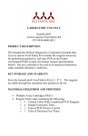
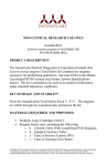
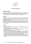
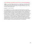
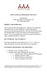
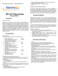
AOAC Official Method 2013.09
Salmonella in Selected Foods
3M™ Molecular Detection Assay (MDA)
Salmonella Method
First Action 2013
Revised First Action 2014
A. Principle
B. Apparatus and Reagents
Items (b)–(g) are available as the 3M MDA Salmonella kit from
3M Food Safety (St. Paul, MN, USA).
(a) 3M Molecular Detection System.—Available from 3M Food
Safety.
(b) 3M MDA Salmonella reagent tubes.—Twelve strips of eight
tubes.
(c) Lysis solution (LS) tubes.—Twelve strips of eight tubes.
(d) Extra caps.—Twelve strips of eight caps.
(e) Negative control (NC).—One vial (2 mL).
(f) Reagent control.—Eight reagent tubes.
Ex A
pe OA
rt C
R R
ev es
ie ea
w rc
Pa h
ne Ins
l U tit
se ute
O
nl
y
[Applicable to detection of Salmonella in raw ground beef
(25, 325, and 375 g), raw ground chicken (25 and 325 g), cooked
breaded chicken (325 g), pasteurized liquid whole egg (100 g), raw
shrimp (head-off, 25 g), fresh spinach (bagged, 25 g), wet dog food
(375 g), pasteurized American cheese (25 g), peanut butter (25 g),
dry dog food (25 and 375 g), sprout irrigation water (375 g), raw
head-on shrimp (25 g), chicken carcass rinsate (30 mL), chicken
carcass sponge, sealed/glazed ceramic tile, concrete, and stainless
steel.]
See Tables 2013.09A and B for a summary of results of the
interlaboratory study.
See Tables 1 and 2 of the Appendix for detailed results of the
interlaboratory study (appendix is available on the J. AOAC Int.
website, http://aoac.publisher.ingentaconnect.com/content/aoac/
jaoac).
efficiency, and rapidity, and bioluminescence to detect the amplified
sequences. Presumptive positive results are reported in real-time;
negative results are displayed after the assay is completed.
The LOD of a method is defined as the lowest concentration
point where reliable analytical results can be obtained. This can
vary with different serotypes. For the 3M MDA Salmonella method
this has been demonstrated to be 1–5 CFU/25 g of sample or
1–5 CFU/swab.
As with all test methods, the source of enrichment medium can
influence the results. The 3M MDA Salmonella method has only
been evaluated for use with the enrichment medium, 3M Buffered
Peptone Water ISO (BPW ISO). Matrixes are incubated in 3M
BPW for 10–24 h to enrich for Salmonella prior to initiating the
assay, with the exception of raw head-on shrimp, which requires
an additional 4–24 h secondary enrichment in RappaportVassiliadis 10 broth (RV10).
The 3M MDA Salmonella method is intended for use with the 3M
Molecular Detection System for the rapid and specific detection of
Salmonella spp. in food, food-related, and environmental samples
after enrichment. The 3M MDA Salmonella test uses isothermal
amplification of unique DNA target sequences with high specificity,
Table 2013.09A. POD summary of raw ground beef (25 g) results for the 3M MDA Salmonella methoda
Inoculation level
Candidate presumptive positive/total No. of samples analyzed
Uninoculated
Low
High
1/120
69/120
120/120
Candidate presumptive (CP) POD
0.01 (0.00, +0.05)
0.58 (+0.48, +0.67)
1.00 (+0.97, +1.00)
srb
0.09 (+0.08, +0.17)
0.51 (+0.45, +0.52)
0.00 (0.00, +0.18)
0.00 (0.00, +0.04)
0.00 (0.00, +0.14)
0.00 (0.00, +0.18)
0.09 (+0.08, +0.10)
0.51 (+0.45, +0.52)
0.00 (0.00, +0.24)
sL
c
sRd
0/120
67/120
120/120
Candidate confirmed (CC) POD
Candidate confirmed positive/total No. of samples analyzed
0.00 (0.00, +0.03)
0.56 (+0.47, +0.65)
1.00 (+0.97, +1.00)
sr
0.00 (0.00, +0.17)
0.51 (+0.45, +0.52)
0.00 (0.00, +0.18)
0.00 (0.00, +0.17)
0.00 (0.00, +0.11)
0.00 (0.00, +0.18)
0.00 (0.00, +0.24)
0.51 (+0.46, +0.52)
0.00 (0.00, +0.24)
0/120
68/120
119/120
sL
sR
Positive reference samples/total No. of samples analyzed
Reference POD
0.00 (0.00, +0.03)
0.57 (+0.48, +0.66)
0.99 (+0.95, +1.00)
sr
0.00 (0.00, +0.17)
0.50 (+0.45, +0.52)
0.09 (+0.08, +0.17)
sL
0.00 (0.00, +0.17)
0.00 (0.00, +0.18)
0.00 (0.00, +0.04)
sR
0.00 (0.00, +0.24)
0.51 (+0.45, +0.52)
0.09 (+0.08, –0.11)
dLPOD (candidate vs reference)
0.00 (–0.03, +0.03)
–0.01 (–0.14, +0.12)
0.01 (–0.02, +0.05)
dLPOD (CP vs CC)
0.01 (–0.02, +0.05)
0.02 (–0.11, +0.15)
0.00 (–0.03, +0.03)
a Results include 95% confidence intervals.
b Repeatability standard deviation.
c Among-laboratory standard deviation.
d Reproducibility standard deviation.
© 2014 AOAC INTERNATIONAL
Table 2013.09B. POD Summary of wet pet food (375 g) results for the 3M MDA Salmonella methoda
Inoculation level
Uninoculated
Candidate presumptive positive/total No. of samples analyzed
Low
High
1/132
65/132
131/132
Candidate presumptive (CP) POD
0.01 (0.00, +0.04)
0.49 (+0.40, +0.58)
0.99 (+0.96, +1.00)
srb
0.09 (+0.08, +0.16)
0.51 (+0.46, +0.52)
0.09 (+0.08, +0.16)
sLc
0.00 (0.00, +0.04)
0.00 (0.00, +0.14)
0.00 (0.00, +0.04)
sR
0.09 (+0.08, +0.10)
0.51 (+0.46, +0.52)
0.09 (+0.08, +0.10)
0/132
65/132
131/132
d
Candidate confirmed positive/total No. of samples analyzed
Candidate confirmed (CC) POD
0.00 (0.00, +0.03)
0.49 (+0.40, +0.58)
0.99 (+0.96, +1.00)
sr
0.00 (0.00, +0.17)
0.51 (+0.46, +0.52)
0.09 (+0.08, +0.16)
sL
0.00 (0.00, +0.17)
0.00 (0.00, +0.14)
0.00 (0.00, +0.04)
sR
0.00 (0.00, +0.23)
0.51 (+0.46, +0.52)
0.09 (+0.08, +0.10)
Reference POD
sr
sL
sR
0/132
70/132
132/132
0.00 (0.00, +0.03)
0.53 (+0.44, +0.62)
1.00 (+0.97, +1.00)
0.00 (0.00, +0.17)
0.52 (+0.46, +0.52)
0.00 (0.00, +0.17)
0.00 (0.00, +0.17)
0.00 (0.00, +0.09)
0.00 (0.00, +0.17)
Ex A
pe OA
rt C
R R
ev es
ie ea
w rc
Pa h
ne Ins
l U tit
se ute
O
nl
y
Positive reference samples/total No. of samples analyzed
0.00 (0.00, +0.23)
0.52 (+0.47, +0.52)
0.00 (0.00, +0.23)
dLPOD (candidate vs reference)
0.00 (–0.03, +0.03)
–0.04 (–0.16, +0.09)
–0.01 (–0.04, +0.02)
dLPOD (CP vs CC)
0.01 (–0.02, +0.05)
0.00 (–0.13, +0.13)
0.00 (–0.03, +0.03)
a
Results include 95% confidence intervals.
b
Repeatability standard deviation.
Among-laboratory standard deviation.
c
d
Reproducibility standard deviation.
(g) Quick start guide.
(h) 3M Molecular Detection Speed Loader Tray.—Available
from 3M Food Safety.
(i) 3M Molecular Detection Chill Block Tray and Chill Block
Insert.—Available from 3M Food Safety.
(j) 3M Molecular Detection Heat Block Insert.—Available
from 3M Food Safety.
(k) 3M Molecular Detection Cap/Decap Tool for reagent
tubes.—Available from 3M Food Safety.
(l) 3M Molecular Detection Cap/Decap Tool for lysis tubes.—
Available from 3M Food Safety.
(m) Empty lysis tube rack.—Available from 3M Food Safety.
(n) Empty reagent tube rack.—Available from 3M Food Safety.
(o) 3M BPW ISO.—Available from 3M Food Safety. Formulation
equivalent to ISO 6579:2002 Annex B (1).
(p) Rappaport-Vassiliadis 10 broth (RV10).—Available from
3M Food Safety.
(q) Disposable pipet.—Capable of 20 µL.
(r) Multichannel (eight-channel) pipet.—Capable of 20 µL.
(s) Sterile filter tip pipet tips.—Capable of 20 µL.
(t) Filter stomacher bags.—Seward Laboratory Systems, Inc.
(Bohemia, NY, USA), or equivalent.
(u) Stomacher.—Seward Laboratory Systems Inc., or
equivalent.
(v) Thermometer.—Calibrated range to include 100 ± 1°C.
(w) Dry double block heater unit or water bath.—Capable of
maintaining 100 ± 1°C.
(x) Incubators.—Capable of maintaining 37 ± 1°C and
41.5 ± 1°C.
(y) Freezer.—Capable of maintaining –10 to –20°C, for storing
the 3M Molecular Detection Chill Block Tray.
(z) Refrigerator.—Capable of maintaining 2–8°C, for storing
the 3M MDA.
(aa) Computer.—Compatible with the 3M Molecular Detection
Instrument.
C. Safety Precautions
The 3M Molecular Detection Instrument is intended for use with
samples that have undergone heat treatment during the assay lysis
step, which is designed to destroy organisms present in the sample.
Samples that have not been properly heat-treated during the assay
lysis step may be considered a potential biohazard and should not
be inserted into the 3M Molecular Detection Instrument. After use,
the enrichment medium and the 3M MDA Salmonella tubes can
potentially contain pathogenic materials. When testing is complete,
follow current industry standards for the disposal of contaminated
waste. Consult the Material Safety Data Sheet for additional
information and local regulations for disposal.
D. Method Preparation and Precautions
The 3M MDA Salmonella is intended for use in a laboratory
environment by professionals trained in laboratory techniques. The
user should read, understand, and follow all safety information in
the instructions for the 3M Molecular Detection System and the
3M MDA Salmonella method and retain the safety instructions for
© 2014 AOAC INTERNATIONAL
Table 2013.09C. Sample enrichment protocols
Sample
size
Enrichment broth
vol., mL
Enrichment
temp., ±1°C
Enrichment
time, h
Raw shrimp (head off)
25 g
225
37
18–24
Fresh spinach (bagged)
25 g
225
37
18–24
Peanut butter
25 g
225
37
18–24
Pasteurized American cheese
25 g
225
37
18–24
Pasteurized liquid whole egg
100 g
900
37
18–24
Cooked breaded chicken
325 g
2925
37
18–24
Dry pet food (dog)
Wet pet food (dog)
Sprout irrigation water
Raw ground beef (27% fat)
Raw ground beef (20% fat)
Raw ground poultry
Chicken carcass rinse
Chicken carcass sponge
Stainless steel
Sealed/glazed ceramic tile
Concrete
Sample matrix
Raw shrimp (head on)
25 g
225
37
18–24
375 g
1500
37
18–24
375 g
3375
37
18–24
375 mL
3375
37
18–24
25 g
225
37
18–24
25 g
225
41.5
10–18
325 g
975
41.5
10–18
375 g
1500
41.5
10–18
Ex A
pe OA
rt C
R R
ev es
ie ea
w rc
Pa h
ne Ins
l U tit
se ute
O
nl
y
Sample matrix
25 g
225
41.5
10–18
325 g
975
41.5
14–18
30 mL
30
41.5
18–24
1 Sponge
50
41.5
18–24
1 Swab
50
41.5
18–24
1 Sponge
50
41.5
18–24
1 Sponge
225 mL
41.5
18–24
Sample
size
Enrichment broth
vol., mL
Enrichment
temp., °C
Enrichment
time, h
Secondary
enrichment
medium, mL
Secondary
enrichment
temp., °C
Secondary
enrichment
time, h
25 g
225
37
18–14
RV R10:
0.1 mL into 10 mL
41.5
4–24
future reference. Follow all instructions carefully. Failure to do so
may lead to inaccurate results.
Store the 3M MDA Salmonella at 2–8°C. Do not freeze. Keep
kit away from light during storage. After opening the kit, check
that the foil pouch is undamaged. If the pouch is damaged, do not
use. After opening, unused reagent tubes should always be stored in
the resealable pouch with the desiccant inside to maintain stability
of the lyophilized reagents. Store resealed pouches at 2–8°C for
no longer than 1 month. Do not use 3M MDA Salmonella past the
expiration date. Expiration date and lot number are noted on the
outside label of the box.
Use proper aseptic technique. Use proper precautions for
Biosafety Level 2 microorganisms. Periodically, laboratory
benches and equipment (pipets, cap/decap tools, etc.) should be
decontaminated with a 1–5% (v/v in water) household bleach
solution or DNA removal solution.
E. Sample Preparation
Table 2013.09C presents guidance for the enrichment of food
and feed samples at a 1:10 dilution. It is the user’s responsibility
to validate alternate sampling protocols or dilution ratios to ensure
this test method meets the user’s criteria.
Prewarm 3M BPW ISO enrichment medium to 37 ± 1°C or 41.5
± 1°C. Aseptically combine the enrichment medium and sample.
Homogenize thoroughly for 2 min. Incubate at 37 ± 1°C or 41.5 ±
1°C. For all meat and highly particulate samples, the use of filter
bags is recommended.
Figure 2013.09A. Transfer of enriched sample to Lysis Solution tube.
© 2014 AOAC INTERNATIONAL
Figure 2013.09B. Sample Lysis.
In an AOAC PTM study, the 3M MDA Salmonella (Certificate
No. 031208) was found to be an effective method for the detection
of Salmonella in the matrixes shown in Table 2013.09C.
takes approximately 20 min and is indicated by an orange light on
the instrument’s status bar. When the instrument is ready to start a
run, the status bar will turn green.
F. Preparation of the 3M™ Molecular Detection Speed Loader
Tray
J. Lysis
Allow the LS tubes to warm up to room temperature by setting
the rack on the laboratory bench for 2 h. Remove the enrichment
broth from the incubator and gently agitate the contents. One LS
tube is required for each sample and the NC sample. LS tube
strips can be cut to desired LS tube number. Select the number of
individual LS tubes or eight-tube strips needed. Place the LS tubes
in an empty rack. To avoid cross-contamination, decap one LS
tubes strip at a time and use a new pipet tip for each transfer step.
Transfer enriched sample to LS tubes as described below:
Note: Transfer each enriched sample into individual LS tube
first. Transfer the NC last.
Use the 3M Molecular Detection Cap/Decap Tool-Lysis to
decap one LS tube strip—one strip at a time. Set the tool with cap
attached aside on a clean surface. Transfer 20 µL of sample into
an LS tube. Repeat until each individual sample has been added
to a corresponding LS tube in the strip. Use the 3M Molecular
Detection Cap/Decap Tool-Lysis to re-cap the LS tube strip. Use
the rounded side of the tool to apply pressure in a back and forth
motion ensuring that the cap is tightly applied (Figure 2013.09A).
Repeat as needed, for the number of samples to be tested. When
all samples have been transferred, transfer 20 µL of NC into an LS
tube. Use the 3M Molecular Detection Cap/Decap Tool-Lysis tool
to re-cap the LS tube. Cover the rack of LS tubes with the rack lid
and firmly invert 3–5 times to mix. Suspension has to flow freely
inside the tube.
Verify that the temperature of the 3M Molecular Detection Heat
Block Insert is at 100 ± 1°C. Place the rack of LS tubes in the
3M Molecular Detection Heat Block Insert and heat for 15 ± 1 min.
Samples that have not been properly heat-treated during the assay
lysis step may be considered a potential biohazard and should not
be inserted into the 3M Molecular Detection Instrument.
Remove the rack of LS tubes from the heating block and allow
to cool in the 3M Molecular Detection Chill Block Insert for 10 ±
1 min. Remove the rack lid during incubation on the 3M Molecular
Detection Chill Block Insert.
Remove the rack of LS tubes from the 3M Molecular Detection
Chill Block Insert/3M Molecular Detection Chill Block Tray
system. Replace the lid on the rack of LS tubes and firmly invert
3–5 times to mix. Suspension has to flow freely inside the tube.
Firmly tap the lysis tubes rack on the laboratory bench 3–5 times.
Place the rack on the laboratory bench. Let it sit undisturbed for at
least 5 min to allow the resin to settle. Do not mix or disturb the
resin at the bottom of the tube (Figure 2013.09B).
(a) Alternatives to equilibrate the LS tubes to room temperature
are to incubate the LS tubes in a 37 ± 1°C incubator for 1 h or at
room temperature overnight (16–18 h).
Ex A
pe OA
rt C
R R
ev es
ie ea
w rc
Pa h
ne Ins
l U tit
se ute
O
nl
y
Wet a cloth or paper towel with a 1–5% (v/v in water) household
bleach solution and wipe the 3M Molecular Detection Speed Loader
Tray. Rinse the 3M Molecular Detection Speed Loader Tray with
water. Use a disposable towel to wipe the 3M Molecular Detection
Speed Loader Tray dry. Ensure the 3M Molecular Detection Speed
Loader Tray is dry before use.
G. Preparation of the 3M™ Molecular Detection Chill Block
Insert
Before using the 3M Molecular Detection Chill Block Insert,
ensure that it has been stored on the 3M Molecular Detection
Chill Block Tray in the freezer (–10 to –20°C) for a minimum
of 2 h before use. When removing the 3M Molecular Detection
Chill Block Insert from the freezer for use, remove it and the
3M Molecular Detection Chill Block Tray together. Use the 3M
Molecular Detection Chill Block Insert/3M Molecular Detection
Chill Block Tray within 20 min.
H. Preparation of the 3M™ Molecular Detection Heat Block
Insert
Place the 3M Molecular Detection Heat Block Insert in a dry
double block heater unit. Turn on the dry block heater unit and set
the temperature to allow the 3M Molecular Detection Heat Block
Insert to reach and maintain a temperature of 100 ± 1°C.
Note: Depending on the heater unit, allow approximately
30–50 min for the 3M Molecular Detection Heat Block Insert to
reach temperature. Using a calibrated thermometer, verify that the
3M Molecular Detection Heat Block Insert is at 100 ± 1°C.
I. Preparation of the 3M Molecular Detection Instrument
Launch the 3M™ Molecular Detection Software and log in. Turn
on the 3M Molecular Detection Instrument. Create or edit a run
with data for each sample. Refer to the 3M Molecular Detection
System User Manual for details.
Note: The 3M Molecular Detection Instrument must reach and
maintain temperature of 60°C before inserting the 3M Molecular
Detection Speed Loader Tray with reaction tubes. This heating step
Figure 2013.09C. Transfer of lysate to reagent tube.
© 2014 AOAC INTERNATIONAL
(b) An alternative to using dry heat for the lysis step is to use
a water bath at 100 ± 1°C. Ensure that sufficient water is used to
cover up to the liquid level in the LS tubes. Place the rack of LS
tubes in the water bath at 100 ± 1°C and heat for 15 ± 1 min.
(c) The LS solution may freeze when processing fewer than 48
LS tubes. Freezing of the LS solution will not affect your test. If
freezing is observed, allow the LS tubes to thaw for 5 min before
mixing.
K. Amplification
Ex A
pe OA
rt C
R R
ev es
ie ea
w rc
Pa h
ne Ins
l U tit
se ute
O
nl
y
Note: It is generally accepted that the matrix may have an impact
on any test method. The 3M Molecular Detection Matrix Control
(MDMC96) is a verification tool that is separate from the specific
pathogen 3M MDAs. The Matrix Control (MC) test is to check
for inhibition by the matrix sample. 3M recommends using the
3M Molecular Detection Matrix Control kit during any validation
period when adopting the 3M method or in the event of testing
new or unknown matrixes or for matrixes that have undergone raw
material or process changes.
A matrix can be defined as a sample drawn from a population
which is meant to represent the whole. Differences between
matrixes may be as simple as the effects caused by differences
in their processing, for example, intact muscle vs ground; raw vs
pasteurized; fresh vs dried, etc.
If using the MC, see the 3M Molecular Detection Matrix Control
product instructions for details. If not, proceed as follows:
One reagent tube is required for each sample and the NC. Reagent
tubes strips can be cut to desired tube number. Select the number
of individual reagent tubes or 8-tube strips needed. Place Reagent
tubes in an empty rack. Avoid disturbing the reagent pellets from
the bottom of the tubes. Select one Reagent Control (RC) tube and
place in rack. To avoid cross-contamination, decap one reagent
tubes strip at a time and use a new pipet tip for each transfer step.
Transfer lysate to reagent tubes and RC tube as follows:
Transfer each sample lysate into individual reagent tubes first
followed by the NC. Hydrate the RC tube last.
Warning: Care must be taken when pipetting LS, as carry-over of
the resin may interfere with amplification.
Use the 3M Molecular Detection Cap/Decap Tool-Reagent to
decap the Reagent tubes, one Reagent tubes strip at a time. Discard
cap. Transfer 20 µL of sample lysate from the upper portion of the
fluid in the LS tube into corresponding reagent tube. Dispense at
an angle to avoid disturbing the pellets. Mix by gently pipetting
up and down 5 times. Repeat until individual sample lysate has
been added to a corresponding reagent tube in the strip. Cover the
reagent tubes with the provided extra cap and use the rounded side
of the 3M Molecular Detection Cap/Decap Tool-Reagent to apply
pressure in a back and forth motion ensuring that the cap is tightly
applied. Repeat as needed for the number of samples to be tested.
When all sample lysates have been transferred, transfer 20 µL of
NC lysate into a reagent tube. Then transfer 20 µL of NC lysate into
an RC tube. Dispense at an angle to avoid disturbing the pellets.
Mix by gently pipetting up and down 5 times. Load capped tubes
into a clean and decontaminated 3M Molecular Detection Speed
Loader Tray. Close and latch the 3M Molecular Detection Speed
Loader Tray lid (Figure 2013.09C).
Review and confirm the configured run in the 3M Molecular
Detection Software. Click the Start button in the software and select
instrument for use. The selected instrument’s lid automatically
opens. Place the 3M Molecular Detection Speed Loader Tray into
the 3M Molecular Detection Instrument and close the lid to start the
assay. Results are provided within 75 min, although positives may
be detected sooner. After the assay is complete, remove the 3M
Molecular Detection Speed Loader Tray from the 3M Molecular
Detection Instrument and dispose of the tubes by soaking in a
1–5% (v/v in water) household bleach solution for 1 h and away
from the assay preparation area.
Notice: To minimize the risk of false positives due to
cross-contamination, never open reagent tubes containing amplified
DNA. This includes RC, Reagent, and MC tubes. Always dispose of
sealed reagent tubes by soaking in a 1–5% (v/v in water) household
bleach solution for 1 h and away from the assay preparation area.
L. Results and Interpretation
An algorithm interprets the light output curve resulting from the
detection of the nucleic acid amplification. Results are analyzed
automatically by the software and are color-coded based on the
result. A positive or negative result is determined by analysis of a
number of unique curve parameters. Presumptive positive results
are reported in real-time while negative and inspect results will be
displayed after the run is completed. Presumptive positive results
should be confirmed using your preferred method or as specified
by local regulations.
Note: Even a negative sample will not give a zero reading as the
system and 3M Molecular Assay Salmonella amplification reagents
have a “background” relative light unit.
In the rare event of any unusual light output, the algorithm labels
this as “Inspect.” 3M recommends that the user repeat the assay for
any Inspect samples. If the result continues to be Inspect, proceed
to confirmation test using your preferred method or as specified by
local regulations
References:(1) International Organization for Standardization
(2002) ISO 6579: Microbiology of Food and
Animal Feeding Stuffs-Horizontal Method
for the Detection of Salmonella spp., 4th Ed.,
Geneva, Switzerland
J. AOAC Int. 96, 1325(2013)
DOI: 10.5740/jaoacint.13-227
J. AOAC Int. 97, 1329(2014)
DOI: 10.5740/jaoacint.14-101
© 2014 AOAC INTERNATIONAL