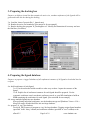
1
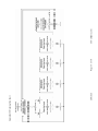
uDock 2.5
User Manual
flexible docking program for MOE/smp clusters
H.Shadnia Sep 2009
Contents
Part A
1- Introduction
2-Theory
Part B
1-Program configuration
2-Preparing the target structure
3- Preparing the docking box
4- Preparing the ligand database
5- Job setup and run
6- Post processing
Part C
Appendix I: Preparation Flowchart
Appendix II: Docking Flowchart
Part D
Selected uDock citations
Part E
Tutorial A: how to render the active site cavity
Tutorial B: how to generate a database of ligand conformers
Tutorial C: Docking of estrogenic ligands into ERa
PART F
Datasets
PART G
FAQ
uDock 2.5
Page 2 of 18
www.shadnia.com
Part A
Introduction
uDock is a flexible ligand-receptor docking program designed for MOE (www.compchem.com). This
program is available for download from www.shadnia.com. The main idea behind uDock is to reduce
the number of commonly used and potentially invalid approximations and cosmetic procedures. In
return, the program requires considerable processing power.
The main workflow in uDock is shown in appendices I,II. In order to run a docking job, three pieces of
data are required:
a- The structure of receptor
This is prepared from the raw crystal coordinates using the PDB-Thaw program and is saved as
a *.moe file. (see section 2)
b- The coordinates of the docking box
This is prepared using the crystal coordinates and the ‘DockingBox’ program. It is saved as a
*.mdb file. (see section 3)
c- The ligand database
This includes all plausible conformers (and sometime even rotamers) of the ligands to be docked
into the active site. It can be prepared in different ways and is saved as a *.mdb file. It also has
to be properly prepared or ‘tagged’ using H_DB_Prepare program. (see section 4)
After the three files were prepared, a job ‘launcher’ job named H_Dock.svl is edited to reflect the
correct filenames, file paths, and some other user settings. Then the docking will be run on a single
machine or a windows / Linux cluster.
The full flowchart of docking is shown in Appendix II. Briefly, random conformations of the each
ligand database entry (individual conformers) are placed inside the docking box, the geometry is
optimized and the energies are recorded before, during and at the end of energy minimization. This is
done for a user defined number of time (usually 10~20 times) which is named the ‘training’ phase. After
that, the program finds typical and minimum energy values for the given complex and tightens the
acceptable energy thresholds for the rest of calculations on the given ligand conformer. The energy
calculations on ligand ‘poses’ which are too high energy will be aborted to save the processing time.
The program will keep generating a user-defined number of ‘good’ solutions for each ligand-conformer
and proceeds to the next entry when done.
The energy minimization steps and the energy criteria used to abort or accept a given pose can be easily
reconfigured for a wide range of applications. For most purposes however, the dynamical ‘Autopilot’
system with the default values is efficient. uDock emphasizes in achieving convergence for each
complex.
uDock 2.5
Page 3 of 18
www.shadnia.com
The docking engine produces a raw output database which includes many conformations of complexes
of ligand-target, with detailed energetics recorded. uDock does not perform any cosmetic ‘cleanup’ on
this database.
A set of ‘post processing’ programs then help to find the best complex, and read the energetics and
perform quantitative calibration or prediction of activities (Section 6 and appendix iii).
Theory
After running extensive tests on different targets, the following rules were used to design uDock:
i – The initial energy of a randomly generated complex has NO meaningful correlation to the final
(energy minimized) value. This is simply because even one single VdW clash between two hydrogens
can raise the energy to hundreds of thousands of kcals/mol. But after a few iterations of energy
minimization, these clashes will relieve and the energy becomes more stable, and comparable to the final
geometry.
ii- The receptor active site can not be treated as rigid (due to the same reason).
iii- Explicit water molecules have to be maintained and treated properly (esp those which make a ‘water
bridge’ between two functional groups. In general they need to be re-arranged or re-docked for each
individual complex. uDock requires the explicit waters to be present but it doesn’t ‘re-dock’ them by
default.
iv- Due to the ‘gearing effect’ [ref] number of possible conformations or ‘poses’ for the simplest ligandreceptor or even host-guest complexes can easy exceed thousands. For this reason uDock tries at least
2~5000 ‘poses’ for each ligand. This is crucial to ensure reaching convergence, i.e. subsequent runs of
the program will not result in a lower energy complex.
Some programs apply ‘smoothing functions’ to VdW terms in order to bypass this effect. Such measures
artificially reduce the complexity of conformational space, but they often diminish the uniqueness of the
active ligands which perfectly fit the grooves of the target active site.
vi- Each conformer of the ligand is a unique chemical entity which may or may not fit to a target. For
this reason all (room temperature) conformers of each ligand need to be individually docked into the
active site. In rare cases where a particular moiety of the receptor is extraordinarily complex, even
different rotamers need to be tried. When docking macromolecular structures as peptides, since it isn’t
possible to generate all plausible conformers, a reasonable number of such conformations generated by
MD may be used.
vii- In theory, the entire conformational space of the target protein need to be explored for each
individual ‘pose’, meaning that the rotatable side chains of the active site residues need to be actively
rotated. The current version of uDock does not perform this, but the program is designed to allow future
implementation of this feature. For many simple active sites (sometimes called ‘rigid active sites’ as
those of nuclear receptors) this is not crucial.
uDock 2.5
Page 4 of 18
www.shadnia.com
viii- The raw crystal coordinates need to be treated properly to generate an energy minimized structure
that is as faithful as possible to the raw coordinates. Three issues are remarkable in this topic:
1 – A regular energy minimization on raw crystal geometry causes unacceptable amount of
perturbation in the geometry, esp. that of buried active sites. For this reason, the ‘PDB Thawing’
program (H_PDB_THAW see www.shadnia.com/H_Thaw ) should be used to energy minimize
the initial PDB file.
2- The limited precision of Cartesian system causes small random perturbations of the
macromolecular geometry independent of ligand binding. For this reason, the energy read outs in
uDock are by default limited to the active site residues (the 1st shell or S1) which are kept fully
flexible. The amino acid residues surrounding the active site (labeled as the 2nd shell or S2) are
kept semi-flexible and their energies are excluded from the final energy calculations. The Atoms
or residues surrounding S2 are kept rigid and can be deleted to accelerate the calculations. The
PDB Thawing program defines these shells, but they can be manually modified too. The
definition of these shells are saved within the ‘receptor’ file that is generated by PDB-Thaw.
3- When comparing different instances of raw crystal structures of supposedly identical
complexes, conformational difference is observed which leads to variation in docking results. A
calibration test can be used to identify and filter out the ‘ill-modeled’ residues in each instance of
crystal structure. This results in dramatic improvement of the docking results [H. Shadnia, J.S.
Wright to be published]
uDock 2.5
Page 5 of 18
www.shadnia.com
PART B
1 – Program configuration
Purpose: installation and configuration of MOE and uDock.
1-a: Program files
The following is the description of the individual programs:
Filename
description
H_Dock.svl
H_Docker.svl
H_Docker_Client.svl
H_DockingBox.svl
H_Host_Tools.svl
H_Toolbox.svl
H_PDB_Thaw.svl
H_DB_Prepare.svl
H_DB_PickMin
H_DB_PicknMin
H_Post_Process
H_IF-E.svl
launcher: configure and launch the docking jobs
Host program
Client program
Graphical interface to define the docking box
Host utilities
Main utilities
Graphical PDB-Thaw program
Graphical program to prepare (tag) the input database
utility to pick the lowest energy conformers
utility to pick n lowest energy conformers
post processing tools
Program to analyze interaction forces and energies
1-b: Setup the environment variable:
For ‘bash’ edit the file .bashrc and add the following lines:
uDock=”~/udock/svl”
export uDock
For other types of shells refer to the operating system manual.
1-c: MOE should be installed and configured to run in SMP mode.
Please refer to MOE manual on ‘Installing and Running MOE/smp’.
1-d: (optional) configure the grid engine (load-balancing system)
1-d:-I: Create a sun-grid-engine file name ‘moe.sge’ that contains the job creation details:
#!/bin/bash
#
# CHOOSE THE NUMBER OF PROCESSOR WHEN YOU RUN THE JOB.
# FOR EXAMPLE TO USE 8 PROCESSORS, TYPE:
#
qsub -pe moe 8 moe.sge
#
#
uDock 2.5
Page 6 of 18
www.shadnia.com
# Tell Gridengine what shell to use:
#$ -S /bin/bash
#
# Start this script from the current working directory:
#$ -cwd
#
# Combine output and error messages into one output file:
#$ -j y
#
# Name of the output file:
#$ -o moe.$JOB_ID.err
#
echo -n "I am running: "
echo $INPUT
/opt/moe/bin/moebatch -mpu $TMPDIR/moemachines -mpulog moe.$JOB_ID.log -run $INPUT
1-d-II: create a job-generation script named ‘runMoe’ as follows:
#!/bin/bash
# usage: runMoe numberOfProcessors inputFile
qsub -v NS=$1,INPUT=$2 -pe moe $1 /opt/moe/bin/moe.sge
2- Preparing the target structure
Purpose: to prepare an energy minimized model of the full or truncated macromolecular target.
2-a: Provide the 3D coordinates
Download the coordinates file from the protein databank (RCSB.ORG) or use the homology
modeling to build it.
2-b: Add hydrogens
In case the hydrogen atoms are missing, add them using MOE>Edit>Hydrogens>Add Hydrogens
or use MOE>Compute>Protonate 3D
2-c: ‘Clean up’ the structure:
3-c-I: Inspect atom types
After selecting the appropriate force-field (MOE>Window>Potential Setup >Force Field), check
the for missing parameters to ensure there are no illegal atom types or undefined force-field
parameters. To do this, use MOE>Window>Potential Setup> Parameters, check ‘Display
Missing Parameters Only’ and under ‘Parameter’ see each individual parameter type, most
importantly ‘Atom Type, Bond-Stretch, Angle Bend,….’ (Fig 1)
uDock 2.5
Page 7 of 18
www.shadnia.com
2-c-II: Inspect missing atoms
Load the ‘sequence editor’ (Main menu > SEQ)
Enable synchronized selection (Sequence Editor > Selection > Synchronize)
Load the ‘Residue Selector’ (Sequence Editor>Selection>Residue Selector)
Under ‘More’ select ‘Amino Acid Atom Data’
Click on each of four classes of missing data to find the missing atoms (see below). Correct the
missing atoms if any.
2-c-III: Delete unrelated chains
Many PDB files include crystallographic or biological dimmers, trimers, or polymers. They
might include co-crystallized small organic molecules or metal ions. Delete all unrelated
structures.
2-d: run the Thawing program.
Select the ligand on the screen and run the ‘H_PDB_Thaw.svl’. Select the appropriate protocol
and click ‘OK’. Refer to http://www.shadnia.com/H_Thaw for more information
3-e: open the thawed file (*_thawed.moe) and delete the ligand. Save this file as ‘_receptor.moe’
or any other name. This is the main ‘target’ file that will be used for docking.
uDock 2.5
Page 8 of 18
www.shadnia.com
3- Preparing the docking box
Purpose: to define a virtual box that contains the active site, random conformers of the ligands will be
generated inside this box during the docking.
3-a. Load the ‘thawed’ protein file (*_thawed.moe)
3-b. Render the active site boundaries [See tutorial A for an example]
3-c. Run the docking box program ’H_DockingBox.svl’. Modify the dimensions if necessary and save
the final box. (See below)
4- Preparing the ligand database
Purpose: to prepare a ‘tagged’ database of all conformers/rotamers of all ligands to be docked into the
receptor.
4-a: build conformers of each ligand
3-a-I. Use the molecular builder module or other ways to draw / import the structure of the
ligand.
3-a-II. Explicit list of conformers/rotamers for each ligand should be prepared. Use the
systematic conformer search, stochastic conformer search, or even MD simulation to build an
MDB containing the conformers for each ligand. [See tutorial B for an example]
4-b: merge multiple ligands into one database
After preparing individual conformers, use the database merge tool [Database Viewer > File >
Merge] to merge all individual files into one large database.
4-c: prepare ( or ‘tag’) the database
Use the H_DB_Prepare.svl program to ‘tag’ the large database. Tagging assigns ligand numbers
(specified in the ‘mseq’ column) and conformer numbers (specified in ‘conf’ column) to the
uDock 2.5
Page 9 of 18
www.shadnia.com
database. uDock uses these tags to monitor different procedures. An example of prepared and
tagged input database is shown here.
5- Job setup and run
5-a: Open the launch program ‘H_Dock.svl’ using the text editor or any other text editor.
5-b: edit the ‘options’ to reflect the correct filenames and desired options. Save the svl file.
5-c: run the launcher program in MOE/smp mode. See MOE’s manual on ‘Installing and Running
MOE/smp’.
5-d how to run the launcher program for HPCVL grid-engine:
5-d-I: To run the program on 24 processors use the following command:
runMoe
24
H_Dock.svl
5-d-II: To monitor the job use ‘qstat’
The following shows an example of the docking job running on 24 processors.
The job has been assigned a job-ID of 5285. It can be killed using ‘qdel 5285’.
job-ID prior
name
user
state submit/start at
queue
slots ja-task-ID
----------------------------------------------------------------------------------------------------------------5285 0.55500 moe.sge
hpc1764
r
09/11/2009 13:23:48 [email protected]
24
5-d-III.To view the docking ‘log’
When docking is running, it creates two files which are named by job-ID as explained above. the error
file contains all debugging messages that would be visible on MOE’s SVL window in graphical mode.
This file would be named ‘moe.5285.err’ for the previous example. You can monitor the progress of
docking by viewing the contents of this file using ‘cat’ or ‘more’ commands or any other method. The
following is an example: (comments are added in italics):
running uDock using the following options:
(input parameters)
[ _program_path:'~/udock/svl/', _project_path:'~/udock/', lig_dbname:'_input.mdb',
output_dbname:'_output.mdb', target_filename:'_receptor_ERa.moe',
docking_box_filename:'_dockingbox_ERa.mdb', _target_good_poses:40, _target_bad_poses:3000,
lig_max_retries:2000, _auto_pilot:'ON', _training_iterations:10, _Threshold_Percent:60,
_forcefield:'$MOE/lib/mmff94s.ff' ]
file transfer adapted to OS: unix
========================= START
uDock 2.5
(automatically assigned)
==================================
Page 10 of 18
www.shadnia.com
running on 64 clients:
(number and list of processing nodes)
compute-1-15
…
compute-1-12.local
================== starting the job spool =========================
running the remote SVL loader
(loading program on processing node)
(v)
OK loading "~/udock/svl/H_Docker_Client.svl" on Clients: .....
(v)
OK loading "~/udock/svl/H_Toolbox.svl" on Clients: .....
Loading Codes on all Clients successful!
>> Scanning the input database: entry 1 of 5
(first ligand/conformer)
!
Training mode: ON
(using 10000kcal/mol limit for energies)
(c)
try #0 to make a good initial pose, max3000 (generate the ligand inside the box)
(c)
rotating .........
(c)
centring .........
(c)
discarding........
(did not fit in the box)
(c)
try #1 to make a good initial pose, max3000
(c)
rotating .........
(c)
centring .........
(c)
good pose found...
(did fit in the box)
:| compute-1-29.local => training (total:1)
.....
:| compute-1-18.local => training (total:9)
:| compute-0-18.local => training (total:10)
[*] Activating AutoPilot
(calculating tight energy thresholds)
== > updating the threshold for task#7 idx=3,value=100000 to 21613.4
== > updating the threshold for task#10 idx=3,value=100000 to 19.2877
== > updating the threshold for task#13 idx=3,value=100000 to -1.13228
training finished, killing all current tasks
(start over with tight thresholds)
:) compute-1-13.local => good pose (total:1)
(good results to be saved in output)
:) compute-0-3.local => good pose (total:2)
:) compute-0-18.local => good pose (total:3)
:( compute-1-30.local => bad pose (total:7)
(bad results will be discarded)
....
:) compute-1-5.local => good pose (total:39)
:) compute-0-13.local => good pose (total:40)
* finished entry #1 in [197] secs, estimated remaining:[788] secs(elapsed\estimated time)
good: 40 [39.2%] , bad: 62 [60.8%], total= 102
(overall statistics)
>> Scanning the input database: entry 2 of 5
(staring up with the next ligand)
....
++
DONE! finished scanning the input database
(the last ligand is sent to the cluster)
>
scanning the input db finished - job spool done
================== Job Spool : Done =========================
Search finished! Aborting further calcs on entry #6
=================================================
= Computations complete
(all calculations are done)
=================================================
total calculation time: 1172.0 seconds
(total time)
good: 200 [30.9%] , bad: 448 [69.1%], wasted:0 , total:648
(performance stats)
The second file contains MOE’s OS level messages and can be used for troubleshooting of MOE/smp
network. This file would be named ‘moe.5285.log’ for the previous example.
6- Post processing
6-a: Understanding the output database
To allow for method development, uDock saves a raw database of the docked poses. The
database contains the following fields:
mseq
uDock 2.5
ligand number
same as the input database
Page 11 of 18
www.shadnia.com
Conf
str_1
str_2
conformer number
//
receptor coordinates at the complex configuration
ligand coordinates at the complex configuration
Thus the most important energies are the ones at the end of the energy minimization (those of
step 4 and 5):
R_4_sel
R_4_int
R_5_sel
The internal energy of the ligand at the bound conformation (EL’)
The interaction energy of the ligand vs. target
(Eint)
The internal energy of the (pseudo) complex. See below for details.
6-b: Quantitative prediction
To allow for fundamental analysis of the results, the raw system energies are recorded in uDock.
This is done in 5 steps, step 1: before any energy minimization, step 2 after 20 iterations of
energy minimization, step 2: after 20 additional iterations, step 3: after 40 more iterations (final
energies). Steps 4,5 at the end of energy minimization. Each energy reading generates three sets
of numbers designated by suffices: _all, _sel, and _int fields which correspond to the total
system energy, the internal energy of the selected atoms and the interaction energy of the
selected atoms vs. the rest of the system. Through step 1 to 4 the ligand structure is selected, and
at step 5 the ligand plus the first shell of amino-acids (active site) are selected.
By definition, the interaction energy is the difference in energy of products (complex) from those
of the reactants (ligand and receptor) in the conformation they hold upon binding:
Eint = EC – (ER’ + EL’)
[1]
In comparison, the binding energy (∆Ebinding) is the difference in energy of products (complex)
from those of the free ligand and receptor:
∆Ebinding = EC – (ER + EL)
[2]
The free ligand and receptor (L, R) are deformed upon binding to form the deformed ligand and
receptor (L’, R’), thus the deformation energies (∆EL, ∆ER) are defined as:
[3]
∆EL = EL’ - EL
∆ER = ER’ - ER
[4]
Thus, from eq. 1:
EC = Eint + ER’ + EL’
From eq.3:
EL = EL’ - ∆EL
From eq.4:
ER = ER’ -∆ER
Now using eq.2:
∆Ebinding = Eint + ER’ + EL’ – (ER’ – ∆ER) – ( EL’ – ∆EL)
= Eint + ER’ + EL’ – ER’ + ∆ER – EL’ + ∆EL
= Eint + ∆ER + ∆EL
Thus, three terms from the docking describe the binding energy
1- Eint
is the ligand interaction energy designated as R_4_int in the raw database.
2- ∆ER
The receptor deformation energy: since the energy of free receptor (ER) and is
arguably difficult to calculate (since it requires explicit solvation of the active site
and more importantly it’s a constant number; from eq.3 the following
approximation can be made:
∆ER = ER’ - const
[5]
uDock 2.5
Page 12 of 18
www.shadnia.com
3- ∆EL
The ligand deformation energy: this can be calculated by subtracting the energy of
ligand in complex by that of the free ligand according to eq.3 .
Two more parameters are required to describe the binding energies
The ligand (de)solvation energy. This can be calculated using Gaussian etc.
4- ∆Gsolv
5- T∆S
Currently there is no easy way to calculate this. When all ligands in a dataset
cause extraction of same number of water molecules from the active site, this term can be
relatively constant. In other cases, for example when a small molecule as phenol is
compared to a larger molecule, this term cannot be ignored. Our group in collaboration
with CCG is developing a method to estimate this term (Oct 2009).
6-c: The detailed analysis of interaction energies
We developed a program to analyze the interaction energy of individual amino acids with
the active site residues. The program calculates and visualizes the interaction energies as
well as forces which allow for detailed analysis and understanding of molecular events
for a given complex. See www.shadnia.com/H_IFE for more details and download.
The analysis of the interaction energies and forces
The red arrows show the atomic forces. The favorable interactions are shown in dark green (0 to -4
kcal/mol) and light green (stronger than -4 kcal/mol). The magnitudes of interactions are displayed
as labels.
We also prepared a comprehensive set of programs also allows automated comparison
and analysis of such forces and energies for an entire set of complexes generated via
docking. Please email the author to obtain this particular package.
uDock 2.5
Page 13 of 18
www.shadnia.com
PART C
Appendix I: Preparation flowchart
uDock 2.5
Page 14 of 18
www.shadnia.com
uDock 2.5
Appendix II: Docking flowchart
Page 15 of 18
www.shadnia.com
PART D
Selected uDock citations
Deconstructing the estradiol ABCD ring structure: A new family of A-CD compounds which are potent and selective
estrogen receptor agonists [Link]
Tony Durst*, Mohammed Asim, James S. Wright, Hooman Shadnia, Christine Pratt, John Katzenellenbogen, Kathryn
Carlsen. Bioorganic and Medicinal Biology, 2009
Investigation of Residues K112, E136, H138, G247, Y248, and D249 in the Active Site of
YeastCystathionine β-Synthase [Link]
Pratik Lodha, Hooman Shadnia, Colleen M. Helferty, James Wright, and Susan M.
Aitken*. Biochemistry and Cell Biology, 2009
Design, synthesis, and biological evaluation of substituted 2-alkylthio-1,5-diarylimidazoles as selectiveCOX-2
inhibitors [Link]
Latifeh Navidpour*, Hooman Shadnia, Hamed Shafaroodi, Mohsen Amini, Ahmad Reza Dehpourd and Abbas
Shafiee.Bioorganic & Medicinal Chemistry, 15, 1976–1982,2007
Protein Targeting with Small Molecules: Chemical Biology Techniques and Applications
Hiroyuki Osada , Wiley, ISBN: 978-0-470-12053-8, Hardcover
uDock 2.5
Page 17 of 18
www.shadnia.com
PART E
Tutorials
[To be added soon!]
Tutorial A: how to render the active site cavity
Tutorial B: how to generate a database of ligand conformers
Tutorial C: Docking of estrogenic ligands into ERa
PART F
Datasets
[To be added soon!]
PART G
FAQ
[To be added soon!]
uDock 2.5
Page 18 of 18
www.shadnia.com