1
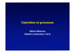
Why do humans
have so many headaches?
Stasha Gominak, M.D.
East Texas Medical Center Neurologic Institute
700 Olympic Plaza, Suite 912 Tyler, Texas 75701
April 25 , 2014
Headache is described in
every human society
throughout written history
Why would it be so common?
Headache is a genetic
disorder
Why would we want to pass on these
horrible headaches to our kids?
We think that genes providing a
survival advantage get spread
throughout the population
What would be the survival advantage of having
a headache?
Could the genes for headache
convey some other thing that
might improve survival?
Why have we taught each
other that “normal
headache” and migraine are
two different things?
What if headache and migraine
are the same?
What if “migraine” and “normal
headache” occur by the same
mechanism?
Why do my patients use the phrase
“normal headaches”?
■
■
■
Why do we think it is
“normal” for the head to
hurt without injury?
Why haven’t we fixed
our most common
neurologic problem?
Are we thinking about it
the wrong way?
What is the evidence that migraine and
headache are on a continuum?
■
■
■
■
All migraine sufferers also have “normal headaches”.
When the triptans became available (triptans are
migraine medications; sumatriptan, rizatriptan,
naratriptan…etc.) we told our patients ”save these for
your migraines”
Their response: “if I take the medicine soon enough it
works.”
The patients found that triptans worked for their milder,
“normal headaches” before they grew into a “migraine”.
The triptans work for “normal
headaches” too
■
■
■
We may not prescribe triptans for normal headaches
because they’re very expensive, but they do work well.
That probably means serotonin plays a role in “normal
headache” as well as migraine
Baby migraine, which is “just a headache”, may grow
into a bigger headache that acquires other features which
make it recognizably “migraine”.
What do we know about the mechanism of
the triptans?
■
■
■
■
Triptans work on 1B and 1D serotonin receptors
1B and 1D receptors are feedback inhibitors; they
decrease serotonin release.
Does that mean that the mechanism of action has to do
with the blood vessels? (Which is what we’ve been
taught.)…… Not necessarily.
Serotonin appears in many areas of the brain.
Serotonin Release
■
■
Most of the serotonin
measured in the brain
originates from the raphe
nuclei in the posterior
brainstem.
Serotonin acts like a neurotransmitter as well as a
hormone. It is released along
the axon as well as at the nerve
terminals bathing the entire
brain in “happy juice” every
few seconds
Sleep and Serotonin
■
■
■
REM sleep and triptans have something in common:
They both drop serotonin levels. In order to enter
REM sleep we must have low serotonin.
Serotonin is high when we are awake but low when we
enter deep sleep.
Your brain wants to be very, very sure that you are
indeed sleeping before you start to dream. Because
REM and awake are similar states the serotonin level
helps the brain know which state you’re in .
Refs: 1. 2
Migraine and Sleep
■
■
If our patients have told us for the last 100 years
that getting into deep sleep is how they “break”
the headache, why are there so few articles
showing us what the sleep looks like in migraine
sufferers?
Why have we been told that sleep disorders only
happen in fat, old men?
Migraine and Sleep
■
■
■
■
I became interested in sleep 10 years ago when one of my
young, normal weight patients insisted that I send her for
a sleep study. Her husband said she “snored like a train”.
She had been on four preventative medicines over a
period of two years and still had daily headache.
She had severe sleep apnea and 6 weeks of sleeping with
CPAP mask completely cleared her headaches.
For the following 5 years I ordered sleep studies on all of
my daily headache patients. They were all abnormal.
Migraine and Sleep
■
■
■
■
■
■
Ten years later there are still few studies looking at the
results of sleep studies in migraine sufferers. Why?
Academic neurologists who are sleep specialists do not
usually study migraine?
Those who are migraine specialists do not study sleep?
Those who study astrocyte anatomy do not see patients?
Most physicians feel more comfortable going along with
the currently accepted medical theories.
But what if the theories don’t explain what the patient
feels?
“Explanations” of headache are theories
■
■
■
■
■
What the patient experiences is the only “truth”.
Headache patients are, by definition “normal” ; normal
scan, normal neuroanatomy. They don’t die from
headache so there are no autopsy studies.
Every one of our explanations is made up; it’s a theory.
There is no user’s manual that confirms which is the
real “truth”.
But my explanation of cause will direct my search for
how to fix it.
Sleep study results in migraineurs
■
■
■
■
■
■
Many of my migraine patients don’t sleep normally. They have
various forms of insomnia, “light sleeper”, “not a morning person”.
All of them had abnormal sleep studies, just not necessarily apnea.
The most common sleep study results in my young, healthy migraine
patients were delayed onset of REM, decreased REM and REM
related apnea. Some slept for 10 hours and had no REM.
We have not been treating migraine by treating sleep because we
haven’t known how.
The sleep medications we have do not produce normal sleep.
But if you know how to fix the sleep, fixing the sleep does indeed fix
the headaches.
Are we the only ones?
I hope to convince you of the following:
■
■
■
■
■
Migraine does not occur in the cerebral blood vessels.
Sleep and migraine are intertwined.
Migraine is a genetic disorder that leads to hyperexcitability of the posterior brainstem and occipital
lobe.
The posterior brainstem sleep nuclei are designed to
turn on and off spontaneously.
That spontaneous “on” signal can accidently “leak” into
the surrounding nuclei causing them to accidently turn
on also, even though they’re not “supposed to”.
The trigemino-vascular theory of
migraine is the old theory
■
■
■
■
This theory, which has been the most popular explanation for
migraine, grew out of the fact that there are no pain fibers in the
brain itself.
The pain fibers are only on the meninges, (the linings that cover
the brain), and on the cerebral blood vessels.
As they are the only pain receptors in the brain the trigeminovascular theory suggests that the pain is experienced in these
receptors.
It proposes that “inflammatory” signals generated in the trigeminal
fibers at the meninges and the blood vessels cause the head pain.
Is there a minority opinion?
■
■
Dr. Michael Welch and Dr. Peter Goadsby have been
the major proponents of an alternative view which
suggests that the trigeminal caudal nucleus and the
occipital lobe are hyper-excitable in migraine patients.
The pain is experienced in the brain stem not in the
blood vessels.
Some of Dr. Welch’s articles establishing hyperexcitability of the brainstem in migraine patients
■
■
■
■
■
■
■
■
■
■
Brain hyperexcitability: the basis for antiepileptic drugs in migraine prevention.Welch KM. Headache. 2005 Apr;
45 Suppl 1:S25-32. Review.
Contemporary concepts of migraine pathogenesis.Welch KM. Neurology. 2003 Oct 28;61(8 Suppl 4):S2-8.
Review.
Functional MRI-BOLD of brainstem structures during visually triggered migraine. Cao Y, Aurora SK, Nagesh
V, Patel SC, Welch KM.Neurology. 2002 Jul 9;59(1):72-8.
Cortical spreading depression and gene regulation: relevance to migraine. Choudhuri R, Cui L, Yong C, Bowyer
S, Klein RM, Welch KM, Berman NE. Ann Neurol. 2002 Apr;51(4):499-506.
Magnetoencephalographic fields from patients with spontaneous and induced migraine aura. Bowyer SM,
Aurora KS, Moran JE, Tepley N, Welch KM. Ann Neurol. 2001 Nov;50(5):582-7.
Periaqueductal gray matter dysfunction in migraine: cause or the burden of illness? Welch KM, Nagesh V,
Aurora SK, Gelman N. Headache. 2001 Jul-Aug;41(7):629-37.
The occipital cortex is hyperexcitable in migraine: experimental evidence. Aurora SK, Cao Y, Bowyer SM,
Welch KM. Headache. 1999 Jul-Aug;39(7):469-76.
MRI of the occipital cortex, red nucleus, and substantia nigra during visual aura of migraine.Welch KM, Cao Y,
Aurora S, Wiggins G, Vikingstad EM. Neurology. 1998 Nov;51(5):1465-9.
Transcranial magnetic stimulation confirms hyperexcitability of occipital cortex in migraine. Aurora SK, Ahmad
BK, Welch KM, Bhardhwaj P, Ramadan NM.Neurology. 1998 Apr;50(4):1111-4.
Brain excitability in migraine: evidence from transcranial magnetic stimulation studies.Aurora SK, Welch KM.
Curr Opin Neurol. 1998 Jun;11(3):205-9. Review.
Dr. Goadsby’s articles regarding hyper-excitability of
the brainstem in migraineurs
■
■
■
■
■
■
■
■
Brain activations in the premonitory phase of nitroglycerin-triggered migraine attacks. Maniyar
FH, Sprenger T, Monteith T, Schankin C, Goadsby PJ. Brain. 2014 Jan;137(Pt 1):232-41.
Diencephalic and brainstem mechanisms in migraine. Akerman S, Holland PR, Goadsby PJ. Nat
Rev Neurosci. 2011 Sep 20;12(10):570-84.
Pathophysiology of migraine. Goadsby PJ. Neurol Clin. 2009 May;27(2):335-60.
Trigeminocervical complex responses after lesioning dopaminergic A11 nucleus are modified by
dopamine and serotonin mechanisms. Charbit AR, Akerman S, Goadsby PJ . Pain 2011 Oct;152
(10):2365-76.
The vascular theory of migraine--a great story wrecked by the facts. Goadsby PJ . Brain 2009 Jan;
132(Pt 1):6-7.
Functional neuroimaging of primary headache disorders. Cohen AS, Goadsby PJ. Curr Pain
Headache Rep. 2005 Apr;9(2):141-6.
A PET study exploring the laterality of brainstem activation in migraine using glyceryl trinitrate.
Afridi SK, Matharu MS, Lee L, Kaube H, Friston KJ, Frackowiak RS, Goadsby PJ. Brain 2005
Apr;128(Pt 4):932-9.
Activation of 5-HT(1B/1D) receptor in the periaqueductal gray inhibits nociception. Bartsch T,
Knight YE, Goadsby PJ . Ann Neurol. 2004 Sep;56(3):371-81
The Minority Opinion:
■
■
Is that migraine is not
experienced at the endings of
the nerves but is instead
experienced in the nucleus
where the wires send their
messages ; the Trigeminal
Nucleus Caudalis.
Unfortunately Dr. Welch, who
originated this viewpoint, has
been effectively drummed out of
the headache meetings because
his ideas are different.
If the blood vessels are the only part of
the brain with pain fibers it seems
perfectly logical to blame them for the
headache
And, by the way, why doesn’t the brain have pain
fibers?
The brain and the spinal cord don’t
have pain fibers in the pinkish- grey
gooey stuff because they don’t need
them
The brain and the spinal cord are the only parts with
the skeleton on the outside
The skull protects the brain from
penetrating objects
■
■
But the skull does not
keep the soft, fragile
brain from banging
against the inside of the
skull when shaken
The pain fibers are on
the vessels and the
meninges to tell us not
to bang our heads.
This still doesn’t tell me why it’s
“normal” to have a headache
(when it’s not normal for any other part of our
body to start hurting for no reason.)
Is there something different about
the pain system of the brain that
would make it more likely to turn
on spontaneously?
The head pain system is in two parts
■
Dorsal horn C1-C4
■
The Trigeminal Nucleus
Caudalis : in blue
perceives pain for the
face and the front of the
scalp shown in pink and
lavender
The dorsal horn of C 1C4 shown in green
perceives pain for the
back of the head and
upper neck.
Headaches happen in head and in
the neck
■Headaches
Dorsal horn C1-C4
start just as
commonly in the neck as in
the head, even though the
neck is not really “in” the
head.
■Why do they both turn on
spontaneously?
■Why those two and not
other nuclei?
Pain system extends down the spinal
cord but does not just “turn on”
■
Dorsal horn C1-C4
■
■
There are analogous cells all the
way down the spinal cord
perceiving pain from the rest of
the body called the dorsal horn
of C4-C8, the dorsal horn of
T1-T12…
Why don’t they turn on
spontaneously too?
What’s the difference between
dorsal horn C1-C4 and those
below?
Why just the trigeminal nucleus and
upper cervical roots and not the rest?
■
■
Could the top two; the trigeminal nucleus and
the top part of the dorsal horn have something
nearby that affects only them, that doesn’t
extend down into the spinal cord?
What about the periaquiductal grey nuclei that
govern sleep, including the raphe serotonergic
nuclei?
Sleep happens here too. Could it affect the
nearby trigeminal and dorsal column nuclei?
Nucleus reticularis pontis oralis
The Periaquiductal Gray runs the timing and
paralysis of sleep
■
■
■
■
The pacemaker cells in the
periaquiductal grey pictured in
red, beat all day all night.
They are the brain clock that
determines when we sleep
The paralysis switch is here also,
Nucleus Reticularis Pontis
Oralis.
The two are heavily intertwined
to be sure that we only get
paralyzed while we are deeply
asleep.
Nucleus Reticularis Pontis Oralis
Why would areas of the
brainstem that are next to each
other affect each other?
(That seems rather sloppy)
Genetic mutations that
cause migraine
(Or, how your hyperactive neighbor in
the brainstem might just make you
cranky too.)
Genes that cause migraine affect the
electrical excitability of brain cells
■
■
■
There are now about 40 genes that are linked to
migraine
All of these genes are mutations in the cellular
apparatus that allows us to turn our cells on and
off:
Ion Channel Mutations.
Ca++ channel in a membrane
■
■
■
■
Our cellular electricity is like a car battery; we use ions floating in
water.
Our brain uses Ca++, K+, Cl-, Na+.
The channels move these ions in and out of our cells to turn them
“on” or “off”.
We have multiple Ca++ channels, K+ channels, etc., each has a
specific role, or several specific roles, in our body.
Ca ++ channels turn cells“on”
Ca++ pumps turn them “off”
■
■
■
■
To turn the cell “on” Ca++
channels open.
The cell is now very positive
inside; it is “on”.
To turn “off” it pumps out
the positive charges.
The mutation leads to a
malfunctioning channel; the
cell goes “on” but can’t turn
“off “ again.
Voltage gated
Ca++ channel
Lots of +’s cell is
++
+ON+++
+
++
++
+
+
+
+
Migraine is a Channel Disorder
■
■
■
There are now multiple Ca++ and Na+
channel mutations linked to migraine.
Also mutations of Ca++ pumps and most
recently Na-K ATPase.
But which cell type has these mutated
channels and how does a malfunctioning
channel produce headache?
Is their proof that the
posterior brainstem is too
“on” in migraine?
PET Scans in Migraine Patients
show that the posterior brain stem is too “on”
Weiller C, May A, Limmroth V, et al. Nature Med 1995;1:658-660
Refs: 5,7,10,21
How do the channel mutations
result in brainstem and occipital
lobe hyper-excitability?
Which brain cell has the mutant
channels? Is it the neurons or some
other cell in the brain?
Are there other imaging procedures
that show what happens in the brain
during a migraine?
1960’s Experiments: “Spreading
Depression”
■
■
Enrolled migraine patients who had a warning, a
visual aura, telling them the headache was about
to happen
They rushed them into a magnetic field as they
were experiencing the visual symptoms to
measure the electrical events during and after the
visual aura.
Magnetic Field Studies
Starting with the visual aura they observed electrical suppression, starting
in the back during visual aura, moving slowly forward taking 15 minutes to
go from back to front
Magnetic Field Studies
electrical suppression, starting in the back during visual aura, moving
slowly forward, 15 minutes to go from back to front
Magnetic Field Studies
electrical suppression, starting in the back during visual aura, moving
slowly forward, 15 minutes to go from back to front
But what did it mean?
■
■
■
■
Why was it moving so slowly, about 3mm/min?
Why was it moving in a wave spreading outward
like a ripple in water instead of jumping from
one place to another like neurons transmit
messages?
What did it have to do with the headache?
What cell in the brain produced this wave?
Spreading Depression of Dr. Leao
observed in rabbit brain slices
Spread of the visual aura was
at the same speed as the
■
■
■
■
Spreading wave in the brain
Stimulating the brain electrically caused a slowly spreading electrical wave.
Traveling 3mm/min, contiguously, taking about 15 minutes to cross the brain
Why is it so slow? What moves at this rate in the brain? It’s too slow for
neurons.
Is it related to migraine in humans?
I always get a headache
when I have to ride in
the car.
Bella can’t tell us if she has a headache and
sometimes she looks a little depressed about it
Astrocytes to the Rescue!
Astrocytes may explain “spreading
depression”
■
■
Confocal microscopes
show us brain cells in 3
dimensions.
We thought these little
“star-like” cells were the
skeletal system of the
brain as they had many
processes spreading out
like a star.
Astrocyte
Neuron
Astrocytes are more influential than
previously imagined
■
■
■
Astrocytes are electrically
active cells that can talk to
one another and other brain
cells.
Their dendrites wrap around
20-30 neurons with multiple
endings on the surface of the
neurons giving excitatory or
inhibitory input to the
neurons.
Each astrocyte is assigned
several neurons and a blood
vessel.
Spreading Depression of Leao is an
inter cellular calcium wave
■
■
■
Astrocytes have gap junctions that
open between adjoining cells
allowing them to directly share
their ionic environments.
Spreading depression may be a
spreading inter-cellular calcium
wave traveling through the
astrocyte population.
The wave travels slowly,
3mm/min, and contiguously,
because it is transmitted by the
astrocytes, not the neurons
The Astrocyte Neurovascular Unit
■
■
■
■
A single astrocyte and it’s
neurons are called “astrocyte
neurovascular unit”
A chemical blood signal can
be received by the astrocyte,
then sent to the neurons
amplifying the message
Thus, spreading depression
has a similar arterial
vasoconstrictive wave that
accompanies it.
The change in mental status
and paralysis is the neuronal
effect not the vascular
effect.
What have we suggested so far?
■
■
■
■
■
Headaches happen equally in the neck and head
Small headaches may grow into big migraine
Brain stem hyper-excitabilty has been observed
in various types of studies.
Astrocyte physiology seems to explain spreading
depression
The astrocytes probably carry the channel
mutations and are the “hyper- excitable “ cells
Are there other migraine symptoms that must
be explained by our theory?
■
■
■
■
■
■
■
■
Dizziness
Hypersensitivity to light sound and smell
Ringing or buzzing in the ears
Visual aura
Nausea
Nasal congestion
Sleepiness
Stroke like episodes; weakness, aphasia
What about the other migraine symptoms? They’re
not in the trigeminal caudal nucleus but they’re right
nearby
■
■
■
Nausea from the
Chemotrigger Zone
Facial congestion from
the Salivatory Nucleus
which innervates the
mucosa of the sinus
cavities .
Several, nearby
brainstem nuclei are
being excited together.
Chemotrigger zone
Can this theory explain the accompanying
symptoms of migraine?
■
■
■
■
■
■
■
■
Dizziness - brain stem cerebellar nuclei
Hypersensitivity to light sound and smell-lat and med. geniculate
Tinnitus - VIIIth nerve nucleus
Visual aura - occipital lobe
Nausea - chemotrigger zone
Nasal congestion - salivatory nucleus
Sleepiness - raphe nuclei
Stroke like episodes; weakness, aphasia- brain stem or cortex
Astrocyte anatomy is regional
■
■
Astrocytes do not follow
neuronal anatomy, they
overlap adjoining nuclei
supplying regions of the
brain.
There may be regional
differences in astrocyte
physiology.
What about the visual “aura” of
migraine
■
■
The visual warning of
migraine is thought to be a
spontaneous electrical
discharge of the occipital
lobe as seen in the spreading
depression experiments.
We know that some
migraines start there and not
in the brainstem, what
would link the brainstem
to the occipital lobe
making both hyperexcitable?
PGO waves
■
■
■
■
Pons Geniculate Occipital Lobe
PGO Waves
Waves that go back and forth
between the brainstem and the
occipital lobe at the rate of REM
eye movements. ( Even while we’re
awake.)
These waves may suggest a special
population of astrocytes linking the
posterior brainstem to the occipital
lobe ( and probably hypothalamus
geniculate ganglia and thalamus).
Ref: 33, 34
PGO waves
■
■
■
Pons Geniculate Occipital Lobe
PGO Waves
Waves that go back and forth
between the brainstem and the
occipital lobe at the rate of REM
eye movements. ( Even while we’re
awake.)
These waves may suggest a special
population of astrocytes linking the
posterior brainstem to the occipital
lobe ( and probably hypothalamus
geniculate ganglia and thalamus).
PGO waves
■
■
■
Pons Geniculate Occipital Lobe
PGO Waves
Waves that go back and forth
between the brainstem and the
occipital lobe at the rate of REM
eye movements. ( Even while we’re
awake.)
These waves may suggest a special
population of astrocytes linking
the posterior brainstem to the
occipital lobe ( and probably
hypothalamus geniculate ganglia
and thalamus).
Why are all these waves
and excitable
astrocytes important?
The channel mutations probably
didn’t arise to cause headaches
■
■
The same astrocyte population which affects the
“excitability” of the sleep switches also affects
the whole posterior brainstem.
Since sleep is the most important thing we do
every day, mutations that improve sleep ( make
it “switch on” easily) might convey a survival
advantage and become common in humans.
Key Points of Brainstem Hyper excitability
● Activation observed in the posterior brain stem on PET
scans in migraine patients.
● Activation of the posterior brain stem can result in pain
anywhere along the trigeminal-cervical network;
including the head, the neck, and the face.
● Activation of the Salivatory Nucleus can lead to sinus
congestion, nausea from the chemotrigger zone,
hypersensitivity to light sound and smell from
connections to the geniculate ganglia.
● Dizziness, tinnitus, double vision, all brain stem nuclei
How do we fix the
headaches?
If I’ve had this “mutation” since I was
born why is it only showing up now?
Fix the sleep fix the headaches
■
■
■
We have to fix the hyper excitability of the brain
stem nuclei, make them go back to “off” .
The pills we’ve used for prevention of migraine
(even before the mutations were described) are
calcium channel blockers like verapamil, and
sodium channel stabilizers like topiramate.
They work by stabilizing cranky, easily excitable
cells that are turning “on “ too easily.
Most people only get
an occasional mild
headache
They have the mechanism to make a
headache but it doesn’t act up all the
time and make their life miserable.
How do I get back to
being one of those
people?
What I learned from sleep apnea masks
■
■
■
■
■
Why did the CPAP mask make my patient’s headaches
better?
The masks are not about getting oxygen to the brain,
that’s what the blood does.
We all get paralyzed in deep sleep and we have to be
paralyzed to repair
Apnea occurs when the paralysis system gets goofed up
and we get too paralyzed in deep sleep
The mask blows air in to allow the brain to stay in deep
sleep long enough to get work done.
The Periaquiductal Gray runs the timing and
paralysis of sleep
■
■
■
■
The pacemaker cells in the
periaquiductal grey pictured in
red, beat all day all night.
They are the brain clock that
determines when we sleep
The paralysis switch is here also,
Nucleus Reticularis Pontis
Oralis.
The two are heavily intertwined
to be sure that we only get
paralyzed while we are deeply
asleep.
Nucleus Reticularis Pontis Oralis
What I learned from CPAP masks
■
■
■
■
My patients returned and said not only were
their headaches gone but they were on fewer
blood pressure meds and they were off their
diabetes pills.
Their knee pain was gone they could think more
clearly and they didn’t feel depressed any more,
And oh, yeah, I think my memory is better too.
Does that mean we repair everything in sleep ,
do we make insulin in sleep, do we make our
serotonin in sleep?
Sleep is not just being unconscious
■
■
■
■
■
We all know what it feels like to wake tired
Sleep is not a passive process
There are specific stages of sleep in which we get
paralyzed and get the “work” of sleep done
I believe that we all make enough chemicals in
sleep to last about 16 hours, then we run out and
we have to go back to sleep to make more.
I believe all repair of all systems only happens
while we’re sleeping
Apnea is not the only sleep disorder
■
■
■
■
My 18 year old patient with daily headache slept for 10
hours during her sleep study but had no deep sleep at
all, no apnea, but also no deep sleep.
Most of my headache patients have reduced or no
REM sleep.
They all say the same thing: “I have a headache every
day, I can’t remember anything and I’m in a bad
mood.”
The chemicals that prevent headache are made in deep
sleep, memories are made in deep sleep serotonin to
make us happy is made in deep sleep.
How do we get the REM back?
■
■
■
■
Why is there no REM ?
Apnea is the end stage terrible disease before we die,
none of us want to get even close to that.
Why does everyone who comes to see me, regardless of
the problem; headache, vertigo, tremor, burning in the
feet, balance difficulty, parkinson’s, seizure, tics, stroke,
all have an abnormal sleep study? Even little 8 year
olds?
This is an epidemic that began in the early 1980’s as did
fibromyalgia and chronic fatigue
Why do I want my REM sleep back?
■
■
■
■
While we’re sleeping we make millions of chemicals
that allow our bodies to run normally.
If you’ve always had the migraine gene mutation but
didn’t always have a headache then you made
modifications in other chemicals that allowed your cells
to run normally, to stay “off” …… until.
You stopped sleeping in deep sleep long enough every
night to make those chemicals that “shored up” your
weakness.
Each of us have genetic weaknesses we’re born with.
Give me back my REM sleep
■
■
■
■
Could it be that each drug that helps migraine is really
just duplicating a chemical the brain ran out of?
And my brain knows how to make my chemical ,which
exactly fits the gene mutation I have, but I only make
that chemical in REM sleep.
40 different channel mutations may explain why I have
only partial success with the medicines I use, and they
“wear off”, the headaches get better, then they’re back
again.
Your brain knows exactly which chemical to make for
you, it’s been doing it since you were born.
Sleep Disorders
■
■
■
■
There are many types of abnormal sleep, one is
“I sleep all night but wake with a headache”.
If you wake up every morning with mild neck
pain or facial pain your sleep is not “normal”
If you sleep all night, wake feeling “fantastic”
and have no pain and no medical problems, then
your sleep is normal.
That is common now in my practice but not
common in the developed world today.
Vitamins are Dangerous
■
■
■
■
Fixing the sleep is not just a matter of taking vitamins,
they are chemicals that have to stay in a specific range
for sleep to occur normally, use them carefully.
But humans and all other animals lived here on this
planet for millions of years before doctors arrived.
They missed those “well rounded diet” lectures, most
of the squirrels still don’t get those lectures.
That means the things our bodies can’t make were
actually partly from the intestinal bacteria and partly
from the food.
Humans lived before doctors
■
■
■
■
Most died of infectious diseases that we have
eliminated to large extent
What remains is slow death by organ failure;
diabetes, heart attack, stroke, parkinsons,
alzheimers, cancer.
All of these diseases result from incomplete
healing during sleep and can be partially or fully
reversed by sleeping normally.
Headache can be cured by sleeping normally
D and B vitamins
■
■
■
■
■
The epidemic of low vitamin D in the developed world
started with computer, television and air conditioning
in the late 70s early 80’s when we all went indoors.
Since there are no drugs to bring back REM what we’re
left with is trying new things.
Go to the vitamin D lecture to see the connection in
detail but vitamin D deficiency is the cause.
Get the D to 60-80 ng/ml and fix the secondary effects
of low D (intestinal bacterial change) so the B’s get
made daily in the right amounts
And the REM comes back and the headaches go away
Why sleeping pills are valuable
■
■
■
■
16 year olds with their first bout of daily headache are
easy to fix with vitamins
52 year olds with daily headache for the last 30 years are
not easy to fix with just vitamins
Every night the brain tries to fix the sleep switches but
doesn’t have the time in deep sleep to succeed.
Daily headache for 30 years means that patient has old,
rusty, poorly functioning sleep switches and even when
those cells get the raw materials they’ve needed, (the
vitamins) they don’t just snap to it and work perfectly.
Long standing sleep disorders
usually require sleeping pills
■
■
■
■
If you give a sleeping pill but don’t fix what’s wrong in
the background they may work for a little while but
then they “wear off” and they need more and they “get
addicted”
They can’t sleep without the medication, but they
couldn’t sleep in the first place.
The medications aren’t bad they just aren’t the whole
answer.
Fix what’s wrong in the background and use the sleep
meds as a bandaid to help while the brain is repairing
It is sleep, not vitamins that cures the
headaches
■
■
■
■
■
Sleep is the cure for headache, if the sleep is not
normal the headaches won’t resolve.
Be patient. If the sleep has been abnormal for 30
years it doesn’t get fixed over night.
Find the sleeping pill that is right for you.
Fall asleep stay asleep and wake feeling great
means that medication is what the brain has
been needing to get into the right phases
They won’t work alone but they are helpful
CPAP is still helpful
■
■
■
■
Anyone with apnea will still get better faster with the
mask on.
Many people who have apnea will not get fixed unless
they are able to wear the mask and sleep
Listen to your body, if you sleep better in the recliner
stay in the recliner.
People end up sleeping on the couch because they sleep
better there than in the bed. The couch keeps them in a
position where their apnea is less.
Listen to your body
■
■
■
■
As you get better, the drugs I have used to help you will
start to have different effects
The sleep medicine that used to help you now makes
you dopey in the morning
Just like taking away the blood pressure meds when the
blood pressure normalizes you need to take away sleep
meds as the sleep gets better
Always wait until your body tells you it doesn’t want
them, they make you feel funny now instead of better
Listen to your body
■
■
■
Once the sleep gets better and the headaches go away
you have to learn what to watch for so they don’t come
back
The vitamin D is hard to keep in range. Every person
needs a different dose depending on their skin color,
where they live, how much they’re in the sun in the
summer and how long they were sick
This means the D dose usually goes down over the first
2-3 years as we get better, we have to have a way to tell
when to get the level checked and catch it before our
sleep falls apart again and the headaches come back.
Little headaches are important
■
■
If you learn that those little “normal headaches”
are warning you that your head pain switch is
getting cranky again and you do something
about it right away, like check the D level, you’ll
fix your sleep before you’ve spent another six
months not sleeping and the headaches are out
of control again.
Each time your sleep gets goofed up for more
than a brief period the headaches will return
until you’ve had months on end of normal sleep.
Headache meds are still important
■
■
■
■
■
The preventatives such as verapamil and topiramate are still
necessary for many patients. There’s nothing wrong with using
them but the improvement won’t last if the sleep remains
abnormal.
The triptans are very important.
The warnings per the FDA are not correct. The receptors that
they work on do not increase serotonin, they decrease serotonin,
and they do indeed cause chest and joint aching but they are
generally very safe.
They do not work in the daily headache sufferers because they
work best when the headache is in the earliest stages.
Even if they failed when the headaches were daily, they will
usually work later when the headaches are once a week.
Always have a CT scan
■
■
Any patient with headaches bad enough to talk
to their doctor or watch this lecture needs a CT
of the head at least once.
There is no difference between the headache of
a brain tumor and “normal headache” at the
beginning. Always confirm that the anatomy is
normal before assuming that it’s migraine.
References
1. Why does serotonergic activity drastically decrease during REM sleep? Sato K . Med
Hypotheses. 2013 Oct;81(4):734-7.
2. Serotonin control of sleep-wake behavior. Monti JM Sleep Med Rev. 2011 Aug;15(4):
269-81
3. Brain hyperexcitability: the basis for antiepileptic drugs in migraine prevention. Welch
KM. Headache. 2005 Apr;45 Suppl 1:S25-32. Review.
4. Contemporary concepts of migraine pathogenesis.Welch KM. Neurology. 2003 Oct 28;
61(8 Suppl 4):S2-8. Review.
5. Functional MRI-BOLD of brainstem structures during visually triggered migraine. Cao
Y, Aurora SK, Nagesh V, Patel SC, Welch KM. Neurology. 2002 Jul 9;59(1):72-8.
6. Cortical spreading depression and gene regulation: relevance to migraine. Choudhuri R,
Cui L, Yong C, Bowyer S, Klein RM, Welch KM, Berman NE. Ann Neurol. 2002 Apr;51
(4):499-506.
7. Magnetoencephalographic fields from patients with spontaneous and induced migraine
aura. Bowyer SM, Aurora KS, Moran JE, Tepley N, Welch KM. Ann Neurol. 2001 Nov;50
(5):582-7.
References
8. Periaqueductal gray matter dysfunction in migraine: cause or the burden of
illness? Welch KM, Nagesh V, Aurora SK, Gelman N. Headache. 2001 Jul-Aug;
41(7):629-37.
9. The occipital cortex is hyperexcitable in migraine: experimental evidence.
Aurora SK, Cao Y, Bowyer SM, Welch KM. Headache. 1999 Jul-Aug;39(7):46976.
10. MRI of the occipital cortex, red nucleus, and substantia nigra during visual
aura of migraine. Welch KM, Cao Y, Aurora S, Wiggins G, Vikingstad EM.
Neurology. 1998 Nov;51(5):1465-9.
11. Transcranial magnetic stimulation confirms hyperexcitability of occipital
cortex in migraine. Aurora SK, Ahmad BK, Welch KM, Bhardhwaj P, Ramadan
NM. Neurology. 1998 Apr;50(4):1111-4.
12. Brain excitability in migraine: evidence from transcranial magnetic stimulation
studies. Aurora SK, Welch KM. Curr Opin Neurol. 1998 Jun;11(3):205-9.
References
13. Brain activations in the premonitory phase of nitroglycerin-triggered migraine
attacks. Maniyar FH, Sprenger T, Monteith T, Schankin C, Goadsby PJ. Brain.
2014 Jan;137(Pt 1):232-41.
14. Diencephalic and brainstem mechanisms in migraine. Akerman S, Holland
PR, Goadsby PJ. Nat Rev Neurosci. 2011 Sep 20;12(10):570-84.
15. Pathophysiology of migraine. Goadsby PJ. Neurol Clin. 2009 May;27(2):33560.
16. Trigeminocervical complex responses after lesioning dopaminergic A11
nucleus are modified by dopamine and serotonin mechanisms. Charbit AR,
Akerman S, Goadsby PJ . Pain 2011 Oct;152 (10):2365-76.
17. The vascular theory of migraine--a great story wrecked by the facts. Goadsby
PJ . Brain 2009 Jan;132(Pt 1):6-7.
18. Functional neuroimaging of primary headache disorders. Cohen AS, Goadsby
PJ. Curr Pain Headache Rep. 2005 Apr;9(2):141-6.
References
19. A PET study exploring the laterality of brainstem activation in migraine using glyceryl
trinitrate. Afridi SK, Matharu MS, Lee L, Kaube H, Friston KJ, Frackowiak RS, Goadsby
PJ. Brain 2005 Apr;128(Pt 4):932-9.
20. Activation of 5-HT(1B/1D) receptor in the periaqueductal gray inhibits nociception.
Bartsch T, Knight YE, Goadsby PJ . Ann Neurol. 2004 Sep;56(3):371-81
21. Brain stem activation in spontaneous human migraine attacks. Weiller C, May A,
Limmroth V, et al. Nat Med. 1995 Jul;1(7):658-60.
22. Joutel A, Bousser MG, Biousse V, et al. A gene for familial hemiplegic migraine maps
to chromosome 19. Nat Genet 1993;5:40-45.[
23. Joutel A, Ducros A, Vahedi K, et al. Genetic heterogeneity of familial hemiplegic
migraine. Am J Hum Genet 1994;55:1166-1172.
24. Ophoff RA, Terwindt GM, Vergouwe MN, et al. Familial hemiplegic migraine and
episodic ataxia type-2 are caused by mutations in the Ca2+ channel gene CACNL1A4.
Cell 1996;87:543-552.
25. Terwindt GM, Ophoff RA, Haan J, et al. Variable clinical expression of mutations in
the P/Q-type calcium channel gene in familial hemiplegic migraine. Neurology 1998;50:
1105-1110.
References
26. Ophoff RA, van Eijk R, Sandkuijl LA, et al. Genetic heterogeneity of familial
hemiplegic migraine. Genomics 1994;22:21-26.
27. Ducros A, Joutel A, Vahedi K, et al. Mapping of a second locus for familial
hemiplegic migraine to 1q21-q23 and evidence of further heterogeneity. Ann Neurol 1997;
42:885-890.
28. Hans M, Luvisetto S, Williams ME, et al. Functional consequences of mutations in the
human alpha1A calcium channel subunit linked to familial hemiplegic migraine. J
Neurosci 1999;19:1610-1619
29. Jurkat-Rott, K., Freilinger, T., Dreier, J. P., Herzog, J., Gobel, H., Petzold, G. C.,
Montagna, P., Gasser, T., Lehmann-Horn, F., Dichgans, M. (2004). Variability of familial
hemiplegic migraine with novel A1A2 Na+/K+-ATPase variants. Neurology 62: 1857-1861
30. Elliott MA, Peroutka SJ, Welch S, May EF. Familial hemiplegic migraine, nystagmus,
and cerebellar atrophy. Ann Neurol 1996;39:100-106.
31. van den Maagdenberg AM, Pietrobon D, Pizzorusso T, Kaja S, Broos LA, Cesetti T,
van de Ven RC, Tottene A, van der Kaa J, Plomp JJ, Frants RR, Ferrari MD. A Cacna1a
knockin migraine mouse model with increased susceptibility to cortical spreading
depression. Neuron. 2004 Mar 4;41(5):701-10.
32. Knight YE, Bartsch T, Kaube H, Goadsby PJ. P/Q Type Calcium-channel blockade in
the periaqueductal gray facilitates trigeminal nociception: A functional genetic link for
migraine? Jour Neurosci 2002 ;22: RC213 1-6.
References
33. Pontogeniculooccipital waves: spontaneous visual system activity during rapid eye
movement sleep. Callaway CW1, Lydic R, Baghdoyan HA, Hobson JA .Cell Mol
Neurobiol. 1987 Jun;7(2):105-49.
34. Waking and dreaming consciousness: neurobiological and functional considerations.
Hobson JA1, Friston KJ. Prog Neurobiol. 2012 Jul;98(1):82-98.
Hormones and Migraine
Menstruation and Releasing Hormones
Hypothalamus
GnRH
Anterior Pituitary
LH/FSH
Ovaries
inhibin, estradiol, progesterone
Adapted from MacGregor EA. Neurologic Clinics 1997;15(1):125-141.
Gonadotropin Releasing Hormones
■
■
■
■
■
The releasing hormones (GnRH) boss the ovaries and the
testicles. GnRH starts to spike in boys and girls at puberty.
GnRH is also a neurotransmitter. There are GnRH receptors
in the brainstem. GnRH levels affect sleep snd brainstem
excitability.
After age 18 the boys have a constant daily testosterone level,
(their GnRH levels stay steady), but their sisters have monthly
GnRH spikes at ovulation and menstruation.
At menopause ovaries are out of eggs, estrogen goes down and
so GnRH levels go up. Low doses of estrogen replacement may
not be enough to inhibit GnRH completely. Women in
menopause can’t stay asleep when their vitamin D is low and
GnRH is high.
Fix the D/B12 system first to get the sleep as good as possible
and the headaches might go away. Estrogen/progesterone
replacement also makes sleep better.
Are there other things like Migraine?
■
■
■
Episodic vertigo is a channel disorder as well. Ca++ or
Na+. (Assumes normal anatomy so always have a
scan.)
Ringing in the ears is a “turning on” of the central
brainstem hearing system and frequently acts like
migraine: i.e., comes on spontaneously for hours to
days, can be daily, gets worse when the sleep is bad.
When it’s both sides, no hearing loss, with or without
“dizzy”, treat it the same way you would migraine;
check the vitamin levels, get the sleep better.
Mouse models of Migraine
■
■
■
■
One of the Ca++ channel
mutations that causes
migraine is found in mice.
Unfortunately the mice can
not tell us if they have a
headache
They do have staggering
episodes and occasionally,
epilepsy.
There are also inherited
epilepsy syndromes and
vertigo syndromes that are
caused by Ca++ channel
mutations.
Boy do I
have a
Headache
!
Epilepsy and Channels
■
■
If you can make a mouse epileptic with a
channel mutation it should not be surprising that
Most of the inherited epilepsies are now known
to be channel disorders as well, usually Na+ or
Cl- channels.
So this is
what they
meant by
“knockout
mouse”