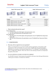
Download TaqMan® hPSC Scorecard™ Panel
Transcript
user guide TaqMan® hPSC Scorecard™ Panel For rapid confirmation of pluripotency and prediction of differentiation potential Catalog Numbers A15870, A15871, A15872, A15876 Publication Number MAN0008384 Revision 1.0 For Research Use Only. Not for use in diagnostic procedures. Information in this document is subject to change without notice. DISCLAIMER LIFE TECHNOLOGIES CORPORATION AND/OR ITS AFFILIATE(S) DISCLAIM ALL WARRANTIES WITH RESPECT TO THIS DOCUMENT, EXPRESSED OR IMPLIED, INCLUDING BUT NOT LIMITED TO THOSE OF MERCHANTABILITY, FITNESS FOR A PARTICULAR PURPOSE, OR NON-INFRINGEMENT. TO THE EXTENT ALLOWED BY LAW, IN NO EVENT SHALL LIFE TECHNOLOGIES AND/OR ITS AFFILIATE(S) BE LIABLE, WHETHER IN CONTRACT, TORT, WARRANTY, OR UNDER ANY STATUTE OR ON ANY OTHER BASIS FOR SPECIAL, INCIDENTAL, INDIRECT, PUNITIVE, MULTIPLE OR CONSEQUENTIAL DAMAGES IN CONNECTION WITH OR ARISING FROM THIS DOCUMENT, INCLUDING BUT NOT LIMITED TO THE USE THEREOF. NOTICE TO PURCHASER: LIMITED USE LABEL LICENSE: Research Use Only The purchase of this product conveys to the purchaser the limited, non-transferable right to use the purchased amount of the product only to perform internal research for the sole benefit of the purchaser. No right to resell this product or any of its components is conveyed expressly, by implication, or by estoppel. This product is for internal research purposes only and is not for use in commercial applications of any kind, including, without limitation, quality control and commercial services such as reporting the results of purchaser’s activities for a fee or other form of consideration. For information on obtaining additional rights, please contact [email protected] or Out Licensing, Life Technologies Corporation, 5791 Van Allen Way, Carlsbad, California 92008. NOTICE TO PURCHASER: LIMITED LICENSE Diagnostic uses of this product require a separate license from Roche. For information on obtaining additional rights, please contact [email protected] or Out Licensing, Life Technologies Corporation, 5791 Van Allen Way, Carlsbad, California 92008. NOTICE TO PURCHASER: LIMITED LICENSE Patents covering oligonucleotide conjugates of Minor Groove Binder (“MGB”) are owned by ELItech Group and licensed to Life Technologies for use in assays whereby the detection is mediated by a 5’ nuclease activity of a polymerase enzyme (the “5’Nuclease Process”). The purchase of this product includes a license to use only this amount of product solely for the purchaser’s own use and may not be used for any other commercial use, including without limitation human in vitro diagnostics, repackaging or resale in any form (including resale by purchasers who are licensed to make and sell kits for use in the 5’ Nuclease Process). Corresponding products conveying commercial and diagnostic use rights for MGB may be obtained from Life Technologies only under a separate agreement. For information on obtaining additional rights, please contact [email protected] or Out Licensing, Life Technologies Corporation, 5791 Van Allen Way, Carlsbad, California 92008. TRADEMARKS The trademarks mentioned herein are the property of Life Technologies Corporation and/or its affiliate(s) or their respective owners. Parafilm is a registered trademark of Bemis Company, Inc. TaqMan is a registered trademark of Roche Molecular Systems, Inc., used under permission and license. © 2013 Life Technologies Corporation. All rights reserved. Table of Contents Preface ................................................................................................................................. 2 About This Guide .........................................................................................................................................2 Product Information ............................................................................................................. 3 Kit Contents and Storage .............................................................................................................................3 Description of the System............................................................................................................................5 TaqMan® hPSC Scorecard™ Panel Workflow ...........................................................................................7 Sample Generation ............................................................................................................... 8 Undifferentiated Cells (Feeder-Dependent Culture) ...............................................................................8 Undifferentiated Cells (Feeder-Free Culture in StemPro® hESC SFM) ......................................................... 11 Undifferentiated Cells (Feeder-Free Culture in Essential 8™ Medium) ...................................................... 14 Randomly Differentiated Cells (Embryoid Bodies) ............................................................................... 17 Sample Preparation ........................................................................................................... 20 Total RNA Isolation by TRIzol® Organic Phase Extraction (Recommended Method) ..................... 20 Optional: DNase Treatment ....................................................................................................................... 22 Total RNA Isolation Using the TRIzol® Plus RNA Purification Kit (Alternative PureLink® Column-Based Method) ............................................................................................................................ 23 RNA Quantification and Quality ............................................................................................................. 25 cDNA Preparation............................................................................................................... 26 Reverse Transcription of Total RNA........................................................................................................ 26 TaqMan® qRT-PCR ............................................................................................................. 28 qRT-PCR Using the TaqMan® hPSC Scorecard™ Panel ......................................................................... 28 Appendix A: Recipes ........................................................................................................... 31 Media and Reagents ................................................................................................................................... 31 Appendix B: Preparing Culture Vessels.............................................................................. 34 Coating Culture Vessels with Geltrex® Matrix ....................................................................................... 34 MEF Culture Dishes ................................................................................................................................... 35 Appendix C: Background Information ................................................................................. 37 TaqMan® Chemistry ................................................................................................................................... 37 Appendix D: Ordering Information ...................................................................................... 38 Products ....................................................................................................................................................... 38 Appendix E: Safety .............................................................................................................. 40 Chemical Safety .......................................................................................................................................... 40 Chemical Waste Safety .............................................................................................................................. 41 Biological Hazard Safety ........................................................................................................................... 42 Documentation and Support ............................................................................................... 43 Technical Support ....................................................................................................................................... 43 1 Preface About This Guide Purpose of this guide This user guide is provides detailed procedures and reference materials for generating and preparing samples from undifferentiated and differentiated ESCs and iPSCs, and for analyzing these samples using the TaqMan® hPSC Scorecard™ Panels for pluripotency and differentiation potential. This user guide does not provide instructions for the hPSC Scorecard™ Analysis Software. Go to www.lifetechnologies.com/scorecardsoftwaremanual for detailed instructions on using the software. To access the hPSC Scorecard™ Analysis Software, go to www.lifetechnologies.com/scorecarddata. User attention words Two user attention words appear in Life Technologies user documentation. Each word implies a particular level of observation or action as described below. Note: Provides information that may be of interest or help but is not critical to the use of the product. IMPORTANT! Provides information that is necessary for proper instrument operation, accurate installation, or safe use of a chemical. Safety alert words Four safety alert words appear in Life Technologies user documentation at points in the document where you need to be aware of relevant hazards. Each alert word—IMPORTANT, CAUTION, WARNING, DANGER—implies a particular level of observation or action, as defined below: IMPORTANT! – Provides information that is necessary for proper instrument operation, accurate installation, or safe use of a chemical. CAUTION! – Indicates a potentially hazardous situation that, if not avoided, may result in minor or moderate injury. It may also be used to alert against unsafe practices. WARNING! – Indicates a potentially hazardous situation that, if not avoided, could result in death or serious injury. DANGER! – Indicates an imminently hazardous situation that, if not avoided, will result in death or serious injury. This signal word is to be limited to the most extreme situations. 2 Product Information Kit Contents and Storage Types of kits This manual is supplied with the products listed below. For a list of components supplied with each catalog number, see below. Product Catalog no. ® ™ A15870 ® ™ A15872 ® ™ A15876 ® ™ A15871 TaqMan hPSC Scorecard Panel 384w TaqMan hPSC Scorecard Kit 384w TaqMan hPSC Scorecard Panel 2 × 96w FAST TaqMan hPSC Scorecard Kit 2 × 96w FAST TaqMan® hPSC Scorecard™ 384w The TaqMan® hPSC Scorecard™ Panel 384w (Cat. no. A15870) and the TaqMan® hPSC Scorecard™ Kit 384w (Cat. no. A15872) contain the components listed below. Each 384-well plate can be used to analyze four cDNA samples. Catalog no. Component ® Composition Amount A15870 A15872 TaqMan hPSC Scorecard™ Panel 384w TaqMan® probes in a 384-well plate 1 plate MicroAmp® Optical Adhesive Film Optical adhesive covers 1 each TaqMan Gene Expression Master Mix* Solution containing AmpliTaq Gold® DNA Polymerase UP (Ultra Pure), Uracil-DNA Glycosylase, dNTPs with dUTP, Passive Reference 1, and optimized mix components 5 mL TaqMan® hPSC Scorecard™ Panel QRC TaqMan® hPSC Scorecard™ Panel Quick Reference Card 1 each ® * TaqMan® Gene Expression Master Mix (Cat. no. 4369016) is also available separately from Life Technologies (see page 38). Continued on next page 3 Kit Contents and Storage, continued TaqMan® hPSC Scorecard™ 2 × 96w FAST The TaqMan® hPSC Scorecard™ Panel 2 × 96w FAST (Cat. no. A15876) and the TaqMan® hPSC Scorecard™ Kit 2 × 96w FAST (Cat. no. A15871) contain the components listed below. Each 96-well plate can be used to analyze one cDNA sample. Catalog no. Component Composition Amount A15876 A15871 2 plates 2 each ® TaqMan hPSC Scorecard™ Panel 96w FAST TaqMan® probes in a 96-well plate MicroAmp® Optical Adhesive Film Optical Adhesive Covers TaqMan Fast Advanced Master Mix* Solution containing: AmpliTaq® Fast DNA Polymerase, Uracil-N glycosylase (UNG), dNTPs with dUTP, ROX™ dye (passive reference), and optimized buffer components TaqMan® hPSC Scorecard™ Panel QRC TaqMan® hPSC Scorecard™ Panel Quick Reference Card ® 2 × 1 mL 1 each * TaqMan® Fast Advanced Master Mix (Cat. no. 4444556) is also available separately from Life Technologies (see page 38). Shipping and storage The TaqMan® hPSC Scorecard™ Panels and Kits are shipped on wet ice. Upon receipt, transfer the entire shipment to 2–8°C for immediate storage. Store the individual components as described below. The performance of the products is guaranteed for 6 months from the date of purchase, if stored and handled properly. Component TaqMan® hPSC Scorecard™ Panel 384w or 96w FAST plates ® MicroAmp Optical Adhesive Film ® Storage and Handling Store at 4°C–30°C. Maintain in foil bag until ready to for use. Briefly spin plates at 400 × g for 2 minutes prior to use. Store at 4°C–30°C. Protect from dust. TaqMan Gene Expression Master Mix Store at 2°C–8°C. DO NOT FREEZE. TaqMan® Fast Advanced Master Mix Store at 2°C–8°C. DO NOT FREEZE. 4 Description of the System TaqMan® hPSC Scorecard™ Panels TaqMan® hPSC Scorecard™ Panel 384w and TaqMan® hPSC Scorecard™ Panel 96w FAST are MicroAmp® Optical 96- or 384-well Reaction Plates, standard or Fast, that contain dried-down TaqMan® Gene Expression Assays specifically formulated for evaluating human embryonic stem cells (ESC) and human induced pluripotent stem cells (iPSC) to confirm their pluripotency and to predict their differentiation potential and outcome. The gene expression assays contain a collection of predesigned, gene-specific primer and probe sets for performing quantitative gene expression studies on the cDNA samples prepared from undifferentiated or differentiated human ESCs and iPSCs. For more information on TaqMan® Gene Expression Assays, see “Appendix C: Background Information” on page 37). How hPSC Scorecard™ Panels work After isolating total RNA from human ESC or iPSC cultures and using it to generate cDNA in a reverse transcription (RT) reaction, TaqMan® hPSC Scorecard™ Panels and associated reagents are used to quantitate RNA expression levels of genetic markers for pluripotency and differentiation potential, as well as endogenous controls. The gene expression data are then analyzed using the web-based hPSC Scorecard™ Analysis Software for the pluripotency and differentiation potential of the cells from which the total RNA is isolated. To do this, you: Types of hPSC Scorecard™ Panels • Isolate total RNA from human ESCs or iPSCs by TRIzol® organic phase extraction or other preferred method. • Prepare each cDNA sample by performing eight reverse transcription reactions per total RNA sample. • Combine the appropriate TaqMan® master mix with your cDNA sample and RNase-free water, and reconstitute each well of the TaqMan® hPSC Scorecard™ Panel by adding 10 μL of the reaction mixture per well. • Load and run the plates on a compatible real-time PCR (RT-PCR) instrument using either standard or Fast thermal cycling conditions. • Analyze the gene expression data using the web-based hPSC Scorecard™ Analysis Software to confirm the pluripotency of the samples and predict their differentiation potential and outcome. The hPSC Scorecard™ Analysis Software is available at www.lifetechnologies.com/scorecarddata. TaqMan® hPSC Scorecard™ Panels are available as 384-well plates (Cat. nos. A15870, A15872) or as 96-well FAST plates (Cat. nos. A15876, A15871) for use with Fast thermal cycling conditions. • TaqMan® hPSC Scorecard™ Panel 384w are 384-well MicroAmp® optical assay plates, which allow the analysis of four separate cDNA samples under standard thermal cycling conditions. • TaqMan® hPSC Scorecard™ Panel 96w FAST are 96-well MicroAmp® optical Fast thermal cycling plates, which reduce quantitative PCR run times to less than 40 minutes when used under Fast thermal cycling conditions in a compatible RT-PCR system. Each 96-well plate allows the analysis of one cDNA sample. Continued on next page 5 Description of the System, continued ® ™ Compatible TaqMan® Each well in a TaqMan hPSC Scorecard Panel contains a pair of unlabeled PCR primers specific to a pluripotency or differentiation marker or endogenous Master Mixes ® control, and a TaqMan probe with a fluorescent dye-label on the 5’ end (e.g., FAM™ or VIC® dye) and a minor groove binder (MGB) and non-fluorescent quencher (NFQ) on the 3’ end (see page 37). The assays in each well are reconstituted to a 1✕ formulation using the compatible TaqMan® master mix as described in this user guide and are designed to run under standard or Fast cycling conditions for two-step RT-PCR. The table below lists the TaqMan® hPSC Scorecard™ Panel and corresponding TaqMan® master mix compatible with it. Note that TaqMan® hPSC Scorecard™ Kits contain the appropriate compatible TaqMan® master mix, which are not supplied with the individually packaged TaqMan® hPSC Scorecard™ Panels and need to be purchased separately from Life Technologies (see page 38 for ordering information). TaqMan® hPSC Scorecard™ Panel Compatible TaqMan® master mix TaqMan® hPSC Scorecard™ Panel 384w TaqMan® Gene Expression Master Mix (for standard cycling) TaqMan® hPSC Scorecard™ Panel 96w FAST TaqMan® Fast Advanced Master Mix (for Fast cycling) ® ™ ® Compatible RT-PCR TaqMan hPSC Scorecard Panels can be used with the Applied Biosystems ® ™ RT-PCR systems listed below. Note that TaqMan hPSC Scorecard Panel 96w instruments FAST must be run on RT-PCR systems that contain Fast thermal cycling blocks. Alternately, TaqMan® hPSC Scorecard™ Panel 384w must be run on RT-PCR systems with standard thermal cycling blocks. Compatible Applied Biosystems® RT-PCR systems TaqMan hPSC Scorecard Panel ® ™ TaqMan® hPSC Scorecard™ Panel 384w • • TaqMan® hPSC Scorecard™ Panel 96w FAST 6 QuantStudio™ 12K Flex System with 384-well Block ViiA™ 7 Real-Time PCR System with 384-well Block • StepOnePlus™ Real-Time PCR System • ViiA™ 7 Real-Time PCR System with Fast 96-well Block TaqMan® hPSC Scorecard™ Panel Workflow Experiment outline The table below describes the major steps needed to generate and prepare cDNA samples for analysis using the TaqMan® hPSC Scorecard™ Panels. For more details, refer to the pages indicated. Step Action Page 1 Harvest ESCs or iPSCs from: Undifferentiated cells (feeder-dependent culture) Undifferentiated cells (feeder-free culture in StemPro® hESC SFM) Undifferentiated cells (feeder-free culture in Essential 8™ medium) Randomly differentiated cells (i.e., embryoid bodies) 8 11 14 17 2 Isolate total RNA from cells 20 3 Optional: Remove contaminating DNA from RNA sample 22 4 Quantitate total RNA and asses its quality 25 5 Generate cDNA by reverse transcription 26 6 7 ® Perform TaqMan qRT-PCR 28 ™ Analyze data using the hPSC Scorecard Analysis Software 30 7 Sample Generation Undifferentiated Cells (Feeder-Dependent Culture) Introduction This section provides instructions on harvesting undifferentiated ESCs or iPSCs that are maintained as feeder-dependent cultures on inactived murine embryonic fibroblast (MEF) feeder cells for total RNA extraction. You will need to harvest at least 5 × 10⁵ cells per sample to isolate sufficient total RNA for the subsequent steps of the workflow. IMPORTANT! Feeder-dependent ESCs or iPSCs should be cultured feeder-free on Geltrex®-matrix coated culture vessels for one passage in MEF-conditioned medium before the cells are harvested and total RNA is isolated. Materials needed Prepare media, reagents, and matrix-coated culture vessels • DMEM/F-12 with GlutaMAX™-I (Cat. no. 10565-018) • KnockOut™ Serum Replacement (KSR) (Cat. no. 10828-010) • MEM Non-Essential Amino Acids Solution (10 mM) (Cat. no. 11140-050) • β-Mercaptoethanol (1000X), liquid (Cat. no. 21985-023) • Basic Fibroblast Growth Factor (bFGF), recombinant human (Cat. no. PGH0264) • Collagenase, Type IV (Cat. no. 17104-019) • Attachment Factor (Cat. no. S-006-100) • DMEM with GlutaMAX™-I (high glucose) (Cat. no. 10569-010) • Fetal Bovine Serum, ESC-Qualified (Cat. no. 16141-079) • DPBS, no Calcium, no Magnesium (Cat. no. 14190-144) • Geltrex®, hESC qualified (Cat. no. A1413302) • TRIzol® Reagent (Cat. no. 18596-018) • Gibco® Mouse Embryonic Fibroblasts (Irradiated) (Cat. no. S1520-100) • Cell Scraper (Falcon, Cat. no. 353085) • Appropriate tissue culture plates and supplies The following media, reagents, and culture vessels are needed for culturing and harvesting undifferentiated, feeder-dependent ESCs or iPSCs. For instructions on preparing the media and reagents listed below, see “Appendix A: Recipes”, page 28. For instructions on coating culture vessels with Geltrex® matrix, see “Appendix B: Preparing Culture Vessels“, page 34. • MEF medium • ESC medium • MEF-conditioned medium (MEF-CM) • 1X Collagenase IV Solution (1 mg/mL) • Geltrex® matrix-coated culture vessels Continued on next page 8 Undifferentiated Cells (Feeder-Dependent Culture), continued Culture and harvest 1. Culture ESCs or iPSCs on inactivated MEF feeder cells using complete ESC medium. cells 2. When the ESC or iPSC culture has reached the desired confluency to yield at least 5 × 10⁵ cells per sample, aspirate off the cell culture medium (i.e., ESC medium). 3. Add Collagenase Type IV (1 mg/mL) solution to the dish containing ESCs or iPSCs. Adjust the volume of Collagenase IV for various dish sizes (refer to Table 1, page 10. 4. Incubate for 30–45 minutes in a 37°C, 5% CO2 incubator. Note: Incubation times may vary among different batches of collagenase; therefore, you need to determine the appropriate incubation time by examining the colonies. Note that the required exposure time shortens as the enzymatic activity of collagenase increases (i.e., 200,000 U/mg at 1 mg/mL concentration will take longer than stocks of 290,000 U/mg at 1 mg/mL concentration). 5. When the edges of the colonies are starting to pull away from the vessel, carefully aspirate the Collagenase IV solution from the vessel without disturbing the attached cell layer and add 3 mL of ESC medium. 6. Dislodge the cells with a 5-mL pipet by gently blowing the cells off the surface of the vessel while pipetting up and down. This will not only dislodge the colonies, but also manually break them up into small fragments to be passaged. Do not create a single cell suspension; usually 5–10 pipetting motions will suffice to dislodge and resuspend the cells. 7. Transfer the cell clumps into a 15-mL tube. Use an additional 2 mL of medium to collect any cell clumps remaining in the vessel and add to the 15-mL conical tube. 8. Allow the cells to gravity sediment for approximately 5 minutes. This will permit the larger clumps to pellet out while allowing the removal of smaller clumps, single cells, and any iMEFs from the feeder layer that were dislodged through the process and still floating in the supernatant. 9. Aspirate the supernatant, and then gently tap the tube to loosen the cell pellet from the bottom of the tube. 10. Add the appropriate amount of ESC medium for your passaging split ratio. At this point you can continue to gently dissociate the cells into small clusters (50–500 cells) by gentle pipetting. Avoid making single cell suspensions. 11. Seed the cell clusters onto Geltrex®-matrix coated culture vessel containing MEF-conditioned medium (MEF-CM). The final volume of the MEF-CM depends on the vessel used (refer to Table 2, page 10, for the final volume of medium required for each vessel). Use a typical split ratio of 1:3 to 1:5 per plate, depending on amount of cell clusters. Continued on next page 9 Undifferentiated Cells (Feeder-Dependent Culture), continued Culture and harvest 12. Move the culture vessel in several quick, short, back-and-forth and side-toside motions to disperse the cells evenly across its surface, and then return cells, continued the vessel to the incubator. 13. Continue to culture the cells until they are 80–90% confluent, changing the spent MEF-CM daily. 14. On the day of harvesting, aspirate the culture medium and wash the cells once with 5 mL of DPBS for 2–3 minutes. 15. Aspirate the DPBS and discard. 16. Add 1 mL of TRIzol® reagent and incubate for 2–3 minutes. Scrape the plate with a sterile cell scraper and collect the slurry into a sterile, RNAse-free microcentrifuge tube. 17. Store the cells at –80°C until ready for RNA isolation. Table 1 Volume of Collagenase IV (1 mg/mL) and MEF-CM required Culture Vessel Surface Area Volume of Collagenase IV Volume of MEF-CM 6-well plate 2 1.0 mL per well 2.0 mL per well 2 12-well plate 4 cm /well 0.5 mL per well 1.0 mL per well 24-well plate 2 cm2/well 0.25 mL per well 0.5 mL per well 35-mm dish 10 cm2 1.0 mL 2.0 mL 60-mm dish 2 2.0 mL 4.0 mL 2 6.0 mL 10.0 mL 100-mm dish 10 10 cm /well 20 cm 60 cm Undifferentiated Cells (Feeder-Free Culture in StemPro® hESC SFM) Introduction This section provides instructions on harvesting undifferentiated ESCs or iPSCs maintained as feeder-free cultures on Geltrex® matrix-coated vessels in complete StemPro® hESC medium for total RNA extraction. You will need to harvest at least 5 × 10⁵ cells per sample to isolate sufficient total RNA for the subsequent steps of the workflow. Materials needed • DMEM/F-12 with GlutaMAX™-I (Cat. no. 10565-018) • StemPro® hESC SFM Kit (Cat. no. A1000701) • β-Mercaptoethanol (1000X), liquid (Cat. no. 21985-023) • Basic Fibroblast Growth Factor (bFGF), recombinant human (Cat. no. PGH0264) • Collagenase, Type IV (Cat. no. 17104-019) • DPBS, no Calcium, no Magnesium (Cat. no. 14190-144) • Geltrex®, hESC qualified (Cat. no. A1413302) • TRIzol® Reagent (Cat. no. 18596-018) • Cell Scraper (Falcon, Cat. no. 353085) • Appropriate tissue culture plates and supplies Prepare media, reagents, and matrix-coated culture vessels The following media, reagents, and culture vessels are needed for culturing and harvesting undifferentiated ESCs or iPSCs maintained as feeder-free cultures in complete StemPro® hESC medium. For instructions on preparing the media and reagents listed below, see “Appendix A: Recipes”, page 28. For instructions on coating culture vessels with Geltrex® matrix, see “Appendix B: Preparing Culture Vessels“, page 34. • StemPro® hESC SFM medium • StemPro® wash solution • 10X Collagenase IV Solution (10 mg/mL) • Geltrex® matrix-coated culture vessels ® Culture and harvest 1. Coat the culture vessels with Geltrex matrix at least 1 hour before passaging the cells. Warm the appropriate amount of 10 mg/mL (10X) Collagenase IV cells ® ® solution, StemPro wash solution, and complete StemPro hESC SFM medium to 37°C in a water bath. 2. When the ESC or iPSC culture has reached the desired confluency to yield at least 5 × 10⁵ cells per sample, aspirate off the cell culture medium (i.e., StemPro® hESC SFM). 3. Gently add 10 mg/mL Collagenase Type IV (10X) solution to the culture vessel containing ESCs or iPSCs. Adjust the volume of Collagenase IV for various dish sizes (refer to Table 2, page 13). Continued on next page 11 Undifferentiated Cells (Feeder-Free Culture in StemPro® hESC SFM), continued Culture and harvest 4. Incubate for 3–5 minutes in a 37°C, 5% CO2 incubator until the edges of the colonies begin to curl. cells, continued Note: Incubation times may vary among different batches of collagenase; therefore, you need to determine the appropriate incubation time by examining the colonies. Note that the required exposure time shortens as the enzymatic activity of collagenase increases (i.e., 200,000 U/mg at 1 mg/mL concentration will take longer than stocks of 290,000 U/mg at 1 mg/mL concentration). 5. When the edges of the colonies are starting to pull away from the vessel, carefully aspirate the Collagenase IV solution from the vessel without disturbing the attached cell layer and gently rinse the cells with 3 mL of pre-warmed StemPro® wash solution. 6. Aspirate the wash solution and add the appropriate volume of pre-warmed complete StemPro® hESC SFM medium (refer to Table 2, page 13). 7. Use a sterile cell scraper to gently remove the cell clumps. Pipette the cell suspension across the plate surface with a 5-mL pipette to break up the detached colonies into smaller clumps. Do not create a single cell suspension; usually 3–5 pipetting motions will suffice to dislodge and resuspend the cells. 8. Gently transfer the cell clumps into a 15-mL tube. Depending on the culture vessel, use an additional 1–3 mL of pre-warmed complete StemPro® hESC SFM medium to collect any cell clumps remaining in the vessel and add to the 15-mL conical tube. 9. Centrifuge cell suspension at 200 × g for 2 minutes or allow the cells to gravity sediment at room temperature for 5–10 minutes. 10. Aspirate off the Geltrex® solution from the fresh Geltrex®-matrix coated vessel and add the appropriate volume of pre-warmed complete StemPro® hESC SFM medium (refer to Table 2, page 13). 11. Following centrifugation or gravity sedimentation of the cell suspension (step 9), aspirate off the supernatant and gently re-suspend the cell clumps with the appropriate amount of complete StemPro® hESC SFM medium. 12. Add the desired amount of cell suspension into each new Geltrex®-matrix coated vessel according to the desired split ratio. Note: The split ratio is variable, though generally between 1:4 and 1:6. If the cells are overly dense and crowding, increase the ratio, and if the cells are sparse, decrease the ratio. Cells will need to be split every 4–6 days based upon their appearance. 13. Move the culture vessel in several quick, short, back-and-forth and side-toside motions to disperse the cells evenly across its surface, and then return the vessel to the incubator. 14. The next day, gently aspirate the medium to remove the non-attached cells, and replace it with fresh complete StemPro® hESC SFM medium. Replace the spent medium with fresh medium every day thereafter. Continued on next page 12 Undifferentiated Cells (Feeder-Free Culture in StemPro® hESC SFM), continued Culture and harvest 15. Continue to culture the cells until they are 80–90% confluent, changing the medium daily. cells, continued 16. On the day of harvesting, aspirate the culture medium and wash the cells once with 5 mL of DPBS for 2–3 minutes. 17. Aspirate the DPBS and discard. 18. Add 1 mL of TRIzol® reagent and incubate for 2–3 minutes. Scrape the plate with a sterile cell scraper and collect the slurry into a sterile, RNAse-free microcentrifuge tube. 19. Store the cells at –80°C until ready for RNA isolation. Table 2 Volume of Collagenase IV (10 mg/mL) and complete StemPro® hESC SFM medium required Culture Vessel Surface Area Volume of Collagenase IV Volume of complete StemPro® hESC SFM 6-well plate 10 cm2/well 1.0 mL per well 2.0 mL per well 0.5 mL per well 1.0 mL per well 12-well plate 24-well plate 2 4 cm /well 2 0.25 mL per well 0.5 mL per well 35-mm dish 2 10 cm 1.0 mL 2.0 mL 60-mm dish 20 cm2 2.0 mL 4.0 mL 2 6.0 mL 10.0 mL 100-mm dish 2 cm /well 60 cm 13 Undifferentiated Cells (Feeder-Free Culture in Essential 8™ Medium) Introduction This section provides instructions on harvesting undifferentiated ESCs or iPSCs maintained as feeder-free cultures on Geltrex® matrix-coated vessels in complete Essential 8™ medium for total RNA extraction. You will need to harvest at least 5 × 10⁵ cells per sample to isolate sufficient total RNA for the subsequent steps of the workflow. Materials needed • Essential 8™ Medium (prototype) (Cat. no. A14666SA) • DMEM/F-12 with GlutaMAX™-I (Cat. no. 10565-018) • Collagenase, Type IV (Cat. no. 17104-019) • DPBS, no Calcium, no Magnesium (Cat. no. 14190-144) • Geltrex®, hESC qualified (Cat. no. A1413302) • TRIzol® Reagent (Cat. no. 18596-018) • Cell Scraper (Falcon, Cat. no. 353085) • Appropriate tissue culture plates and supplies Prepare media, reagents, and matrix-coated culture vessels The following media, reagents, and culture vessels are needed for culturing and harvesting undifferentiated ESCs or iPSCs maintained as feeder-free cultures on in complete Essential 8™ medium. For instructions on preparing the media and reagents listed below, see “Appendix A: Recipes”, page 28. For instructions on coating culture vessels with Geltrex® matrix, see “Appendix B: Preparing Culture Vessels“, page 34. • Essential 8™ Medium • 10X Collagenase IV Solution (10 mg/mL) • Geltrex® matrix-coated culture vessels ® Culture and harvest 1. Coat the culture vessels with Geltrex matrix at least 1 hour before passaging the cells. Warm the appropriate amount of 10 mg/mL (10X) Collagenase IV cells solution and DMEM/F-12 to 37°C in a water bath. 2. Warm complete Essential 8™ medium required for that day at room temperature until it is no longer cool to the touch, approximately 20–30 minutes. Do not warm the Essential 8™ medium at 37°C. 3. When the ESC or iPSC culture has reached the desired confluency to yield at least 5 × 10⁵ cells per sample, aspirate off the cell culture medium (i.e., Essential 8™ medium). 4. Gently add 10 mg/mL Collagenase Type IV (10X) solution to the culture vessel containing ESCs or iPSCs. Adjust the volume of Collagenase IV for various dish sizes (refer to Table 3, page 16). Continued on next page 14 Undifferentiated Cells (Feeder-Free Culture in Essential 8™ Medium), continued Culture and harvest 5. Incubate for 3–5 minutes in a 37°C, 5% CO2 incubator until the edges of the colonies begin to curl. cells, continued Note: Incubation times may vary among different batches of collagenase; therefore, you need to determine the appropriate incubation time by examining the colonies. Note that the required exposure time shortens as the enzymatic activity of collagenase increases (i.e., 200,000 U/mg at 1 mg/mL concentration will take longer than stocks of 290,000 U/mg at 1 mg/mL concentration). 6. When the edges of the colonies are starting to pull away from the vessel, carefully aspirate the Collagenase IV solution from the vessel without disturbing the attached cell layer and gently rinse the cells with 3 mL of pre-warmed DMEM/F-12 solution. 7. Aspirate the wash solution and add the appropriate volume of pre-warmed complete Essential 8™ medium (refer to Table 3, page 16). 8. Use a sterile cell scraper to gently remove the cell clumps. Pipette the cell suspension across the plate surface with a 5-mL pipette to break up the detached colonies into smaller clumps. Do not create a single cell suspension; usually 3–5 pipetting motions will suffice to dislodge and resuspend the cells. 9. Gently transfer the cell clumps into a 15-mL tube. Depending on the culture vessel, use an additional 1–3 mL of pre-warmed complete StemPro® hESC SFM medium to collect any cell clumps remaining in the vessel and add to the 15-mL conical tube. 10. Centrifuge cell suspension at 200 × g for 2 minutes or allow the cells to gravity sediment at room temperature for 5–10 minutes. 11. Aspirate off the Geltrex® solution from the fresh Geltrex®-matrix coated vessel and add the appropriate volume of pre-warmed complete Essential 8™ medium (refer to Table 3, page 16). 12. Following centrifugation or gravity sedimentation of the cell suspension (step 10), aspirate off the supernatant and gently re-suspend the cell clumps with the appropriate amount of complete Essential 8™ medium. 13. Add the desired amount of cell suspension into each new Geltrex®-matrix coated vessel according to the desired split ratio. Note: The split ratio is variable, though generally between 1:4 and 1:6. If the cells are overly dense and crowding, increase the ratio, and if the cells are sparse, decrease the ratio. Cells will need to be split every 4–6 days based upon their appearance. 14. Move the culture vessel in several quick, short, back-and-forth and side-toside motions to disperse the cells evenly across its surface, and then return the vessel to the incubator. 15. The next day, gently aspirate the medium to remove the non-attached cells, and replace it with fresh complete Essential 8™ medium. Replace the spent medium with fresh medium every day thereafter. Continued on next page 15 Undifferentiated Cells (Feeder-Free Culture in Essential 8™ Medium), continued Culture and harvest 16. Continue to culture the cells until they are 80–90% confluent, changing the medium daily. cells, continued 17. On the day of harvesting, aspirate the culture medium and wash the cells once with 5 mL of DPBS for 2–3 minutes. 18. Aspirate the DPBS and discard. 19. Add 1 mL of TRIzol® reagent and incubate for 2–3 minutes. Scrape the plate with a sterile cell scraper and collect the slurry into a sterile, RNAse-free microcentrifuge tube. 20. Store the cells at –80°C until ready for RNA isolation. Table 3 Volume of Collagenase IV (10 mg/mL) and complete Essential 8™ medium required Culture Vessel Surface Area Volume of Collagenase IV Volume of complete Essential 8™ medium 6-well plate 10 cm2/well 1.0 mL per well 2.0 mL per well 0.5 mL per well 1.0 mL per well 12-well plate 24-well plate 4 cm /well 2 0.25 mL per well 0.5 mL per well 35-mm dish 2 10 cm 1.0 mL 2.0 mL 60-mm dish 20 cm2 2.0 mL 4.0 mL 2 6.0 mL 10.0 mL 100-mm dish 16 2 2 cm /well 60 cm Randomly Differentiated Cells (Embryoid Bodies) Introduction This section provides instructions on generating embryoid bodies (EBs) for random differentiation from ESCs or iPSCs maintained as feeder-dependent or feeder-free cultures, and their subsequent harvest for total RNA extraction. IMPORTANT! There are several methods for creating EBs for random differentiation. Here we cover suspension EB culture, which is recommended for a maximum of 7 days. If extended EB culture is desired, we recommend seeding the EBs on a Geltrex®-matrix coated culture vessel and continuing as an adherent culture as described in “Harvest cells (Option 2: 4 day EB suspension and adherent culture)”, page 19, until total RNA isolation. Materials needed Prepare media and matrix-coated culture vessels Generate EBs from ESCs/iPSCs • DMEM/F-12 with GlutaMAX™-I (Cat. no. 10565-018) • KnockOut™ Serum Replacement (KSR) (Cat. no. 10828-010) • MEM Non-Essential Amino Acids Solution (10 mM) (Cat. no. 11140-050) • β-Mercaptoethanol (1000X), liquid (Cat. no. 21985-023) • DPBS, no Calcium, no Magnesium (Cat. no. 14190-144) • TRIzol® Reagent (Cat. no. 18596-018) • Cell Scraper (Falcon, Cat. no. 353085) • 60-mm Petri dish (non-tissue culture treated) or ultra-low binding dish • Appropriate tissue culture plates and supplies • Geltrex®, hESC qualified (Cat. no. A1413302; for optional adherent EB culture) The following media and matrix-coated culture vessels are needed for creating and harvesting EBs for total RNA isolation. For instructions on preparing the media listed below, see “Appendix A: Recipes”, page 28. For instructions on coating culture vessels with Geltrex® matrix, see “Appendix B: Preparing Culture Vessels“, page 34. • EB medium • Geltrex® matrix-coated culture vessels (for optional adherent EB culture) Embryoid Bodies (EBs) are generated at a normally scheduled passage by plating ESCs or iPSCs into non-tissue culture-treated dishes to prevent attachment and allowing them to aggregate to form EBs. 1. Culture ESCs or iPSCs on MEF feeder cells or in the desired feeder-free condition (StemPro® hESC SFM, MEF-CM, or Essential 8™ Medium) until the cells are approximately 90% confluent. 2. Aspirate off the culture medium from the culture plates or dishes, and then add 1 mL pre-warmed EB medium to each well of 6-well plate or to each 35-mm dish, 2 mL to each 60-mm dish, or 3 mL to each 100-mm dish. Continued on next page 17 Randomly Differentiated Cells (Embryoid Bodies), continued Generate EBs from ESCs/iPSCs, 3. Roll the StemPro® EZPassage™ disposable stem cell passaging tool across the entire dish or plate in one direction (left to right). Rotate the culture dish or plate 90 degree, and roll the StemPro® EZPassage™ disposable stem cell passaging tool across the entire dish or plate. 4. Use a cell scraper to gently detach the cells off the surface of the culture vessel. 5. Gently transfer the cell clumps using a 5-mL pipette into a 15-mL conical tube. Do not break the cell clumps into small pieces. 6. Depending on the culture vessel, use an additional 1–3 mL of pre-warmed EB medium (see step 2, page 17) to collect any cell clumps remaining in the vessel and add to the 15-mL conical tube. 7. Transfer the cell clumps to a 60-mm Petri dish (non-tissue culture treated) or ultra-low binding dish in a total of 5 mL of EB medium. If necessary, add more EB medium into the 15-mL tube to bring up the volume to 5 mL. continued Note: If using a different size vessel, add more pre-warmed EB medium into the 15-mL tube to bring up the volume of the cell suspension to the appropriate level for the vessel used (refer to Table 4, below). Generally use one plate of ESCs or iPSCs per one plate of EBs to be formed. 8. Place Petri dish containing the cells in a 37°C, 5% CO2 incubator to allow the cells to form EBs. 9. The next day and every other day thereafter, replace the spent medium with fresh, pre-warmed EB medium. To replace the medium, gently transfer the EB suspension into a 15-mL conical tube and allow the EBs to sediment down by gravity for 10–15 minutes. Then gently aspirate off the supernatant and re-suspend the EBs with fresh, pre-warmed EB medium. Return the EB suspension into the same culture vessel for continued growth. 10. On day 7 of incubation, harvest the cells as described in “Harvest cells (Option 1: 7 day EB suspension)”, page 19. Note: Culturing EBs in suspension for 7 days is usually sufficient for the analysis of random differentiation. If extended culture is desired, use the optional protocol for adherent EB culture as described in “Harvest cells (Option 2: 4 day EB suspension and adherent culture)”, page 19. Table 4 Volume of EB medium required Culture Vessel Surface Area Final volume of cell suspension in EB medium 6-well plate 10 cm2/well 2.0 mL per well 12-well plate 24-well plate 35-mm dish 60-mm dish 100-mm dish 2 4 cm /well 2 2 cm /well 1.0 mL per well 0.5 mL per well 2 2.0 mL 2 5.0 mL 2 12.0 mL 10 cm 20 cm 60 cm Continued on next page 18 Randomly Differentiated Cells (Embryoid Bodies), continued Harvest cells (Option 1: 7 day EB suspension) Harvest cells (Option 2: 4 day EB suspension and adherent culture) 1. On day 7 of EB suspension, gently transfer the cells and the medium from the Petri dish into a 15-mL conical tube. Use an additional 5 mL of DPBS to collect any remaining EBs from the culture dish and add into the conical tube. 2. Allow the EBs to sediment down by gravity for 10–15 minutes, and then aspirate off the supernatant (i.e., spent EB medium). 3. Using a P1000 pipettor, add 1 mL of TRIzol® reagent and pipette up and down to assist in properly breaking up the cell clumps. 4. Incubate the EBs for 2–3 minutes. Repeat pipetting and incubation, if the EBs require more time to be lysed. Collect the slurry into a sterile RNAse-free microcentrifuge tube. Store at –80°C until ready for RNA isolation. 1. On Day 4, gently transfer the cells and the medium from the Petri dish into a 15-mL conical tube. Use an additional 5 mL of DPBS to collect any remaining EBs from the culture dish and add into the conical tube. 2. Allow the EBs to sediment down by gravity for 10–15 minutes in the cell culture hood. 3. Aspirate the supernatant (i.e., spent EB medium) and replace it with 5 mL of fresh EB medium. 4. Transfer the cells to a fresh 60-mm tissue culture-treated dish coated with Geltrex®-matrix. If using a different size culture dish, refer to Table 4 for the volume EB of medium needed. Place the dish containing the cells in the 37°C, 5% CO2 incubator and change the spent medium every other day. 5. Allow the EBs to attach and the contents of the EBs to grow out from the EBs to obtain adherent cell types. 6. On Day 7 and 14 of total differentiation (or as desired), aspirate off the EB medium and wash the cells once with 5 mL of DPBS for 2–3 minutes. 7. Aspirate off the DPBS wash. 8. Add 1 mL of TRIzol® reagent and incubate for 2–3 minutes. Scrape the plate with a sterile cell scraper and collect the slurry into an RNAse-free microcentrifuge tube. Store at –80°C until ready for RNA isolation. 19 Sample Preparation Total RNA Isolation by TRIzol® Organic Phase Extraction (Recommended Method) Introduction This section provides instructions on extracting total RNA from the ESCs and iPSCs by TRIzol® organic phase extraction to use as a template for synthesis of single-stranded cDNA. You will need at least 5 × 10⁵ cells harvested per sample to isolate sufficient total RNA for the reverse transcription reaction. Note: For optimal performance, we recommend isolating total RNA by organic phase extraction using the TRIzol® reagent. Note that column based purification methods (see page 23) may also yield high-quality RNA. Materials needed • TRIzol® reagent (Cat. no. 15596-026) • Chloroform (Sigma, Cat. no. C-2432) • Isopropanol (Sigma, Cat. no. I9516) • Ethanol (Sigma, Cat. no. E7023) • UltraPure™ DNase/RNase-Free Distilled Water (Cat. no. 10977) CAUTION! TRIzol® Reagent contains phenol (toxic and corrosive) and guanidine isothiocyanate (an irritant), and may be a health hazard if not handled properly. Always work with TRIzol® Reagent in a fume hood, and always wear a lab coat, gloves and safety glasses. For more information, refer to the TRIzol® Reagent SDS (Safety Data Sheet), available from our website at www.lifetechnologies.com/support. Isolate total RNA by TRIzol® organic phase extraction 1. Incubate the lysate with TRIzol® reagent (from the last step of the harvesting procedure) at room temperature for 5 minutes to allow complete dissociation of nucleoprotein complexes. 2. To the cells in TRIzol® reagent add 0.2 mL of chloroform per 1 mL of TRIzol® reagent, and shake the tube vigorously by hand for 15 seconds. 3. Incubate the sample at room temperature for 2–3 minutes and centrifuge at 12,000 × g for 15 minutes at 4°C. Note: The mixture separates into a lower red phenol-chloroform phase, an interphase, and a colorless upper aqueous phase. RNA remains exclusively in the aqueous phase. The upper aqueous phase is ~50% of the total volume. 4. Carefully remove the upper aqueous phase and transfer to a new tube. Avoid drawing any of the interphase or organic layer into the pipette when removing the aqueous phase. Continued on next page 20 Total RNA Isolation by TRIzol® Organic Phase Extraction, continued Isolate total RNA by TRIzol® organic phase extraction, continued 5. Add 0.5 mL of 100% isopropanol to the aqueous phase per 1 mL of TRIzol® reagent, and incubate at room temperature for 10 minutes. 6. Centrifuge at 12,000 × g for 10 minutes at 4°C. 7. Carefully remove the supernatant from the RNA pellet and wash with 1 ml 75% ethanol. Note: The RNA is often invisible prior to centrifugation, and forms a gel-like pellet on the side and at the bottom of the tube upon centrifugation. 8. Centrifuge the tube at 7500 × g for 5 minutes at 4°C. 9. Discard the supernatant and air dry the RNA pellet for 5–10 minutes. 10. Resuspend the RNA pellet in 20–50 µL RNase-free water. 21 Optional: DNase Treatment Introduction One key variable to the success of any RT-PCR experiment is the quality of the template RNA. DNA removal is critical for ensuring high-quality RNA, because DNA can serve as a template during the PCR portion of the experiment, resulting in false positives, background, etc. Ideally, the total RNA sample should have less than 0.005% of genomic DNA by weight. We recommend treating the isolated total RNA with the DNA-free™ Kit, which digests the contaminating DNA to levels below the limit of detection by routine PCR. Materials needed • DNA-free™ Kit (Cat. no. AM1906; contains rDNase I, 10X DNase I Buffer, DNase Inactivation Reagent, and nuclease–free water) Guidelines for using the DNA-free™ Kit • We recommend conducting the reactions in 0.5 mL tubes to facilitate removal of the supernatant after treatment with the DNase Inactivation Reagent. • DNA-free™ reactions can be conducted in 96-well plates. We recommend using V-bottom plates because their shape makes it easier to remove the RNA from the pelleted DNase Inactivation Reagent at the end of the procedure. • The recommended reaction size is 10–100 μL. A typical reaction is 50 µL. • Routine DNase treatment removes 2 µg of genomic DNA from 50 µL reaction with ≤ 200 µg/mL nucleic acid; refer to product insert if more rigorous DNase treatment is needed. 1. For a 50-µL reaction, combine the following reagents in a clean, DNase- and RNase-free 0.5-mL microcentrifuge tube, and mix gently. DNA-free™ Kit procedure RNA Sample 10X DNase I Reaction Buffer rDNase I (2 Units) DEPC-treated water to bring reaction to 50 µL Total 22 1–10 µg 5 µL 1 µL X µL 50 µL 2. Incubate the tube at 37°C for 20–30 minutes. 3. Resuspend the DNase Inactivation Reagent by flicking or vortexing the tube, add 5 µL (0.1 volume) of the resuspended inactivation reagent to the reaction mix, and mix well. 4. Incubate for 2 minutes at room temperature, mixing the reaction occasionally. 5. Centrifuge at 10,000 × g for 1.5 minutes and transfer the RNA to a fresh tube. 6. RNA is now ready for reverse transcription. Total RNA Isolation Using the TRIzol® Plus RNA Purification Kit (Alternative PureLink® Column-Based Method) Introduction The TRIzol® Plus RNA Purification Kit provides a simple, reliable, and rapid method for isolating high-quality total RNA from a wide variety of samples, including animal and plant cells and tissue, bacteria, and yeast. The kit utilizes the strong lysis capability of TRIzol® reagent, followed by a convenient and time-saving silica-cartridge purification protocol from the PureLink® RNA Mini Kit, to purify total RNA. Materials needed • TRIzol® Plus RNA Purification kit (Cat. no. 12183-555) • Chloroform (Sigma, Cat. no. C-2432) • Ethanol (Sigma, Cat. no. E7023) • UltraPure™ DNase/RNase-Free Distilled Water (Cat. no. 10977) 1. Incubate the lysate with TRIzol® reagent (from the last step of the harvesting procedure) at room temperature for 5 minutes to allow complete dissociation of nucleoprotein complexes. 2. To the cells in TRIzol® reagent add 0.2 mL of chloroform per 1 mL of TRIzol® reagent, and shake the tube vigorously by hand for 15 seconds. 3. Incubate the sample at room temperature for 2–3 minutes and centrifuge at 12,000 × g for 15 minutes at 4°C. 4. Transfer ~600 µL of the colorless, upper phase containing the RNA to a fresh RNase-free tube and add an equal volume of 100% ethanol to obtain a final ethanol concentration of 50%. Mix well by vortexing. 5. Invert the tube to disperse any visible precipitate that may form after adding ethanol. Proceed to next step using the column. 6. Transfer up to 700 µL of sample to a Spin Cartridge (with a Collection Tube) and centrifuge at 12,000 × g for 15 seconds at room temperature. Discard the flow–through and reinsert the Spin Cartridge into the same Collection Tube. 7. Repeat above two steps until the entire sample has been processed. 8. Optional: If your downstream application requires DNA-free total RNA, proceed to “On-Column PureLink® DNase Treatment during RNA Purification” at this time (for details, see the PureLink® RNA Mini Kit user guide, available from www.lifetechnologies.com). 9. Add 700 μL Wash Buffer I to the Spin Cartridge. Centrifuge at 12,000 × g for 15 seconds at room temperature. Isolate total RNA using the TRIzol® Plus RNA Purification Kit 10. Discard the flow-through and the Collection Tube. Insert the Spin Cartridge into a new Collection Tube. Continued on next page 23 Total RNA Isolation Using the TRIzol® Plus RNA Purification Kit, continued Isolate total RNA using the TRIzol® Plus RNA Purification Kit, continued 11. Add 500 μL Wash Buffer II with ethanol to the Spin Cartridge. 12. Centrifuge at 12,000 × g for 15 seconds at room temperature. Discard the flow-through, and reinsert the Spin Cartridge into the same Collection Tube. 13. Repeat Steps 5–6 once. 14. Centrifuge the Spin Cartridge and Collection Tube at 12,000 × g for 1 minute at room temperature to dry the membrane with attached RNA. Discard the Collection Tube and insert the Spin Cartridge into a Recovery Tube. 15. Add 30–100 µL RNase-Free Water to the center of the Spin Cartridge. Incubate at room temperature for 1 minute. 16. Centrifuge the Spin Cartridge with the Recovery Tube for 2 minutes at ≥ 12,000 × g at room temperature. The recovery tube contains the purified total RNA. 24 RNA Quantification and Quality Introduction We recommend using total RNA that is: • Between 0.002 and 0.2 μg/μL • Less than 0.005% of genomic DNA by weight • Dissolved in a PCR-compatible buffer • Free of RNase activity • Free of inhibitors of reverse transcription and PCR • Nondenatured IMPORTANT! Denaturation of the RNA is not necessary and may reduce the yield of cDNA for some gene targets. Asses total RNA amount and quality • Use NanoDrop to quantify extracted RNA sample. Quality of RNA is best assessed using A260/280, with the recommended value close to 2.0. • RNA integrity can be further assessed by running the samples on a 1% Agarose gel and assessing the 2:1 ratio of the 28s and 18s RNA bands and the absence of degraded RNA that appears as small molecular weight smear. • If using Bioanalyzer, a RIN (RNA integrity number) value of higher than 5 maybe sufficient, but higher than 8 is ideal for downstream applications. 25 cDNA Preparation Reverse Transcription of Total RNA Introduction This section provides instructions on generating single-stranded cDNA from the total RNA by reverse transcription (RT) using the High-capacity cDNA Reverse Transcription kit with RNase Inhibitor. Materials needed • High-capacity cDNA Reverse Transcription Kit with RNase Inhibitor (Cat. no. 4374966) Perform RT reaction 1. Allow the components of the High-capacity cDNA Reverse Transcription Kit with RNase Inhibitor to thaw on ice 2. Prepare 2X RT master mix by mixing the following components: Per well 1 sample (8 wells) 4 samples (8 wells/sample) 10X TaqMan ® RT Buffer 5 µL 50 µL 190 µL 25X dNTP Mix 2 µL 20 µL 76 µL 10X Random Primers 5 µL 50 µL 190 µL MultiScribe Reverse Transcriptase (50 U/µL) 2.5 µL 25 µL 95 µL RNase Inhibitor (20 U/ µL) 2.5 µL 25 µL 95 µL RNase-free water 8 µL 80 µL 304 µL TOTAL 25 µL 250 µL 950 µL Component ™ 3. Place the 2X RT master mix on ice and mix gently. 4. Prepare RNA samples by diluting 1 µg total RNA in a total of 225 µL of RNase-free water. 5. Add 225 µL of 2X RT master mix to the diluted RNA and mix well. 6. Aliquot 50 µL of the above RNA plus RT mix in 8 vertical wells of a 96-well plate or an 8-strip PCR tube (see image below). Continued on next page 26 Reverse Transcription of Total RNA, continued Perform RT reaction, continued 7. 8. Run the RT reaction in a thermal cycler using conditions as listed below. Step Temperature Time 1 25°C 10 minutes 2 37°C 120 minutes 3 85°C 5 minutes 4 4°C hold Proceed to TaqMan® qRT-PCR, page xx. If you do not proceed immediately to PCR amplification, store all cDNA samples at −15°C to −25°C. To minimize freeze-thaw cycles, store the cDNA in smaller aliquots. 27 TaqMan® qRT-PCR qRT-PCR Using the TaqMan® hPSC Scorecard™ Panel Introduction This section provides instructions on analyzing your cDNA samples by qRT-PCR using the TaqMan® hPSC Scorecard™ Panel. Materials needed • TaqMan® hPSC Scorecard™ Panel (TaqMan® hPSC Scorecard™ Panel 384w or TaqMan® hPSC Scorecard™ Panel 96w FAST) • TaqMan® Fast Advanced Master Mix (96-well format for running in FAST mode using the TaqMan® hPSC Scorecard™ Panel 96w FAST) • TaqMan® Gene Expression Master Mix (384-well format using the TaqMan® hPSC Scorecard™ Panel 384w) • MicroAmp® Optical Adhesive Film 1. Dilute each well containing 50 µL cDNA with 20 µL PCR water for a final volume of 70 µL. 2. Add 70 µL 2X TaqMan® Gene Expression Master Mix (if using the TaqMan® hPSC Scorecard™ Panel 384w) or 70 µL 2X TaqMan® Fast Advanced Master Mix (if using the TaqMan® hPSC Scorecard™ Panel 96w FAST). 3. Load 10 µL per well using multichannel pipette onto the 384-well or the 96-well plate using fresh tips each time as shown below. For 96-well plates, one well is sufficient to load one row of the plate. Run the qRT-PCR Continued on next page 28 qRT-PCR Using the TaqMan® hPSC Scorecard™ Panel, continued Run the qRT-PCR, 4. Seal the plate with the MicroAmp® Optical Adhesive Film, and centrifuge it at 600 × g for 2 minutes. 5. Place the plate in a compatible RT-PCR instrument equipped with the appropriate thermal block. continued IMPORTANT! TaqMan® hPSC Scorecard™ Panel 96w FAST must be run on RT-PCR systems that contain Fast thermal cycling blocks and the TaqMan® hPSC Scorecard™ Panel 384w must be run on systems with standard thermal cycling blocks. For a list of Applied Biosystems® RT-PCR systems compatible with TaqMan® hPSC Scorecard™ Panels, see page 6. 6. Open the experiment template file and save a separate copy with your experimental details. Run the experiment using Standard method for 384-well plates with the TaqMan® Gene Expression Master Mix and Fast mode for 96-well plates with the TaqMan® Fast Advanced Master Mix, using the cycling parameters listed below. Note: The experiment template files (.eds) are available at www.lifetechnologies.com/scorecardinstrument. Refer to the appropriate instrument user guide for information on how to set up the plate document/experiment or create a template from the setup file. TaqMan® hPSC Scorecard™ 384w Run mode (Ramp rate): Standard Step Temperature Time Cycles Hold 50°C 2 minutes — Hold 95°C 10 minutes — Melt 95°C 15 seconds Anneal/Extend 60°C 1 minute 40 TaqMan® hPSC Scorecard™ 96w FAST Run mode (Ramp rate): Fast Step Temperature Time Cycles Hold 50°C 20 seconds — Melt 95°C 1 second Anneal/Extend 60°C 20 seconds 40 IMPORTANT! Be sure to run your qRT-PCR experiment using Standard Curve method. Do not use ∆∆Ct comparative PCR. 29 qRT-PCR Using the TaqMan® hPSC Scorecard™ Panel, continued Analyze the results Analyze the gene expression data from the TaqMan® hPSC Scorecard™ Panels using the web-based hPSC Scorecard™ Analysis Software, available at www.lifetechnologies.com/scorecarddata. The hPSC Scorecard™ Analysis Software summarizes all key experimental results, including pluripotency and differentiation potential on a single dashboard. It also allows you to tag and filter experiments, view expression, correlation, and box plots, and export experimental results and data as a PDF or as a spreadsheet. 30 Appendix A: Recipes Media and Reagents Basic FGF stock solution 1. To prepare 10 mL of 10-μg/mL Basic FGF solution, aseptically mix the following components: Basic FGF DPBS without Ca2+ and Mg2+ 10% BSA 0.5 mM EDTA in DPBS 100 µg 9.8 mL 100 µL 2. Aliquot and store the Basic FGF solution at –20°C for up to 6 months. 1. To prepare 50 mL of 0.5 mM EDTA in DPBS, aseptically mix the following components in a 50-mL conical tube: DPBS without Ca2+ and Mg2+ 0.5 M EDTA 2. 50 mL 50 µL Filter-sterilize the solution through a 0.22-µm filter and store at room temperature for up to 6 months. Collagenase Type IV 10X Collagenase Type IV solution (10 mg/mL, for 50 mL) solution 1. Add 50 mL of DMEM/F-12 to 500 mg of Collagenase Type IV to make a 10 mg/mL stock solution (10X). 2. Gently vortex to suspend, and filter sterilize the solution through a 0.22-µm filter. This solution can be stored at 2–8°C for up to 2 weeks, or it can be aliquoted and stored frozen at –20°C until use. 1X Collagenase Type IV solution (1 mg/mL, for 50 mL) MEF medium 3. To prepare a 1 mg/mL working solution of Collagenase Type IV, dilute the 10X stock solution 1:10 in DMEM/F-12. 4. The working solution can be used for 2 weeks if properly stored at 2–8°C (store in aliquots to avoid repeated warming). To prepare 100 mL of complete MEF medium, aseptically mix the components listed below. Complete MEF medium can be stored at 2–8°C for up to 1 week. Component ™ Volume DMEM/F-12 (1X) with GlutaMAX -I 89 mL FBS, ESC-Qualified 10 mL MEM Non-essential Amino Acids Solution (10 mM) 1 mL β-Mercaptoethanol (1000X) 100 µL Continued on next page 31 Media and Reagents, continued ESC medium To prepare 100 mL of complete ESC medium, aseptically mix the components listed below. Complete ESC medium can be stored at 2–8°C for up to 1 week. Component ™ DMEM/F-12 (1X) with GlutaMAX -I ™ Volume 79 mL KnockOut Serum Replacement (KSR) 20 mL MEM Non-essential Amino Acids Solution (10 mM) 1 mL β-Mercaptoethanol (1000X) 100 µL Basic FGF* (10 µg/mL) 40 µL * Prepare the iPSC Medium without bFGF, and then supplement with fresh bFGF to a final concentration of 4 ng/mL when the medium is used. MEF-conditioned medium (MEF-CM) 1. Cover the whole surface of each new culture vessel with Attachment Factor (AF) solution and incubate the vessels for 30 minutes at 37°C or for 1 hour at room temperature. For MEF-CM generation, a T-175 flask is recommended. Note: AF (Cat. no. S-006-100) is a sterile 1X solution containing 0.1 % gelatin available from Life Technologies (see page 39 for ordering information). 2. Using sterile technique in a laminar flow culture hood, completely remove the AF solution from the culture vessel by aspiration just prior to use. Coated vessels may be used immediately or stored at room temperature for up to 24 hours. 3. Plate 9.4 × 10⁶ Mitomycin C-treated or irradiated MEFs in a T-175 flask coated with AF and containing 45 mL of MEF medium. Allow the cells to attach overnight in the incubator under normal growth conditions. 4. The following day, replace the MEF medium with 90 mL of ESC medium. 5. Collect the ESC medium, now considered MEF-CM, from the flasks after 24 hours of conditioning. This method of producing MEF-CM can be repeated up to seven days in a row. 6. Each day, filter-sterilize the collected MEF-CM through a 0.22 µM filter. Filtered MEF-CM can be used immediately or stored at –20°C until use. 7. At the time of use, supplement the MEF-CM with fresh bFGF at a final concentration of 4 ng/mL. Note: It is not necessary to wash the culture surface before adding cells or medium. Continued on next page 32 Media and Reagents, continued StemPro® hESC medium To prepare 100 mL of complete StemPro® hESC medium, aseptically mix the following components. StemPro® hESC medium (without bFGF) can be stored at 2–8°C for up to 2 weeks. Component Volume DMEM/F-12 with HEPES 90.8 mL ® StemPro hESC Supplement 2.0 mL BSA 25% 7.2 mL β-Mercaptoethanol (55 mM) 182 μL Basic FGF (10 µg/mL)* 80 µL ® * Prepare the StemPro hESC medium without bFGF, and then supplement with fresh bFGF to a final concentration of 8 ng/mL when the medium is used. StemPro® wash solution 1. To prepare 100 mL of StemPro® wash solution, aseptically mix the following components. DMEM/F-12 with HEPES BSA 25% Essential 8™ medium 100 mL 0.2 mL 2. Filter-sterilize the solution through a 0.22-µm filter and store at 2–8°C for up to 2 weeks. 1. Thaw the frozen Essential 8™ Supplement at 2–8°C overnight. Do not thaw the frozen supplement at 37°C. 2. Mix the thawed supplement by gently inverting the vial a couple of times, remove 10 mL from the bottle of DMEM/F-12 (HAM) 1:1, and then aseptically transfer the entire contents of the Essential 8™ Supplement to the bottle of DMEM/F-12 (HAM) 1:1. 3. Swirl the bottle to mix and to obtain 500 mL of homogenous complete medium. 4. Complete Essential 8™ medium can be stored at 2–8°C for up to 2 weeks. Before use, warm complete medium required for that day at room temperature until it is no longer cool to the touch. Do not warm the medium at 37°C. Embryoid Body (EB) To prepare 100 mL of complete EB medium, aseptically mix the components listed below. Complete EB medium can be stored at 2–8°C for up to 1 week. medium Component ™ DMEM/F-12 (1X) with GlutaMAX -I ™ Volume 79 mL KnockOut Serum Replacement (KSR) 20 mL MEM Non-essential Amino Acids Solution (10 mM) 1 mL β-Mercaptoethanol (1000X) 100 µL 33 Appendix B: Preparing Culture Vessels Coating Culture Vessels with Geltrex® Matrix Coating protocol 1. Thaw a 5-mL bottle of Geltrex® LDEV-Free hESC-Qualified Reduced Growth Factor Basement Membrane Matrix at 2–8°C overnight. 2. Dilute the thawed Geltrex® matrix solution 1:1 with cold sterile DMEM/F-12 to prepare 1-mL aliquots in tubes chilled on ice. These aliquots can be frozen at –20°C or used immediately. Note: The aliquot volumes of the 1:1 diluted Geltrex® matrix solution may be adjusted according to your needs. 3. To create working stocks, dilute an aliquot of Geltrex® matrix solution 1:50 with cold DMEM on ice, for a total dilution of 1:100. Note: An optimal dilution of the Geltrex® matrix solution may need to be determined for each cell line. Try various dilutions from 1:30 to 1:100. 4. Quickly cover the whole surface of each culture dish with the Geltrex® matrix solution (see table below). 5. Incubate the dishes in a 37°C, 5% CO2 incubator for 1 hour. 6. Geltrex® matrix-coated culture dishes can now be used or stored at 2–8°C for up to a week. Do not allow dishes to dry. 7. Aspirate the diluted Geltrex® matrix solution from the culture dish and discard. You do not need to rinse off the Geltrex® matrix solution from the culture dish after removal. Cells can now be passaged directly onto the Geltrex® matrix-coated culture dish. Culture vessel Surface area Volume of Geltrex® matrix dilution 6-well plate 10 cm2/well 1.5 mL/well 12-well plate 24-well plate 4 cm /well 2 2 cm /well 750 µL/well 350 µL/well 2 1.5 mL 60-mm dish 2 20 cm 3.0 mL 100-mm dish 60 cm2 6.0 mL 35-mm dish 34 2 10 cm MEF Culture Dishes Gelatin coating culture vessels 1. Cover the whole surface of each culture vessel with Attachment Factor (AF) solution and incubate the vessels for 30 minutes at 37°C or for 2 hours at room temperature. Note: AF (Cat. no. S-006-100) is a sterile 1X solution containing 0.1 % gelatin available from Life Technologies (see page 39 for ordering information). 2. Using sterile technique in a laminar flow culture hood, completely remove the AF solution from the culture vessel by aspiration. Note: It is not necessary to wash the culture surface before adding cells or medium. Coated vessels may be used immediately or wrapped in Parafilm® sealing film and stored at room temperature for up to 24 hours. Thawing MEFs 1. Remove the cryovial containing inactivated MEFs from the liquid nitrogen storage tank. 2. Briefly roll the vial between hands to remove frost, and swirl it gently in a 37°C water bath. 3. When only a small ice crystal remains in the vial, remove it from water bath. Spray the outside of the vial with 70% ethanol before placing it in the cell culture hood. 4. Pipet the thawed cells gently into a 15-mL conical tube. 5. Rinse the cryovial with 1 mL of pre-warmed MEF medium (see page 31). Transfer the medium to the same 15-mL tube containing the cells. 6. Add 4 mL of pre-warmed MEF medium dropwise to the cells. Gently mix by pipetting up and down. Note: Adding the medium slowly helps the cells to avoid osmotic shock. 7. Centrifuge the cells at 200 × g for 5 minutes. 8. Aspirate the supernatant and resuspend the cell pellet in 5 mL of prewarmed MEF medium. 9. Remove 20 μL of the cell suspension and determine the viable cell count using your method of choice (e.g., Countess® Automated Cell Counter). Continued on next page 35 MEF Culture Dishes, continued Plating MEFs 1. Centrifuge the remaining cell suspension (step 9, previous page) at 200 × g for 5 minutes at room temperature. 2. Aspirate the supernatant. Resuspend the cell pellet in MEF medium (see page 31) to a density of 2.5 × 10⁶ cells/mL. 3. Aspirate the gelatin solution from the gelatin coated culture vessel. 4. Add the appropriate amount of MEF medium into each culture vessel (refer to the table below). 5. Into each of these culture vessels, add the appropriate amount of MEF suspension (refer to the table below). Note: The recommended plating density for Gibco® Mouse Embryonic Fibroblasts (Irradiated) (Cat. no. S1520-100) is 2.5 × 10⁴ cells/cm2. Vessel size 24-well plate 12-well plate 6-well plate 60-mm dish 100-mm dish 2 25-cm flask 2 75-cm flask 36 6. Move the culture vessels in several quick back-and-forth and side-to-side motions to disperse the cells across the surface of the vessels. 7. Incubate the cells in a 37°C incubator with a humidified atmosphere of 5% CO2. 8. Use the MEF culture vessels within 3–4 days after plating. Approximate growth area 2 2 cm /well 2 4 cm /well 2 10 cm /well 2 20 cm 2 60 cm 2 25 cm 2 75 cm Volume of MEF medium Number of MEFs Volume of MEF suspension 0.5 mL 5.0 × 10⁴/well 20 µL 1 mL 2 mL 5 mL 10 mL 5 mL 15 mL 5 1.0 × 10 /well 5 2.5 × 10 /well 40 µL 0.1 mL 5.0 × 10 5 0.2 mL 1.5 × 10 6 0.6 mL 6.3 × 10 5 0.25 mL 1.9 × 10 6 0.75 mL Appendix C: Background Information TaqMan® Chemistry TaqMan® probes TaqMan® probes are dual labeled, hydrolysis probes that increase the specificity of real-time PCR assays. TaqMan® probes contain: • A reporter dye (for example, FAM™ dye) linked to the 5′ end of the probe • A non-fluorescent quencher (NFQ) at the 3′ end of the probe • MGB moiety attached to the NFQ TaqMan MGB probes also contain a minor groove binder (MGB) at the 3´ end of the probe. MGBs increase the melting temperature (Tm) without increasing probe length; allowing for the design of shorter probes. How TaqMan® realtime chemistry works 1. An oligonucleotide probe is constructed with a fluorescent reporter dye bound to the 5′ end and a quencher on the 3′ end. While the probe is intact, the proximity of the quencher greatly reduces the fluorescence emitted by the reporter dye by fluorescence resonance energy transfer through space. 2. If the target sequence is present, the probe anneals between primer sites and is cleaved by the 5′ nuclease activity of the Taq DNA polymerase during extension. This cleavage of the probe: 3. • Separates the reporter dye from the quencher, increasing the reporter dye signal. • Removes the probe from the target strand, allowing primer extension to continue to the end of the template strand. Thus, inclusion of the probe does not inhibit the overall PCR process. Additional reporter dye molecules are cleaved from their respective probes with each cycle, resulting in an increase in fluorescence intensity proportional to the amount of amplicon produced. The higher the starting copy number of the nucleic acid target, the sooner a significant increase in fluorescence is observed. 37 Appendix D: Ordering Information Products TaqMan® hPSC Scorecard™ Panel products Various components of the TaqMan® hPSC Scorecard™ Panels are also available separately from Life Technologies. For more information about the following products, refer to our website at www.lifetechnologies.com or contact Technical Support (page 43). Product Quantity Catalog no. 1 plate A15870 1 kit A15872 2 plates A15876 1 kit A15871 25 covers 100 covers 4360954 4311971 TaqMan® Gene Expression Master Mix, 1-Pack (1 × 5 mL) 200 reactions 4369016 TaqMan® Fast Advanced Master Mix (1 × 1 mL) 100 reactions 4444556 ® ™ TaqMan hPSC Scorecard Panel 384w TaqMan® hPSC Scorecard™ Kit 384w ® ™ TaqMan hPSC Scorecard Panel 2 × 96w FAST ® ™ TaqMan hPSC Scorecard Kit 2 × 96w FAST MicroAmp® Optical Adhesive Film Accessory products For more information about the following products, refer to our website at www.lifetechnologies.com or contact Technical Support (page 43). Product High Capacity cDNA Reverse Transcription Kit with RNase Inhibitor TRIzol® Reagent DNA-free™ Kit ™ UltraPure DNase/RNase-Free Distilled Water Cells Quantity Catalog no. 200 reactions 1000 reactions 4374966 4374967 100 mL 200 mL 15596-026 15596-018 50 reactions AM1906 500 mL 10 × 500 mL 10977-015 10977-023 For more information about the following products, refer to our website at www.lifetechnologies.com or contact Technical Support (page 43). Product ® Gibco Mouse Embryonic Fibroblasts (Irradiated) (1 × 10⁶ cells/mL) Quantity Catalog no. 1 mL S1520-100 Continued on next page 38 Accessory Products, continued Media, sera, and supplements For more information about the following products, refer to our website at www.lifetechnologies.com or contact Technical Support (page 43). Product Quantity Catalog no. Dulbecco’s Modified Eagle Medium (DMEM) with GlutaMAX™-I (high glucose) 500 mL 10569-010 DMEM/F-12 with GlutaMAX™-I 500 mL 10565-018 KnockOut™ Serum Replacement 100 mL 500 mL 10828-010 10828-028 Essential 8™ Medium (Prototype) (50X) 500 mL A14666SA 1 kit A1000701 100 mL 11140-050 10 μg PHG0264 Bovine Albumin Fraction V Solution (BSA), 7.5% 100 mL 15260-037 Fetal Bovine Serum (FBS), ES-Cell Qualified 500 mL 16141-079 β-Mercaptoethanol (1000X), liquid 50 mL 21985-023 DPBS, no Calcium, no Magnesium 500 mL 14190-144 ® StemPro hESC SFM Kit (for 500 mL of complete StemPro® hESC SFM) MEM Non-Essential Amino Acids Solution (10 mM) Basic Fibroblast Growth Factor (bFGF), recombinant human Matrices and dissociation reagents For more information about the following products, refer to our website at www.lifetechnologies.com or contact Technical Support (page 43). Product ® Geltrex hESC-qualified Reduced Growth Factor Basement Membrane Matrix Attachment Factor ™ UltraPure 0.5 M EDTA, pH 8.0 0.05% Trypsin-EDTA (1X), Phenol Red Collagenase, Type IV, powder Equipment Quantity Catalog no. 5 mL A1413302 100 mL S-006-100 4 × 100 mL 15575-020 100 mL 25300-054 1g 17104-019 For more information about the following products, refer to our website at www.lifetechnologies.com or contact Technical Support (page 43). Product ® Countess Automated Cell Counter ® ™ StemPro EZPassage Disposable Stem Cell Passaging Tool Quantity Catalog no. 1 unit C10227 10 units 23181-010 39 Appendix E: Safety Chemical Safety Chemical Hazard Warning WARNING! CHEMICAL HAZARD. Before handling any chemicals, refer to the Safety Data Sheet (SDS) provided by the manufacturer, and observe all relevant precautions. WARNING! CHEMICAL HAZARD. All chemicals in the instrument, including liquid in the lines, are potentially hazardous. Always determine what chemicals have been used in the instrument before changing reagents or instrument components. Wear appropriate eyewear, protective clothing, and gloves when working on the instrument. WARNING! CHEMICAL STORAGE HAZARD. Never collect or store waste in a glass container because of the risk of breaking or shattering. Reagent and waste bottles can crack and leak. Each waste bottle should be secured in a low-density polyethylene safety container with the cover fastened and the handles locked in the upright position. Wear appropriate eyewear, clothing, and gloves when handling reagent and waste bottles. General Safety Guidelines 40 To minimize the hazards of chemicals: • Read and understand the Safety Data Sheets (SDSs) provided by the chemical manufacturer before you store, handle, or work with any chemicals or hazardous materials. (See “Safety Data Sheets (SDS)”, page 43) • Minimize contact with chemicals. Wear appropriate personal protective equipment when handling chemicals (for example, safety glasses, gloves, or protective clothing). For additional safety guidelines, consult the SDS. • Minimize the inhalation of chemicals. Do not leave chemical containers open. Use only with adequate ventilation (for example, fume hood). For additional safety guidelines, consult the SDS. • Check regularly for chemical leaks or spills. If a leak or spill occurs, follow the manufacturer’s cleanup procedures as recommended in the SDS. • Comply with all local, state/provincial, or national laws and regulations related to chemical storage, handling, and disposal. Chemical Waste Safety Chemical Waste Hazard Chemical Waste Safety Guidelines Waste Disposal CAUTION! HAZARDOUS WASTE. Refer to Safety Data Sheets (SDSs) and local regulations for handling and disposal. To minimize the hazards of chemical waste: • Read and understand the Safety Data Sheets (SDSs) provided by the manufacturers of the chemicals in the waste container before you store, handle, or dispose of chemical waste. • Provide primary and secondary waste containers. (A primary waste container holds the immediate waste. A secondary container contains spills or leaks from the primary container. Both containers must be compatible with the waste material and meet federal, state, and local requirements for container storage.) • Minimize contact with chemicals. Wear appropriate personal protective equipment when handling chemicals (for example, safety glasses, gloves, or protective clothing). For additional safety guidelines, consult the SDS. • Minimize the inhalation of chemicals. Do not leave chemical containers open. Use only with adequate ventilation (for example, fume hood). For additional safety guidelines, consult the SDS. • Handle chemical wastes in a fume hood. • After emptying the waste container, seal it with the cap provided. • Dispose of the contents of the waste tray and waste bottle in accordance with good laboratory practices and local, state/provincial, or national environmental and health regulations. If potentially hazardous waste is generated when you operate the instrument, you must: • Characterize (by analysis, if necessary) the waste generated by the particular applications, reagents, and substrates used in your laboratory. • Ensure the health and safety of all personnel in your laboratory. • Ensure that the instrument waste is stored, transferred, transported, and disposed of according to all local, state/provincial, and/or national regulations. IMPORTANT! Radioactive or biohazardous materials may require special handling, and disposal limitations may apply. 41 Biological Hazard Safety WARNING! BIOHAZARD. Biological samples such as tissues, body fluids, and blood of humans and other animals have the potential to transmit infectious diseases. Follow all applicable local, state/provincial, and/or national regulations. Wear appropriate protective eyewear, clothing, and gloves. Read and follow the guidelines in these publications: In the U.S.: • U.S. Department of Health and Human Services guidelines published in Biosafety in Microbiological and Biomedical Laboratories (stock no. 017-040-00547-4; www.cdc.gov/OD/ohs/biosfty/bmbl4/bmbl4toc.htm) • Occupational Safety and Health Standards, Bloodborne Pathogens (29 CFR§1910.1030; www.access.gpo.gov/nara/cfr/waisidx_01/29cfr1910a_01.html) • Your company’s/institution’s Biosafety Program protocols for working with/handling potentially infectious materials. • Additional information about biohazard guidelines is available at: www.cdc.gov In the EU: • Check your local guidelines and legislation on biohazard and biosafety precaution, and the best practices published in the World Health Organisation (WHO) Laboratory Biosafety Manual, third edition: www.who.int/csr/resources/publications/biosafety/WHO_CDS_CSR_LYO_ 2004_11/en/ 42 Documentation and Support Technical Support Obtaining support For the latest services and support information for all locations, go to www.lifetechnologies.com. At the website, you can: • Access worldwide telephone and fax numbers to contact Technical Support and Sales facilities • Search through frequently asked questions (FAQs) • Submit a question directly to Technical Support ([email protected]) • Search for user documents, SDSs, vector maps and sequences, application notes, formulations, handbooks, certificates of analysis, citations, and other product support documents • Obtain information about customer training • Download software updates and patches Safety data sheets (SDS) Safety Data Sheets (SDSs) are available at www.lifetechnologies.com/sds. Certificate of analysis The Certificate of Analysis provides detailed quality control and product qualification information for each product. Certificates of Analysis are available on our website. Go to www.lifetechnologies.com/support and search for the Certificate of Analysis by product lot number, which is printed on the box. Limited product warranty Life Technologies Corporation and/or its affiliate(s) warrant their products as set forth in the Life Technologies’ General Terms and Conditions of Sale found on Life Technologies’ website at www.lifetechnologies.com/termsandconditions. If you have any questions, please contact Life Technologies at www.lifetechnologies.com/support. 43 Headquarters 5791 Van Allen Way | Carlsbad, CA 92008 USA | Phone +1 760 603 7200 | Toll Free in USA 800 955 6288 For support visit lifetechnologies.com/support or email [email protected] lifetechnologies.com 13 June 2013


