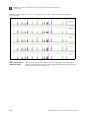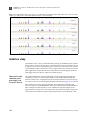
1
USER GUIDE
AmpFlSTR® MiniFiler™ PCR Amplification Kit
for use with:
100 reaction kit (Part no. 4373872)
Publication Part Number 4374618 Rev. F
Revision Date August 2012
For Forensic or Paternity Use Only.
Information in this document is subject to change without notice.
LIFE TECHNOLOGIES CORPORATION AND/OR ITS AFFILIATE(S) DISCLAIM ALL WARRANTIES WITH RESPECT TO THIS DOCUMENT, EXPRESSED OR IMPLIED,
INCLUDING BUT NOT LIMITED TO THOSE OF MERCHANTABILITY, FITNESS FOR A PARTICULAR PURPOSE, OR NON-INFRINGEMENT. TO THE EXTENT
ALLOWED BY LAW, IN NO EVENT SHALL LIFE TECHNOLOGIES AND/OR ITS AFFILIATE(S) BE LIABLE, WHETHER IN CONTRACT, TORT, WARRANTY, OR
UNDER ANY STATUTE OR ON ANY OTHER BASIS FOR SPECIAL, INCIDENTAL, INDIRECT, PUNITIVE, MULTIPLE OR CONSEQUENTIAL DAMAGES IN
CONNECTION WITH OR ARISING FROM THIS DOCUMENT, INCLUDING BUT NOT LIMITED TO THE USE THEREOF.
TRADEMARKS
The trademarks mentioned herein are the property of Life Technologies Corporation or their respective owners.
Windows and Windows Vista are registered trademarks of Microsoft Corporation.
FTA is a registered trademark of Whatman International Ltd.
TaqMan is a registered trademark of Roche Molecular Systems, Inc.
© 2012 Life Technologies Corporation. All rights reserved.
Contents
About This Guide . . . . . . . . . . . . . . . . . . . . . . . . . . . . . . . . . . . . . . . . . . . . . . . . . 9
Revision history . . . . . . . . . . . . . . . . . . . . . . . . . . . . . . . . . . . . . . . . . . . . . . . . . . . . . . . . . . . . . . . . . . . . . . . 9
Purpose . . . . . . . . . . . . . . . . . . . . . . . . . . . . . . . . . . . . . . . . . . . . . . . . . . . . . . . . . . . . . . . . . . . . . . . . . . . . . 9
■ CHAPTER 1
Overview . . . . . . . . . . . . . . . . . . . . . . . . . . . . . . . . . . . . . . . . . . 11
Product overview . . . . . . . . . . . . . . . . . . . . . . . . . . . . . . . . . . . . . . . . . . . . . . . . . . . . . . . . . . . . . . . . . . . .
Purpose . . . . . . . . . . . . . . . . . . . . . . . . . . . . . . . . . . . . . . . . . . . . . . . . . . . . . . . . . . . . . . . . . . . . . . . .
Product description . . . . . . . . . . . . . . . . . . . . . . . . . . . . . . . . . . . . . . . . . . . . . . . . . . . . . . . . . . . . . .
About the primers . . . . . . . . . . . . . . . . . . . . . . . . . . . . . . . . . . . . . . . . . . . . . . . . . . . . . . . . . . . . . . .
Loci amplified by the kit . . . . . . . . . . . . . . . . . . . . . . . . . . . . . . . . . . . . . . . . . . . . . . . . . . . . . . . . . .
Allelic ladder . . . . . . . . . . . . . . . . . . . . . . . . . . . . . . . . . . . . . . . . . . . . . . . . . . . . . . . . . . . . . . . . . . .
11
11
11
11
12
13
Workflow overview . . . . . . . . . . . . . . . . . . . . . . . . . . . . . . . . . . . . . . . . . . . . . . . . . . . . . . . . . . . . . . . . . . . 14
Instrument and software overview . . . . . . . . . . . . . . . . . . . . . . . . . . . . . . . . . . . . . . . . . . . . . . . . . . . . .
Data collection and analysis software . . . . . . . . . . . . . . . . . . . . . . . . . . . . . . . . . . . . . . . . . . . . . .
Instrument and software compatibility . . . . . . . . . . . . . . . . . . . . . . . . . . . . . . . . . . . . . . . . . . . . .
About multicomponent analysis . . . . . . . . . . . . . . . . . . . . . . . . . . . . . . . . . . . . . . . . . . . . . . . . . . .
How multicomponent analysis works . . . . . . . . . . . . . . . . . . . . . . . . . . . . . . . . . . . . . . . . . . . . . .
15
15
15
15
15
Materials and equipment . . . . . . . . . . . . . . . . . . . . . . . . . . . . . . . . . . . . . . . . . . . . . . . . . . . . . . . . . . . . . 16
Kit contents and storage . . . . . . . . . . . . . . . . . . . . . . . . . . . . . . . . . . . . . . . . . . . . . . . . . . . . . . . . . 16
Standards for samples . . . . . . . . . . . . . . . . . . . . . . . . . . . . . . . . . . . . . . . . . . . . . . . . . . . . . . . . . . . 17
■ CHAPTER 2
Perform PCR . . . . . . . . . . . . . . . . . . . . . . . . . . . . . . . . . . . . . . 19
Required user-supplied reagents . . . . . . . . . . . . . . . . . . . . . . . . . . . . . . . . . . . . . . . . . . . . . . . . . . . . . . 19
DNA quantification . . . . . . . . . . . . . . . . . . . . . . . . . . . . . . . . . . . . . . . . . . . . . . . . . . . . . . . . . . . . . . . . . . . 19
Importance of quantification . . . . . . . . . . . . . . . . . . . . . . . . . . . . . . . . . . . . . . . . . . . . . . . . . . . . . . 19
Methods of quantifying DNA . . . . . . . . . . . . . . . . . . . . . . . . . . . . . . . . . . . . . . . . . . . . . . . . . . . . . . 20
Prepare the amplification kit reactions . . . . . . . . . . . . . . . . . . . . . . . . . . . . . . . . . . . . . . . . . . . . . . . . . . 21
Perform PCR . . . . . . . . . . . . . . . . . . . . . . . . . . . . . . . . . . . . . . . . . . . . . . . . . . . . . . . . . . . . . . . . . . . . . . . . 22
Amplification using bloodstained FTA® cards . . . . . . . . . . . . . . . . . . . . . . . . . . . . . . . . . . . . . . . . . . . . 22
■ CHAPTER 3
Perform Electrophoresis . . . . . . . . . . . . . . . . . . . . . . . . . . . 25
Allelic ladder requirements . . . . . . . . . . . . . . . . . . . . . . . . . . . . . . . . . . . . . . . . . . . . . . . . . . . . . . . . . . . 26
Section 3.1 3100/3100-Avant and 3130/3130xl instruments . . . . . . . . . . . . . . . . . . . . . . . 27
Set up the 3100/3100-Avant and 3130/3130xl instruments for electrophoresis . . . . . . . . . . . . . . . . 27
Reagents and parts . . . . . . . . . . . . . . . . . . . . . . . . . . . . . . . . . . . . . . . . . . . . . . . . . . . . . . . . . . . . . . 27
Electrophoresis software setup and reference documents . . . . . . . . . . . . . . . . . . . . . . . . . . . . 27
Prepare samples for electrophoresis on the 3100/3100-Avant or 3130/3130xl instruments . . . . . 28
AmpFlSTR® MiniFiler™ PCR Amplification Kit User Guide
3
Contents
Section 3.2 3500/3500xL instruments . . . . . . . . . . . . . . . . . . . . . . . . . . . . . . . . . . . . . . . . . 29
Set up the 3500/3500xL instruments for electrophoresis . . . . . . . . . . . . . . . . . . . . . . . . . . . . . . . . . . . 29
Reagents and parts . . . . . . . . . . . . . . . . . . . . . . . . . . . . . . . . . . . . . . . . . . . . . . . . . . . . . . . . . . . . . . 29
Electrophoresis software setup and reference documents . . . . . . . . . . . . . . . . . . . . . . . . . . . . . 29
Prepare samples for electrophoresis on the 3500/3500xL instruments . . . . . . . . . . . . . . . . . . . . . . 29
Section 3.3 310 Instrument . . . . . . . . . . . . . . . . . . . . . . . . . . . . . . . . . . . . . . . . . . . . . . . . . . 31
Set up the 310 instrument for electrophoresis . . . . . . . . . . . . . . . . . . . . . . . . . . . . . . . . . . . . . . . . . . . . 31
Reagents and parts . . . . . . . . . . . . . . . . . . . . . . . . . . . . . . . . . . . . . . . . . . . . . . . . . . . . . . . . . . . . . . 31
Electrophoresis software setup and reference documents . . . . . . . . . . . . . . . . . . . . . . . . . . . . . 31
Prepare samples for electrophoresis on the 310 instrument . . . . . . . . . . . . . . . . . . . . . . . . . . . . . . . . 31
■ CHAPTER 4
Analyze Data . . . . . . . . . . . . . . . . . . . . . . . . . . . . . . . . . . . . . 33
Section 4.1 GeneMapper® ID Software . . . . . . . . . . . . . . . . . . . . . . . . . . . . . . . . . . . . . . . . 33
Overview of GeneMapper® ID Software . . . . . . . . . . . . . . . . . . . . . . . . . . . . . . . . . . . . . . . . . . . . . . . . . . 33
Instruments . . . . . . . . . . . . . . . . . . . . . . . . . . . . . . . . . . . . . . . . . . . . . . . . . . . . . . . . . . . . . . . . . . . . . 33
Before you start . . . . . . . . . . . . . . . . . . . . . . . . . . . . . . . . . . . . . . . . . . . . . . . . . . . . . . . . . . . . . . . . . 33
Set up GeneMapper® ID Software for data analysis . . . . . . . . . . . . . . . . . . . . . . . . . . . . . . . . . . . . . . . .
File names . . . . . . . . . . . . . . . . . . . . . . . . . . . . . . . . . . . . . . . . . . . . . . . . . . . . . . . . . . . . . . . . . . . . . .
Before using the software for the first time . . . . . . . . . . . . . . . . . . . . . . . . . . . . . . . . . . . . . . . . . .
Import panels and bins . . . . . . . . . . . . . . . . . . . . . . . . . . . . . . . . . . . . . . . . . . . . . . . . . . . . . . . . . . .
Create an analysis method . . . . . . . . . . . . . . . . . . . . . . . . . . . . . . . . . . . . . . . . . . . . . . . . . . . . . . . .
General tab settings . . . . . . . . . . . . . . . . . . . . . . . . . . . . . . . . . . . . . . . . . . . . . . . . . . . . . . . . . . . . . .
Allele tab settings . . . . . . . . . . . . . . . . . . . . . . . . . . . . . . . . . . . . . . . . . . . . . . . . . . . . . . . . . . . . . . . .
Peak Detector tab settings . . . . . . . . . . . . . . . . . . . . . . . . . . . . . . . . . . . . . . . . . . . . . . . . . . . . . . . .
Peak Quality tab settings . . . . . . . . . . . . . . . . . . . . . . . . . . . . . . . . . . . . . . . . . . . . . . . . . . . . . . . . . .
Quality Flags tab settings . . . . . . . . . . . . . . . . . . . . . . . . . . . . . . . . . . . . . . . . . . . . . . . . . . . . . . . . .
Create a size standard . . . . . . . . . . . . . . . . . . . . . . . . . . . . . . . . . . . . . . . . . . . . . . . . . . . . . . . . . . . .
34
34
34
34
38
38
39
40
41
42
43
Analyze and edit sample files with GeneMapper® ID Software . . . . . . . . . . . . . . . . . . . . . . . . . . . . . . 44
Examine and edit a project . . . . . . . . . . . . . . . . . . . . . . . . . . . . . . . . . . . . . . . . . . . . . . . . . . . . . . . . . . . . . 45
For more information . . . . . . . . . . . . . . . . . . . . . . . . . . . . . . . . . . . . . . . . . . . . . . . . . . . . . . . . . . . . . . . . . 45
Section 4.2 GeneMapper® ID-X Software . . . . . . . . . . . . . . . . . . . . . . . . . . . . . . . . . . . . . . . 46
Overview of GeneMapper® ID-X Software . . . . . . . . . . . . . . . . . . . . . . . . . . . . . . . . . . . . . . . . . . . . . . . . 46
Instruments . . . . . . . . . . . . . . . . . . . . . . . . . . . . . . . . . . . . . . . . . . . . . . . . . . . . . . . . . . . . . . . . . . . . . 46
Before you start . . . . . . . . . . . . . . . . . . . . . . . . . . . . . . . . . . . . . . . . . . . . . . . . . . . . . . . . . . . . . . . . . 46
Set up GeneMapper® ID-X Software for data analysis . . . . . . . . . . . . . . . . . . . . . . . . . . . . . . . . . . . . . .
Panel, bin, and stutter file version . . . . . . . . . . . . . . . . . . . . . . . . . . . . . . . . . . . . . . . . . . . . . . . . . .
Before using the software for the first time . . . . . . . . . . . . . . . . . . . . . . . . . . . . . . . . . . . . . . . . . .
Check panel, bin, and stutter file version . . . . . . . . . . . . . . . . . . . . . . . . . . . . . . . . . . . . . . . . . . . .
Import panels, bins, and marker stutter . . . . . . . . . . . . . . . . . . . . . . . . . . . . . . . . . . . . . . . . . . . .
Create an analysis method . . . . . . . . . . . . . . . . . . . . . . . . . . . . . . . . . . . . . . . . . . . . . . . . . . . . . . . .
General tab settings . . . . . . . . . . . . . . . . . . . . . . . . . . . . . . . . . . . . . . . . . . . . . . . . . . . . . . . . . . . . . .
Allele tab settings . . . . . . . . . . . . . . . . . . . . . . . . . . . . . . . . . . . . . . . . . . . . . . . . . . . . . . . . . . . . . . .
Peak Detector tab settings . . . . . . . . . . . . . . . . . . . . . . . . . . . . . . . . . . . . . . . . . . . . . . . . . . . . . . . .
4
47
47
47
47
48
53
54
55
56
AmpFlSTR® MiniFiler™ PCR Amplification Kit User Guide
Contents
Peak Quality tab settings . . . . . . . . . . . . . . . . . . . . . . . . . . . . . . . . . . . . . . . . . . . . . . . . . . . . . . . . . 57
SQ & GQ tab settings . . . . . . . . . . . . . . . . . . . . . . . . . . . . . . . . . . . . . . . . . . . . . . . . . . . . . . . . . . . . 58
Create size standard (optional) . . . . . . . . . . . . . . . . . . . . . . . . . . . . . . . . . . . . . . . . . . . . . . . . . . . . 58
Analyze and edit sample files with GeneMapper® ID-X Software . . . . . . . . . . . . . . . . . . . . . . . . . . . . 60
Examine and edit a project . . . . . . . . . . . . . . . . . . . . . . . . . . . . . . . . . . . . . . . . . . . . . . . . . . . . . . . . . . . . 61
For more information . . . . . . . . . . . . . . . . . . . . . . . . . . . . . . . . . . . . . . . . . . . . . . . . . . . . . . . . . . . . . . . . . 62
■ CHAPTER 5
Experiments and Results . . . . . . . . . . . . . . . . . . . . . . . . . . . 63
Section 5.1 Developmental Validation . . . . . . . . . . . . . . . . . . . . . . . . . . . . . . . . . . . . . . . . . 65
Overview . . . . . . . . . . . . . . . . . . . . . . . . . . . . . . . . . . . . . . . . . . . . . . . . . . . . . . . . . . . . . . . . . . . . . . . . . . . .
Experiments using the MiniFiler™ Kit . . . . . . . . . . . . . . . . . . . . . . . . . . . . . . . . . . . . . . . . . . . . . . .
Importance of validation . . . . . . . . . . . . . . . . . . . . . . . . . . . . . . . . . . . . . . . . . . . . . . . . . . . . . . . . . .
Experiment conditions . . . . . . . . . . . . . . . . . . . . . . . . . . . . . . . . . . . . . . . . . . . . . . . . . . . . . . . . . . .
65
65
65
65
Developmental validation . . . . . . . . . . . . . . . . . . . . . . . . . . . . . . . . . . . . . . . . . . . . . . . . . . . . . . . . . . . . .
SWGDAM guideline 1.2.1 . . . . . . . . . . . . . . . . . . . . . . . . . . . . . . . . . . . . . . . . . . . . . . . . . . . . . . . . .
SWGDAM guideline 2.10.1 . . . . . . . . . . . . . . . . . . . . . . . . . . . . . . . . . . . . . . . . . . . . . . . . . . . . . . . .
PCR components . . . . . . . . . . . . . . . . . . . . . . . . . . . . . . . . . . . . . . . . . . . . . . . . . . . . . . . . . . . . . . . .
Thermal cycler parameters . . . . . . . . . . . . . . . . . . . . . . . . . . . . . . . . . . . . . . . . . . . . . . . . . . . . . . .
PCR cycle number . . . . . . . . . . . . . . . . . . . . . . . . . . . . . . . . . . . . . . . . . . . . . . . . . . . . . . . . . . . . . . .
65
65
65
66
66
67
Accuracy, precision, and reproducibility . . . . . . . . . . . . . . . . . . . . . . . . . . . . . . . . . . . . . . . . . . . . . . . . .
SWGDAM guideline 2.9 . . . . . . . . . . . . . . . . . . . . . . . . . . . . . . . . . . . . . . . . . . . . . . . . . . . . . . . . . . .
Accuracy . . . . . . . . . . . . . . . . . . . . . . . . . . . . . . . . . . . . . . . . . . . . . . . . . . . . . . . . . . . . . . . . . . . . . . .
Precision and size windows . . . . . . . . . . . . . . . . . . . . . . . . . . . . . . . . . . . . . . . . . . . . . . . . . . . . . . .
68
68
68
69
Extra peaks in the electropherogram . . . . . . . . . . . . . . . . . . . . . . . . . . . . . . . . . . . . . . . . . . . . . . . . . . . 75
Causes of extra peaks . . . . . . . . . . . . . . . . . . . . . . . . . . . . . . . . . . . . . . . . . . . . . . . . . . . . . . . . . . . 75
Artifacts . . . . . . . . . . . . . . . . . . . . . . . . . . . . . . . . . . . . . . . . . . . . . . . . . . . . . . . . . . . . . . . . . . . . . . . . 79
Characterization of loci . . . . . . . . . . . . . . . . . . . . . . . . . . . . . . . . . . . . . . . . . . . . . . . . . . . . . . . . . . . . . . .
SWGDAM guideline 2.1 . . . . . . . . . . . . . . . . . . . . . . . . . . . . . . . . . . . . . . . . . . . . . . . . . . . . . . . . . .
Nature of the polymorphisms . . . . . . . . . . . . . . . . . . . . . . . . . . . . . . . . . . . . . . . . . . . . . . . . . . . . .
Inheritance . . . . . . . . . . . . . . . . . . . . . . . . . . . . . . . . . . . . . . . . . . . . . . . . . . . . . . . . . . . . . . . . . . . . .
Mapping . . . . . . . . . . . . . . . . . . . . . . . . . . . . . . . . . . . . . . . . . . . . . . . . . . . . . . . . . . . . . . . . . . . . . . .
81
81
81
81
81
Species specificity . . . . . . . . . . . . . . . . . . . . . . . . . . . . . . . . . . . . . . . . . . . . . . . . . . . . . . . . . . . . . . . . . . . 82
SWGDAM Guideline 2.2 . . . . . . . . . . . . . . . . . . . . . . . . . . . . . . . . . . . . . . . . . . . . . . . . . . . . . . . . . . 82
Sensitivity . . . . . . . . . . . . . . . . . . . . . . . . . . . . . . . . . . . . . . . . . . . . . . . . . . . . . . . . . . . . . . . . . . . . . . . . . . . 83
SWGDAM guideline 2.3 . . . . . . . . . . . . . . . . . . . . . . . . . . . . . . . . . . . . . . . . . . . . . . . . . . . . . . . . . . . 83
Effect of DNA quantity on results . . . . . . . . . . . . . . . . . . . . . . . . . . . . . . . . . . . . . . . . . . . . . . . . . . 83
Stability . . . . . . . . . . . . . . . . . . . . . . . . . . . . . . . . . . . . . . . . . . . . . . . . . . . . . . . . . . . . . . . . . . . . . . . . . . . . .
SWGDAM guideline 2.4 . . . . . . . . . . . . . . . . . . . . . . . . . . . . . . . . . . . . . . . . . . . . . . . . . . . . . . . . . . .
Degraded DNA . . . . . . . . . . . . . . . . . . . . . . . . . . . . . . . . . . . . . . . . . . . . . . . . . . . . . . . . . . . . . . . . . .
Effect of inhibitors – Hematin . . . . . . . . . . . . . . . . . . . . . . . . . . . . . . . . . . . . . . . . . . . . . . . . . . . . .
Effect of inhibitors – Humic Acid . . . . . . . . . . . . . . . . . . . . . . . . . . . . . . . . . . . . . . . . . . . . . . . . . . .
84
84
84
86
88
Mixture studies . . . . . . . . . . . . . . . . . . . . . . . . . . . . . . . . . . . . . . . . . . . . . . . . . . . . . . . . . . . . . . . . . . . . . . 90
SWGDAM guideline 2.8 . . . . . . . . . . . . . . . . . . . . . . . . . . . . . . . . . . . . . . . . . . . . . . . . . . . . . . . . . . . 90
AmpFlSTR® MiniFiler™ PCR Amplification Kit User Guide
5
Contents
Mixture studies . . . . . . . . . . . . . . . . . . . . . . . . . . . . . . . . . . . . . . . . . . . . . . . . . . . . . . . . . . . . . . . . . . 90
Resolution of genotypes in mixed samples . . . . . . . . . . . . . . . . . . . . . . . . . . . . . . . . . . . . . . . . . . 91
Limit of detection of the minor component . . . . . . . . . . . . . . . . . . . . . . . . . . . . . . . . . . . . . . . . . . 92
Population data . . . . . . . . . . . . . . . . . . . . . . . . . . . . . . . . . . . . . . . . . . . . . . . . . . . . . . . . . . . . . . . . . . . . . .
SWGDAM guideline 2.7 . . . . . . . . . . . . . . . . . . . . . . . . . . . . . . . . . . . . . . . . . . . . . . . . . . . . . . . . . . .
Overview . . . . . . . . . . . . . . . . . . . . . . . . . . . . . . . . . . . . . . . . . . . . . . . . . . . . . . . . . . . . . . . . . . . . . . . .
Concordance studies . . . . . . . . . . . . . . . . . . . . . . . . . . . . . . . . . . . . . . . . . . . . . . . . . . . . . . . . . . . . .
93
93
93
94
Mutation rate . . . . . . . . . . . . . . . . . . . . . . . . . . . . . . . . . . . . . . . . . . . . . . . . . . . . . . . . . . . . . . . . . . . . . . . . 95
Estimating germ-line mutations . . . . . . . . . . . . . . . . . . . . . . . . . . . . . . . . . . . . . . . . . . . . . . . . . . . 95
Additional mutation studies . . . . . . . . . . . . . . . . . . . . . . . . . . . . . . . . . . . . . . . . . . . . . . . . . . . . . . . 95
Probability of identity . . . . . . . . . . . . . . . . . . . . . . . . . . . . . . . . . . . . . . . . . . . . . . . . . . . . . . . . . . . . . . . . . . 95
Probability of paternity exclusion . . . . . . . . . . . . . . . . . . . . . . . . . . . . . . . . . . . . . . . . . . . . . . . . . . . . . . . 96
Section 5.2 Performance Validation After Buffer and Enzyme Component
Replacement . . . . . . . . . . . . . . . . . . . . . . . . . . . . . . . . . . . . . . . . . . . . . . . . . . . . . . . . . . . . . . . .98
Overview . . . . . . . . . . . . . . . . . . . . . . . . . . . . . . . . . . . . . . . . . . . . . . . . . . . . . . . . . . . . . . . . . . . . . . . . . . . . 98
Experiments . . . . . . . . . . . . . . . . . . . . . . . . . . . . . . . . . . . . . . . . . . . . . . . . . . . . . . . . . . . . . . . . . . . . . . . . . 98
Reproducibility study . . . . . . . . . . . . . . . . . . . . . . . . . . . . . . . . . . . . . . . . . . . . . . . . . . . . . . . . . . . . . . . . . . 99
Intracolor balance . . . . . . . . . . . . . . . . . . . . . . . . . . . . . . . . . . . . . . . . . . . . . . . . . . . . . . . . . . . . . . . 99
Stutter percentages . . . . . . . . . . . . . . . . . . . . . . . . . . . . . . . . . . . . . . . . . . . . . . . . . . . . . . . . . . . . . . 99
Artifacts . . . . . . . . . . . . . . . . . . . . . . . . . . . . . . . . . . . . . . . . . . . . . . . . . . . . . . . . . . . . . . . . . . . . . . . 100
Sensitivity study . . . . . . . . . . . . . . . . . . . . . . . . . . . . . . . . . . . . . . . . . . . . . . . . . . . . . . . . . . . . . . . . . . . . .
Mean referenced peak height . . . . . . . . . . . . . . . . . . . . . . . . . . . . . . . . . . . . . . . . . . . . . . . . . . . . .
DNA concentration and peak height . . . . . . . . . . . . . . . . . . . . . . . . . . . . . . . . . . . . . . . . . . . . . . .
Allelic dropout . . . . . . . . . . . . . . . . . . . . . . . . . . . . . . . . . . . . . . . . . . . . . . . . . . . . . . . . . . . . . . . . . .
Genotype concordance . . . . . . . . . . . . . . . . . . . . . . . . . . . . . . . . . . . . . . . . . . . . . . . . . . . . . . . . . .
101
101
102
103
105
Degraded DNA study . . . . . . . . . . . . . . . . . . . . . . . . . . . . . . . . . . . . . . . . . . . . . . . . . . . . . . . . . . . . . . . . .
Intracolor balance . . . . . . . . . . . . . . . . . . . . . . . . . . . . . . . . . . . . . . . . . . . . . . . . . . . . . . . . . . . . . .
Mean referenced peak height . . . . . . . . . . . . . . . . . . . . . . . . . . . . . . . . . . . . . . . . . . . . . . . . . . . . .
Allelic dropout . . . . . . . . . . . . . . . . . . . . . . . . . . . . . . . . . . . . . . . . . . . . . . . . . . . . . . . . . . . . . . . . . .
105
105
106
107
Inhibition study . . . . . . . . . . . . . . . . . . . . . . . . . . . . . . . . . . . . . . . . . . . . . . . . . . . . . . . . . . . . . . . . . . . . . . 108
Mean peak height, minimum mean peak height, and intracolor balance . . . . . . . . . . . . . . . . 108
Allelic dropout . . . . . . . . . . . . . . . . . . . . . . . . . . . . . . . . . . . . . . . . . . . . . . . . . . . . . . . . . . . . . . . . . . 112
Conclusions . . . . . . . . . . . . . . . . . . . . . . . . . . . . . . . . . . . . . . . . . . . . . . . . . . . . . . . . . . . . . . . . . . . . . . . . 112
■ APPENDIX A
Troubleshooting . . . . . . . . . . . . . . . . . . . . . . . . . . . . . . . . . 113
■ APPENDIX B
The 3rd Order Least Squares Sizing Method . . . . . . . 115
When to use . . . . . . . . . . . . . . . . . . . . . . . . . . . . . . . . . . . . . . . . . . . . . . . . . . . . . . . . . . . . . . . . . . . . . . . . 115
About the Local Southern algorithm . . . . . . . . . . . . . . . . . . . . . . . . . . . . . . . . . . . . . . . . . . . . . . . . . . . 115
Comparing genotyping accuracy . . . . . . . . . . . . . . . . . . . . . . . . . . . . . . . . . . . . . . . . . . . . . . . . . . . . . . . 116
6
AmpFlSTR® MiniFiler™ PCR Amplification Kit User Guide
Contents
■ APPENDIX C
Ordering Information . . . . . . . . . . . . . . . . . . . . . . . . . . . . 117
Equipment and materials not included . . . . . . . . . . . . . . . . . . . . . . . . . . . . . . . . . . . . . . . . . . . . . . . . . 117
■ APPENDIX D
PCR Work Areas . . . . . . . . . . . . . . . . . . . . . . . . . . . . . . . . 121
Work area setup and lab design . . . . . . . . . . . . . . . . . . . . . . . . . . . . . . . . . . . . . . . . . . . . . . . . . . . . . . . 121
PCR setup work area . . . . . . . . . . . . . . . . . . . . . . . . . . . . . . . . . . . . . . . . . . . . . . . . . . . . . . . . . . . . . . . . 121
Amplified DNA work area . . . . . . . . . . . . . . . . . . . . . . . . . . . . . . . . . . . . . . . . . . . . . . . . . . . . . . . . . . . . 122
■ APPENDIX E
Safety . . . . . . . . . . . . . . . . . . . . . . . . . . . . . . . . . . . . . . . . . 123
Chemical safety . . . . . . . . . . . . . . . . . . . . . . . . . . . . . . . . . . . . . . . . . . . . . . . . . . . . . . . . . . . . . . . . . . . . 124
Specific chemical handling . . . . . . . . . . . . . . . . . . . . . . . . . . . . . . . . . . . . . . . . . . . . . . . . . . . . . . 124
Biological hazard safety . . . . . . . . . . . . . . . . . . . . . . . . . . . . . . . . . . . . . . . . . . . . . . . . . . . . . . . . . . . . . . 125
Bibliography . . . . . . . . . . . . . . . . . . . . . . . . . . . . . . . . . . . . . . . . . . . . . . . . . . 127
Documentation and Support . . . . . . . . . . . . . . . . . . . . . . . . . . . . . . . . . . . . . 131
Related documentation . . . . . . . . . . . . . . . . . . . . . . . . . . . . . . . . . . . . . . . . . . . . . . . . . . . . . . . . . . . . . . 131
Obtain SDSs . . . . . . . . . . . . . . . . . . . . . . . . . . . . . . . . . . . . . . . . . . . . . . . . . . . . . . . . . . . . . . . . . . . . . . . . 132
Obtain support . . . . . . . . . . . . . . . . . . . . . . . . . . . . . . . . . . . . . . . . . . . . . . . . . . . . . . . . . . . . . . . . . . . . . 132
Limited Product Warranty . . . . . . . . . . . . . . . . . . . . . . . . . . . . . . . . . . . . . . . . . . . . . . . . . . . . . . . . . . . . 132
Index . . . . . . . . . . . . . . . . . . . . . . . . . . . . . . . . . . . . . . . . . . . . . . . . . . . . . . . . . 133
AmpFlSTR® MiniFiler™ PCR Amplification Kit User Guide
7
Contents
8
AmpFlSTR® MiniFiler™ PCR Amplification Kit User Guide
About This Guide
IMPORTANT! Before using this product, read and understand the information the
“Safety” appendix in this document.
Revision history
Revision
Date
Description
A
October 2006
New document.
B
March 2007
Add Experiments and Results chapter.
C
December 2010
Change to limited licensing information.
D
April 2011
Change to limited licensing information.
E
March 2012
Change to limited licensing information.
F
August 2012
• Add 3500 and 3500xL Genetic Analyzer
information. Add GeneMapper® ID-X Software
information.
• Add validation experiments and results for
buffer and enzyme kit component changes.
Purpose
The AmpFlSTR® MiniFiler™ PCR Amplification Kit User Guide provides information
about the Life Technologies instruments, chemistries, and software associated with the
AmpFlSTR® MiniFiler™ PCR Amplification Kit.
AmpFlSTR® MiniFiler™ PCR Amplification Kit User Guide
9
About This Guide
Purpose
10
AmpFlSTR® MiniFiler™ PCR Amplification Kit User Guide
1
Overview
■
Product overview . . . . . . . . . . . . . . . . . . . . . . . . . . . . . . . . . . . . . . . . . . . . . . . . . . . . . 11
■
Workflow overview . . . . . . . . . . . . . . . . . . . . . . . . . . . . . . . . . . . . . . . . . . . . . . . . . . . 14
■
Instrument and software overview . . . . . . . . . . . . . . . . . . . . . . . . . . . . . . . . . . . . . . 15
■
Materials and equipment . . . . . . . . . . . . . . . . . . . . . . . . . . . . . . . . . . . . . . . . . . . . . . 16
Product overview
Purpose
The AmpFlSTR® MiniFiler™ PCR Amplification Kit is an assay optimized for
genotyping degraded and/or inhibited DNA samples. It is a short tandem repeat (STR)
multiplex assay optimized to allow direct amplification of single-source samples:
The MiniFiler™ Kit amplifies eight autosomal STR loci (D13S317, D7S820, D2S1338,
D21S11, D16S539, D18S51, CSF1PO and FGA) and the sex-determining marker,
Amelogenin, in a single PCR reaction. The loci span a range between 70 to 283
nucleotides with the aid of non-nucleotide linkers to achieve appropriate spacing
between loci.
Product
description
The MiniFiler™ Kit contains all the necessary reagents for the amplification of human
genomic DNA.
The reagents are designed for use with the following instruments:
• Applied Biosystems® 3100/3100-Avant Genetic Analyzer
• Applied Biosystems® 3130/3130xl Genetic Analyzer
• Applied Biosystems® 3500/3500xL Genetic Analyzer
• Veriti® 96-Well Thermal Cycler (Part no. 4375786)
• GeneAmp® PCR System 9700 with the Silver 96-Well Block
• GeneAmp® PCR System 9700 with the Gold-plated Silver 96-Well Block
About the primers
The MiniFiler™ Kit uses primers closely flanking the STR repetitive regions (miniSTRs)
of the DNA. This amplification results in amplicons that are significantly shorter in
length than those produced in the AmpFlSTR® Identifiler® and SGM Plus® PCR
Amplification Kits. The comparison is shown in Table 1 on page 12. Several
laboratories confirm that MiniSTRs have a higher success rate for DNA analysis of
degraded DNA samples (Butler et al., 2003; Chung et al., 2004; Coble and Butler, 2005;
Drabek et al., 2004; Grubwieser et al., 2006; Wiegand et al., 2001).
To prevent overlap of the miniSTR amplicons in the multiplex, non-nucleotide linkers
are used in primer synthesis for the following loci: CSF1PO, FGA, D16S539, D18S51,
Amelogenin, D2S1338, D21S11, and D7S820. For these primers, non-nucleotide linkers
are placed between the primers and the fluorescent dye during oligonucleotide
synthesis (Butler 2005, Grossman et al., 1994, and Baron et al., 1996). Non-nucleotide
AmpFlSTR® MiniFiler™ PCR Amplification Kit User Guide
11
1
Chapter 1 Overview
Product overview
linkers enable reproducible positioning of the alleles to facilitate inter-locus spacing.
The combination of a five-dye fluorescent system and the inclusion of non-nucleotide
linkers allows for simultaneous amplification and efficient separation of the eight STR
loci and Amelogenin during automated DNA fragment analysis.
Loci amplified by
the kit
Locus
MiniFiler Kit Amplicon Length Reduction
(nt)
D7S820
−129
D13S317
−99
D21S11
−33
D2S1338
−183
Amelogenin
0
D18S51
−168
D16S539
−157
FGA
−87
CSF1PO
−201
Table 1 shows the loci amplified, their chromosomal locations, and the corresponding
fluorescent marker dyes. The AmpFlSTR® MiniFiler™ Allelic Ladder is used to
genotype the analyzed samples. The alleles contained in the allelic ladder, and the
genotype of the AmpFlSTR® Control DNA 007, are also listed in the table.
Table 1 AmpFlSTR® MiniFiler™ PCR Amplification Kit loci and alleles
Locus designation
Chromosome
location
Alleles included in Allelic Ladder
Dye
label
6-FAM™
Control
DNA 007
D13S317
13q22-31
8, 9, 10, 11, 12, 13, 14, 15
D7S820
7q11.21-22
6, 7, 8, 9, 10, 11, 12, 13, 14, 15
Amelogenin
X:p22.1-22.3
Y:p11.2
X, Y
D2S1338
2q35-37.1
15, 16, 17, 18, 19, 20, 21, 22, 23, 24, 25, 26,
27, 28
20, 23
D21S11
21q11.2-q21
24, 24.2, 25, 26, 27, 28, 28.2, 29, 29.2, 30,
30.2, 31, 31.2, 32, 32.2, 33, 33.2, 34, 34.2, 35,
35.2, 36, 37, 38
28, 31
D16S539
16q24-qter
5, 8, 9, 10, 11, 12, 13, 14, 15
D18S51
18q21.3
7, 9, 10, 10.2, 11, 12, 13, 13.2, 14, 14.2, 15,
16, 17, 18, 19, 20, 21, 22, 23, 24, 25, 26, 27
CSF1PO
5q33.3-34
6, 7, 8, 9, 10, 11, 12, 13, 14, 15
FGA
4q28
17, 18, 19, 20, 21, 22, 23, 24, 25, 26, 26.2, 27,
28, 29, 30, 30.2, 31.2, 32.2, 33.2, 42.2, 43.2,
44.2, 45.2, 46.2, 47.2, 48.2, 50.2, 51.2
12
11
7, 12
VIC®
NED™
X, Y
9, 10
12, 15
PET®
11, 12
24, 26
AmpFlSTR® MiniFiler™ PCR Amplification Kit User Guide
Chapter 1 Overview
Product overview
Allelic ladder
1
Figure 1 shows the allelic ladder for the MiniFiler™ Kit. See “Allelic ladder
requirements” on page 26 for information on ensuring accurate genotyping.
Figure 1 GeneMapper® ID-X Software plot of the AmpFlSTR® MiniFiler™ Allelic Ladder
AmpFlSTR® MiniFiler™ PCR Amplification Kit User Guide
13
1
Chapter 1 Overview
Workflow overview
Perform
PCR
Extract
DNA
Workflow overview
Quantify
DNA
AutoMate Express™ System + PrepFiler® Express Kit
Perform
PCR
Prepare
reactions
Quantifiler® Duo DNA Quantification Kit
AmpFlSTR® MiniFiler™ PCR Amplification Kit
GeneAmp® PCR System 9700 Cycler
Veriti® 96-Well Thermal Cycler
Perform
electrophoresis
3100/3100-Avant
Genetic Analyzer
3130/3130xl
Genetic Analyzer
3500/3500xL
Genetic Analyzer
310 Genetic
Analyzer
Analyze
data
GeneMapper® ID-X or GeneMapper® ID Software
14
AmpFlSTR® MiniFiler™ PCR Amplification Kit User Guide
Chapter 1 Overview
Instrument and software overview
1
Instrument and software overview
This section provides information about the data collection and analysis software
versions required to run the this kit on specific instruments.
Data collection and
analysis software
Instrument and
software
compatibility
The data collection software provides instructions to firmware running on the
instrument and displays instrument status and raw data in real time. As the
instrument measures sample fluorescence with its detection system, the data collection
software collects the data and stores it. The data collection software stores information
about each sample in a sample file (.fsa files for 31xx instruments and .hid files for 3500
instruments), which is then analyzed by the analysis software.
Instrument
3100/3100Avant
Operating system
Windows® NT
3500/3500xL
1.1 (3100)
1.0 (3100-Avant)
Windows®
3130/3130xl†
2000
Windows XP
2.0
Analysis software
• GeneMapper® ID
Software v3.2.1
• GeneMapper® ID-X
Software v1.0.1 or higher
3.0
3500 Series Data
Collection
Software v1.0
GeneMapper® ID-X Software
v1.2 or higher
Windows XP
3.1
GeneMapper ID 3.2
Windows NT and
Windows 2000
3.0
GeneMapper ID 3.2
•
Windows®
XP
or
• Windows Vista®
310
Data collection
software
† We conducted validation studies for the MiniFiler™ Kit using these configurations.
About
multicomponent
analysis
Life Technologies fluorescent multi-color dye technology allows the analysis of
multiple loci, including loci that have alleles with overlapping size ranges. Alleles for
overlapping loci are distinguished by labeling locus-specific primers with different
colored dyes.
Multicomponent analysis is the process that separates the five different fluorescent
dye colors into distinct spectral components. The four dyes used in the MiniFiler™ Kit
to label samples are 6-FAM™, VIC®, NED™, and PET® dyes. The fifth dye, LIZ®, is
used to label the GeneScan™ 500 LIZ® Size Standard or the GeneScan™ 600 LIZ® Size
Standard v2.0.
How
multicomponent
analysis works
Each of these fluorescent dyes emits its maximum fluorescence at a different
wavelength. During data collection on the Life Technologies instruments, the
fluorescence signals are separated by a diffraction grating according to their
wavelengths and projected onto a charge-coupled device (CCD) camera in a
predictably spaced pattern. The 6-FAM™ dye emits at the shortest wavelength and is
displayed as blue, followed by the VIC® dye (green), NED™ dye (yellow), PET® dye
(red), and LIZ® dye (orange).
Although each of these dyes emits its maximum fluorescence at a different
wavelength, there is some overlap in the emission spectra between the dyes (Figure 2).
The goal of multicomponent analysis is to correct for spectral overlap.
AmpFlSTR® MiniFiler™ PCR Amplification Kit User Guide
15
1
Chapter 1 Overview
Materials and equipment
Figure 2 Emission spectra of the five dyes used in the MiniFiler™ Kit
Dyes
Normalized Emission
6-FAM
VIC
NED PET
LIZ
100
80
60
40
20
0
500
550
600
650
700
Wavelength (nm)
Materials and equipment
Kit contents and
storage
The MiniFiler™ Kit (Part no. 4373872) contains sufficient quantities of the following
reagents to perform 100 reactions at 25 µL/reaction:
IMPORTANT! The fluorescent dyes attached to the primers are light-sensitive. Protect
the primer set, amplified DNA, allelic ladder, and size standard from light when not in
use. Keep freeze-thaw cycles to a minimum.
Table 2 Kit contents and storage
Component
Description
100 reaction
AmpFlSTR® MiniFiler™
Master Mix
Contains enzyme, salts, dNTPs, carrier
protein, and 0.05% sodium azide
2 tubes,
0.5 mL/tube
AmpFlSTR® Control
DNA 007
Contains 0.10 ng/µL human male genomic
DNA in 0.05% sodium azide and buffer.†
1 tube, 0.3 mL
Storage
–15 to –25°C upon
receipt, 2 to 8°C after
initial use
See Table 1 on page 12 for profile.
AmpFlSTR® MiniFiler™
Primer Set
Contains forward and reverse primers to
amplify human DNA targets.
1 tube, 0.5 mL
–15 to –25°C upon
receipt.
AmpFlSTR® MiniFiler™
Allelic Ladder
Contains amplified alleles.
1 tube, 0.05 mL
2 to 8°C after initial
use.
See Table 1 on page 12 for a list of alleles
included in the allelic ladder.
Store protected from
light.
† The Control DNA 007 is included at a concentration appropriate to its intended use as an amplification control (i.e., to provide confirmation of
the capability of the kit reagents to generate a profile of expected genotype). The Control DNA 007 is not designed to be used as a DNA
quantitation control and you may see variation from the labelled concentration when quantitating aliquots of the Control DNA 007.
16
AmpFlSTR® MiniFiler™ PCR Amplification Kit User Guide
Chapter 1 Overview
Materials and equipment
Standards for
samples
1
For the MiniFiler™ Kit, the panel of standards needed for PCR amplification, PCR
product sizing, and genotyping are:
• AmpFlSTR® Control DNA 007 – A positive control for evaluating the efficiency
of the amplification step and STR genotyping using the AmpFlSTR® MiniFiler™
Allelic Ladder.
• GeneScan™ 500 LIZ® Size Standard or GeneScan™ 600 LIZ® Size Standard
v2.0 – Used for obtaining sizing results. These standards, which have been
evaluated as internal size standards, yield precise sizing results for MiniFiler™ Kit
PCR products. Order the GeneScan™ 500 LIZ® Size Standard (Part no. 4322682) or
the GeneScan™ 600 LIZ® Size Standard v2.0 (Part no. 4408399) separately.
• AmpFlSTR® MiniFiler™ Allelic Ladder – Developed for accurate
characterization of the alleles amplified by the MiniFiler™ Kit. The Allelic Ladder
contains most of the alleles reported for the 16 autosomal loci. See page 12 for a
list of the alleles included in the Allelic Ladder.
AmpFlSTR® MiniFiler™ PCR Amplification Kit User Guide
17
1
18
Chapter 1 Overview
Materials and equipment
AmpFlSTR® MiniFiler™ PCR Amplification Kit User Guide
2
Perform PCR
■
Required user-supplied reagents . . . . . . . . . . . . . . . . . . . . . . . . . . . . . . . . . . . . . . . . 19
■
DNA quantification . . . . . . . . . . . . . . . . . . . . . . . . . . . . . . . . . . . . . . . . . . . . . . . . . . . 19
■
Prepare the amplification kit reactions . . . . . . . . . . . . . . . . . . . . . . . . . . . . . . . . . . . 21
■
Perform PCR . . . . . . . . . . . . . . . . . . . . . . . . . . . . . . . . . . . . . . . . . . . . . . . . . . . . . . . . . 22
■
Amplification using bloodstained FTA® cards . . . . . . . . . . . . . . . . . . . . . . . . . . . . 22
Required user-supplied reagents
In addition to the Identifiler® Kit reagents, the use of low-TE buffer (10 mM Tris,
0.1 mM EDTA, pH 8.0) is recommended. You can prepare the buffer as described in the
procedure below or order it from Teknova (Cat # T0223).
To prepare low-TE buffer:
1. Mix together:
• 10 mL of 1 M Tris-HCl, pH 8.0
• 0.2 mL of 0.5 M EDTA, pH 8.0
• 990 mL glass-distilled or deionized water
Note: Adjust the volumes accordingly for specific needs.
2. Aliquot and autoclave the solutions.
3. Store at room temperature.
DNA quantification
Importance of
quantification
Quantifying the amount of DNA in a sample before amplification allows you to
determine whether or not sufficient DNA is present to permit amplification and to
calculate the optimum amount of DNA to add to the reaction. The optimum amount of
DNA for the Identifiler® Kit is 1.0 ng in a maximum input volume of 10 µL for 28 PCR
cycles.
AmpFlSTR® MiniFiler™ PCR Amplification Kit User Guide
19
2
Chapter 2 Perform PCR
DNA quantification
If too much DNA is added to the PCR reaction, then the increased amount of PCR
product that is generated can result in:
• Fluorescence intensity that exceeds the linear dynamic range for detection by the
instrument (“off-scale” data). Off-scale data are problematic because:
– Quantitation (peak height and area) for off-scale peaks is not accurate. For
example, an allele peak that is off-scale can cause the corresponding stutter
peak to appear higher in relative intensity, thus increasing the calculated
percent stutter.
– Multicomponent analysis of off-scale data is not accurate, and it results in
poor spectral separation (“pull-up”).
• Incomplete A-nucleotide addition.
When the total number of allele copies added to the PCR is extremely low, allelic
dropout can occur resulting in a partial profile.
Methods of
quantifying DNA
Life Technologies provides several kits for quantifying DNA in samples. See the
references cited in the following table for details about these kits.
Product
Quantifiler® Human DNA
Quantification Kit
(Part no. 4343895)
and
Description
Properties:
The Quantifiler® Human and Quantifiler® Y Human Male Kits are highly specific for
human DNA, and they individually detect total human or male DNA, respectively. The
kits detect single-stranded and degraded DNA.
Quantifiler® Y Human Male
DNA Quantification Kit
(Part no. 4343906)
How they work:
For more information, see
Quantifiler® Human DNA
Quantification Kits User’s Manual
(Pub. no. 4344790)
The Quantifiler® Human and Quantifiler® Y Human Male Kits contain different targetspecific assays (human DNA or human male DNA, respectively) that each consist of two
locus-specific PCR primers and one TaqMan® MGB probe labeled with FAM™ dye for
detecting the amplified sequence. The kits each contain a separate internal PCR control
(IPC) assay, which consists of an IPC template DNA (a synthetic sequence not found in
nature), two primers for amplifying the IPC template, and one TaqMan® MGB probe
labeled with VIC® dye for detecting the amplified IPC.
Quantifiler® Duo DNA
Quantification Kit
(Part no. 4387746)
Properties:
For more information, see
Quantifiler® Duo DNA
Quantification Kit User's Manual
(Pub. no.4391294)
The Quantifiler® DNA Quantification Kits consist of target-specific and internal control
5' nuclease assays.
The Quantifiler® Duo Kit is highly specific for human DNA. This kit combines the
detection of both total human and male DNA in one PCR reaction.The kit detects singlestranded and degraded DNA.
How it works:
The Quantifiler® Duo DNA Quantification Kit consists of target-specific and internal
control 5' nuclease assays.
The Quantifiler® Duo kit combines two human-specific assays in one PCR reaction (for
total human DNA and human male DNA). The two human DNA specific assays each
consist of two PCR primers and a TaqMan® probe. The TaqMan® probes for the human
DNA and human male DNA assays are labeled with VIC® and FAM™ dyes, respectively.
In addition, the kit contains an internal PCR control (IPC) assay similar in principle to
that used in the other Quantifiler kits, but labeled with NED™ dye.
20
AmpFlSTR® MiniFiler™ PCR Amplification Kit User Guide
Chapter 2 Perform PCR
Prepare the amplification kit reactions
2
Prepare the amplification kit reactions
1. Calculate the volume of each component needed to prepare the reactions, using
the table below.
DNA sample
Volume per reaction
AmpFlSTR® MiniFiler™ Master Mix
10.0 µL
AmpFlSTR® MiniFiler™ Primer Set
5.0 µL
Note: The volumes indicated above include a slight overfill to account for the loss
that occurs during reagent transfers.
2. Prepare reagents. Thaw the PCR Reaction Mix and the Identifiler® Primer Set,
then vortex all reagent tubes, including the enzyme, for 3 seconds and centrifuge
briefly before opening the tubes.
IMPORTANT! Thawing is required only during first use of the Primer Set and PCR
Reaction Mix. After first use, these reagents are stored at 2 to 8 °C and do not
require subsequent thawing. Do not refreeze these reagents.
3. Prepare the reaction mix: Pipette the required volumes of components into an
appropriately sized polypropylene tube.
4. Vortex the reaction mix for 3 seconds, then centrifuge briefly.
5. Dispense 15 µL of the reaction mix into each reaction well of a MicroAmp®
Optical 96-Well Reaction Plate or each MicroAmp® tube.
6. Prepare the DNA samples:
DNA sample
To prepare...
Negative control
Add 10 µL of low-TE buffer (10mM Tris, 0.1mM EDTA, pH 8.0).
Test sample
Dilute a portion of the test DNA sample with low-TE buffer so
that 0.5 to 0.75 ng of total DNA is in a final volume of 10 µL. Add
10 µL of the diluted sample to the reaction mix.
Positive control
Combine 5 µL of control DNA (0.1 ng/µL) with 5 μL of low-TE
buffer for a total volume of 10 µL. (The final sample
concentration is 0.05 ng/µL.) Add to the reaction mix.
The final reaction volume (sample or control plus reaction mix) is 25 µL.
7. Seal the plate with MicroAmp® Clear Adhesive Film or MicroAmp® Optical
Adhesive Film, or cap the tubes.
8. Centrifuge the tubes at 3000 rpm for about 20 seconds in a tabletop centrifuge
(with plate holders if using 96-well plates).
9. Amplify the samples in a GeneAmp® PCR System 9700 with the silver or
gold-plated silver 96-well blocks or a Veriti® 96-Well Thermal Cycler.
AmpFlSTR® MiniFiler™ PCR Amplification Kit User Guide
21
2
Chapter 2 Perform PCR
Perform PCR
Perform PCR
1. Program the thermal cycling conditions:
• When using the GeneAmp PCR System 9700 with either 96-well silver or
gold-plated silver block, select the 9600 Emulation Mode.
• When using the Veriti® 96-Well Thermal Cycler, refer to the following
document for instructions on how to configure the Veriti instrument to run
in the 9600 Emulation Mode: User Bulletin: Veriti® 96-Well Thermal Cycler
AmpFlSTR® Kit Validation (Pub. no.4440754).
Initial
incubation step
Denature
HOLD
95°C
11 min
Anneal
Extend
CYCLE (30)
94°C
20 sec
59°C
2 min
72°C
1 min
Final
extension
Final hold
HOLD
HOLD
60°C
45 min
4°C
∞
2. Load the plate into the thermal cycler and close the heated cover.
IMPORTANT! If using the 9700 thermal cycler with silver or gold-plated silver
block and adhesive clear film instead of caps to seal the plate wells, be sure to
place a MicroAmp® compression pad (Part no. 4312639) on top of the plate to
prevent evaporation during thermal cycling. The Veriti® Thermal Cycler does not
require a compression pad.
3. Start the run.
4. On completion of the run, store the amplified DNA and protect from light.
If you are storing the DNA...
Then place at...
< 2 weeks
2 to 8°C
> 2 weeks
–15 to –25 °C
IMPORTANT! Store the amplified products so that they are protected from light.
Amplification using bloodstained FTA® cards
FTA® cards can be useful for collecting, storing, and processing biological samples. A
small punch disc of the card containing the sample can be placed directly into an
amplification tube, purified, and amplified, without transferring the disc. Our studies
indicate that a 1.2-mm bloodstained disc contains approximately 5 to 20 ng DNA. An
appropriate cycle number for this high quantity of DNA is 24 cycles as determined by
our validation studies. However, it is recommended that each laboratory determine
the optimum cycle number based on internal validation studies.
In the example shown in Figure 3, a 1.2-mm disc of a bloodstained FTA® card was
purified using three washes with FTA® Purification Reagent and two washes with
1✕ low-TE buffer. The purified punch disc was then amplified in the MicroAmp® tube
for 24 cycles.
22
AmpFlSTR® MiniFiler™ PCR Amplification Kit User Guide
Chapter 2 Perform PCR
Amplification using bloodstained FTA® cards
2
Figure 3 MiniFiler™ Kit results from a 1.2-mm FTA® bloodstain disc (24-cycle amplification), analyzed on the Applied
Biosystems 3130xl Genetic Analyzer
AmpFlSTR® MiniFiler™ PCR Amplification Kit User Guide
23
2
24
Chapter 2 Perform PCR
Amplification using bloodstained FTA® cards
AmpFlSTR® MiniFiler™ PCR Amplification Kit User Guide
3
Perform Electrophoresis
Allelic ladder requirements. . . . . . . . . . . . . . . . . . . . . . . . . . . . . . . . . . . . . . . . . . . . . .26
■
Section 3.1 3100/3100-Avant and 3130/3130xl instruments . . . . . . . . . . . . . . . . . . 27
Set up the 3100/3100-Avant and 3130/3130xl instruments for electrophoresis . . . 27
Prepare samples for electrophoresis on the 3100/3100-Avant or 3130/3130xl
instruments . . . . . . . . . . . . . . . . . . . . . . . . . . . . . . . . . . . . . . . . . . . . . . . . . . . . . . . . . . .28
■
Section 3.2 3500/3500xL instruments . . . . . . . . . . . . . . . . . . . . . . . . . . . . . . . . . . . . 29
Set up the 3500/3500xL instruments for electrophoresis . . . . . . . . . . . . . . . . . . . . . 29
Prepare samples for electrophoresis on the 3500/3500xL instruments. . . . . . . . . . 29
■
Section 3.3 310 Instrument . . . . . . . . . . . . . . . . . . . . . . . . . . . . . . . . . . . . . . . . . . . . . 31
Set up the 310 instrument for electrophoresis . . . . . . . . . . . . . . . . . . . . . . . . . . . . . . 31
Prepare samples for electrophoresis on the 310 instrument . . . . . . . . . . . . . . . . . . 31
AmpFlSTR® MiniFiler™ PCR Amplification Kit User Guide
25
3
Chapter 3 Perform Electrophoresis
Allelic ladder requirements
Allelic ladder requirements
To accurately genotype samples, you must run an allelic ladder sample along with the
unknown samples.
Instrument
Number of
allelic ladders
to run
One
injection
equals
3100-Avant or 3130
1 per 4 injections
4 samples
15 samples + 1 allelic ladder
3100 or 3130xl
1 per injection
16 samples
15 samples + 1 allelic ladder
3500
1 per 3 injections
8 samples
23 samples + 1 allelic ladder
3500xL
1 per injection
24 samples
23 samples + 1 allelic ladder
Number of samples per allelic
ladder(s)
IMPORTANT! Variation in laboratory temperature can cause changes in fragment
migration speed and sizing variation between both single- and multiple-capillary runs
(with larger size variations seen between samples injected in multiple-capillary runs).
We recommend the above frequency of allelic ladder injections, which should account
for normal variation in run speed. However, during internal validation studies, verify
the required allelic ladder injection frequency to ensure accurate genotyping of all
samples in your laboratory environment.
It is critical to genotype using an allelic ladder run under the same conditions as the
samples, because size values obtained for the same sample can differ between
instrument platforms because of different polymer matrices and electrophoretic
conditions.
26
AmpFlSTR® MiniFiler™ PCR Amplification Kit User Guide
Section 3.1 3100/3100-Avant and 3130/3130xl instruments
Set up the 3100/3100-Avant and 3130/3130xl instruments for electrophoresis
3
Section 3.1 3100/3100-Avant and 3130/3130xl
instruments
Reagents and parts
Appendix C, “Ordering Information” on page 117 lists the required materials not
supplied with this kit.
IMPORTANT! The fluorescent dyes attached to the primers are light-sensitive. Protect
the primer set, amplified DNA, allelic ladder, and size standard from light when not in
use. Keep freeze-thaw cycles to a minimum.
Electrophoresis
software setup and
reference
documents
Genetic
Analyzer
The following table lists data collection software and the run modules that can be used
to analyze PCR products generated by this kit. For details on the procedures, refer to
the documents listed in the table.
Data
Collection
Software
Applied
Biosystems®
3100-Avant
1.0
Applied
Biosystems®
3100
2.0
Operating
System
Run modules and conditions
References
Windows®
NT
• GeneScan36Avb_DyeSetG5Module
Injection condition: 3 kV/5sec
3100/3100-Avant Genetic Analyzers
Protocols for Processing AmpFlSTR®
PCR Amplification Kit PCR Products
User Bulletin (Part no. 4332345)
• GS600v2.0Analysis.gsp
Windows®
2000
• HIDFragmentAnalysis36_POP4_1
Injection condition: 3kV/10 sec
• Dye Set G5
1.1
Windows®
NT
• GeneScan36vb_DyeSetG5Module
Injection condition: 3kV/10 sec
• GS600v2.0Analysis.gsp
Applied
Biosystems®
3130/3130xl†
3.0
Windows®
XP
• HIDFragmentAnalysis36_POP4_1
Injection conditions:
– 3130 = 3 kV/5 sec
– 3130xl = 3 kV/10 sec
• Dye Set G5
3100/3100-Avant Genetic Analyzers
Using Data Collection Software v2.0,
Protocols for Processing AmpFlSTR®
PCR Amplification Kit PCR Products
User Bulletin (Part no. 4350218)
3100/3100-Avant Genetic Analyzers
Protocols for Processing AmpFlSTR®
PCR Amplification Kit PCR Products
User Bulletin (Part no. 4332345)
Applied Biosystems 3130/3130xl
Genetic Analyzers Using Data
Collection Software v3.0, Protocols for
Processing AmpFlSTR® PCR
Amplification Kit PCR Products User
Bulletin (Part no. 4363787)
† We conducted validation studies for the MiniFiler™ Kit using the 3130xl configuration.
AmpFlSTR® MiniFiler™ PCR Amplification Kit User Guide
27
3100/3100-Avant and 3130/3130xl Instruments
Set up the 3100/3100-Avant and 3130/3130xl instruments for
electrophoresis
3
Chapter 3 Perform Electrophoresis
Prepare samples for electrophoresis on the 3100/3100-Avant or 3130/3130xl instruments
Prepare samples for electrophoresis on the 3100/3100-Avant or
3130/3130xl instruments
Prepare the samples for electrophoresis immediately before loading.
1. Calculate the volume of Hi-Di™ Formamide and size standard needed to prepare
the samples:
Reagent
Volume per
reaction
GeneScan™ 500
LIZ® Size Standard
0.3 µL
Hi-Di™ Formamide
8.7 µL
Reagent
OR
Volume per
reaction
GeneScan™ 600
LIZ® Size Standard
v2.0
0.5 µL
Hi-Di™ Formamide
8.5 µL
Note: Include additional samples in your calculations to provide excess volume
for the loss that occurs during reagent transfers.
IMPORTANT! The volume of size standard indicated in the table is a suggested
amount. Determine the appropriate amount of size standard based on your
experiments and results.
2. Pipet the required volumes of components into an appropriately sized
polypropylene tube.
3. Vortex the tube, then centrifuge briefly.
4. Into each well of a MicroAmp® Optical 96-Well Reaction Plate, add:
• 9 µL of the formamide:size standard mixture
• 1 µL of PCR product or Allelic Ladder
Note: For blank wells, add 10 µL of Hi-Di™ Formamide.
5. Seal the reaction plate with appropriate septa, then briefly vortex and centrifuge
the plate to ensure that the contents of each well are mixed and collected at the
bottom.
6. Heat the reaction plate in a thermal cycler for 3 minutes at 95°C.
7. Immediately place the plate on ice for 3 minutes.
8. Prepare the plate assembly on the autosampler.
9. Start the electrophoresis run.
28
AmpFlSTR® MiniFiler™ PCR Amplification Kit User Guide
Chapter 3 3500/3500xL Instruments
Set up the 3500/3500xL instruments for electrophoresis
3
Section 3.2 3500/3500xL instruments
Set up the 3500/3500xL instruments for electrophoresis
Appendix C, “Ordering Information” on page 117 lists the required materials not
supplied with this kit.
IMPORTANT! The fluorescent dyes attached to the primers are light-sensitive. Protect
the primer set, amplified DNA, allelic ladder, and size standard from light when not in
use. Keep freeze-thaw cycles to a minimum.
Electrophoresis
software setup and
reference
documents
Genetic
Analyzer
Applied
Biosystems®
3500
Applied
Biosystems®
3500xL
The following table lists data collection software and the run modules that you can use
to analyze PCR products generated by this kit. For details on the procedures, refer to
the documents listed in the table.
Data
Collection
Software
1.0
Operating
System
Run modules and conditions
Windows®
XP
• HID36_POP4
Injection conditions: 1.2kV/15 sec
or
• Dye Set G5
Windows
Vista ®
• HID36_POP4
Injection conditions: 1.2kV/24 sec
References
Applied Biosystems® 3500/
3500xL Genetic Analyzer User
Guide (Part no. 4401661)
3500 and 3500xL Genetic
Analyzers Quick Reference Card
(Part no. 4401662)
• Dye Set G5
Prepare samples for electrophoresis on the 3500/3500xL
instruments
Prepare the samples for electrophoresis immediately before loading.
1. Calculate the volume of Hi-Di™ Formamide and GeneScan™ 600 LIZ® Size
Standard v2.0 needed to prepare the samples:
Reagent
Volume per reaction
GeneScan™ 600 LIZ® Size Standard v2.0
0.5 µL
Hi-Di™
8.5 µL
Formamide
Note: Include additional samples in your calculations to provide excess volume
for the loss that occurs during reagent transfers.
IMPORTANT! The volume of size standard indicated in the table is a suggested
amount. Determine the appropriate amount of size standard based on your
experiments and results.
AmpFlSTR® MiniFiler™ PCR Amplification Kit User Guide
29
3500/3500 xL Instruments
Reagents and parts
3
Chapter 3 3500/3500xL Instruments
Prepare samples for electrophoresis on the 3500/3500xL instruments
2. Pipet the required volumes of components into an appropriately sized
polypropylene tube.
3. Vortex the tube, then centrifuge briefly.
4. Into each well of a MicroAmp® Optical 96-Well Reaction Plate, add:
• 9 µL of the formamide:size standard mixture
• 1 µL of PCR product or Allelic Ladder
Note: For blank wells, add 10 µL of Hi-Di™ Formamide.
5. Seal the reaction plate with appropriate septa, then briefly vortex and centrifuge
the plate to ensure that the contents of each well are mixed and collected at the
bottom.
6. Heat the plate in a thermal cycler for 3 minutes at 95°C.
7. Immediately place the plate on ice for 3 minutes.
8. Place the sample tray on the autosampler.
9. Start the electrophoresis run.
30
AmpFlSTR® MiniFiler™ PCR Amplification Kit User Guide
Chapter 3 Perform Electrophoresis
Set up the 310 instrument for electrophoresis
3
Section 3.3 310 Instrument
Set up the 310 instrument for electrophoresis
Reagents and parts
“Ordering Information” on page 117 lists the required materials not supplied with the
AmpFlSTR® Identifiler® PCR Amplification Kit.
Electrophoresis
software setup and
reference
documents
Data
Collection
Software
The following table lists Data Collection Software and the run modules that can be
used to analyze Identifiler® Kit PCR products. For details on the procedures, refer to
the documents listed in the table.
Operating
System
Run modules and conditions
3.1†
or
Windows XP
or
• GS STR POP4 (1mL) G5 v2.md5
Injection condition:
3.0
Windows®
NT and
Windows
2000
References
310 Genetic Analyzer User’s Manual (Windows)
(Pub. no. 4317588)
310 Protocols for Processing AmpFlSTR® PCR
Amplification Kit Products with Microsoft Windows
NT Operating System: User Bulletin
(Pub. no. 4341742)
15 kV/5 sec
† We conducted concordance studies for the Identifiler® Kit using this configuration.
Prepare samples for electrophoresis on the 310 instrument
Prepare the samples for electrophoresis immediately before loading.
1. Calculate the volume of Hi-Di™ Formamide and size standard needed to prepare
the samples:
Reagent
GeneScan™ 500 LIZ® Size Standard or
GeneScan™
600
LIZ®
Hi-Di™ Formamide
Volume per reaction
0.5 µL
Size Standard v2.0
24.5 µL
Note: Include additional samples in your calculations to provide excess volume
for the loss that occurs during reagent transfers.
AmpFlSTR® MiniFiler™ PCR Amplification Kit User Guide
31
310 Instruments
IMPORTANT! The fluorescent dyes attached to the primers are light sensitive. Protect
the primer set, amplified DNA, allelic ladder, and size standard from light when not in
use. Keep freeze-thaw cycles to a minimum.
3
Chapter 3 Perform Electrophoresis
Prepare samples for electrophoresis on the 310 instrument
IMPORTANT! The volume of size standard indicated in the table is a suggested
amount. Determine the appropriate amount of size standard based on your
results and experiments.
2. Pipette the required volumes of components into an appropriately sized
polypropylene tube.
3. Vortex the tube, then centrifuge briefly.
4. Into each 0.2 mL sample tube, add:
• 25 µL of the formamide:size standard mixture
• 1.5 µL of PCR product or allelic ladder
Note: For blank wells, add 25 µL of Hi-Di™ Formamide.
5. Seal the tubes with the appropriate septa, then briefly centrifuge to ensure that
the contents of each tube are mixed and collected at the bottom.
6. Heat the tubes in a thermal cycler for 3 minutes at 95°C.
7. Immediately place the tubes on ice for 3 minutes.
8. Place the sample tray on the autosampler.
9. Ensure that an injection list is prepared.
10. Start the electrophoresis run.
32
AmpFlSTR® MiniFiler™ PCR Amplification Kit User Guide
4
Analyze Data
■
Section 4.1 GeneMapper® ID Software. . . . . . . . . . . . . . . . . . . . . . . . . . . . . . . . . . . 33
Overview of GeneMapper® ID Software . . . . . . . . . . . . . . . . . . . . . . . . . . . . . . . . . 33
Set up GeneMapper® ID Software for data analysis . . . . . . . . . . . . . . . . . . . . . . . . 34
Analyze and edit sample files with GeneMapper® ID Software. . . . . . . . . . . . . . 44
Examine and edit a project . . . . . . . . . . . . . . . . . . . . . . . . . . . . . . . . . . . . . . . . . . . . . 45
For more information. . . . . . . . . . . . . . . . . . . . . . . . . . . . . . . . . . . . . . . . . . . . . . . . . . 45
■
Section 4.2 GeneMapper® ID-X Software . . . . . . . . . . . . . . . . . . . . . . . . . . . . . . . . 48
Overview of GeneMapper® ID-X Software . . . . . . . . . . . . . . . . . . . . . . . . . . . . . . . 48
Set up GeneMapper® ID-X Software for data analysis . . . . . . . . . . . . . . . . . . . . . 49
Analyze and edit sample files with GeneMapper® ID-X Software . . . . . . . . . . . 61
Examine and edit a project . . . . . . . . . . . . . . . . . . . . . . . . . . . . . . . . . . . . . . . . . . . . . 62
For more information. . . . . . . . . . . . . . . . . . . . . . . . . . . . . . . . . . . . . . . . . . . . . . . . . . 45
Section 4.1 GeneMapper® ID Software
Overview of GeneMapper® ID Software
GeneMapper® ID Software is an automated genotyping software for forensic
casework, databasing, and paternity data analysis.
After electrophoresis, the Data Collection Software stores information for each sample
in an .fsa file. Using GeneMapper® ID Software v3.2.1 software, you can then analyze
and interpret the data from the .fsa files.
Instruments
Refer to “Instrument and software overview” on page 16 for a list of compatible
instruments.
Before you start
When using GeneMapper® ID Software v3.2.1 to perform human identification (HID)
analysis with AmpFlSTR® kits, be aware that:
• HID analysis requires at least one allelic ladder sample per run folder. Perform
the appropriate internal validation studies if you want to use multiple ladder
samples in an analysis.
For multiple ladder samples, the GeneMapper® ID Software calculates allelic bin
offsets by using an average of all ladders that use the same panel within a run
folder.
• Allelic ladder samples in an individual run folder are considered to be from a
single run.
AmpFlSTR® MiniFiler™ PCR Amplification Kit User Guide
33
4
Chapter 4 Analyze Data
Set up GeneMapper® ID Software for data analysis
When the software imports multiple run folders into a project, only the ladder(s)
within their respective run folders are used for calculating allelic bin offsets and
subsequent genotyping.
• Allelic ladder samples must be labeled as “Allelic Ladder” in the Sample Type
column in a project. Failure to apply this setting for ladder samples results in
failed analysis.
• Injections containing the allelic ladder must be analyzed with the same analysis
method and parameter values that are used for samples, to ensure proper allele
calling.
• Alleles that are not in the AmpFlSTR® Allelic Ladders do exist. Off-ladder (OL)
alleles may contain full and/or partial repeat units. An off-ladder allele is an allele
that occurs outside the ±0.5-nt bin window of any known allelic ladder allele or
virtual bin.
Note: If a sample allele peak is called as an off-ladder allele, verify the sample
result according to your laboratory’s protocol.
Set up GeneMapper® ID Software for data analysis
File names
The file names shown in this section may differ from the file names you see when you
download or import files. If you need help determining the correct files to use, contact
your local Life Technologies Human Identification representative, or go to
www.lifetechnologies.com/supportSoftware, Patches & UpdatesGeneMapper®
ID Software.
Before using the
software for the
first time
To analyze sample files (.fsa) using GeneMapper® ID Software v3.2.1 for the first time:
• Import panels and bins into the Panel Manager, as explained in “Import panels
and bins” on page 34.
• Create an analysis method, as explained in “Create an analysis method” on
page 38.
• Create a size standard, as explained in “Create a size standard” on page 43.
• Define custom views of analysis tables.
Refer to the GeneMapper® ID Software Versions 3.1 and 3.2 Human Identification
Analysis Tutorial (Part no. 4335523) for more information.
• Define custom views of plots.
Refer to the GeneMapper® ID Software Versions 3.1 and 3.2 Human Identification
Analysis Tutorial (Part no. 4335523) for more information.
Import panels and
bins
To import the latest panel and bin set from the our web site into the GeneMapper® ID
Software v3.2.1 database:
1. Download and open the file containing panels and bins:
a. Go to www.lifetechnologies.com/supportSoftware, Patches &
UpdatesGeneMapper® ID Software. Download the file
GMID_MiniFiler_files.
b. Unzip the file.
34
AmpFlSTR® MiniFiler™ PCR Amplification Kit User Guide
Section 4.1 GeneMapper® ID Software
Set up GeneMapper® ID Software for data analysis
4
2. Start the GeneMapper® ID Software, then log in with the appropriate user name
and password.
IMPORTANT! For logon instructions, refer to the GeneMapper® ID Software Version
3.1 Human Identification Analysis User Guide (Part no. 4338775).
3. Select ToolsPanel Manager.
GeneMapper® ID Software
4. Find, then open the folder containing the panels and bins:
a. Select Panel Manager in the navigation pane.
b. Select FileImport Panels to open the
Import Panels dialog box.
c. Navigate to, then open the MiniFiler
Analysis Files GMID folder that you
unzipped in step 1 on page 34.
5. Select AmpFLSTR_MiniFiler_GS500_Panels_v1.txt, then click Import.
Note: Importing this file creates a new folder in the navigation pane of the Panel
Manager, AmpFlSTR_MiniFiler_GS500_Panels_v1. This folder contains the panel
and associated markers.
6. Import the bins file:
a. Select the
AmpFlSTR_MiniFiler_GS500_Panels_v1 folder
in the navigation pane.
b. Select FileImport Bin Set to open the Import
Bin Set dialog box.
c. Navigate to, then open the MiniFiler Analysis Files GMID folder.
AmpFlSTR® MiniFiler™ PCR Amplification Kit User Guide
35
4
Chapter 4 Analyze Data
Set up GeneMapper® ID Software for data analysis
d. Select AmpFLSTR_MiniFiler_GS500_Bins_v1.txt, then click Import.
Note: Importing this file associates the bin set with the panels in the
AmpFlSTR_MiniFiler_GS500_Panels_v1 folder.
7. View the imported panels in the navigation pane:
a. Double-click the AmpFlSTR_MiniFiler_GS500_Panels_v1 folder to view
the MiniFiler_GS500_v1 folder.
b. Double-click the MiniFiler_GS500_v1 folder to display the panel
information in the right pane.
36
AmpFlSTR® MiniFiler™ PCR Amplification Kit User Guide
Section 4.1 GeneMapper® ID Software
Set up GeneMapper® ID Software for data analysis
4
8. Select D2S1338 to display the Bin view for the marker in the right pane.
GeneMapper® ID Software
9. Click Apply, then OK to add the panel and bin set to the GeneMapper® ID
Software database.
IMPORTANT! If you close the Panel Manager without clicking OK, the panels and
bins are not imported into the GeneMapper® ID Software database.
AmpFlSTR® MiniFiler™ PCR Amplification Kit User Guide
37
4
Chapter 4 Analyze Data
Set up GeneMapper® ID Software for data analysis
Create an analysis
method
Use the following procedure to create an analysis method.
1. Select ToolsGeneMapper Manager to open the GeneMapper Manager.
2. Select the Analysis Methods tab, then click New to open the New Analysis
Method dialog box.
3. Select HID and click OK to open the Analysis Method Editor with the General tab
selected.
4. Enter the settings shown in the figures on the following pages.
Note: The Analysis Method Editor closes when you save your settings. To
complete this step quickly, do not save the analysis method until you finish
entering settings in all of the tabs.
5. After you enter settings in all tabs, click Save.
General tab
settings
In the Name field, either type the name as shown for consistency with files supplied
with other AmpFlSTR® kits, or enter a name of your choosing. The Description and
Instrument fields are optional.
38
AmpFlSTR® MiniFiler™ PCR Amplification Kit User Guide
Section 4.1 GeneMapper® ID Software
Set up GeneMapper® ID Software for data analysis
4
Allele tab settings
GeneMapper® ID Software
• In the Bin Set field, select the AmpFLSTR_MiniFiler_Bins_v1 bin set imported
previously and configure the stutter distance parameters as shown.
• GeneMapper® ID Software v3.2.1 allows you to specify four types of marker
repeat motifs: tri, tetra, penta, and hexa. You can enter parameter values for each
type of repeat in the appropriate column.
• Specify the peak filter ratios:
– To apply the stutter ratios listed in the Allele tab for single-source data,
deselect the “Use marker-specific stutter ratio if available” check box
(selected by default). Perform appropriate internal validation studies to
determine the appropriate filter setting to use.
Note: Applying global stutter ratios may reduce the editing required for
single-source sample data.
– To apply the stutter ratios contained in the MiniFiler_GS500_v1 file, select
the “Use marker-specific stutter ratio if available” check box (selected by
default). Perform appropriate internal validation studies to determine the
appropriate filter setting to use.
• To specify an Amelogenin Cutoff Ratio, enter the appropriate ratio into the
Amelogenin Cutoff field.
Note: Do not use an Amelogenin cutoff for data that may contain mixtures of
male and female DNA
AmpFlSTR® MiniFiler™ PCR Amplification Kit User Guide
39
4
Chapter 4 Analyze Data
Set up GeneMapper® ID Software for data analysis
Peak Detector tab
settings
Perform
internal
validation
studies to
determine
settings
IMPORTANT! Perform the appropriate internal validation studies to determine the
peak amplitude thresholds for interpretation of data.
Fields include:
• Peak amplitude thresholds – The software uses these parameters to specify the
minimum peak height, in order to limit the number of detected peaks. Although
GeneMapper® ID Software displays peaks that fall below the specified amplitude
in electropherograms, the software does not label or determine the genotype of
these peaks.
• Size calling method – This kit has been validated using the Local Southern sizing
method. Before using alternative sizing methods, perform the appropriate
internal validation studies.
40
AmpFlSTR® MiniFiler™ PCR Amplification Kit User Guide
Section 4.1 GeneMapper® ID Software
Set up GeneMapper® ID Software for data analysis
4
Peak Quality tab
settings
GeneMapper® ID Software
Perform
internal
validation
studies to
determine
settings
IMPORTANT! Perform the appropriate internal validation studies to determine the
minimum heterozygous and homozygous minimum peak height thresholds and the
minimum peak height ratio threshold that allow for reliable interpretation of data.
AmpFlSTR® MiniFiler™ PCR Amplification Kit User Guide
41
4
Chapter 4 Analyze Data
Set up GeneMapper® ID Software for data analysis
Quality Flags tab
settings
IMPORTANT! The values shown are the software defaults and are the values we used
during developmental validation. Perform the appropriate internal validation studies
to determine the appropriate values to use in your laboratory.
42
AmpFlSTR® MiniFiler™ PCR Amplification Kit User Guide
Section 4.1 GeneMapper® ID Software
Set up GeneMapper® ID Software for data analysis
Create a size
standard
4
The GeneScan™ 600 LIZ® Size Standard v2.0 contains the following size standard
peaks:
GeneScan™ 600 LIZ® Size Standard v2.0 peak sizes
60, 80, 100, 114, 120, 140, 160, 180, 200, 214, 220, 240, 250, 260, 280, 300, 314, 320, 340, 360,
380, 400, 414, 420, 440 and 460
GeneMapper® ID Software
To create the size standard definition file:
1. Select ToolsGeneMapper Manager to open the GeneMapper Manager.
2. Select the Size Standards tab, then click New.
3. Enter a name as shown below or enter a name of your choosing. In the Size
Standard Dye field, select Orange. In the Size Standard Table, enter the sizes
specified above.
AmpFlSTR® MiniFiler™ PCR Amplification Kit User Guide
43
4
Chapter 4 Analyze Data
Analyze and edit sample files with GeneMapper® ID Software
Analyze and edit sample files with GeneMapper® ID Software
1. In the Project window, select FileAdd Samples to Project, then navigate to the
disk or directory containing the sample files.
2. Apply analysis settings to the samples in the project. The names of the settings
shown are the names suggested in the sections above. If you named the settings
differently, select the names you specified.
Parameter
Settings
Sample Type
Select the sample type.
Analysis Method
MiniFiler_AnalysisMethod_v1 (or the name of the analysis method you
created)
Panel
MiniFiler_GS500_v1
Size Standard
CE_G5_HID_GS500 (or the name of the size standard you created)
For more information about how the Size Caller works, refer to the GeneScan®
Analysis Software for the Windows NT® Operating System Overview of the Analysis
Parameters and Size Caller User Bulletin (Part no. 4335617). For additional
information about size standards, refer to the GeneMapper® ID Software Version 3.1
Human Identification Analysis User Guide (Part no. 4338775).
3. Click
(Analyze), enter a name for the project (in the Save Project dialog box),
then click OK to start analysis. During analysis:
• The status bar displays the progress of analysis as both:
– A completion bar extending to the right with the percentage completed
indicated
– Text messages on the left
• The table displays the row of the sample currently being analyzed in green
(or red if analysis failed for the sample).
• The Genotypes tab becomes available after analysis.
44
AmpFlSTR® MiniFiler™ PCR Amplification Kit User Guide
Section 4.1 GeneMapper® ID Software
Examine and edit a project
4
The following figure shows the analysis summary window after analysis.
GeneMapper® ID Software
Examine and edit a project
You can display electropherogram plots from the Samples and Genotypes tabs of the
Project window to examine the data. These procedures start with the Samples tab of
the Project window (assuming the analysis is complete).
For more information
For details about GeneMapper® ID Software features, allele filters, peak detection
algorithms, and project editing, refer to:
• GeneMapper® ID Software Versions 3.1 and 3.2 Human Identification Analysis Tutorial
(Part no. 4335523)
• GeneMapper® ID Software Version 3.1 Human Identification Analysis User Guide (Part
no. 4338775)
• Installation Procedures and New Features for GeneMapper® ID Software Software
Version v3.2 User Bulletin (Part no. 4352543)
AmpFlSTR® MiniFiler™ PCR Amplification Kit User Guide
45
4
Chapter 4 GeneMapper® ID-X Software
Overview of GeneMapper® ID-X Software
Section 4.2 GeneMapper® ID-X Software
Overview of GeneMapper® ID-X Software
GeneMapper® ID-X Software is an automated genotyping software for forensic
casework, databasing, and paternity data analysis.
After electrophoresis, the data collection software stores information for each sample
in a .fsa or .hid file. Using GeneMapper® ID-X Software v1.0.1 or higher you can then
analyze and interpret the data from the .fsa or .hid files.
Instruments
Refer to “Instrument and software overview” on page 16 for a list of compatible
instruments.
Before you start
When using GeneMapper® ID-X Software v1.0.1 or higher to perform human
identification (HID) analysis with AmpFlSTR® kits, be aware that:
• HID analysis requires at least one allelic ladder sample per run folder. Your
laboratory can use multiple ladder samples in an analysis, provided individual
laboratories conduct the appropriate validation studies.
For multiple ladder samples, the GeneMapper® ID-X Software calculates allelic
bin offsets by using an average of all ladders that use the same panel within a run
folder.
• Allelic ladder samples in an individual run folder are considered to be from a
single run.
When the software imports multiple run folders into a project, only the ladder(s)
within their respective run folders are used for calculating allelic bin offsets and
subsequent genotyping.
• Allelic ladder samples must be labeled as “Allelic Ladder” in the Sample Type
column in a project. Failure to apply this setting for ladder samples results in
failed analysis.
• Injections containing the allelic ladder must be analyzed with the same analysis
method and parameter values that are used for samples to ensure proper allele
calling.
• Alleles that are not in the AmpFlSTR® Allelic Ladders do exist. Off-ladder (OL)
alleles may contain full and/or partial repeat units. An off-ladder allele is an allele
that occurs outside the ±0.5-nt bin window of any known allelic ladder allele or
virtual bin.
Note: If a sample allele peak is called as an off-ladder allele, the sample result
needs to be verified according to the laboratory’s protocol.
46
AmpFlSTR® MiniFiler™ PCR Amplification Kit User Guide
Section 4.2 GeneMapper® ID-X Software
Set up GeneMapper® ID-X Software for data analysis
4
Set up GeneMapper® ID-X Software for data analysis
Panel, bin, and
stutter file version
The instructions and examples in this section refer to the latest version of panel, bin,
and stutter file available at the time of publication.
Before using the
software for the
first time
Before you use GeneMapper® ID-X Software (v1.0.1 or higher for .fsa files, v1.2 or
higher for .hid files) to analyze data for the first time, you must do the following:
1. Check the version of panel, bin, and stutter files installed with the GeneMapper®
ID-X Software as explained in “Check panel, bin, and stutter file version” below.
2. Check www.lifetechnologies.com/supportSoftware, Patches &
UpdatesGeneMapper® ID-X Software to determine if newer files are available.
3. If updated files are available, download and import the files into the
GeneMapper® ID-X Software, as explained in “Import panels, bins, and marker
stutter” on page 48.
Note: When downloading new versions of analysis files, refer to the associated
Read Me file for details of changes between software file versions. If you have
validated previous file versions for data analysis, conduct the appropriate
internal verification studies before using new file versions for operational
analysis.
4. Create an analysis method, as explained in “Create an analysis method” on
page 53.
5. Define custom views of analysis tables.
Refer to Chapter 1 of the GeneMapper® ID-X Software Version 1.0 Getting Started
Guide (Pub. no. 4375574) for more information.
6. Define custom views of plots.
Refer to Chapter 1 of the GeneMapper® ID-X Software Version 1.0 Getting Started
Guide (Pub. no. 4375574) for more information.
Check panel, bin,
and stutter file
version
1. Start the GeneMapper® ID-X Software, then log in with the appropriate user
name and password.
IMPORTANT! For logon instructions, refer to the GeneMapper® ID-X Software
Version 1.0 Getting Started Guide (Pub. no. 4375574).
2. Select ToolsPanel Manager.
AmpFlSTR® MiniFiler™ PCR Amplification Kit User Guide
47
GeneMapper® ID-X Software
The file names shown in this section may differ from the file names you see when you
download or import files. If you need help determining the correct files to use, contact
your local Life Technologies Human Identification representative, or go to
www.lifetechnologies.com/supportSoftware, Patches & UpdatesGeneMapper®
ID-X Software.
4
Chapter 4 GeneMapper® ID-X Software
Set up GeneMapper® ID-X Software for data analysis
3. Check the version of files imported into the Panel Manager:
a. Select Panel Manager in the navigation pane.
b. Expand the Panel Manager folder and any subfolders to identify the analysis file version already
installed for your kit choice.
4. Check the version of files available for import into the
Panel Manager:
a. Select Panel Manager, then select FileImport Panels to open the Import
Panels dialog box.
b. Navigate to, then open the Panels folder and check the version of panel, bin,
and stutter files installed.
5. If newer versions are available on the website, download and import as described
below.
Import panels,
bins, and marker
stutter
To import the MiniFiler™ Kit panel, bin set, and marker stutter from our web site into
the GeneMapper® ID-X Software database:
1. Download and open the file containing panels, bins, and marker stutter:
a. Go to www.lifetechnologies.com/supportSoftware, Patches &
UpdatesGeneMapper® ID-X Software. Download the file AmpFLSTR
Analysis Files GMIDX.
b. Unzip the file.
2. Start the GeneMapper® ID-X Software, then log in with the appropriate user
name and password.
IMPORTANT! For logon instructions, refer to the GeneMapper® ID-X Software
Version 1.0 Getting Started Guide (Pub. no. 4375574).
3. Select ToolsPanel Manager.
4. Find, then open the folder containing the panels, bins, and marker stutter:
a. Select Panel Manager in the navigation pane.
b. Select FileImport Panels to open the Import
Panels dialog box.
c. Navigate to, then open the AmpFLSTR Analysis
Files GMIDX folder that you unzipped in step 1
on page 48.
48
AmpFlSTR® MiniFiler™ PCR Amplification Kit User Guide
Section 4.2 GeneMapper® ID-X Software
Set up GeneMapper® ID-X Software for data analysis
4
5. Select AmpFLSTR_Panels_v2X (or the version you installed), then click Import.
Note: Importing this file creates a new folder in the navigation pane of the Panel
Manager “AmpFLSTR_Panels_v2X”. This folder contains panels for multiple
AmpFlSTR® kits and associated markers.
GeneMapper® ID-X Software
6. Import AmpFLSTR_Bins_v2X.txt:
a. Select the AmpFLSTR_Panels_v2X folder in the navigation pane.
b. Select File Import Bin Set to open the Import Bin Set dialog box.
c. Navigate to, then open the AmpFLSTR Analysis Files GMIDX folder.
AmpFlSTR® MiniFiler™ PCR Amplification Kit User Guide
49
4
Chapter 4 GeneMapper® ID-X Software
Set up GeneMapper® ID-X Software for data analysis
d. Select AmpFLSTR_Bins_v2X.txt, then click Import.
Note: Importing this file associates the bin set with the panels in the
AmpFLSTR_Panels_v2X folder.
7. View the imported panels in the navigation pane:
a. Double-click the AmpFLSTR_Panels_v2X folder.
b. Double-click the MiniFiler_v1.1X folder to display the panel information in
the right pane.
50
AmpFlSTR® MiniFiler™ PCR Amplification Kit User Guide
Section 4.2 GeneMapper® ID-X Software
Set up GeneMapper® ID-X Software for data analysis
4
8. Select and expand MiniFiler_v1.1X in the navigation pane, then select D13S317 to
display the Bin view for the marker in the right pane.
GeneMapper® ID-X Software
9. Import AmpFLSTR_Stutter_v2X:
a. Select the AmpFLSTR_Panels_v2X folder in the navigation panel.
b. Select FileImport Marker Stutter to open the Import Marker Stutter dialog
box.
c. Navigate to, then open the AmpFLSTR Analysis Files GMIDX folder.
AmpFlSTR® MiniFiler™ PCR Amplification Kit User Guide
51
4
Chapter 4 GeneMapper® ID-X Software
Set up GeneMapper® ID-X Software for data analysis
d. Select AmpFLSTR_Stutter_v2X, then click Import.
Note: Importing this file associates the marker stutter ratio with the bin set
in the AmpFLSTR_Panels_v2X folder.
10. View the imported marker stutters in the navigation pane:
a. Double-click the AmpFLSTR_Panels_v2X folder to display its list of kits in
the right pane.
b. Double-click the MiniFiler_v1.1X folder to display its list of markers below
it.
c. Double-click D21S11 to display the Stutter Ratio & Distance view for the
marker in the right pane.
11. Click Apply, then OK to add the MiniFiler™ Kit panel, bin set, and marker stutter
to the GeneMapper® ID-X Software database.
IMPORTANT! If you close the Panel Manager without clicking Apply, the panels,
bin sets, and marker stutter will not be imported into the GeneMapper® ID-X
Software database.
52
AmpFlSTR® MiniFiler™ PCR Amplification Kit User Guide
Section 4.2 GeneMapper® ID-X Software
Set up GeneMapper® ID-X Software for data analysis
Create an analysis
method
4
Use the following procedure to create an analysis method for the SGM Plus® Kit.
IMPORTANT! Analysis methods are version-specific, so you must create an analysis
method for each version of the software. For example, an analysis method created for
GeneMapper® ID-X version 1.2 is not compatible with earlier versions of
GeneMapper® ID-X Software or with GeneMapper® ID Software version 3.2.1.
GeneMapper® ID-X Software
1. Select ToolsGeneMapper® ID-X Manager to open the
GeneMapper® ID-X Manager.
2. Select the Analysis Methods tab, then click New to open the Analysis Method
Editor with the General tab selected.
The figures below show the settings for each tab of the Analysis Method Editor.
Configure the Analysis Method Editor tab settings as shown in the figures below,
unless the instructions state otherwise.
Note: The Analysis Method Editor closes when you save your settings. To
complete this step quickly, do not save the analysis method until you finish
entering settings in all of the tabs.
3. After you enter settings in all tabs, click Save.
AmpFlSTR® MiniFiler™ PCR Amplification Kit User Guide
53
4
Chapter 4 GeneMapper® ID-X Software
Set up GeneMapper® ID-X Software for data analysis
General tab
settings
In the Name field, either type the name as shown or enter a name of your choosing. In
the Security Group field, select the Security Group appropriate to your software
configuration from the dropdown list. The Description and Instrument fields are
optional.
54
AmpFlSTR® MiniFiler™ PCR Amplification Kit User Guide
Section 4.2 GeneMapper® ID-X Software
Set up GeneMapper® ID-X Software for data analysis
4
Allele tab settings
GeneMapper® ID-X Software
• In the Bin Set field, select the AmpFLSTR_Bins_v2X bin set and configure the
stutter distance parameters as shown.
• GeneMapper® ID-X Software v1.0.1 or higher allows you to specify 4 types of
marker repeat motifs: tri, tetra, penta and hexa. You can enter parameter values
for each type of repeat in the appropriate column.
• Specify the stutter ratio:
– To apply the stutter ratios listed in the Allele tab for single-source data,
deselect the “Use marker-specific stutter ratio if available” check box
(selected by default). Perform appropriate internal validation studies to
determine the appropriate filter setting to use.
Note: Applying global stutter ratios may reduce the editing required for
single-source sample data.
– To apply the stutter ratios contained in the AmpFLSTR_Stutter_v2X file,
select the “Use marker-specific stutter ratio if available” check box (selected
by default). Perform appropriate internal validation studies to determine the
appropriate filter setting to use.
AmpFlSTR® MiniFiler™ PCR Amplification Kit User Guide
55
4
Chapter 4 GeneMapper® ID-X Software
Set up GeneMapper® ID-X Software for data analysis
Peak Detector tab
settings
Perform
internal
validation
studies to
determine
settings
IMPORTANT! Perform the appropriate internal validation studies to determine the
appropriate peak amplitude thresholds for interpretation of MiniFiler™ Kit data.
Fields include:
• Peak amplitude thresholds – The software uses these parameters to specify the
minimum peak height, in order to limit the number of detected peaks. Although
GeneMapper® ID-X Software displays peaks that fall below the specified
amplitude in electropherograms, the software does not label or determine the
genotype of these peaks.
• Size calling method – The MiniFiler™ Kit has been validated using the 3rd Order
Least Squares sizing method with the GeneScan™ 500 LIZ® Size Standard. If you
use the GeneScan™ 600 LIZ® Size Standard v2.0, select the Local Southern
Method. Select alternative sizing methods only after performing the appropriate
internal validation studies.
• Normalization – A Normalization checkbox is available on this tab in
GeneMapper® ID-X Software v1.2 for use in conjunction with data run on the
Applied Biosystems® 3500 Series Genetic Analyzers. Normalization cannot be
applied to 4-dye data, so this feature is not for use with MiniFiler™ Kit data.
56
AmpFlSTR® MiniFiler™ PCR Amplification Kit User Guide
Section 4.2 GeneMapper® ID-X Software
Set up GeneMapper® ID-X Software for data analysis
4
Peak Quality tab
settings
GeneMapper® ID-X Software
Perform
internal
validation
studies to
determine
settings
IMPORTANT! Perform the appropriate internal validation studies to determine the
minimum heterozygous and homozygous minimum peak height thresholds,
maximum peak height threshold and the minimum peak height ratio threshold for
interpretation of MiniFiler™ Kit data.
AmpFlSTR® MiniFiler™ PCR Amplification Kit User Guide
57
4
Chapter 4 GeneMapper® ID-X Software
Set up GeneMapper® ID-X Software for data analysis
SQ & GQ tab
settings
IMPORTANT! The values shown are the software defaults and are the values we used
during developmental validation. Perform appropriate internal validation studies to
determine the appropriate values to use.
Create size
standard (optional)
The CE_G5_GS500 (75–450) size standard definition is installed with the software for
use with the MiniFiler™ Kit and contains the following size standard peaks:
GeneScan™ 500 LIZ® Size Standard peak
sizes
GeneScan™ 600 LIZ® Size Standard v2.0
peak sizes
75, 100, 139, 150, 160, 200, 300, 350, 400, and
450
80, 100, 114, 120, 140, 160, 180, 200, 214, 220,
240, 250, 260, 280, 300, 314, 320, 340, 360,
380, 400, 414, 420, 440 and 460
Note: The 250-nt and the 340-nt peaks in the GeneScan™ 500 LIZ® Size Standard are
not included in the size standard definition. These peaks can be used as an indicator of
precision within a run.
Use the following procedure if you want to create your own size standard:
1. Select ToolsGeneMapper Manager to open the GeneMapper Manager.
58
AmpFlSTR® MiniFiler™ PCR Amplification Kit User Guide
Section 4.2 GeneMapper® ID-X Software
Set up GeneMapper® ID-X Software for data analysis
4
2. Select the Size Standards tab, then click New.
GeneMapper® ID-X Software
3. Enter a name. In the Size Standard Dye field, select Orange. In the Size Standard
Table, enter the sizes specified in on page 58. The example below is for the
GeneScan™ 500 LIZ® Size Standard.
AmpFlSTR® MiniFiler™ PCR Amplification Kit User Guide
59
4
Chapter 4 GeneMapper® ID-X Software
Analyze and edit sample files with GeneMapper® ID-X Software
Analyze and edit sample files with GeneMapper® ID-X Software
1. In the Project window, select FileAdd Samples to Project, then navigate to the
disk or directory containing the sample files.
2. Apply analysis settings to the samples in the project.
Parameter
Settings
Sample Type
Select the sample type.
Analysis Method
MiniFiler_AnalysisMethod_v2X (or the name of the analysis
method you created)
Panel
AmpFLSTR_Panels_v2X
Size Standard
CE_G5_HID_GS500 (or the name of the size standard you
created)
Note: For more information about how the Size Caller works, refer to the
GeneScan™ Analysis Software for the Windows® NT Operating System Overview of the
Analysis Parameters and Size Caller User Bulletin (Pub. no. 4335617).
60
AmpFlSTR® MiniFiler™ PCR Amplification Kit User Guide
Section 4.2 GeneMapper® ID-X Software
Examine and edit a project
4
3. Click
(Analyze), enter a name for the project (in the Save Project dialog box),
then click OK to start analysis.
• The status bar displays the progress of analysis as a completion bar
extending to the right with the percentage indicated.
• The table displays the row of the sample currently being analyzed in green
(or red if analysis failed for the sample).
GeneMapper® ID-X Software
• The Analysis Summary tab is displayed and the Genotypes tab becomes
available upon completion of the analysis.
Analysis summary window after analysis
Examine and edit a project
You can display electropherogram plots from the Samples and Genotypes tabs of the
Project window to examine the data. These procedures start with the Analysis
Summary tab of the Project window (assuming the analysis is complete).
AmpFlSTR® MiniFiler™ PCR Amplification Kit User Guide
61
4
Chapter 4 GeneMapper® ID-X Software
For more information
For more information
For more information, refer to:
• GeneMapper® ID-X Software Version 1.0 Getting Started Guide (Pub. no. 4375574)
• GeneMapper® ID-X Software Version 1.0 Quick Reference Guide (Pub. no. 4375670)
• GeneMapper® ID-X Software Version 1.0 Reference Guide (Pub. no. 4375671)
• GeneMapper® ID-X Software Version 1.1(Mixture Analysis) Getting Started Guide
(Pub. no. 4396773)
• GeneMapper® ID-X Software Version 1.2 Reference Guide (Pub. no. 4426481)
• GeneMapper® ID-X Software Version 1.2 Quick Reference Guide (Pub. no. 4426482)
62
AmpFlSTR® MiniFiler™ PCR Amplification Kit User Guide
5
Experiments and Results
■
Section 5.1 Developmental Validation . . . . . . . . . . . . . . . . . . . . . . . . . . . . . . . . . . . 65
Overview . . . . . . . . . . . . . . . . . . . . . . . . . . . . . . . . . . . . . . . . . . . . . . . . . . . . . . . . . . . . 65
Developmental validation . . . . . . . . . . . . . . . . . . . . . . . . . . . . . . . . . . . . . . . . . . . . . . 65
Accuracy, precision, and reproducibility . . . . . . . . . . . . . . . . . . . . . . . . . . . . . . . . . 68
Extra peaks in the electropherogram. . . . . . . . . . . . . . . . . . . . . . . . . . . . . . . . . . . . . 75
Characterization of loci . . . . . . . . . . . . . . . . . . . . . . . . . . . . . . . . . . . . . . . . . . . . . . . . 81
Species specificity . . . . . . . . . . . . . . . . . . . . . . . . . . . . . . . . . . . . . . . . . . . . . . . . . . . . . 82
Sensitivity . . . . . . . . . . . . . . . . . . . . . . . . . . . . . . . . . . . . . . . . . . . . . . . . . . . . . . . . . . . 83
Stability . . . . . . . . . . . . . . . . . . . . . . . . . . . . . . . . . . . . . . . . . . . . . . . . . . . . . . . . . . . . . 84
Mixture studies . . . . . . . . . . . . . . . . . . . . . . . . . . . . . . . . . . . . . . . . . . . . . . . . . . . . . . . 90
Population data . . . . . . . . . . . . . . . . . . . . . . . . . . . . . . . . . . . . . . . . . . . . . . . . . . . . . . . 93
Mutation rate. . . . . . . . . . . . . . . . . . . . . . . . . . . . . . . . . . . . . . . . . . . . . . . . . . . . . . . . . 95
Probability of identity . . . . . . . . . . . . . . . . . . . . . . . . . . . . . . . . . . . . . . . . . . . . . . . . . 95
Probability of paternity exclusion . . . . . . . . . . . . . . . . . . . . . . . . . . . . . . . . . . . . . . . 96
■
Section 5.2 Performance Validation After Buffer and Enzyme Component
Replacement . . . . . . . . . . . . . . . . . . . . . . . . . . . . . . . . . . . . . . . . . . . . . . . . . . . . . . . . . . 98
Overview . . . . . . . . . . . . . . . . . . . . . . . . . . . . . . . . . . . . . . . . . . . . . . . . . . . . . . . . . . . . 98
Experiments. . . . . . . . . . . . . . . . . . . . . . . . . . . . . . . . . . . . . . . . . . . . . . . . . . . . . . . . . . 98
Reproducibility study . . . . . . . . . . . . . . . . . . . . . . . . . . . . . . . . . . . . . . . . . . . . . . . . . 99
Sensitivity study . . . . . . . . . . . . . . . . . . . . . . . . . . . . . . . . . . . . . . . . . . . . . . . . . . . . . 101
Degraded DNA study . . . . . . . . . . . . . . . . . . . . . . . . . . . . . . . . . . . . . . . . . . . . . . . . 105
Inhibition study . . . . . . . . . . . . . . . . . . . . . . . . . . . . . . . . . . . . . . . . . . . . . . . . . . . . . 108
Conclusions . . . . . . . . . . . . . . . . . . . . . . . . . . . . . . . . . . . . . . . . . . . . . . . . . . . . . . . . . 112
AmpFlSTR® MiniFiler™ PCR Amplification Kit User Guide
63
5
64
Chapter 5 Experiments and Results
AmpFlSTR® MiniFiler™ PCR Amplification Kit User Guide
Section 5.1 Developmental Validation
Overview
5
Section 5.1 Developmental Validation
Overview
This chapter provides results of the developmental validation experiments we
performed using the AmpFlSTR® MiniFiler™ PCR Amplification Kit.
Importance of
validation
Validation of a DNA typing procedure for human identification applications is an
evaluation of the procedure’s efficiency, reliability, and performance characteristics. By
challenging the procedure with samples commonly encountered in forensic and
parentage laboratories, the validation process uncovers attributes and limitations that
are critical for sound data interpretation in casework (Sparkes, Kimpton, Watson et al.,
1996; Sparkes, Kimpton, Gilbard et al., 1996; Wallin et al., 1998).
Experiment
conditions
The experiments to evaluate the performance of the MiniFiler™ Kit were performed
according to the DNA Advisory Board (DAB) Quality Assurance Standards, effective
October 1, 1998 (DNA Advisory Board, 1998). The DAB standards describe the quality
assurance requirements that a laboratory should follow to ensure the quality and
integrity of the data and competency of the laboratory.
We performed additional experiments according to the revised guidelines from the
Scientific Working Group on DNA Analysis Methods (SWGDAM, July 10, 2003). Based
on these guidelines, we conducted experiments that comply with guidelines 1.0 and
2.0 and its associated subsections. This DNA methodology is not novel. (Moretti et al.,
2001; Frank et al., 2001; Wallin et al., 2002; and Holt et al., 2000)
This chapter discusses many of the experiments we performed and provides examples
of results obtained. We chose conditions that produced optimum PCR product yield
and that met reproducible performance standards. It is our opinion that while these
experiments are not exhaustive, they are appropriate for a manufacturer of STR kits
intended for forensic and/or parentage testing use.
IMPORTANT! Each laboratory using the MiniFiler™ Kit must perform internal
validation studies.
Developmental validation
SWGDAM guideline
1.2.1
“Developmental validation is the demonstration of the accuracy, precision, and
reproducibility of a procedure by the manufacturer, technical organization, academic
institution, government laboratory, or other party.” (SWGDAM, July 2003)
SWGDAM guideline
2.10.1
“The reaction conditions needed to provide the required degree of specificity and
robustness must be determined. These include thermocycling parameters, the
concentration of primers, magnesium chloride, DNA polymerase, and other critical
reagents.” (SWGDAM, July 2003)
AmpFlSTR® MiniFiler™ PCR Amplification Kit User Guide
65
Developmental Validation
Experiments using
the MiniFiler™ Kit
5
Chapter 5 Experiments and Results
Developmental validation
PCR components
We examined the concentration of each component of the MiniFiler™ Kit and
established that the concentration of each component was within the range where data
indicated that the amplification met the required performance criteria for specificity,
sensitivity, and reproducibility. For example, various magnesium chloride
concentrations were tested on the Applied Biosystems® 3130xl Genetic Analyzer. The
amplification of 0.50 ng of the control DNA 007 is shown in Figure 4 on page 66. We
observed that the performance of the multiplex is most robust within a ± 20% window
of magnesium chloride.
For example, blood and buccal samples on treated paper substrates or swab sample
lysates were amplified in the presence of varying concentrations of magnesium
chloride, and the results were analyzed on an Applied Biosystems® 3130xl or 3500xL
Genetic Analyzer. Results are shown in Figure 4. The performance of the multiplex is
robust within ±20% of the optimal magnesium chloride concentration.
Figure 4 A 0.50 ng sample of Contol DNA 007 amplified with the MiniFiler™ Kit in the presence of varying concentrations of
magnesium chloride and analyzed on an Applied Biosystems® 3130xl Genetic Analyzer (y-axis 5000 RFU).
+30%
+20%
+10%
Optimal
–10%
–20%
–30%
Thermal cycler
parameters
Thermal cycling parameters were established for amplification of the MiniFiler™ Kit.
Thermal cycling times and temperatures of GeneAmp® PCR systems were verified.
Varying annealing and denaturation temperature windows were tested to verify that a
specific PCR product with the desired sensitivity of at least 0.50 ng of Control
DNA 007 was produced.
For example, annealing temperatures of 55, 57, 59, 61, and 63°C were tested for twominute hold times in the Silver 96-Well GeneAmp® PCR System 9700 (Figure 5). The
PCR products were analyzed using an Applied Biosystems® 3130xl Genetic Analyzer.
66
AmpFlSTR® MiniFiler™ PCR Amplification Kit User Guide
Section 5.1 Developmental Validation
Developmental validation
5
Of the tested annealing temperatures, 55 to 61°C produced robust profiles. At 63°C the
yield of the majority loci was significantly reduced. No preferential amplification was
observed at the standard annealing temperature of 59°C. Thermal cycler temperature
is critical to assay performance; therefore, routine, regularly scheduled thermal cycler
calibration is strongly recommended.
55°C
57°C
59°C
61°C
63°C
PCR cycle number
MiniFiler™ Kit reactions were amplified for 28, 29, 30, 31 and 32 cycles on the Silver 96Well GeneAmp® PCR System 9700 using 0.50 ng from three DNA samples. As
expected, the amount of PCR product increased with the number of cycles. A full
profile was generated 28 cycles and off-scale data were collected for several allele
peaks at 32 cycles (Figure 6).
While none of the cycle numbers tested produced nonspecific peaks, 30 cycles was
found to give optimal sensitivity when the amplified products were examined on
Applied Biosystems® 3130xl Genetic Analyzers.
AmpFlSTR® MiniFiler™ PCR Amplification Kit User Guide
67
Developmental Validation
Figure 5 Electropherograms obtained from amplification of 0.50 ng of Control DNA 007 at annealing temperatures of 55, 57,
59, 61, and 63°C, analyzed on an Applied Biosystems® 3130xl Genetic Analyzer (y-axis 4000 RFU).
5
Chapter 5 Experiments and Results
Accuracy, precision, and reproducibility
Figure 6 Representative MiniFiler™ Kit profiles obtained from amplification of 0.50 ng DNA template using 28, 29, 30, 31,
and 32 cycles, analyzed on an Applied Biosystems® 3130xl Genetic Analyzer (y-axis 4000 RFU)
28 cycles
29 cycles
30 cycles
31 cycles
32 cycles
Accuracy, precision, and reproducibility
SWGDAM guideline
2.9
“The extent to which a given set of measurements of the same sample agree with their
mean and the extent to which these measurements match the actual values being
measured should be determined.” (SWGDAM, July 2003)
Accuracy
Laser-induced fluorescence detection of length polymorphism at short tandem repeat
loci is not a novel methodology (Holt et al., 2000; and Wallin et al., 2002). However,
accuracy and reproducibility of MiniFiler™ Kit profiles have been determined from
various sample types. Figure 7 shows the size differences that are typically observed
between sample alleles and allelic ladder alleles on the Applied Biosystems® 3130xl
Genetic Analyzer with POP-4® polymer. The x axis in Figure 7 represents the nominal
nucleotide sizes for the AmpFlSTR® MiniFiler™ Allelic Ladder. The dashed lines
parallel to the x axis represent ±0.25-nt windows. The y axis represents the deviation of
each sample allele size from the corresponding Allelic Ladder allele size. All sample
alleles are within ±0.5 nt from a corresponding allele in the Allelic Ladder.
68
AmpFlSTR® MiniFiler™ PCR Amplification Kit User Guide
Section 5.1 Developmental Validation
Accuracy, precision, and reproducibility
5
Figure 7 Allele Size vs. Allelic Ladder Sizing for 42 samples analyzed on an Applied Biosystems® 3130xl Genetic Analyzer
0.75
CSF1PO
FGA
D18S51
D16S539
AMEL
D2S1338
D21S11
D13S317
D7S820
Developmental Validation
Size Difference (nt)
0.50
0.25
0.00
-0.25
-0.50
-0.75
Precision and size
windows
80
100
120
140
160
180
200
Allele Size (nt)
220
240
260
280
Sizing precision enables the determination of accurate and reliable genotypes. Sizing
precision was measured on an Applied Biosystems® 3130xl Genetic Analyzer. The
recommended method for genotyping is to employ a ±0.5-nt “window” around the
size obtained for each allele in the AmpFlSTR® MiniFiler™ Allelic Ladder. A ±0.5-nt
window allows for the detection and correct assignment of alleles. Any sample allele
that sizes outside the specified window could be:
• An “off-ladder” allele, that is, an allele of a size that is not represented in the
AmpFlSTR® MiniFiler™ Allelic Ladder
or
• An allele that does correspond to an Allelic Ladder allele, but whose size is just
outside a window because of measurement error
The measurement error inherent in any sizing method can be defined by the degree of
precision in sizing an allele multiple times. Precision is measured by calculating the
standard deviation in the size values obtained for an allele that is run in several
injections on a capillary instrument.
Table 3 on page 70 shows typical precision results obtained from five runs
(16 capillaries/run) of the AmpFlSTR® MiniFiler™ Allelic Ladder on an Applied
Biosystems® 3130xl Genetic Analyzer (36-cm capillary and POP-4® polymer), using
the GeneScan™ 500 LIZ® Size Standard. The results were obtained within a set of
injections on a single capillary array.
Sample alleles may occasionally size outside of the ±0.5-nt window for a respective
Allelic Ladder allele because of measurement error. The frequency of such an
occurrence is lowest in detection systems having the smallest standard deviations in
sizing. Figure 7 on page 69 illustrates the tight clustering of allele sizes obtained on the
AmpFlSTR® MiniFiler™ PCR Amplification Kit User Guide
69
5
Chapter 5 Experiments and Results
Accuracy, precision, and reproducibility
Applied Biosystems® 3130xl Genetic Analyzer, where the standard deviation in sizing
is typically less than 0.15 nt. The instance of a sample allele sizing outside the ±0.5-nt
window because of measurement error is relatively rare when the standard deviation
in sizing is approximately 0.15 nt or less (Smith, 1995).
For sample alleles that do not size within a ±0.5-nt window, the PCR product must be
rerun to distinguish between a true off-ladder allele versus measurement error of a
sample allele that corresponds with an allele in the Allelic Ladder. Repeat analysis,
when necessary, provides an added level of confidence in the final allele assignment.
GeneMapper® ID Software and GeneMapper® ID-X Software automatically flag
sample alleles that do not size within the prescribed window around an allelic ladder
allele by labelling the allele as OL (off-ladder).
Maximum sizing precision is obtained within the same set of capillary injections.
Cross-platform sizing differences occur due to a number of factors including type and
concentration of polymer, run temperature, and electrophoresis conditions. Variations
in sizing can also occur between runs on the same instrument and between runs on
different instruments of the same platform type because of these factors.
We strongly recommend that the allele sizes be compared to the sizes obtained for
known alleles in the AmpFlSTR® MiniFiler™ Allelic Ladder from the same run and
then be converted to genotypes as described in “Before you start” on page 33
(GeneMapper® ID Software) and “Before you start” on page 46 (GeneMapper® ID-X
Software). See Table 3 for the results of five runs of the AmpFlSTR® MiniFiler™ Allelic
Ladder on an Applied Biosystems® 3130xl Genetic Analyzer. For more information on
precision and genotyping, see Lazaruk et al., 1998 and Mansfield et al., 1998.
In Table 3, the mean sizes for all the alleles in each run (16 capillaries) were calculated.
The mean range shown in the table represents the lowest and highest mean size values
obtained across all five runs. Similarly, the standard deviation for the allele sizing was
calculated for all the alleles in each run. The standard deviation range shown in Table 3
represents the lowest and highest standard deviation values obtained across all five
runs.
Table 3 Precision results of five runs (16 capillaries/run) of the AmpFlSTR® MiniFiler™ Allelic
Ladder
Allele
Mean
Standard Deviation
X
101.54–101.59
0.024–0.037
Y
107.51–107.56
0.029–0.038
6
86.65–86.67
0.027–0.038
7
90.70–90.72
0.026–0.038
8
94.72–94.77
0.023–0.036
9
98.76–98.79
0.033–0.041
10
102.79–102.81
0.028–0.038
11
106.80–106.85
0.031–0.044
12
110.82–110.85
0.030–0.043
13
114.83–114.88
0.027–0.045
AMEL
CSF1PO
70
AmpFlSTR® MiniFiler™ PCR Amplification Kit User Guide
Section 5.1 Developmental Validation
Accuracy, precision, and reproducibility
Allele
Mean
Standard Deviation
14
118.83–118.87
0.023–0.041
15
122.83–122.89
0.031–0.041
8
103.25–103.28
0.029–0.039
9
107.38–107.43
0.028–0.042
10
111.50–111.54
0.035–0.044
11
115.63–115.66
0.031–0.045
12
119.73–119.78
0.037–0.044
13
123.82–123.85
0.038–0.047
14
127.83–127.88
0.038–0.049
15
131.93–131.97
0.035–0.051
5
74.96–75.01
0.033–0.047
8
87.58–87.61
0.030–0.044
9
91.78–91.81
0.021–0.039
10
95.91–95.95
0.038–0.046
11
100.06–100.09
0.038–0.046
12
104.20–104.22
0.041–0.045
13
108.30–108.36
0.031–0.044
14
112.42–112.46
0.037–0.050
15
116.52–116.58
0.037–0.048
7
124.68–124.73
0.035–0.060
9
132.53–132.57
0.044–0.059
10
136.46–136.50
0.040–0.056
10.2
138.37–138.42
0.040–0.056
11
140.38–140.43
0.038–0.055
12
144.33–144.37
0.039–0.059
13
148.27–148.31
0.048–0.054
13.2
150.19–150.22
0.040–0.062
14
152.21–152.24
0.043–0.057
14.2
154.14–154.18
0.035–0.054
15
156.17–156.20
0.042–0.061
16
160.13–160.16
0.047–0.060
17
164.06–164.10
0.046–0.057
18
168.05–168.06
0.039–0.058
19
172.00–172.02
0.041–0.054
20
175.97–175.99
0.035–0.061
5
D13S317
Developmental Validation
D16S539
D18S51
AmpFlSTR® MiniFiler™ PCR Amplification Kit User Guide
71
5
Chapter 5 Experiments and Results
Accuracy, precision, and reproducibility
Allele
Mean
Standard Deviation
21
179.93–179.96
0.045–0.055
22
183.92–183.95
0.048–0.064
23
187.89–187.94
0.043–0.062
24
191.87–191.91
0.045–0.058
7
124.68–124.73
0.035–0.060
9
132.53–132.57
0.044–0.059
10
136.46–136.50
0.040–0.056
10.2
138.37–138.42
0.040–0.056
11
140.38–140.43
0.038–0.055
12
144.33–144.37
0.039–0.059
13
148.27–148.31
0.048–0.054
13.2
150.19–150.22
0.040–0.062
14
152.21–152.24
0.043–0.057
14.2
154.14–154.18
0.035–0.054
15
156.17–156.20
0.042–0.061
16
160.13–160.16
0.047–0.060
17
164.06–164.10
0.046–0.057
18
168.05–168.06
0.039–0.058
19
172.00–172.02
0.041–0.054
20
175.97–175.99
0.035–0.061
21
179.93–179.96
0.045–0.055
22
183.92–183.95
0.048–0.064
23
187.89–187.94
0.043–0.062
24
191.87–191.91
0.045–0.058
25
195.85–195.87
0.053–0.070
26
199.86–199.89
0.047–0.063
27
203.84–203.88
0.044–0.070
24
186.96–186.99
0.056–0.067
24.2
188.95–188.97
0.047–0.066
25
190.89–190.92
0.051–0.071
26
194.82–194.84
0.052–0.063
27
198.67–198.7
0.053–0.071
28
202.71–202.74
0.055–0.065
28.2
204.63–204.66
0.060–0.070
29
206.73–206.77
0.048–0.072
29.2
208.50–208.55
0.051–0.077
D18S51
D21S11
72
AmpFlSTR® MiniFiler™ PCR Amplification Kit User Guide
Section 5.1 Developmental Validation
Accuracy, precision, and reproducibility
Mean
Standard Deviation
30
210.59–210.63
0.053–0.075
30.2
212.53–212.59
0.058–0.067
31
214.54–214.59
0.054–0.069
31.2
216.51–216.55
0.054–0.075
32
218.48–218.55
0.051–0.067
32.2
220.48–220.53
0.057–0.073
33
222.46–222.51
0.055–0.073
33.2
224.52–224.57
0.052–0.078
34
226.35–226.39
0.054–0.063
34.2
228.42–228.47
0.049–0.072
35
230.35–230.40
0.047–0.081
35.2
232.38–232.45
0.055–0.083
36
234.42–234.48
0.053–0.080
37
238.31–238.36
0.057–0.074
38
242.41–242.47
0.063–0.077
15
120.06–120.10
0.032–0.042
16
124.11–124.16
0.028–0.044
17
128.07–128.12
0.031–0.044
18
132.04–132.08
0.036–0.051
19
136.05–136.08
0.028–0.046
20
140.00–140.04
0.028–0.045
21
144.01–144.04
0.034–0.043
22
147.99–148.02
0.029–0.039
23
151.96–151.99
0.034–0.044
24
155.95–155.97
0.029–0.042
25
159.93–159.94
0.038–0.049
26
163.91–163.94
0.032–0.055
27
167.99–168.01
0.033–0.052
28
172.24–172.26
0.038–0.052
6
149.69–149.73
0.032–0.051
7
153.65–153.68
0.036–0.051
8
157.62–157.65
0.031–0.051
9
161.59–161.62
0.032–0.057
10
165.55–165.57
0.035–0.046
11
169.53–169.54
0.037–0.050
12
173.50–173.52
0.034–0.055
Developmental Validation
Allele
5
D2S1338
D7S820
AmpFlSTR® MiniFiler™ PCR Amplification Kit User Guide
73
5
Chapter 5 Experiments and Results
Accuracy, precision, and reproducibility
Allele
Mean
Standard Deviation
13
177.48–177.50
0.041–0.047
14
181.46–181.49
0.034–0.050
15
185.45–185.47
0.034–0.053
17
150.52–150.55
0.031–0.040
18
154.26–154.29
0.031–0.043
19
158.03–158.04
0.029–0.047
20
161.78–161.80
0.033–0.044
21
165.55–165.57
0.030–0.042
22
169.32–169.34
0.031–0.047
23
173.11–173.12
0.032–0.041
24
176.88–176.91
0.034–0.048
25
180.68–180.70
0.025–0.045
26
184.49–184.51
0.031–0.047
26.2
186.29–186.34
0.027–0.049
27
188.34–188.37
0.022–0.047
28
192.20–192.25
0.037–0.047
29
195.97–196.02
0.032–0.046
30
199.69–199.74
0.032–0.047
30.2
202.12–202.17
0.034–0.055
31.2
205.94–205.98
0.034–0.055
32.2
209.74–209.80
0.034–0.051
33.2
213.57–213.64
0.035–0.064
42.2
248.46–248.55
0.042–0.064
43.2
252.35–252.43
0.038–0.067
44.2
256.39–256.46
0.043–0.064
45.2
260.28–260.36
0.043–0.054
46.2
263.89–263.95
0.040–0.055
47.2
267.71–267.77
0.039–0.057
48.2
271.69–271.76
0.040–0.058
50.2
279.48–279.54
0.036–0.062
51.2
283.23–283.28
0.041–0.061
FGA
74
AmpFlSTR® MiniFiler™ PCR Amplification Kit User Guide
Section 5.1 Developmental Validation
Extra peaks in the electropherogram
5
Extra peaks in the electropherogram
Causes of extra
peaks
Peaks other than the target alleles may be detected on the electropherogram. Causes
for the appearance of extra peaks include stutter products, incomplete 3´ A nucleotide
addition (at the n-1 position), dye artifacts, and mixed DNA samples (see DAB
Standard 8.1.2.2).
Stutter is a well-characterized PCR artifact that refers to the appearance of a minor
peak one repeat unit smaller (or less frequently, one repeat larger) than the major STR
product (Butler, 2005; Mulero et al., 2006). Sequence analysis of stutter products at
tetranucleotide STR loci has revealed that the stutter product is missing a single
tetranucleotide core repeat unit relative to the main allele (Walsh et al., 1996).
The proportion of the stutter product relative to the main allele (stutter percent) is
measured by dividing the height of the stutter peak by the height of the main allele
peak. Peak heights were measured for amplified samples (n=967) at the loci used in the
MiniFiler™ Kit. All data were generated on the Applied Biosystems® 3130xl Genetic
Analyzer.
Some conclusions from these measurements and observations are:
• For each MiniFiler™ Kit locus, the stutter percentage generally increases with
allele length, as shown in Figure 8 on page 76 through Figure 11 on page 77.
• Smaller alleles display a lower level of stutter relative to the longer alleles within
each locus.
• Each allele within a locus displays a consistent stutter percentage.
• Stutter filter sets in GeneMapper® ID and GeneMapper® ID-X Software,
calculated as the mean stutter for the locus plus three standard deviations
(N = 668), are shown in Table 4 on page 78. Peaks in the stutter position that are
above the stutter filter percentage specified in the software are not filtered. Peaks
in the stutter position that have not been filtered and remain labeled can be
further evaluated.
• The measurement of stutter percentage for allele peaks that are off-scale may be
unusually high.
AmpFlSTR® MiniFiler™ PCR Amplification Kit User Guide
75
Developmental Validation
Stutter products
5
Chapter 5 Experiments and Results
Extra peaks in the electropherogram
Figure 8 Stutter percentages for D13S317 and D7S820 loci
18
17
16
15
14
13
12
Percent Stutter
11
10
9
8
7
6
5
4
3
2
1
0
7
8
9
10
11
12
D13S317
13
14
15
16
5
6
7
8
9
10
11
D7S820
12
13
14
15
Figure 9 Stutter percentages for the D2S1338 and D21S11 loci
18
17
16
15
14
13
12
Percent Stutter
11
10
9
8
7
6
5
4
3
2
1
0
14 15 16 17 18 19 20 21 22 23 24 25 26 27 28 29
D2S1338
76
23 24 25 26 27 28 29 30 31 32 33 34 35 36 37 38 39
D21S11
AmpFlSTR® MiniFiler™ PCR Amplification Kit User Guide
Section 5.1 Developmental Validation
Extra peaks in the electropherogram
5
Figure 10 Stutter percentages for the D16S539 and D18S51 loci
18
17
16
15
14
13
Developmental Validation
12
Percent Stutter
11
10
9
8
7
6
5
4
3
2
1
0
5
6
7
8
9 10 11 12 13 14 15 16
D16S539
7
8
9 10 11 12 13 14 15 16 17 18 19 20 21 22 23 24 25 26 27
D18S51
Figure 11 Stutter percentages for the CSF1PO and FGA loci
18
17
16
15
14
13
12
Percent Stutter
11
10
9
8
7
6
5
4
3
2
1
0
5 6 7 8 9 10 11 12 13 14 15 16
CSF1P0
AmpFlSTR® MiniFiler™ PCR Amplification Kit User Guide
16 17 18 19 20 21 22 23 24 25 26 27 28 29 30 31 32 33 34 35
FGA
42 43 44 45 46 47 48 49 50 51 52
77
5
Chapter 5 Experiments and Results
Extra peaks in the electropherogram
Table 4 Marker-specific stutter filter percentages for MiniFiler™ Kit loci
Locus†
% Stutter
D13S317
14
D7S820
11
D2S1338
18
D21S11
16
D16S539
15
D18S51
18
CSF1PO
14
FGA
15
† These percentages are used as stutter filters in GeneMapper® ID v3.2.1 AmpFLSTR_MiniFiler_Panels_v1.txt
and GeneMapper® ID-X AmpFLSTR_Stutter_v2X.
IMPORTANT! The values shown are the values we determined during developmental
validation studies. We recommend that laboratories perform internal validation
studies to determine the appropriate values to use.
Addition of 3´ A nucleotide
Many DNA polymerases can catalyze the addition of a single nucleotide
(predominantly adenosine) to the 3´ ends of double-stranded PCR products (Clark,
1988; Magnuson et al., 1996). This nontemplate addition results in a PCR product that
is one nucleotide longer than the actual target sequence. The PCR product with the
extra nucleotide is referred to as the “+A” form.
The efficiency of +A addition is related to the particular sequence of the DNA at the 3´
end of the PCR product. The MiniFiler™ Kit includes two main design features that
promote maximum +A addition:
• The primer sequences have been optimized to encourage +A addition.
• The new, highly robust PCR chemistry allows complete +A addition with a short
final incubation at 60°C for 5 minutes.
This final extension step gives the DNA polymerase additional time to complete +A
addition to all double-stranded PCR products. STR systems, where each allele is
represented by two peaks one nucleotide apart, that have not been optimized for +A
addition may have “split peaks.”
Figure 12 shows examples of incomplete and normal +A addition.
78
AmpFlSTR® MiniFiler™ PCR Amplification Kit User Guide
Section 5.1 Developmental Validation
Extra peaks in the electropherogram
5
Figure 12 Omission of the final extension step resulted in split peaks due to incomplete A
nucleotide addition. These data were generated on the 310 Genetic Analyzer using another
AmpFlSTR®kit.
Developmental Validation
Lack of complete +A nucleotide addition may be observed in MiniFiler™ Kit results
when the amount of input DNA is greater than the recommended protocols, because
more time is needed for the DNA Polymerase to add the +A nucleotide to all molecules
as more PCR product is generated. Amplification of too much input DNA may also
result in off-scale data.
Artifacts
Artifacts and anomalies are seen in all molecular biological systems. Artifacts are
typically reproducible while anomalies are non-reproducible, intermittent occurrences
that are not observed consistently in a system (for example, spikes and baseline noise).
Artifacts have been seen in data produced on genetic analyzers when using the
MiniFiler™ Kit. In amplified samples, artifacts in the non-calling region may appear in
the blue (70 nt) and VIC® (80 nt) dyes. Low-level artifacts in the calling region may
appear in the blue (117 and 127 nt), green (118 nt), and black (166 nt) dyes, depending
on the sensitivity of the instrument.
AmpFlSTR® MiniFiler™ PCR Amplification Kit User Guide
79
5
Chapter 5 Experiments and Results
Extra peaks in the electropherogram
Figure 13 shows examples of baseline noise and artifacts in an electropherogram while
using the MiniFiler™ Kit. You should consider possible noise and artifacts when
interpreting data from the MiniFiler™ Kit on the Applied Biosystems® 3130xl Genetic
Analyzer.
Note: A high degree of magnification (y-axis) is used in Figure 13 to illustrate these
artifacts (data produced on capillary electrophoresis instrument platforms).
Figure 13 Examples of baseline noise and reproducible artifacts in data produced on an Applied
Biosystems® 3130xl Genetic Analyzer
80
AmpFlSTR® MiniFiler™ PCR Amplification Kit User Guide
Section 5.1 Developmental Validation
Characterization of loci
5
Another type of PCR artifact occurs when the amount of input DNA exceeds the
recommended amount (0.50 to 0.75 ng). These artifacts were characterized as
secondary stutter products in D13S317 and D21S11 as shown in the bottom example in
Figure 13. Their mobility varies with that of the main amplification product.
Genotyping may result in the detection of these artifacts as off-ladder alleles, or “OL
Alleles”. This occurs if the recommended amount of input DNA is exceeded and
off-scale data is obtained.
SWGDAM guideline
2.1
“The basic characteristics of a genetic marker must be determined and documented.”
(SWGDAM, July 2003)
This section describes basic characteristics of the eight loci and the sex-determining
marker, Amelogenin, which are amplified with the MiniFiler™ Kit. Most of these loci
have been extensively characterized by other laboratories.
Nature of the
polymorphisms
The primers for the Amelogenin locus flank a six-nucleotide deletion within intron 1 of
the X homologue. Amplification results in 104-nt and 110-nt products from the X and Y
chromosomes, respectively. (Sizes are the actual nucleotide size according to
sequencing results, including 3´ A nucleotide addition.) The remaining MiniFiler™ Kit
loci are all tetranucleotide short tandem repeat (STR) loci. The length differences
among alleles of a particular locus result from differences in the number of 4-nt repeat
units.
We have sequenced all the alleles in the AmpFlSTR® MiniFiler™ Allelic Ladder. In
addition, other groups in the scientific community have sequenced alleles at some of
these loci. Among the various sources of sequence data on the MiniFiler™ Kit loci,
there is consensus on the repeat patterns and structure of the STRs.
Inheritance
The Centre d'Etude du Polymorphisme Humain (CEPH) has collected DNA from
families of Utah Mormon, French Venezuelan, and Amish descent. These DNA sets
have been extensively studied all over the world and are routinely used to characterize
the mode of inheritance of various DNA loci. Each family set contains three
generations, generally including four grandparents, two parents, and several
offspring. Consequently, the CEPH family DNA sets are ideal for studying inheritance
patterns (Begovich et al., 1992).
Three CEPH family DNA sets were examined. 0.50 ng of DNA from each sample was
amplified using the MiniFiler™ Kit and Identifiler® Kit, followed by analysis using an
Applied Biosystems® 3130xl Genetic Analyzer. The families examined included #1333
(9 offspring), #1340 (7 offspring), and #1345 (7 offspring), representing 23 meiotic
divisions. The results showed concordance between MiniFiler™ Kit and Identifiler®
Kit genotypes and confirmed that the loci are inherited according to Mendelian rules,
as expected.
Mapping
The MiniFiler™ Kit loci have been mapped, and the chromosomal locations have been
published (Nakahori et al., 1991; Edwards et al., 1992; Kimpton et al., 1992; Mills et al.,
1992; Sharma and Litt, 1992; Li et al., 1993; Straub et al., 1993; Barber and Parkin, 1996).
AmpFlSTR® MiniFiler™ PCR Amplification Kit User Guide
81
Developmental Validation
Characterization of loci
5
Chapter 5 Experiments and Results
Species specificity
Species specificity
SWGDAM Guideline
2.2
“For techniques designed to type human DNA, the potential to detect DNA from
forensically relevant nonhuman species should be evaluated.” (SWGDAM, July 2003)
The MiniFiler™ Kit provides the required specificity for primates. Other species do not
amplify for the loci tested.
Nonhuman studies
The following species were tested (in the specified amounts) using standard PCR and
capillary electrophoresis conditions for the MiniFiler™ Kit.
• Primates: gorilla, chimpanzee, orangutan, and macaque (0.50 ng each)
• Non-primates: mouse, dog, pig, cat, horse, hamster, rat, chicken and cow
(10 ng each)
• Microorganisms: Candida alb icans, Escherichia coli, Lactob acillus casei, Staphylococcus
aureus, Neisseria gonorrhoeae, Bacillus sub tilis, and Lactob acillus rhamnosus
(equivalent to 105 copies).
The chimpanzee and gorilla DNA samples produced partial profiles within the
70 to 283 nucleotide region.
The remaining species tested did not yield reproducible detectable products.
Figure 14 on page 82 shows example electropherogram results from the species
specificity tests.
Figure 14 Representative electropherograms for some species tested in a species specificity study including positive and
non-template controls (NTC)
Human Control
DNA 007
Chimpanzee
Cat
Dog
Microbial Pool
Negative Control
82
AmpFlSTR® MiniFiler™ PCR Amplification Kit User Guide
Section 5.1 Developmental Validation
Sensitivity
5
Sensitivity
“When appropriate, the range of DNA quantities able to produce reliable typing
results should be determined.” (SWGDAM, July 2003)
Effect of DNA
quantity on results
If too much DNA is added to the PCR reaction, the increased amount of PCR product
that is generated can result in:
• Fluorescence intensity that exceeds the linear dynamic range for detection by the
instrument (“off-scale” data).
Off-scale data is a problem because:
– Quantitation (peak height and area) for off-scale peaks is not accurate. For
example, an allele peak that is off-scale can cause the corresponding stutter
peak to appear higher in relative intensity, thus increasing the calculated
percent stutter.
– Multicomponent analysis of off-scale data is not accurate. This inaccuracy
results in poor spectral separation (“pull-up”).
• Incomplete +A nucleotide addition.
The sample can be re-amplified using less DNA.
When the total number of allele copies added to the PCR is extremely low, unbalanced
amplification of the alleles may occur because of stochastic fluctuation.
Individual laboratories may find it useful to determine an appropriate minimum peak
height threshold based on their own results and instruments using low amounts of
input DNA.
Figure 15 Effect of amplifying varying amounts of Control DNA 007
1 ng
0.50 ng
0.25 ng
0.125 ng
0.062 ng
Note that the y-axis scale is magnified for the lower amounts of DNA, analyzed using
the Applied Biosystems® 3130xl Genetic Analyzer.
AmpFlSTR® MiniFiler™ PCR Amplification Kit User Guide
83
Developmental Validation
SWGDAM guideline
2.3
5
Chapter 5 Experiments and Results
Stability
Stability
SWGDAM guideline
2.4
“The ability to obtain results from DNA recovered from biological samples deposited
on various substrates and subjected to various environmental and chemical insults has
been extensively documented. In most instances, assessment of the effects of these
factors on new forensic DNA procedures is not required. However, if substrates and/
or environmental and/or chemical insults could potentially affect the analytical
process, then the process should be evaluated using known samples to determine the
effects of such factors.” (SWGDAM, July 2003)
Degraded DNA
As the average size of degraded DNA approaches the size of the target sequence, the
amount of PCR product generated is reduced, due to the reduced number of intact
templates in the size range necessary for amplification.
Degraded DNA was prepared to examine the potential for preferential amplification
of loci. High molecular weight Raji DNA was sonicated and incubated with increasing
doses of DNase I (0 to 6 Units) for 20 minutes (Bender et al., 2004). The DNA was
examined by capillary electrophoresis analysis to determine the average size of the
DNA fragments at each time point.
One nanogram of degraded DNA was amplified using the MiniFiler™ Kit and
Identifiler® Kit. Two nanograms of degraded DNA was amplified using the SGM
Plus® PCR Amplification Kit. As the DNA became increasingly degraded, the larger
size Identifiler® Kit and SGM Plus® Kit loci became undetectable. However, the
amplification with the MiniFiler™ Kit resulted in an increased overall typing success
rate.
84
AmpFlSTR® MiniFiler™ PCR Amplification Kit User Guide
Section 5.1 Developmental Validation
Stability
5
Figure 16 Amplification of Raji DNA samples untreated or sonicated for 5 minutes and
incubated with DNase I
1 ng DNA
Untreated
1 ng DNA
5 Units
DNase I
2 ng DNA
Untreated
SGM Plus® Kit
2 ng DNA
5 Units
DNase I
1 ng DNA
Untreated
MiniFiler™ Kit
1 ng DNA
5 Units
DNase I
The performance of the three kits was compared in a simulated model of DNA
degradation (sonication and DNase I treatments). Only those loci (>50 RFUs)
represented in the MiniFiler™ Kit were measured in the Identifiler® Kit and SGM
Plus® Kit (see Table 5). A complete profile with Raji DNA yields 14 peaks using the
MiniFiler™ Kit.
AmpFlSTR® MiniFiler™ PCR Amplification Kit User Guide
85
Developmental Validation
Identifiler® Kit
5
Chapter 5 Experiments and Results
Stability
Table 5 Comparison of MiniFiler™, Identifiler®, and SGM Plus® Kit performance in simulated
model of DNA degradation (n = 3)
Effect of inhibitors
– Hematin
DNase I
MiniFiler™ Kit
Identifiler® Kit
SGM Plus® kit
0 units
14/14, 14/14, 14/14
14/14, 14/14, 14/14
10/10, 10/10, 10/10
4 units
14/14, 14/14, 14/14
8/14, 3/14, 4/14
2/10, 4/10, 5/10
5 units
14/14, 14/14, 14/14
3/14, 4/14, 4/14
2/10, 2/10, 2/10
6 units
14/14, 14/14, 13/14
0/14, 0/14, 0/14
0/10, 1/10, 1/10
Heme compounds have been identified as PCR inhibitors in DNA samples extracted
from bloodstains (DeFranchis et al., 1988; Akane et al., 1994). It is believed that the
inhibitor is co-extracted and co-purified with the DNA and subsequently interferes
with PCR by inhibiting polymerase activity.
To examine the effects of hematin on the amplification results obtained by the
MiniFiler™ Kit, male Control DNA 007 (1 ng input DNA for the MiniFiler™ Kit and the
Identifiler® Kit and 2 ng for the SGM Plus® Kit) was amplified with increasing
concentrations of hematin. The concentrations of hematin used were 0 µM, 20 µM,
40 µM, 60 µM, and 80 µM. No preferential amplification was observed in the presence
of increasing amounts of hematin (Figure 17 on page 87).
86
AmpFlSTR® MiniFiler™ PCR Amplification Kit User Guide
Section 5.1 Developmental Validation
Stability
5
Figure 17 Amplification of Control DNA 007 in the presence of hematin analyzed on the Applied
Biosystems® 3130xl Genetic Analyzer
1 ng DNA 007
Untreated
1 ng DNA 007
80 μM Hematin
2 ng DNA 007
Untreated
SGM Plus® Kit
2 ng DNA 007
80 μM Hematin
1 ng DNA 007
Untreated
MiniFiler™ Kit
1 ng DNA 007
80 μM Hematin
Comparison of performance of the three kits in a simulated model of hematin
inhibition. Only those loci (>50 RFU) represented in the MiniFiler™ Kit were measured
in the Identifiler® Kit and SGM Plus® Kit (see Table 6). A complete profile with Control
DNA 007 yields 17 peaks using the MiniFiler™ Kit.
AmpFlSTR® MiniFiler™ PCR Amplification Kit User Guide
87
Developmental Validation
Identifiler® Kit
5
Chapter 5 Experiments and Results
Stability
Table 6 Comparison of MiniFiler™, Identifiler®, and SGM Plus® Kit performance in simulated
model of hematin inhibition (n = 3)
Hematin
Effect of inhibitors
– Humic Acid
88
MiniFiler™ Kit
Identifiler® Kit
SGM Plus® kit
20 μM
17/17, 17/17, 17/17
17/17, 17/17, 17/17
14/14, 14/14, 14/14
40 μM
17/17, 17/17, 17/17
17/17, 17/17, 9/17
14/14, 14/14, 14/14
60 μM
17/17, 17/17, 17/17
2/17, 2/17, 0/17
2/14, 1/14, 2/14
80 μM
17/17, 17/17, 17/17
0/17, 0/17, 0/17
0/14, 0/14, 0/14
Traces of humic acid may inhibit the PCR amplification of DNA evidence collected
from soil. In this study, we tested increasing amounts of humic acid in the PCR
amplification of 1 ng of Control DNA 007 with the Identifiler® Kit and the MiniFiler™
Kit and 2 ng with the SGM Plus® Kit. As the concentration of humic acid increased in
the reaction, the larger Identifiler® Kit and SGM Plus® Kit loci failed to amplify.
However, the MiniFiler™ Kit loci efficiently amplified the DNA at concentrations of
humic acid that inhibited the amplification of DNA with the Identifiler® Kit and SGM
Plus® Kit (Figure 18 on page 89). The concentrations of humic acid tested were 0, 10,
30, and 50 ng/µL.
AmpFlSTR® MiniFiler™ PCR Amplification Kit User Guide
Section 5.1 Developmental Validation
Stability
5
Figure 18 Amplification of Control DNA 007 in the presence of humic acid analyzed on the
Applied Biosystems® 3130xl Genetic Analyzer
1 ng DNA 007
Untreated
Developmental Validation
Identifiler® Kit
1 ng DNA 007
30 ng/μL
Humic acid
2 ng DNA 007
Untreated
SGM Plus® Kit
2 ng DNA 007
30 ng/μL
Humic acid
1 ng DNA 007
Untreated
MiniFiler™ Kit
1 ng DNA 007
30 ng/μL
Humic acid
Comparison of performance of the three kits in a simulated model of humic acid
inhibition. Only those loci (>50 RFU) represented in the MiniFiler™ Kit were measured
in the Identifiler® Kit and SGM Plus® Kit (see Table 7). A complete profile with control
007 DNA yields 17 peaks using the MiniFiler™ Kit.
AmpFlSTR® MiniFiler™ PCR Amplification Kit User Guide
89
5
Chapter 5 Experiments and Results
Mixture studies
Table 7 Comparison of MiniFiler™, Identifiler®, and SGM Plus® Kit performance in simulated
model of humic acid inhibition (n = 5)
MiniFiler™ Kit
Identifiler® Kit
SGM Plus® kit
10 ng/μL
17/17, 17/17, 17/17,
17/17, 17/17
17/17, 17/17, 17/17,
17/17, 14/17
14/14, 14/14, 14/14,
14/14, 14/14
30 ng/μL
17/17, 17/17, 17/17,
17/17, 17/17
0/17, 0/17, 0/17, 0/
17, 0/17
0/14, 0/14, 0/14, 0/
14, 0/14
50 ng/μL
17/17, 17/17, 17/17,
17/17, 14/17
0/17, 0/17, 0/17, 0/
17, 0/17
0/14, 0/14, 0/14, 0/
14, 0/14
Humic Acid
Mixture studies
SWGDAM guideline
2.8
“The ability to obtain reliable results from mixed source samples should be
determined.” (SWGDAM, July 2003)
Evidence samples may contain DNA from more than one individual. The possibility of
multiple contributors should be considered when interpreting the results. We
recommend that individual laboratories assign a minimum peak height threshold
based on validation experiments performed in each laboratory to avoid typing when
stochastic effects are likely to interfere with accurate interpretation of mixtures.
Mixture studies
Evidence samples that contain body fluids and/or tissues originating from more than
one individual are an integral component of forensic casework. Therefore, it is
essential to ensure that the DNA typing system is able to detect DNA mixtures. Mixed
samples can be distinguished from single source samples in a variety of ways:
• The presence of greater than two alleles at a locus.
• The presence of a peak at a stutter position that is significantly greater in
percentage than what is typically observed in a single-source sample.
• Significantly imbalanced alleles for a heterozygous genotype.
The peak height ratio is defined as the height of the lower peak (in RFU) divided by the
height of the higher peak (in RFU), expressed as a percentage. Mean, median,
minimum, and maximum peak height ratios observed for alleles in the MiniFiler™ Kit
loci in unmixed population database samples are shown in Table 8.
90
AmpFlSTR® MiniFiler™ PCR Amplification Kit User Guide
Section 5.1 Developmental Validation
Mixture studies
5
Table 8 Peak height ratios for 0.50 ng input DNA
Mean
Median
Minimum
Maximum
CSF1PO
781
87.9
89.3
57.9
100.0
D2S1338
911
87.0
88.8
52.3
100.0
D7S820
820
88.1
89.7
58.4
100.0
D13S317
733
88.4
90.4
50.5
100.0
D16S539
804
87.5
89.1
46.1
100.0
D18S51
906
87.9
89.2
55.1
100.0
D21S11
856
88.2
90.0
47.1
100.0
FGA
904
88.0
89.4
53.9
100.0
Developmental Validation
Number of
Observatio
ns (n)
Allele
If an unusually low peak height ratio is observed for one locus, and there are no other
indications that the sample is a mixture, the sample may be reamplified and
reanalyzed to determine if the imbalance is reproducible. Possible causes of imbalance
at a locus are:
• Degraded DNA
• Presence of inhibitors
• Extremely low amounts of input DNA
• SNP in one of the primer binding sites
• Presence of an allele containing a rare sequence that does not amplify as
efficiently as the other allele
Resolution of
genotypes in mixed
samples
A sample containing DNA from two sources can be comprised (at a single locus) of
any of the seven genotype combinations listed below.
• Heterozygote + heterozygote, no overlapping alleles (four peaks)
• Heterozygote + heterozygote, one overlapping allele (three peaks)
• Heterozygote + heterozygote, two overlapping alleles (two peaks)
• Heterozygote + homozygote, no overlapping alleles (three peaks)
• Heterozygote + homozygote, overlapping allele (two peaks)
• Homozygote + homozygote, no overlapping alleles (two peaks)
• Homozygote + homozygote, overlapping allele (one peak)
Specific genotype combinations and input DNA ratios of the samples contained in a
mixture determine whether it is possible to resolve the genotypes of the major and
minor component(s) at a single locus.
The ability to obtain and compare quantitative values for the different allele peak
heights on Life Technologies instruments provides additional valuable data to aid in
resolving mixed genotypes. This quantitative value is much less subjective than
comparing relative intensities of bands on a stained gel.
AmpFlSTR® MiniFiler™ PCR Amplification Kit User Guide
91
5
Chapter 5 Experiments and Results
Mixture studies
Ultimately, the likelihood that any sample is a mixture must be determined by the
analyst in the context of each particular case, including the information provided from
known reference sample(s).
Figure 19 Amplification of DNA mixtures at various ratios
1:0
15:1
7:1
3:1
1:1
0:1
Limit of detection
of the minor
component
Mixtures of two DNA samples were examined at various ratios (0:1, 1:1, 3:1, 7:1, 15:1,
1:0). The total amount of genomic input DNA mixed at each ratio was 1 ng. The
samples were amplified in a GeneAmp® PCR System 9700 and were electrophoresed
and detected using an Life Technologies 3130xl Genetic analyzer.
The results of the mixed DNA samples are shown in Figure 19 above, where samples A
and B were mixed according to the ratios provided. The minor component allele calls
at non-overlapping loci are highlighted. The amplification of the minor contributor at
3:1 and 7:1 (0.875:0.125 ng) mixture ratios was readily typeable. 15:1 ratios generally
resulted in partial profiles for the minor component.
The profiles of the samples in Figure 19 are shown in Table 9.
92
AmpFlSTR® MiniFiler™ PCR Amplification Kit User Guide
Section 5.1 Developmental Validation
Population data
5
Table 9 Genotypes of mixed DNA samples
Profile Sample A
(Control DNA 007)
Profile Sample B
D13S317
11
12, 14
D7S820
7, 12
8, 9
Amelogenin
X, Y
X, Y
D2S1338
20, 23
20, 21
D21S11
28, 31
28, 30
D16S539
9, 10
12, 13
D18S51
12, 15
17, 19
CSF1PO
11, 12
10
FGA
24, 26
21, 22
Developmental Validation
Allele
The MiniFiler kit has been optimized to amplify and type approximately 0.50 to
0.75 ng of single source DNA reliably.
Population data
SWGDAM guideline
2.7
“The distribution of genetic markers in populations should be determined in relevant
population groups.” (SWGDAM, July 2003)
Overview
To interpret the significance of a match between genetically typed samples, you must
know the population distribution of alleles at each locus in question. If the genotype of
the relevant evidence sample is:
• Different from the genotype of a suspects reference sample, then the suspect is
excluded as the donor of the biological evidence that was tested. An exclusion is
independent of the frequency of the two genotypes in the population.
• The same as the genotype of a suspects reference sample, then the suspect is
included as a possible source of the evidence sample.
The probability that another, unrelated individual would also match the evidence
sample is estimated by the frequency of that genotype in the relevant population(s).
The MiniFiler™ Kit contains loci for which extensive population data are available. For
additional information on 11 loci shared between many of the AmpFlSTR® kits, see the
population data and additional studies section of the AmpFlSTR® Identifiler® PCR
Amplification Kit User Guide (Part no. 4323291).
Analyzing the four databases
Analysis across the four databases of 2274 total chromosomes per locus revealed the
following number of different alleles: 10 CSF1PO alleles, 13 D2S1338 alleles, 9 D7S820
alleles, 8 D13S317 alleles, 8 D16S539 alleles, 20 D18S51 alleles, 26 D21S11 alleles, and 31
FGA alleles.
AmpFlSTR® MiniFiler™ PCR Amplification Kit User Guide
93
5
Chapter 5 Experiments and Results
Population data
In addition to the alleles that were observed and recorded in the Life Technologies
databases, other alleles have been published or reported to us by other laboratories
(see the STRBase at www.cstl.nist.gov/div831/strbase).
Low-frequency alleles
Some alleles of the MiniFiler™ Kit loci occur at a low frequency. For these alleles, a
minimum frequency (five divided by 2n, where n equals the number of individuals in
the database) was assigned for the MiniFiler™ Kit African-American, U.S. Caucasian,
U.S. Hispanic and Native American databases, as suggested in the 1996 report of the
Committee on DNA Forensic Science (National Research Council, 1996). These
databases are summarized in Chapter 5 of the AmpFlSTR® Identifiler® PCR
Amplification Kit User Guide (Part no. 4323291). The minimum reportable genotype
frequency at each locus is as follows:.
• 1.19 x 10⁻⁴ for the African-American database
• 1.19 x 10⁻⁴ for the U.S. Caucasian database
• 1.70 x 10⁻⁴ for the U.S. Hispanic database; and
• 2.97 x 10⁻⁴ for the Native American database
[p2 + p(1–p) θ, where θ = 0.01].
Therefore, the minimum combined multilocus genotype frequency at 8 loci is as
follows:
• 4.02 x 10⁻³² for the African-American database;
• 4.02 x 10⁻³² for the U.S. Caucasian database;
• 6.98 x 10⁻³¹ for the U.S. Hispanic database; and
• 6.05 x 10⁻²⁹ for the Native American database
Concordance
studies
Primer relocation in the MiniFiler™ Kit could unintentionally lead to allele imbalance
or allele dropouts that are not found in the Identifiler® kit. These may be caused by a
SNP or a deletion in the primer binding site. Experimental data was used to quantitate
allele calling differences between the MiniFiler™ Kit and the Identifiler® Kit.
We analyzed 1,064 samples (353 Caucasians, 347 African Americans, 207 Hispanics,
and 157 Asians) by comparing allele calls between the Identifiler® Kit and MiniFiler™
Kit. In the majority of samples analyzed, the results were found to be concordant
between the kits with minor discordancy found in few exceptions. Discordant data
were found in D13S317 (1.73% in African Americans, 0.57% in Caucasians, and 1.45%
in Hispanics), D7S820 (0.29% in African Americans), CSF1PO (0.48% in Hispanics),
D16S539 (1.73% in African Americans and 0.64% in Asians), and D18S51 (0.48% in
Hispanics). The variants leading to discordant genotypes in the D13S317 locus have
been characterized previously (Drabek, 2004).
94
AmpFlSTR® MiniFiler™ PCR Amplification Kit User Guide
Section 5.1 Developmental Validation
Mutation rate
5
Mutation rate
Estimating germline mutations
Estimation of spontaneous or induced germ-line mutation at genetic loci can be
achieved by comparing the genotypes of offspring to those of their parents. From such
comparisons the number of observed mutations are counted directly.
Additional
mutation studies
Additional studies (Edwards et al., 1991; Edwards et al., 1992; Weber and Wong, 1993;
Hammond et al., 1994; Brinkmann et al., 1995; Chakraborty et al., 1996; Chakraborty et
al., 1997; Brinkmann et al., 1998; Momhinweg et al., 1998; Szibor et al., 1998) of direct
mutation rate counts produced:
• Larger sample sizes for some of the MiniFiler™ Kit loci.
• Methods for modifications of these mutation rates (to infer mutation rates
indirectly for those loci where the rates are not large enough to be measured
directly and/or to account for those events undetectable as Mendelian errors).
Probability of identity
Table 10 shows the Probability of identity (PI) values of the MiniFiler™ Kit loci
individually and combined. The PI value is the probability that two individuals
selected at random will have an identical MiniFiler™ Kit genotype (Sensabaugh, 1982).
The PI values for the populations described in this section are then approximately
1/1.53 ✕ 10 10 (African-American), 1/1.22 ✕ 10 10 (U.S. Caucasian), 1/9.57 ✕ 10 9 (U.S.
Hispanic), and 1/4.82 ✕ 10 9 (Native American).
Table 10 Probability of identity (PI) values for the MiniFiler™ Kit loci
AfricanAmerican
Caucasian
Hispanic
Native
American
CSF1PO
0.079
0.132
0.141
0.123
D2S1338
0.023
0.027
0.038
0.043
D7S820
0.085
0.063
0.083
0.081
D13S317
0.132
0.079
0.056
0.056
D16S539
0.077
0.097
0.090
0.082
D18S51
0.033
0.031
0.031
0.046
D21S11
0.037
0.044
0.047
0.074
FGA
0.034
0.035
0.032
0.031
6.52 x 10⁻¹¹
8.21 x 10⁻¹¹
1.05 x 10⁻¹⁰
2.08 x 10⁻¹⁰
Locus
Combined
AmpFlSTR® MiniFiler™ PCR Amplification Kit User Guide
95
Developmental Validation
In previous studies, genotypes of ten STR loci that were amplified by the AmpFlSTR
SGM Plus® PCR Amplification Kit were determined for a total of 146 parent-offspring
allelic transfers (meioses) at the Forensic Science Service, Birmingham, England. One
length-based STR mutation was observed at the D18S11 locus; mutations were not
detected at any of the other nine STR loci. The D18S11 mutation was represented by an
increase of one 4-nt repeat unit, allele 17 was inherited as allele 18 (single-step
mutation). The maternal/paternal source of this mutation could not be distinguished.
5
Chapter 5 Experiments and Results
Probability of paternity exclusion
Probability of paternity exclusion
Table 11 shows the Probability of paternity exclusion (PE) values of the MiniFiler™ Kit
STR loci individually and combined.
Table 11 Probability of paternity exclusion (PE) values for the MiniFiler™ Kit loci
AfricanAmerican
Caucasian
Hispanic
Native
American
CSF1PO
0.545
0.496
0.450
0.409
D2S1338
0.748
0.725
0.671
0.399
D7S820
0.591
0.582
0.574
0.492
D13S317
0.383
0.487
0.638
0.370
D16S539
0.649
0.566
0.567
0.428
D18S51
0.760
0.731
0.767
0.329
D21S11
0.737
0.708
0.586
0.399
FGA
0.760
0.766
0.739
0.309
0.99985
0.99976
0.99970
0.98188
Locus
Combined
The PE value is the probability, averaged over all possible mother-child pairs, that a
random alleged father will be excluded from paternity after DNA typing using the
MiniFiler™ Kit STR loci (Chakraborty, Stivers, and Zhong, 1996).
96
AmpFlSTR® MiniFiler™ PCR Amplification Kit User Guide
Section 5.1 Developmental Validation
Probability of paternity exclusion
5
Developmental Validation
AmpFlSTR® MiniFiler™ PCR Amplification Kit User Guide
97
5
Chapter 5 Performance Validation After Buffer and Enzyme Component Replacement
Overview
Section 5.2 Performance Validation After Buffer and
Enzyme Component Replacement
Overview
As part of an ongoing program to exercise greater control over raw materials used in
the AmpFlSTR® PCR Amplification Kits, manufacturing of the AmpliTaq Gold®
enzyme and 10✕ PCR Buffer II (Tris-KCl buffer) components is transitioning from
Roche Molecular Systems to Life Technologies. Manufacturing of both components by
Life Technologies will be conducted according to the same specifications used
previously by Roche. The in-house components are established raw materials in our
next generation kits (for example, the NGM™, NGM SElect™ and Identifiler® Plus
Kits).
Experiments
We performed studies to compare the performance of the MiniFiler™ Kit containing
the in-house components (updated kit) with the performance of the original kit,
focussing on studies most relevant to forensic DNA testing (see SWGDAM Guidelines
effective January 1, 2011). These studies, while not exhaustive, are in our opinion
appropriate for a manufacturer.
Our studies compared the performance of two Roche-manufactured enzyme and
buffer lots (Control mixes) with three new lots of buffer and two new lots of enzyme
manufactured by Life Technologies (Test mixes). Studies were performed using Test
mixes containing both the enzyme and buffer manufactured by Life Technologies.
Test
Material
Control A mix
Control B mix
Test A mix
Test B mix
Test C mix
Buffer
Control Buffer
Lot 1
Control Buffer
Lot 2
Test Buffer
Lot 1
Test Buffer
Lot 2
Test Buffer
Lot 3
Enzyme
Control
Enzyme
Lot 1
Control
Enzyme
Lot 2
Test Enzyme
Lot 1
Control
Enzyme
Lot 2
Test Enzyme
Lot 2
Each of the five mixes listed above were used to conduct reproducibility, sensitivity,
degraded DNA, and inhibition studies. All amplifications were performed using a
GeneAmp® PCR System 9700 with either silver or gold-plated silver block using the
recommended amplification conditions and cycle number for the MiniFiler™ Kit. All
data was run on an Applied Biosystems® 3130xl Genetic Analyzer running Data
Collection Software v3.0 and analyzed using GeneMapper® ID-X Software.
Subsequent data analysis was performed using Minitab® Statistical Software.
98
AmpFlSTR® MiniFiler™ PCR Amplification Kit User Guide
Section 5.2 Performance Validation After Buffer and Enzyme Component Replacement
Reproducibility study
5
Reproducibility study
Intracolor balance
No significant difference (<10% increase or decrease) in the level of intracolor balance
was observed between the Test and Control mixes with the exception of Control B Mix,
which showed slightly increased levels of intracolor balance for the FAM™ dye (blue)
but decreased intracolor balance results for the PET® dye (red) (Figure 20). The levels
of intracolor balance obtained for all Test and Control mixes fall within the expected
range of performance for the MiniFiler™ Kit.
Figure 20 Reproducibility study: intracolor balance
Stutter
percentages
Stutter percentage results for each marker were comparable across all Test and Control
mixes (Figure 21).
AmpFlSTR® MiniFiler™ PCR Amplification Kit User Guide
99
Performance Validation: Buffer and Enzyme Replacement
For the reproducibility study, 12 replicates of control DNA 007 at 0.5 ng input and
three negative control replicates were amplified. The results were evaluated for
intracolor balance, stutter percentage, and the presence, signal intensity, and location
of artifacts.
5
Chapter 5 Performance Validation After Buffer and Enzyme Component Replacement
Reproducibility study
Figure 21 Reproducibility study: mean stutter percentage
Artifacts
Known artifacts observed showed the same morphology, signal intensity, and location
in all Test and Control mixes and do not exceed 50 RFU (Figure 22). No new artifacts
were observed in the Test mixes.
No artifacts were observed in the Test and Control mixes for the 6-FAM™ dye (blue),
NED™ dye (yellow), and PET® (red) dye. A very low level artifact was visible in the
VIC® (green) dye at ~115 bp for all Test and Control mixes but did not exceed 50 RFU.
Figure 22 Reproducibility study: known artifact VIC® dye (Y-scale 50 RFU)
VIC® dye labeled artifacts at ~115 bp
Control A
Control B
Test A
Test B
Test C
100
AmpFlSTR® MiniFiler™ PCR Amplification Kit User Guide
Section 5.2 Performance Validation After Buffer and Enzyme Component Replacement
5
Sensitivity study
Sensitivity study
Mean referenced
peak height
Overall mean referenced peak height observations were consistent between all Test
and Control mixes (Figure 23) demonstrating equivalent performance (Figure 24).
Figure 23 Sensitivity study: mean peak heights for three genomic DNA samples
AmpFlSTR® MiniFiler™ PCR Amplification Kit User Guide
101
Performance Validation: Buffer and Enzyme Replacement
For the sensitivity study, dilution series of three genomic DNA samples were
amplified: 0.75 ng (three replicates), 0.5 ng, 0.25 ng, and 0.125 ng (four replicates each).
The results were evaluated for mean referenced peak height, degree of linearity
between input DNA concentration and peak height, level of allelic dropout at 125 pg,
and genotype concordance.
5
Chapter 5 Performance Validation After Buffer and Enzyme Component Replacement
Sensitivity study
Figure 24 Sensitivity study: representative electropherograms for Sample 2 amplified using 250 pg input DNA
(Y-scale 500 RFU)
Control A
Control B
Test A
Test B
Test C
DNA concentration
and peak height
102
The calculated slope and R2 values for each of the plotted curves are equivalent,
showing comparable relationships between peak height and DNA input amount for
the Test and Control mixes (Figure 25).
AmpFlSTR® MiniFiler™ PCR Amplification Kit User Guide
Section 5.2 Performance Validation After Buffer and Enzyme Component Replacement
5
Sensitivity study
Figure 25 Sensitivity study: linear regression plot of combined mean peak height for three
genomic DNA samples
Performance Validation: Buffer and Enzyme Replacement
Allelic dropout
Allelic dropout was only visible at 125 pg input DNA concentration. Levels of allelic
dropout at 125 pg were comparable across all Test and Control mixes and are
compared in Table 12. Examples of allelic dropout are shown in Figure 26 and
Figure 27.
Table 12 Sensitivity study: summary of allelic dropout observed at 125 pg input DNA
concentration
Reagent Mix
Number of
samples
Number of
alleles
expected
Number of
alleles
dropped
Percent of
alleles
dropped
Test A
3
51
2
4%
Test B
4
68
4
6%
Test C
4
68
2
3%
Control A
4
68
2
3%
Control B
4
68
4
6%
AmpFlSTR® MiniFiler™ PCR Amplification Kit User Guide
103
5
Chapter 5 Performance Validation After Buffer and Enzyme Component Replacement
Sensitivity study
Figure 26 Sensitivity study: electropherogram of 125 ng Sample 3 amplified with Control B Mix.
Two alleles are below the analysis threshold of 50 RFU: at the D18S51 locus in the NED™ dye
(yellow); at the FGA locus in the PET® dye (red) (Y-scale 400 RFU)
Figure 27 Sensitivity study: electropherogram of 125 ng Sample 3 amplified with Test B Mix. One
allele at the D7S820 locus in the FAM™ dye (blue) is below the analysis threshold of 50 RFU:
(Y-scale 300 RFU)
104
AmpFlSTR® MiniFiler™ PCR Amplification Kit User Guide
Section 5.2 Performance Validation After Buffer and Enzyme Component Replacement
5
Degraded DNA study
Genotype
concordance
Genotypes for Test and Control mixes were 100% concordant (Table 13).
Table 13 Sensitivity study: genotype concordance
Reagent Mix
Genotype Concordance
125 pg
Test A
100%
Test B
100%
Test C
100%
Control A
100%
Control B
100%
Test A
100%
Test B
100%
Test C
100%
Control A
100%
Control B
100%
Test A
100%
Test B
100%
Test C
100%
Control A
100%
Control B
100%
Test A
100%
Test B
100%
Test C
100%
Control A
100%
Control B
100%
250 pg
500 pg
750 pg
Degraded DNA study
To reflect the specific design of the MiniFiler™ Kit for degraded samples, 5 replicates of
0.5 ng degraded Control DNA 007 DNA and 5 replicates of 0.5 ng pristine Control
DNA 007 were amplified. Results were evaluated for intracolor balance, mean
referenced peak height, and levels of allelic dropout (degraded DNA replicates only).
Degraded DNA was prepared by first sonicating the DNA then treating with 1 U
DNAse I enzyme for increasing time increments to simulate increasing levels of
degradation. A final input DNA concentration of 500 pg was used for all
amplifications.
Intracolor balance
No significant difference (<10% increase of decrease) in the level of intracolor balance
was observed between the Test and Control mixes on either degraded or pristine DNA
with the exception of Test A Mix, which showed higher levels of intracolor balance for
the NED™ (yellow) dye in degraded samples. The levels of intracolor balance obtained
for all Test and Control mixes fall within the expected range of performance for the
MiniFiler™ Kit.
AmpFlSTR® MiniFiler™ PCR Amplification Kit User Guide
105
Performance Validation: Buffer and Enzyme Replacement
DNA Input Amount
5
Chapter 5 Performance Validation After Buffer and Enzyme Component Replacement
Degraded DNA study
Figure 28 Degraded DNA study: intracolor balance 0.5 ng input DNA amount
Mean referenced
peak height
Overall mean referenced peak height observations were consistent between all Test
and Control mixes with the exception of Test C Mix on pristine DNA, which showed
slightly higher referenced peak heights overall. The mean referenced peak height
results for all Test and Control mixes fall within the expected range of performance for
the MiniFiler™ Kit.
Figure 29 Degraded DNA study: intracolor balance 0.5 ng input DNA amount
106
AmpFlSTR® MiniFiler™ PCR Amplification Kit User Guide
Section 5.2 Performance Validation After Buffer and Enzyme Component Replacement
5
Degraded DNA study
Allelic dropout
Degraded DNA study: summary of Allelic Dropout observed at 125 pg input DNA concentration
Reagent Mix
Number of
samples
Number of
alleles
expected
Number of
alleles
dropped
Percent of
alleles
dropped
Control A
5
85
3
4%
Control B
5
85
9
11%
Test A
5
85
12
14%
Test B
5
85
5
6%
Test C
5
85
13
15%
Figure 30 Degraded DNA study: representative electropherograms from 500 pg input DNA amplifications of simulated
degraded DNA samples (Y-scale 1000 RFU)
Control A
Control B
Test A
Test B
Test C
AmpFlSTR® MiniFiler™ PCR Amplification Kit User Guide
107
Performance Validation: Buffer and Enzyme Replacement
Simulated degraded samples showed an overall drop in peak height compared to
pristine DNA samples. Peak height of the higher molecular weight loci showed a
greater drop in peak height compared to lower molecular weight loci, in some cases
leading to allelic dropout. This is representative of the typical pattern observed in
partially degraded samples. Profile morphology and levels of allelic dropout were
comparable across all Test and Control mixes demonstrating equivalent performance.
5
Chapter 5 Performance Validation After Buffer and Enzyme Component Replacement
Inhibition study
Figure 31 Degraded DNA study: representative electropherograms from 500 pg input DNA amplifications of pristine DNA
samples for comparison to simulated degraded samples (Y-scale 5000 RFU)
Control A
Control B
Test A
Test B
Test C
Inhibition study
An inhibition series of 0.5 ng control DNA 007 consisting of uninhibited control, humic
acid at a final concentration of 50 ng/µL, and hematin at a final concentration of 45 µM
in replicates of five were amplified using each of Test and Control mixes. The amount
of each inhibitor tested was titrated to cause an approximate 50% reduction in overall
peak height of the samples. Results were evaluated for mean peak height, minimum
peak height, intracolor balance, and levels of allelic dropout.
Mean peak height,
minimum mean
peak height, and
intracolor balance
No significant difference in mean peak height or mean minimum peak height was
observed for any Test or Control mixes tested on Control DNA 007 inhibited with
hematin or humic acid. A significant increase in intracolor balance was observed only
for Control A Mix on Control DNA 007 inhibited with Hematin (Figure 32, 33, and 34).
More variation was seen in mean peak height, mean minimum peak height, and
intracolor balance on uninhibited DNA. This is not unexpected because the MiniFiler™
Kit was designed and developed for use on inhibited or degraded samples and is
optimized for performance on such sample types. All results obtained for all Test and
Control mixes fall within the expected range of performance for the MiniFiler™ Kit.
108
AmpFlSTR® MiniFiler™ PCR Amplification Kit User Guide
Section 5.2 Performance Validation After Buffer and Enzyme Component Replacement
Inhibition study
5
Figure 32 Inhibition study: mean referenced peak height
Performance Validation: Buffer and Enzyme Replacement
Figure 33 Inhibition study: minimum referenced peak height
AmpFlSTR® MiniFiler™ PCR Amplification Kit User Guide
109
5
Chapter 5 Performance Validation After Buffer and Enzyme Component Replacement
Inhibition study
Figure 34 Inhibition study: intracolor balance
Representative electropherograms from the inhibition study are shown in Figure 35,
Figure 36, and Figure 37.
Figure 35 Inhibition study: representative electropherograms using uninhibited Control DNA 007 (Y-scale 3000 RFU)
Control A
Control B
Test A
Test B
Test C
110
AmpFlSTR® MiniFiler™ PCR Amplification Kit User Guide
Section 5.2 Performance Validation After Buffer and Enzyme Component Replacement
Inhibition study
5
Figure 36 Inhibition study: representative electropherograms using Control DNA 007 inhibited with 45 μM Hematin (Y-scale
3000 RFU)
Performance Validation: Buffer and Enzyme Replacement
Control A
Control B
Test A
Test B
Test C
AmpFlSTR® MiniFiler™ PCR Amplification Kit User Guide
111
5
Chapter 5 Performance Validation After Buffer and Enzyme Component Replacement
Conclusions
Figure 37 Inhibition study: representative electropherograms using Control DNA 007 inhibited with 50 ng/μL Humic Acid
(Y-scale 4000 RFU)
Control A
Control B
Test A
Test B
Test C
Allelic dropout
No allelic dropout events were seen for any Test or Control mixes tested on
uninhibited Control DNA 007 and Control DNA 007 inhibited with hematin or humic
acid.
Conclusions
Laboratories can expect to obtain equivalent quality profiles across a wide range of
forensic samples when using the MiniFiler™ Kit containing the AmpliTaq Gold®
enzyme and 10✕ PCR Buffer II manufactured by Life Technologies as compared to the
original MiniFiler™ Kit containing AmpliTaq Gold® enzyme and 10✕ PCR Buffer II
manufactured by Roche Molecular Systems.
112
AmpFlSTR® MiniFiler™ PCR Amplification Kit User Guide
A
Troubleshooting
Follow the actions recommended in this appendix to troubleshoot problems that occur
during analysis.
Table 14 Troubleshooting
Observation
Faint or no signal from
both the Control
DNA 007 and the DNA
test samples at all loci
Positive signal from
Control DNA 007 but
partial or no signal from
DNA test samples
Possible causes
Recommended actions
Incorrect volume or absence of
Master Mix or Primer Set
Repeat amplification.
No activation of DNA Polymerase
Repeat amplification, making sure to hold
reactions initially at 95°C for 1 minute.
Master Mix not vortexed thoroughly
before aliquoting
Vortex the Master Mix thoroughly.
Primer Set exposed to too much light
Store the Primer Set protected from light.
PCR System malfunction
Refer to the thermal cycler user’s manual and
check instrument calibration.
Use of incorrect thermal cycling
parameters
Check the protocol for correct thermal cycling
parameters.
MicroAmp® Base used with tray/
retainer set and tubes in GeneAmp®
9700
Remove MicroAmp Base from tray/retainer set
and repeat test.
Insufficient PCR product
electrokinetically injected
Prepare PCR product as described in Chapter 3,
“Perform Electrophoresis” on page 25.
Degraded formamide
Check the storage of formamide; do not thaw and
refreeze multiple times. Try Hi-Di™ Formamide.
Quantity of test DNA sample is below
assay sensitivity
Quantitate DNA and add 0.5 to 0.75 ng of DNA.
Repeat test.
Test sample contains high
concentration of PCR inhibitor (e.g.,
heme compounds, certain dyes)
Quantitate DNA and add minimum necessary
volume. Repeat test.
Test sample DNA is severely
degraded
If possible, evaluate the quality of DNA sample by
running an agarose gel. If DNA is degraded,
re-amplify with an increased amount of DNA.
Dilution of test sample DNA in water
or wrong buffer (for example, wrong
EDTA concentration)
Redilute DNA using TE Buffer (with 0.1-mM
EDTA).
AmpFlSTR® MiniFiler™ PCR Amplification Kit User Guide
Wash the sample in a Centricon®-100. Repeat
test.
113
A
Appendix A Troubleshooting
Observation
More than two alleles
present at a locus
Possible causes
Recommended actions
Presence of exogenous DNA
Use appropriate techniques to avoid introducing
foreign DNA during laboratory handling.
Too much DNA in reaction
Use recommended amount of template DNA
(0.5 to 0.75 ng).
Mixed sample
See “Stutter products” on page 75.
Amplification of stutter product
(1 repeat unit position)
Incomplete 3´ A base addition (n–1 nt
position)
See “Experiments and Results” on page 63. Be
sure to include the final extension step of 60°C for
5 minutes in the PCR.
Signal exceeds dynamic range of
instrument (off-scale data)
Ensure cycle number is optimized according to
instructions on page 19. Repeat PCR amplification
using fewer PCR cycles or use your laboratory’s
SOP to analyze off-scale data.
Poor spectral separation (bad matrix)
Follow the steps for creating a spectral file.
Confirm that Filter Set G5 modules are installed
and used for analysis.
Some but not all loci
visible on
electropherogram of
DNA Test Samples
114
Test sample DNA is severely
degraded
If possible, evaluate the quality of DNA sample by
running an agarose gel. If DNA is degraded, reamplify with an increased amount of DNA.
Test sample contains high
concentrations of a PCR inhibitor for
example, heme compounds, certain
dyes)
Quantitate DNA and add minimum necessary
volume. Repeat test.
Wash the sample in a Centricon-100.
AmpFlSTR® MiniFiler™ PCR Amplification Kit User Guide
B
The 3rd Order Least Squares Sizing
Method
You can use the 3rd Order Least Squares Sizing Method as an alternative to the Local
Southern method when analyzing MiniFiler™ Kit data.
When to use
We recommend using the 3rd Order Least Squares method to size data obtained when
analyzing MiniFiler™ Kit data using the GeneScan™ 500 LIZ® Size Standard.
The Least Squares methods (both 2nd Order and 3rd Order) use regression analysis to
build a best-fit size-calling curve. This method is used to extrapolate sizes that extend
beyond the physical range of the size standard. Small (< 75 nt) fragments generated by
the MiniFiler™ Kit can be sized using the GeneScan™ 500 LIZ® Size Standard.
About the Local Southern algorithm
For the highest level of accuracy, the Local Southern method requires two
size-standard fragments below the smallest unknown fragment and two size-standard
fragments above the largest unknown fragment. All AmpFlSTR ® kits except the
MiniFiler™ Kit, have an allele size range between 100 to 360 nt. Optimal genotyping
accuracy of all kits other than MiniFiler, require detection of all of the GeneScan™ 500
LIZ® Size Standard (or ROX™) fragments between 75 and 450 nt (or 75 and 400 nt
depending on the kit).
In the MiniFiler™ Kit, the amplicon sizes for the large AmpFlSTR ® loci have been
reduced to improve genotyping performance with degraded and inhibited DNA
samples. The allele range for the MiniFiler™ Kit is 70 to 283 nt. In order to use the Local
Southern algorithm, at least one peak <70 nt would have to be detected.
Although the GeneScan™ 500 LIZ® Size Standard does contain 50-nt and 35-nt
size-standard peaks, they are often difficult or impossible to detect. The fragments are
obscured by the primer front associated with the MiniFiler™ Kit amplifications.
Because fragment sizes cannot be extrapolated when using the Local Southern
algorithm, we recommend the 3rd Order Least Squares algorithm as an alternative
sizing method for the MiniFiler™ Kit.
For a full description of the Least Squares Method refer to the GeneMapper® ID Software
Version 3.1 Human Identification Analysis: User Guide (Part no. 4338775).
AmpFlSTR® MiniFiler™ PCR Amplification Kit User Guide
115
B
Appendix B The 3rd Order Least Squares Sizing Method
Comparing genotyping accuracy
Comparing genotyping accuracy
We compared the Local Southern and 3rd Order Least Squares methods for
genotyping accuracy, using a data set of 1,156 Identifiler ® Kit amplifications. The
amplified samples were from a single source, electrophoresed on an Applied
Biosystems® 3100 or 3130xl instruments, and sized with the GeneScan™ 500 LIZ® Size
Standard. The samples were analyzed using both sizing methods and their allele calls
were compared. The size standard definitions for both methods include all the peaks
from 75 to 450 nt, with the exception of the 250-nt peak.
The genotyping results (n=36,000 alleles) for the two methods were compared for
concordance. The genotyping accuracy rates for the Local Southern and 3rd Order
Least Squares algorithms were equivalent. No alleles were labeled with an incorrect
genotype and only a very small percentage (Local Southern, 0.05%; 3rd Order Least
Squares, 0.008%) of the alleles were designated as off-ladder when they did not
represent a true micovariant allele. All of the discordant off-ladder allele calls were
within 0.08 nt of the ±0.5 nt off set for the bin sizing window.
116
AmpFlSTR® MiniFiler™ PCR Amplification Kit User Guide
C
Ordering Information
Equipment and materials not included
Table 15 Equipment
Equipment
Applied
Biosystems®
3100/3100-Avant Genetic Analyzer
Applied Biosystems® 3500/3500xL Genetic Analyzer
Applied Biosystems® 3130/3130xl Genetic Analyzer
Veriti® 96-Well Thermal Cycler
Source
Contact your local Life
Technologies sales
representative
4375786
GeneAmp®
PCR System 9700 with the Silver 96-Well Block
GeneAmp®
PCR System 9700 with the Gold-plated Silver 96-Well Block
Silver 96-Well Sample Block
N8050001
4314878
N8050251
Gold-plated Silver 96-Well Sample Block
4314443
Tabletop centrifuge with 96-Well Plate Adapters (optional)
MLS (major laboratory
supplier)
Table 16 User-supplied materials
Item†
AmpFlSTR®
MiniFiler™
PCR Amplification Kit
Source
4373872
3100 Analyzer materials
96-Well Plate Septa
4315933
Reservoir Septa
4315932
3100/3130xl Genetic Analyzer Capillary Array, 36-cm
4315931
POP-4® Polymer for 3100/3100-Avant Genetic Analyzers
4316355
3100/3100-Avant Genetic Analyzer Autosampler Plate Kit, 96-well
4316471
GeneScan™
4322682
500
LIZ®
Size Standard
OR
OR
GeneScan™ 600 LIZ® Size Standard v2.0
4408399
Running Buffer, 10✕
402824
Hi-Di™ Formamide
4311320
DS-33 Matrix Standard Kit (Dye Set G5)
4345833
MicroAmp®
Optical 96-Well Reaction Plate
250-µL Glass Syringe (array-fill syringe)
AmpFlSTR® MiniFiler™ PCR Amplification Kit User Guide
N8010560
4304470
117
C
Appendix C Ordering Information
Equipment and materials not included
Item†
Source
5.0-mL Glass Syringe (polymer-reserve syringe)
628-3731
For a complete list of parts and accessories for the 3100 instrument, refer to Appendix B of the 3100 Genetic Analyzer and
3100-Avant Genetic Analyzer User Reference Guide (Part no. 4335393).
3130xl Analyzer materials
96-Well Plate Septa
4315933
Reservoir Septa
4315932
3100/3130xl Genetic Analyzer Capillary Array, 36-cm
4315931
POP-4®
4352755
Polymer for 3130/3130xl Genetic Analyzers
3100/3100-Avant Genetic Analyzer Autosampler Plate Kit, 96-well
4316471
GeneScan™ 500 LIZ® Size Standard
4322682
OR
OR
GeneScan™ 600 LIZ® Size Standard v2.0
4408399
Running Buffer, 10✕
402824
DS-33 Matrix Standard Kit (Dye Set G5)
4345833
MicroAmp®
Hi-Di™
Optical 96-Well Reaction Plate
N8010560
Formamide
4311320
For a complete list of parts and accessories for the 3130xl instrument, refer to Appendix A of the Applied Biosystems®
3130/3130xl Genetic Analyzers Maintenance, Troubleshooting, and Reference Guide (Part no. 4352716).
3500/3500xL Analyzer materials
Anode buffer container (ABC)
4393927
Cathode buffer container (CBC)
4408256
DS-33 Matrix Standard Kit (Dye Set G5)
4345833
POP-4®
polymer (960 samples) for 3500/3500xL Genetic Analyzers
4393710
POP-4®
polymer (384 samples) for 3500/3500xL Genetic Analyzers
4393715
GeneScan™
600
LIZ®
Size Standard v2.0
4408399
Conditioning reagent
4393718
8-Capillary array, 36 cm for 3500 Genetic Analyzers
4404683
24-Capillary array, 36 cm for 3500xL Genetic Analyzers
4404687
96-well retainer & base set (Standard) 3500/3500xL Genetic Analyzers
4410228
8-Tube retainer & base set (Standard) for 3500/3500xL Genetic Analyzers
4410231
8-Strip Septa for 3500/3500xL Genetic Analyzers
4410701
96-Well Septa for 3500/3500xL Genetic Analyzers
4412614
Septa Cathode Buffer Container, 3500 series
4410715
For a complete list of parts and accessories for the 3500/3500xL instrument, refer to the Applied Biosystems® 3500/3500xL
Genetic Analyzer User Guide (PN 4401661).
118
AmpFlSTR® MiniFiler™ PCR Amplification Kit User Guide
Appendix C Ordering Information
Equipment and materials not included
Item†
C
Source
310 Analyzer materials
310 DNA Analyzer capillary array, 47-cm
402839
0.5 mL sample tray
5572
96-well tray adaptor (for 9700 thermal cycler trays)
4305051
GeneScan™
4322682
500
LIZ®
Size Standard
OR
OR
GeneScan™ 600 LIZ® Size Standard v2.0
4408399
Running Buffer, 10✕
4335643
Genetic analyzer septa retainer clips for 96-tube sample tray
402866
Genetic analysis sample tubes (0.5-mL)
401957
Septa for 0.5-mL sample tubes
DS-33 Matrix Standard Set
(6-FAM™,
401956
VIC®,
NED™,
PET®,
and
LIZ®
dyes) for 310/377 systems
4318159
MicroAmp®
8-tube strip, 0.2-mL
N8010580
MicroAmp®
96-well base (holds 0.2-mL reaction tubes)
N8010531
MicroAmp® 96-well full plate cover
N8010550
MicroAmp® 96-well tray/retainer set
403081
POP-4® polymer for the 310 Genetic Analyzer
402838
For a complete list of parts and accessories for the 310 instrument, refer to Appendix B of the 310 Genetic Analyzer User
Guide (Pub. no. 4317588).
PCR Amplification
MicroAmp® 96-Well Tray
N8010541
MicroAmp®
Reaction Tube with Cap, 0.2-mL
N8010540
MicroAmp®
8-Tube Strip, 0.2-mL
N8010580
MicroAmp®
8-Cap Strip
N8010535
MicroAmp® 96-Well Tray/Retainer Set
MicroAmp® 96-Well Base
403081
N8010531
MicroAmp® Clear Adhesive Film
4306311
MicroAmp®
Optical Adhesive Film
4311971
MicroAmp®
Optical 96-Well Reaction Plate
N8010560
Other user-supplied materials
Hi-Di™ Formamide, 25-mL
4311320
Aerosol resistant pipette tips
MLS
Microcentrifuge tubes
MLS
Pipettors
MLS
Tape, labeling
MLS
Tube, 50-mL Falcon
MLS
Tube decapper, autoclavable
MLS
Deionized water, PCR grade
MLS
AmpFlSTR® MiniFiler™ PCR Amplification Kit User Guide
119
C
Appendix C Ordering Information
Equipment and materials not included
Item†
Vortex
Source
MLS
† For the Safety Data Sheet (SDS) of any chemical not distributed by Life Technologies, contact the chemical manufacturer. Before handling any
chemicals, refer to the SDS provided by the manufacturer, and observe all relevant precautions.
120
AmpFlSTR® MiniFiler™ PCR Amplification Kit User Guide
D
PCR Work Areas
■
Work area setup and lab design . . . . . . . . . . . . . . . . . . . . . . . . . . . . . . . . . . . . . . . . 121
■
PCR setup work area . . . . . . . . . . . . . . . . . . . . . . . . . . . . . . . . . . . . . . . . . . . . . . . . . 121
■
Amplified DNA work area . . . . . . . . . . . . . . . . . . . . . . . . . . . . . . . . . . . . . . . . . . . . 122
Work area setup and lab design
Many resources are available for the appropriate design of a PCR laboratory. If you are
using an AmpFlSTR® Kit for:
• Forensic DNA testing, refer to “Forensic Laboratories: Handbook for Facility
Planning, Design, Construction and Moving,” National Institute of Justice, 1998
• Parentage DNA testing, refer to the “Guidance for Standards for Parentage
Relationship Testing Laboratories,” American Association of Blood Banks, 7th
edition, 2004
The sensitivity of AmpFlSTR® Kits (and other PCR-based tests) enables amplification
of minute quantities of DNA, necessitating precautions to avoid contamination of
samples yet to be amplified (Kwok and Higuchi, 1989).
Also take care while handling and processing samples to prevent contamination by
human DNA. Wear gloves at all times and change them frequently. Close sample tubes
when not in use. Limit aerosol dispersal by handling sample tubes and reagents
carefully.
Note: We do not intend these references for laboratory design to constitute all
precautions and care necessary for using PCR technology.
PCR setup work area
IMPORTANT! These items should never leave the PCR Setup Work Area.
• Calculator
• Gloves, disposable
• Marker pen, permanent
• Microcentrifuge
• Microcentrifuge tubes, 1.5-mL, or 2.0-mL, or other appropriate clean tube (for
Master Mix preparation)
• Microcentrifuge tube rack
• Pipette tips, sterile, disposable hydrophobic filter-plugged
• Pipettors
AmpFlSTR® MiniFiler™ PCR Amplification Kit User Guide
121
D
Appendix D PCR Work Areas
Amplified DNA work area
• Tube decapper, autoclavable
• Vortex
Amplified DNA work area
IMPORTANT! Place the thermal cyclers in the Amplified DNA Work Area.
You can use the following systems:
• Veriti® 96-Well Thermal Cycler (Part no. 4375786)
• GeneAmp® PCR System 9700 with the Silver 96-Well Block
• GeneAmp® PCR System 9700 with the Gold-plated Silver 96-Well Block
IMPORTANT! The MiniFiler™ Kit is validated for use with the Veriti® 96-well Thermal
Cycler (Part no. 4375786) and the GeneAmp® PCR System 9700 with the silver block
(Part no. N8050251) or the gold-plated silver block (Part no. 4314443).
It is not verified for use with the Veriti® 96-Well Fast Thermal Cycler (Part no. 4375305)
or the GeneAmp® PCR System 9700 with the aluminium block (Part no. 4314879).
122
AmpFlSTR® MiniFiler™ PCR Amplification Kit User Guide
E
Safety
WARNING! GENERAL SAFETY. Using this product in a manner not specified
in the user documentation may result in personal injury or damage to the
instrument or device. Ensure that anyone using this product has received
instructions in general safety practices for laboratories and the safety
information provided in this document.
• Before using an instrument or device, read and understand the safety
information provided in the user documentation provided by the
manufacturer of the instrument or device.
• Before handling chemicals, read and understand all applicable Safety Data
Sheets (SDSs) and use appropriate personal protective equipment (gloves,
gowns, eye protection, etc). To obtain SDSs, see the “Documentation and
Support” section in this document.
AmpFlSTR® MiniFiler™ PCR Amplification Kit User Guide
123
E
Appendix E Safety
Chemical safety
Chemical safety
WARNING! GENERAL CHEMICAL HANDLING. To minimize hazards,
ensure laboratory personnel read and practice the general safety guidelines for
chemical usage, storage, and waste provided below, and consult the relevant
SDS for specific precautions and instructions:
• Read and understand the Safety Data Sheets (SDSs) provided by the
chemical manufacturer before you store, handle, or work with any chemicals
or hazardous materials. To obtain SDSs, see the “Documentation and
Support” section in this document.
• Minimize contact with chemicals. Wear appropriate personal protective
equipment when handling chemicals (for example, safety glasses, gloves, or
protective clothing).
• Minimize the inhalation of chemicals. Do not leave chemical containers
open. Use only with adequate ventilation (for example, fume hood).
• Check regularly for chemical leaks or spills. If a leak or spill occurs, follow
the manufacturer's cleanup procedures as recommended in the SDS.
• Handle chemical wastes in a fume hood.
• Ensure use of primary and secondary waste containers. (A primary waste
container holds the immediate waste. A secondary container contains spills
or leaks from the primary container. Both containers must be compatible
with the waste material and meet federal, state, and local requirements for
container storage.)
• After emptying a waste container, seal it with the cap provided.
• Characterize (by analysis if necessary) the waste generated by the particular
applications, reagents, and substrates used in your laboratory.
• Ensure that the waste is stored, transferred, transported, and disposed of
according to all local, state/provincial, and/or national regulations.
• IMPORTANT! Radioactive or biohazardous materials may require special
handling, and disposal limitations may apply.
Specific chemical
handling
CAS
26628-22-8
124
Chemical
Sodium Azide
Phrase
Sodium azide may react with lead and copper
plumbing to form highly explosive metal azides.
AmpFlSTR® MiniFiler™ PCR Amplification Kit User Guide
Appendix E Safety
Biological hazard safety
E
Biological hazard safety
WARNING! Potential Biohazard. Depending on the samples used on this
instrument, the surface may be considered a biohazard. Use appropriate
decontamination methods when working with biohazards.
WARNING! BIOHAZARD. Biological samples such as tissues, body fluids,
infectious agents, and blood of humans and other animals have the potential to
transmit infectious diseases. Follow all applicable local, state/provincial, and/or
national regulations. Wear appropriate protective equipment, which includes
but is not limited to: protective eyewear, face shield, clothing/lab coat, and
gloves. All work should be conducted in properly equipped facilities using the
appropriate safety equipment (for example, physical containment devices).
Individuals should be trained according to applicable regulatory and company/
institution requirements before working with potentially infectious materials.
Read and follow the applicable guidelines and/or regulatory requirements in
the following:
In the U.S.:
• U.S. Department of Health and Human Services guidelines published in
Biosafety in Microbiological and Biomedical Laboratories found at:
www.cdc.gov/biosafety
• Occupational Safety and Health Standards, Bloodborne Pathogens
(29 CFR§1910.1030), found at: www.access.gpo.gov/nara/cfr/waisidx_01/
29cfr1910a_01.html
• Your company’s/institution’s Biosafety Program protocols for working with/
handling potentially infectious materials.
• Additional information about biohazard guidelines is available at:
www.cdc.gov
In the EU:
Check local guidelines and legislation on biohazard and biosafety precaution
and refer to the best practices published in the World Health Organization
(WHO) Laboratory Biosafety Manual, third edition, found at: www.who.int/
csr/resources/publications/biosafety/WHO_CDS_CSR_LYO_2004_11/en/
AmpFlSTR® MiniFiler™ PCR Amplification Kit User Guide
125
E
126
Appendix E Safety
Biological hazard safety
AmpFlSTR® MiniFiler™ PCR Amplification Kit User Guide
Bibliography
Akane, A., Matsubara, K., Nakamura, H., Takahashi, S., and Kimura, K. 1994.
Identification of the heme compound copurified with deoxyribonucleic acid (DNA)
from bloodstains, a major inhibitor of polymerase chain reaction (PCR) amplification.
J. Forensic Sci. 39:362–372.
Barber, M.D. and Parkin, B.H. 1996. Sequence analysis and allelic designation of the
two short tandem repeat loci D18S51 and D8S1179. Intl. J. Legal Med. 109:62–65.
Baron, H., Fung, S., Aydin, A., Bahrig, S., Luft, F.C., Schuster, H. 1996. Oligonucleotide
ligation assay (OLA) for the diagnosis of familial hypercholesterolemia. Nat. Biotechnol.
14:1279–1282.
Begovich A.B., McClure G.R., Suraj V.C., Helmuth R.C., Fildes N., Bugawan T.L.,
Erlich H.A., Klitz W. 1992. Polymorphism, recombination, and linkage disequilibrium
within the HLA class II region. J. Immunol. 148:249–58.
Bender, K., Farfan, M.J., Schneider, P.M. 2004. Preparation of degraded human DNA
under controlled conditions. Forensic Sci. Int. 139:134–140.
Brinkman, B., Klintschar, M., Neuhuber, F., Huhne, J. and Rolf, B. 1998. Mutation rate
in human microsatellites: Influence of the structure and length of the tandem repeat.
Am. J. Hum. Genet. 62:1408–1415.
Brinkman, B., Moller, A. and Wiegand, P. 1995. Structure of new mutations in 2 STR
systems. Intl. J. Legal Med. 107:201–203.
Butler, J.M. 2005. Forensic DNA Typing. Burlington, MA:Elsevier Academic Press.
Butler, J.M., Shen, Y., McCord, B.R. 2003. The development of reduced size STR
amplicons as tools for analysis of degraded DNA. J. Forensic Sci. 48:1054–1064.
Chakraborty, R. Kimmel, M., Stivers, D., Davison, L., and Deka, R. 1997. Relative
mutation rates at di-, tri-, and tetranucleotide microsatellite loci. Proc. Natl. Acad. Sci.
USA 94:1041–1046.
Chakraborty, R., Stivers, D., and Zhong, Y. 1996. Estimation of mutation rates from
parentage exclusion data: applications to STR and VNTR loci. Mutat. Res. 354:41–48.
Chung, D.T., Drabek, J., Opel, K.L., Butler, J.M. and McCord, B.R. 2004. A study of the
effects of degradation and template concentration on the amplification efficiency of the
Miniplex primer sets. J. Forensic Sci. 49:733–740.
Clark J.M. 1988. Novel non-templated nucleotide addition reactions catalyzed by
procaryotic and eucaryotic DNA polymerases. Nucleic Acids Res. 16:9677–9686.
Coble, M.D. and Butler, J.M. 2005. Characterization of new miniSTR loci to aid analysis
of degraded DNA. J. Forensic Sci. 50:43–53.
DeFranchis, R., Cross, N.C.P., Foulkes, N.S., and Cox, T.M. 1988. A potent inhibitor of
Taq DNA polymerase copurifies with human genomic DNA. Nucleic Acids Res.
16:10355.
AmpFlSTR® MiniFiler™ PCR Amplification Kit User Guide
127
Bibliography
DNA Advisory Board, Federal Bureau of Investigation, U.S. Department of Justice.
1998. Quality assurance standards for forensic DNA testing laboratories.
Drabek, J., Chung, D.T., Butler, J.M., McCord, B.R. 2004. Concordance study between
Miniplex assays and a commercial STR typing kit. J. Forensic Sci. 49:859–860.
Edwards, A., Civitello, A., Hammond, H., and Caskey, C. 1991. DNA typing and
genetic mapping with trimeric and tetrameric tandem repeats. Am. J. Hum. Genet.
49:746–756.
Edwards, A., Hammond, H.A., Lin, J., Caskey, C.T., and Chakraborty, R. 1992. Genetic
variation at five trimeric and tetrameric tandem repeat loci in four human population
groups. Genomics 12:241–253.
Frank, W., Llewellyn, B., Fish, P., et al. 2001. Validation of the AmpFlSTR ® Profiler
Plus™ PCR Amplification Kit for use in forensic casework. J. Forensic Sci. 46:642–646.
Grossman, P.D., Bloch, W., Brinson, E., Chang, C.C., Eggerding, F.A., Fung, S.,
Iovannisci, D.M., Woo, S., Winn-Deen, E.S. 1994. High-density multiplex detection of
nucleic acid sequences: oligonucleotide ligation assay and sequence-coded separation.
Nucleic Acids Res. 22:4527–4534.
Grubwieser, P. Muhlmann, R., Berger, B., Niederstatter, H., Palvic, M., Parson, W.
2006. A new “mini-STR-multiplex” displaying reduced amplicon lengths for the
analysis of degraded DNA. Int. J. Legal Med. 120:115–120.
Hammond, H., Jin, L., Zhong, Y., Caskey, C., and Chakraborty, R. 1994. Evaluation of
13 short tandem repeat loci for use in personal identification applications. Am J. Hum.
Genet. 55:175–189.
Holt, C., Stauffer, C., Wallin, J., et al. 2000. Practical applications of genotypic Surveys
for forensic STR testing. Forensic Sci. Int. 112:91–109.
Kimpton, C., Walton, A., and Gill, P. 1992. A further tetranucleotide repeat
polymorphism in the vWF gene. Hum. Mol. Genet. 1:287.
Kwok, S., and Higuchi, R. 1989. Avoiding false positives with PCR. Nature 339:237–238.
Lazaruk, K., Walsh, P.S., Oaks, F., Gilbert, D., Rosenblum, B.B., Menchen, S., Scheibler,
D., Wenz, H.M., Holt, C., Wallin, J. 1998. Genotyping of forensic short tandem repeat
(STR) systems based on sizing precision in a capillary electrophoresis instrument.
Electrophoresis 19:86–93.
Li, H. Schmidt, L., Wei, M-H., Hustad, T. Leman, M.I., Zbar, B. and Tory, K. 1993. Three
tetranucleotide polymorphisms for loci:D3S1352; D3S1358; D3S1359. Hum. Mol. Genet.
2:1327.
Magnuson, V.L., Ally, D.S., Nylund, S.J., Karanjawala, Z.E., Rayman, J.B., Knapp, J.I.,
Lowe, A.L., Ghosh, S., Collins, F.S. 1996. Substrate nucleotide-determined nontemplated addition of adenine by Taq DNA polymerase: implications for PCR-based
genotyping and cloning. Biotechniques 21:700–709.
Mansfield, E.S., Robertson, J.M., Vainer, M., Isenberg, A.R., Frazier, R.R., Ferguson, K.,
Chow, S., Harris, D.W., Barker, D.L., Gill, P.D., Budowle, B., McCord, B.R. 1998.
Analysis of multiplexed short tandem repeat (STR) systems using capillary array
electrophoresis. Electrophoresis 19:101–107.
Mills, K.A., Even, D., and Murrau, J.C. 1992. Tetranucleotide repeat polymorphism at
the human alpha fibrinogen locus (FGA). Hum. Mol. Genet. 1:779.
128
AmpFlSTR® MiniFiler™ PCR Amplification Kit User Guide
Bibliography
Momhinweg, E., Luckenbach, C., Fimmers, R., and Ritter, H. 1998. D3S1358: sequence
analysis and gene frequency in a German population. Forensic Sci. Int. 95:173–178.
Moretti, T., Baumstark, A., Defenbaugh, D., Keys, K., Smerick, J., and Budowle, B.
2001. Validation of short tandem repeats (STRs) for forensic usage: Performance testing
of fluorescent multiplex STR systems and analysis of authentic and simulated forensic
samples. J. Forensic Sci. 46(3):647–660.
Mulero, J.J., Chang, C.W., and Hennessy, L.K. 2006. Characterization of N+3 stutter
product in the trinucleotide repeat locus DYS392. J. Forensic Sci. 51:826–830.
Nakahori, Y., Takenaka, O., and Nakagome, Y. 1991. A human X-Y homologous region
encodes amelogenin. Genomics 9:264–269.
Revised Validation Guidelines-Scientific Working Group on DNA Analysis Methods
(SWGDAM). Forensic Science Communications (July 2004) Volume 6 (3). Available at
www.fbi.gov/hq/lab/fsc/current/standards/2004_03_standards02.htm
Sensabaugh, G.F. 1982. Biochemical markers of individuality. In: Saferstein, R., ed.
Forensic Science Handb ook. Prentice-Hall, Inc., New York, pp. 338–415.
Sharma, V. and Litt, M. 1992. Tetranucleotide repeat polymorphism at the D21S11
locus. Hum Mol. Genet. 1:67.
Smith, R.N. 1995. Accurate size comparison of short tandem repeat alleles amplified by
PCR. Biotechniques 18:122–128.
Sparkes, R., Kimpton, C., Watson, S., Oldroyd, N., Clayton, T., Barnett, L., Arnold, J.,
Thompson, C., Hale, R., Chapman, J., Urquhart, A., and Gill, P. 1996a. The validation
of a 7-locus multiplex STR test for use in forensic casework. (I). Mixtures, ageing,
degradation and species studies. Int. J. Legal Med. 109:186–194.
Sparkes, R., Kimpton, C., Gilbard, S., Carne, P., Andersen, J., Oldroyd, N., Thomas, D.,
Urquhart, A., and Gill, P. 1996b. The validation of a 7-locus multiplex STR test for use
in forensic casework. (II), Artifacts, casework studies and success rates. Int. J. Legal
Med. 109:195–204.
Straub, R.E., Speer, M.C., Luo, Y., Rojas, K., Overhauser, J., Ott, J., and Gilliam, T.C.
1993. A microsatellite genetic linkage map of human chromosome 18. Genomics 15:48–
56.
Szibor, R., Lautsch, S., Plate, I., Bender, K., and Krause, D. 1998. Population genetic
data of the STR HumD3S1358 in two regions of Germany. Intl. J. Legal Med. 111:160–
161.
Wallin, J.M., Buoncristiani, M.R., Lazaruk, K.D., Fildes, N., Holt, C.L., Walsh, P.S. 1998.
SWGDAM validation of the AmpFlSTR blue PCR amplification kit for forensic
casework analysis. J. Forensic Sci. 43:854–870.
Wallin, J.M., Holt, C.L., Lazaruk, K.D., Nguyen, T.H., Walsh, P.S. 2002. Constructing
universal multiplex PCR systems for comparative genotyping. J. Forensic Sci. 47:52–65.
Walsh, P.S., Fildes, N.J., Reynolds, R. 1996. Sequence analysis and characterization of
stutter products at the tetranucleotide repeat locus vWA. Nucleic Acids Res. 24:2807–
2812.
Weber, J. and Wong, C. 1993. Mutation of human short tandem repeats. Hum. Mol.
Genet. 2:1123–1128.
Weir, B. 1990. Genetic Data Analysis. Sinauer Associates Sunderland, MA
AmpFlSTR® MiniFiler™ PCR Amplification Kit User Guide
129
Bibliography
Wiegand, P. and Kleiber, M. 2001. Less is more—length reduction of STR amplicons
using redesigned primers. Int. J. Legal Med. 114:285–287.
130
AmpFlSTR® MiniFiler™ PCR Amplification Kit User Guide
Documentation and Support
Related documentation
Part
number
Document title
3100/3100-Avant Data Collection v2.0 User Guide
4347102
3100/3100-Avant Genetic Analyzers Using Data Collection Software v2.0 User Bulletin
4350218
3100 Genetic Analyzer User Manual (Data Collection v1.1)
4315834
3100/3100-Avant Genetic Analyzers Protocols for Processing AmpFlSTR® PCR Amplification Kit PCR Products
User Bulletin
4332345
Applied Biosystems® 3130/3100xl Genetic Analyzers Using Data Collection Software v3.0 User Bulletin
4363787
Applied
Biosystems®
3130/3130xl Genetic Analyzers Getting Started Guide
4352715
Applied
Biosystems®
3130/3130xl Genetic Analyzers Maintenance, Troubleshooting, and Reference Guide
4352716
Applied
Biosystems®
3130/3130xl Genetic Analyzers Quick Reference Card
4362825
Applied
Biosystems®
3130/3130xl Genetic Analyzers AB Navigator Software Administrator Guide
4359472
Applied
Biosystems®
3130/3100xl DNA Analyzers User Guide
4331468
Applied Biosystems® 3500/3500xL Genetic Analyzer Quick Reference Card
4401662
Applied Biosystems® 3500/3500xL Genetic Analyzer User Guide, Data Collection v1.0
4401661
Life Technologies 3500/3500xL Genetic Analyzer User Bulletin: Solutions to issues related to software, data,
hardware, and consumables
4445098
Note: Additional user bulletins may be available at www.lifetechnologies.com
Life Technologies 3730/3730xl Genetic Analyzer Getting Started Guide
GeneAmp®
4359476
PCR System 9700 Base Module User’s Manual
Quantifiler®
Quantifiler®
Kits:
Human DNA Quantification Kit and
Quantification Kit User’s Manual
N805-0200
Quantifiler®
Y Human Male DNA
4344790
GeneMapper® ID Software Version 3.1 Human Identification Analysis User Guide
4338775
GeneMapper® ID Software Versions 3.1 and 3.2 Human Identification Analysis Tutorial
4335523
Installation Procedures and New Features for GeneMapper® ID Software v3.2 User Bulletin
4352543
GeneMapper® ID-X Software Version 1.0 Getting Started Guide
4375574
GeneMapper®
ID-X Software Version 1.0 Quick Reference Guide
4375670
GeneMapper®
ID-X Software Version 1.0 Reference Guide
4375671
GeneMapper®
ID-X Software Version 1.1 (Mixture Analysis) Getting Started Guide
4396773
GeneMapper®
ID-X Software Version 1.1 (Mixture Analysis) Quick Reference Guide
4402094
GeneMapper®
ID-X Software Version 1.2 Reference Guide
4426481
GeneMapper® ID-X Software Version 1.2 Quick Reference Guide
AmpFlSTR® MiniFiler™ PCR Amplification Kit User Guide
4426482
131
Documentation and Support
Obtain SDSs
Portable document format (PDF) versions of this guide and the documents listed
above are available at www.lifetechnologies.com.
Note: To open the user documentation available from the our web site, use the
Adobe® Acrobat® Reader® software available from www.adobe.com.
Obtain SDSs
Safety Data Sheets (SDSs) are available from www.lifetechnologies.com/support.
Note: For the SDSs of chemicals not distributed by Life Technologies, contact the
chemical manufacturer.
Obtain support
For HID support:
• In North America – Send an email to [email protected], or call
888-821-4443 option 1.
• Outside North America – Contact your local support office.
For the latest services and support information for all locations, go to:
www.lifetechnologies.com
At the website, you can:
• Access worldwide telephone and fax numbers to contact Technical Support and
Sales facilities
• Search through frequently asked questions (FAQs)
• Submit a question directly to Technical Support
• Search for user documents, SDSs, vector maps and sequences, application notes,
formulations, handbooks, certificates of analysis, citations, and other product
support documents
• Obtain information about customer training
• Download software updates and patches
Limited Product Warranty
Life Technologies Corporation and/or its affiliate(s) warrant their products as set forth
in the Life Technologies' General Terms and Conditions of Sale found on Life
Technologies’ website at www.lifetechnologies.com/termsandconditions. If you have
any questions, please contact Life Technologies at www.lifetechnologies.com/support.
132
AmpFlSTR® MiniFiler™ PCR Amplification Kit User Guide
Index
Symbols
D
.fsa sample files 34
+A nucleotide addition
defined 78
efficiency of 78
data
accuracy, precision, reproducibility 68
for different populations 90, 93
Data Collection Software 15
degraded DNA 105
developmental validation 65
DNA
degraded 105
effect of quantity, figure 83
negative-control reaction 21
positive-control reaction 21
quantification 19
quantification methods 20
sample preparation 21
sensitivity 83
test sample 21
documentation, related 131
Numerics
310 instrument 31
3100/3100-Avant instruments 27
3130/3130xl instruments 27
3500/3500xL instruments 29
A
accuracy and reproducibility 68
alleles, off-ladder 69
allelic dropout 103
allelic ladder
profile 13
requirements for accurate genotyping 26
volume per reaction 28, 30, 32
amplification using bloodstained FTA cards 22
artifacts 79
B
baseline noise, examples 80
bins
check version 47
import 34, 48
biohazard safety 125
buffer, new 98
C
characterization of loci, validation 81
chemical safety 124
contents of kit 16
control DNA 9947A 17
control DNA, about 17
cycle number, validation 67
AmpFlSTR® MiniFiler™ PCR Amplification Kit User Guide
E
electrophoresis
Data Collection Software 27, 29, 31
prepare samples 28, 29
preparing samples on the 310 instrument 31
reagents and parts 31
references 27, 29, 31
run module 27, 29, 31
set up 31
emission spectra 16
enzyme, new 98
equipment, not included with kit 117
extra peaks, causes 75
F
five-dye fluorescent system 12
fluorescent dyes 15
FTA cards
amplification 22
bloodstained 22
133
Index
G
GeneMapper ID Software
analyze project 44
create analysis method 38
create size standard 43
examine and edit project 45
import panels and bins 34
overview 33
set up 34
GeneMapper ID-X Software
analyze project 60
check version of panels, bins, and stutter 47
create analysis method 53
create size standard 58
examine and edit project 61
import panels, bins, and stutter 48
overview 46
set up 47
size standard, create 58
GeneScan size standard
about 17
dye label 15
volume per reaction 28, 29, 31
H
Hi-Di formamide, volume per reaction 28, 29, 31
I
identity, probability of 95
import
size standard 58
inheritance 81
instrumentation
310 genetic analyzer 31
3100/3100-Avant 27
3130/3130xl 27
3500/3500xL 29
software compatibility 15
K
kit
contents 16
description 11
fluorescent dyes 15
instruments for use with 11
loci amplified 12
master mix 16
134
PCR reaction mix 21
primers 11, 16, 20
purpose 11
reagents 16
storage 16
thermal cyclers for use with 122
kit performance, comparison
hematin, table 88
humic acid, table 90
L
Limited Product Warranty 132
LIZ size standard
about 17
volume per reaction 28, 29, 31
loci
characterization 81
chromosomal location 12
dye label 12
mapping 81
low-TE buffer 19
M
materials and equipment 16
multicomponent analysis 15
mutation studies 95
N
negative control, sample preparation 21
O
off-ladder alleles 69
operating systems 15, 27, 29, 31
ordering information 117
P
panel
check version 47
import 48
panels
import 34
part numbers 117
paternity exclusion, probability of 96
PCR
components, validation of 66
AmpFlSTR® MiniFiler™ PCR Amplification Kit User Guide
Index
cycle number, validation 67
performing 22
setup 121
thermal cycling conditions, programming 22
PCR work areas 117, 121
percent stutter 75
positive control, sample preparation 21
primers
about 11
primers, volume per reaction 21
probability of identity, definition 95
T
technical support 132
Terms and Conditions 132
thermal cyclers
for use with kit 122
programming conditions 22
training, information on 132
troubleshooting 113
U
user-supplied reagents 19
Q
quantification, DNA 19
R
reaction mix, volume per reaction 21
reactions, preparing for PCR 21
reagents, user supplied 19
reproducibility 68
run module, electrophoresis 27, 29, 31
S
safety
biohazard 125
chemical 124
Safety Data Sheets (SDSs), obtaining 132
sample files
.fsa 34
sample preparation 21
DNA negative control 21
DNA positive control 21
size deviation, sample alleles and ladder alleles 68, 69
size standard, create 58
sizing precision 69
species specificity 82
split peaks, +A nucleotide addition 78
standards for samples 17
STRBase 94
stutter
check version 47
import 48
stutter peak or products 75
stutter percentages, marker-specific 78
support, obtaining 132
AmpFlSTR® MiniFiler™ PCR Amplification Kit User Guide
V
validation
accuracy, precision, reproducibility 68
annealing temperatures 66, 67
characterization of loci 81
conditions 65
developmental 65
effect of DNA quantity 83
loci characterization 81
mutation rate 95
PCR cycle number 67
performance after buffer and enzyme
replacement 98
population data 93
probability of identity 95
probability of paternity exclusion 96
sensitivity 83
size deviation, sample and ladder alleles 68, 69
species specificity 82
stutter 75
thermal cycling parameters 66
W
warranty 132
work area
amplified DNA 122
PCR setup 121
setup and lab design 121
workflow overview 14
135
Headquarters
5791 Van Allen Way | Carlsbad, CA 92008 USA | Phone +1 760 603 7200 | Toll Free in USA 800 955 6288
For support visit www.appliedbiosystems.com/support
www.lifetechnologies.com