1
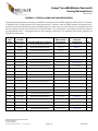
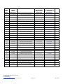
Vantage™ microRNA Multiplex Detection Kit
Oncology Detection Panel 1
Store at 2-8ºC
Catalog No. 11831-050
50 Reactions
Overview and Intended Use
The Vantage™ microRNA Detection Kit offers researchers a fast
and simple method for profiling the expression levels of multiple
microRNAs from many different sample types including total RNA,
enriched low molecular weight (LMW) RNA, and degraded RNA.
The assays are configured on the xMAP® bead array allowing for
the detection of multiple microRNAs in one sample. In addition,
the 96-well format allows many samples to be analyzed in one run.
MicroRNAs are a class of small molecules, about 21-23
nucleotides in length that regulate gene expression by various
methods including translational repression, mRNA cleavage,
methylation, and deadenylation. Differences in the expression
levels of microRNAs have been associated with the pathogenesis
of many diseases, including cancer. By measuring the expression
levels of microRNAs, researchers obtain a better understanding of
the processes involved in tumor development and progression. In
addition, researchers can observe distinct expression patterns
associated with particular stages of disease.
The specific microRNAs detected in the Vantage™ Oncology
Detection Panel 1 are given in Appendix A. These microRNAs have
been associated with a variety of solid tumors (ref.1-5.)
Principle of Method
The Vantage™ microRNA Detection Kit utilizes a simple,
hybridization procedure where samples are ready for detection on
the Luminex® reader within 90 minutes. The samples are first
labeled with multiple biotins using the Vantage™ microRNA
Labeling Kit (Kit sold separately. See Cat. No. 11820-025). The
biotinylated samples are incubated with a Bead Mix containing a
mixture of different fluorescently dyed xMAP® beads. Each
distinct xMAP® bead is coupled with a unique probe that
recognizes a specific microRNA. The beads and sample are
incubated at 60ºC allowing the microRNAs present in the sample
to hybridize to the specific probes. Following hybridization, the
samples are subjected to a high stringency wash to remove any
non-specific binding. Finally, the samples are incubated with
streptavidin-phycoerythrin (SAPE), which binds to the biotinylated
microRNA hybridized to the xMAP® bead. The samples are read
on Luminex® or Luminex-based instruments (e.g. BioPlex®) that
detect the specific microRNAs present in the sample by their
unique bead region and quantify the microRNAs by the intensity of
the SAPE signal.
Terms and Conditions
By opening this Assay Product (which contains fluorescently
labeled microsphere beads authorized by Luminex Corporation) or
2502 Urbana Pike Ijamsville, MD 21754 USA
301-874-4990 (phone)
301-874-4993 (fax)
[email protected] www.marligen.com
using this Assay Product in any manner, you are consenting to be
bound by the following terms and conditions. You are also
agreeing that the following terms and conditions constitute a
legally valid and binding contract that is enforceable against you.
If you do not agree to all of the terms and conditions set forth
below, you must promptly return this Assay Product for a full
refund prior to using it in any manner. You, the customer, acquire
the right under Luminex Corporation’s patent rights, if any, to use
this Assay Product or any portion of this Assay Product, including
without limitation the microsphere beads contained herein, only
with Luminex Corporation’s laser based fluorescent under the
name Luminex Instrument.
Safety and Use Statement
All biological materials should be handled as potentially
hazardous. Follow universal precautions as established by the
Centers for Disease Control and Prevention and by the
Occupational Safety and Health Administration when handling and
disposing of potentially infectious or hazardous agents.
This product is authorized for laboratory research use only. The
product has not been qualified or found safe and effective for any
human or animal diagnostic application. Uses other than the
labeled intended use may be a violation of applicable law. If you
have any questions concerning the use of this product, please
contact Marligen Biosciences, Inc. at (866) 464 4990 or visit
www.marligen.com.
Components included with this kit:
Component
Hybridization Buffer
Oncology Panel 1 Bead Mix
Detection Reagent
Wash Buffer
SAPE Diluent
Aluminum Plate Sealers
Filter Plate
Amount
1.25 mL
400 μL
55 μL
2 x 10 mL
25 mL
2 Each
1 Each
Storage Conditions:
Store all components at 2-8ºC.
Handling Instructions: The kit is shipped on ice packs. Upon
receipt, the components should be stored at 2-8ºC.
Page 1 of 8
20215 Rev3
Materials and Equipment Required But Not Supplied:
Nuclease-free PCR stripwell plate or nuclease-free PCR tubes
1.5 mL RNase free microfuge tubes
Plugged micropipette tips
Nuclease-Free water (Ambion Cat. No. AM9934 or equivalent)
Microcentrifuge
Thermocycler or heating block at 60ºC
Plate Shaker
Vortex Mixer
Sonicating waterbath
96-well filter plate vacuum manifold
Luminex Instrument
Optional: RNase Inhibitor (Superase-In, Ambion Cat. No. AM2694
or equivalent)
Important Information
READ ENTIRE PROTOCOL BEFORE USE
ADDITIONAL PRECAUTIONS SHOULD BE TAKEN TO PREVENT THE
DEGRADATION OF RNA:
RNases are very stable and robust enzymes that degrade RNA.
Autoclaving solutions and glassware is not always sufficient to
actively remove these enzymes. The first step when preparing to
work with RNA is to create an RNase-free environment. The
following precautions are recommended as your best defense
against these enzymes.
1. The RNA area should be located away from microbiological
work stations.
2. Clean, disposable gloves should be worn at all times when
handling reagents, samples, pipettes, disposable tubes, etc. It
is recommended that gloves are changed frequently to avoid
contamination.
3. There should be designated solutions, tips, tubes, lab coats,
pipettes, etc. for RNA only.
4. All RNA solutions should be prepared using at least 0.05%
DEPC-treated autoclaved water or molecular biology grade
nuclease-free water.
5. Clean all surfaces with commercially available RNase
decontamination solutions.
6. When working with purified RNA samples, ensure that they
remain on ice during downstream applications.
Add 20 μL of the Vantage™ miR-plex Control into 33 μL of the
Hybridization/Bead Mix at Hybridization Step 6, then follow
protocol as described. Expected results for this assay control is
shown in Appendix B.
Note: There is no need to label this sample as the MiR-plex
Control is already biotinylated.
Set-up Prior to Starting Detection Protocol
1. Prepare labeled RNA. Prior to using this detection kit the
microRNAs present in the samples must be labeled with biotin.
To obtain optimal results, it is recommended that the
Vantage™ microRNA Labeling Kit (Cat. No. 11820-025) is used
to label samples.
2. Use 0.5-2 μg of labeled RNA per reaction. If duplicates are to be
performed in the detection assay, double the amount of input
RNA to be labeled and split the sample accordingly.
Notes: Less than 0.5 μg/reaction may be used for some samples.
However, it is recommended that a pilot study is carried out to
determine the optimal amount of labeled RNA for a particular
sample type. Refer to the protocol for Vantage™ microRNA
Labeling Kit for further details on sample labeling.
3. Set thermocycler or heating block to 60°C.
4. Warm up the Luminex or Luminex-based instrument.
Luminex Instrument Setup
A. Set up the instrument as described in the user’s manual.
Setup details specific to this kit are described below:
1. The XY platform heater should be off.
2. Set the events/bead to 50.
3. Set the minimum events to 20.
4. Enter the number of samples.
5. Set the sample size to 50 μL.
6. Set the flow rate to Fast.
7. Enter the bead region numbers as indicated in the table in
Appendix A.
8. Check the probe height and adjust it, if necessary, to
accommodate the filter plate.
9. Perform 1 prime with sheath fluid, 1 alcohol flush, and 2 sheath
fluid washes.
B. Adjust Luminex Instrument to High Gain Setting
A high gain setting for the Luminex instrument is recommended to
provide the best results. Each specific software used with the
Luminex or Luminex-based instrument may have different
instructions for obtaining the high gain setting. Below are
instructions using the Luminex 2.3™ software. Please see
manufacturer’s guidelines for instrument/software specific
instructions (e.g. BioPlex®).
Assay controls
1. Control 1 (bead 49) detects 5.8S RNA that is ubiquitously
expressed in mammalian cells and is selected as a housekeeping gene for the Vantage™ Detection Kits. A signal of
4000-10000 MFI is typically observed when using 1-2 μg of
high quality (RIN >8) total RNA.
2. An additional assay control is available from Marligen. The
Vantage™ MiR-plex Control (Cat. No. 11830-001) contains 7 1. Create a new lot number for CAL2 and enter lot number with an
synthetic biotinlyated RNAs including 5.8S, miR-21, and miR-9
HG at the end to designate High Gain.
that are detected by the Vantage™ Oncology Detection Panel 1.
2502 Urbana Pike Ijamsville, MD 21754 USA
301-874-4990 (phone)
301-874-4993 (fax)
[email protected] www.marligen.com
Page 2 of 8
20215 Rev3
2. Record the CAL2 Calibrator target “RP1” which is usually around
3832.
3. Multiply the CAL2 Calibrator target “RP1” by 4.55 to get a new
target value of approximately 17,436.
4. Enter the new Calibrator target “RP1” as the value for your New
CAL2 lot.
5. Run the CAL2 calibration.
Part II: Detection
During this step, non-specific binding is removed by
subjecting the reactions to high stringency washes. The
specific microRNAs present in the sample are then
detected by labeling the biotins with SAPE.
Usage Notes
1. IMPORTANT: Do not allow the filter membrane to dry
throughout the transfer, wash and detection steps.
Detection Protocol
2. It is highly recommended to add 1 unit/μL of RNase Inhibitor
to the SAPE Diluent.
Part I: Hybridization
3. It is important to apply a slight vacuum of ~2-3 in Hg during all
During this step the microRNAs present in the sample are
wash steps. Higher vacuum may result in the loss of beads
hybridized to their complimentary sequences on the
and reduce bead count.
xMAP® beads.
4. During all wash steps, cover unused wells with a plate sealer
to ensure a seal necessary to pull a vacuum.
1. Vortex the Bead Mix vigorously for 20 seconds.
2. Sonicate the Bead Mix in a sonicating waterbath for
A. Washes
2 minutes.
1. Pre-wet the wells in filter plate with 100 μL Wash Buffer.
3. Prepare the Hybridization/Bead Mix based on the number of
2. Transfer the hybridized reactions to each pre-wet well, cover
reactions to be run in the assay as illustrated in Table 1.
unused wells with a plate and apply vacuum to evacuate.
3. Remove vacuum and immediately add 100 μL of Wash Buffer
Table 1
Hybridization/Bead Mix Preparation
to each well and apply vacuum to remove buffer. Repeat this
Volume per
Volume per
Volume per
wash step for total of 3 washes.
Component
reaction
25 reactions
50 reactions
Optional: For customer convenience, all washes are performed at
Hybridization
25 µL
625 µL
1250 µL
room temperature. As shown in Appendix B, increased assay
Buffer
specificity may be achieved by increasing temperature of wash
Bead Mix
8 µL
200 µL
400 µL
buffer to 60°C. However, please note that increasing the wash
temperature may reduce overall assay signal.
4. Vortex the Hybridization/Bead Mix to ensure that it is fully 4. Remove vacuum and add 100 μL SAPE Diluent to each well and
mixed.
apply vacuum to remove diluent. Remove plate from manifold.
5. Add 33 μL of the Hybridization/Bead Mix into each well of a 5. Blot the bottom of the filter plate dry on a clean paper towel.
nuclease-free PCR stripwell plate or into each nuclease-free
PCR tube.
B. SAPE Detection
6. Transfer 20 μL of the labeled RNA sample (prepared using
1. Prepare SAPE Detection Reagent as shown in Table 2.
Vantage™ microRNA Labeling Kit) into the 33 μL of the
Hybridization/Bead Mix in the PCR wells or tubes, mix by
Table 2 SAPE Detection Reagent Preparation
pipeting up and down.
Note: If duplicates are to be performed, add 10 μL of the labeled
RNA sample to the 33 μL of the Hybridization/Bead Mix in the
PCR wells or tubes, bring the volume to 53 μL by adding 10 μL
nuclease-free water, and mix by pipeting up and down.
Component
Detection
reagent
SAPE Diluent
Volume per
reaction
Volume per
25 reactions
Volume per 50
reactions
1 μL
25 μL
50 μL
100 μL
2.5 mL
5 mL
IMPORTANT: As noted in the Set-up, duplicates should have
double the amount of input RNA labeled with the Vantage™ Optional: Add 1 unit of RNase Inhibitor (e.g. Superase-In, Ambion
Cat. No. AM2694 or equivalent) per microliter of SAPE Diluent.
microRNA Labeling Kit.
2. Mix the SAPE Detection Reagent by vortexing.
7. Hybridize the reactions at 60ºC by using a thermocycler or 3. Add 100 μL of SAPE Detection Reagent into the washed well of
heating block for one hour with continuous shaking at 400 rpm.
the filter plate.
Protect the reactions from light during this incubation.
4. Incubate the filter plate in dark for 30 minutes at room
[Note: If shaking is not possible during this step, the MFI signals
temperature.
may be slightly reduced. However the overall results will not be [Note: To increase MFI signal, the plate may be shaken at 400 rpm
affected.]
during this incubation. Protect the reactions from light.]
2502 Urbana Pike Ijamsville, MD 21754 USA
301-874-4990 (phone)
301-874-4993 (fax)
[email protected] www.marligen.com
Page 3 of 8
20215 Rev3
Related Products:
To see our full line of Vantage™ microRNA analysis products visit
our website at www.marligen.com.
5. Add 100 μL of SAPE Diluent to each well and apply vacuum to
remove buffer. Repeat this wash step for total of 3 washes.
6. Blot the bottom of the filter plate dry on a clean paper towel.
7. Add 100 μL of SAPE Diluent into each well to resuspend the
beads in the filter plate by shaking for 2 minutes at 400 rpm or
by pipeting up and down.
8. Read the filter plate in Luminex instrument at high gain setting
(see Luminex Instrument Set-up).
DATA Analysis
1. Use the MFI data output from the Luminex Instrument to collect
the raw MFI.
2. Subtract the background MFI in negative control from the
Sample MFI. Note: If after subtraction of background MFI
signal is negative, regard results as zero i.e., that specific
microRNA is not detectable in the sample.
3. Control 1 (bead 49) detects 5.8S RNA that is ubiquitously
expressed in mammalian cells and is selected as a housekeeping microRNA gene for the Vantage™ Oncology Panel 1.
When comparing two samples, normalize the MFI with Control
1.
Normalization factor =
Limited Use License and Use Restrictions
A limited, research use only, license is conveyed to the purchaser
of this product.
Trademarks
Vantage™ is a trademark of Marligen Biosciences, Inc. Luminex®
and xMAP® are trademarks of Luminex Corporation.
References
1. Calin GA, Sevignani C, Dumitru CD, Hyslop T, Noch E,
Yendamuri S, Shimizu M, Rattan S, Bullrich F, Negrini M, Croce
CM. (2004). Human microRNA genes are frequently located at
fragile sites and genomic regions involved in cancers. Proc.
Natl. Acad. Sci., 104(19) pp8017-8022.
2. Volinia S, Calin GA, Liu CG, Ambs S, Cimmino A, Petrocca F,
Visone R, Iorio M, Roldo C, Ferracin M, Prueitt RL, Yanaihara N,
Lanza G, Scarpa A, Vecchione A, Negrini M, Harris CC, Croce
CM. (2006). A microRNA expression signature of human solid
tumors defines cancer gene targets. Proc. Natl. Acad. Sci.,
103(7) pp2257-2261.
3. Blower PE, Verducci JS, Lin S, Zhou J, Chung JH, Dai Z, Liu CG,
Reinhold W, Lorenzi PL, Kaldjian EP, Croce CM, Weinstein JN,
Sadee W. (2007). MicroRNA expression profiles for the NCI-60
cancer cell panel. Mol. Cancer Ther. 6 pp1483-1491.
4. Gaur A, Jewell DA, Liang Y, Ridzon D, Moore JH, Chen C,
Ambros VR, Israel MA. (2007). Characterization of microRNA
expression levels and their biological correlates in human
cancer cell lines. Cancer Res. 67 pp2456-2468.
5. Yu SL, Chen HY, Chang GC, Chen CY, Chen HW, Singh S, Cheng
CL, Yu CJ, Lee YC, Chen HS, Su TJ, Chiang CC, Li HN, Hong QS,
Su HY, Chen CC, Chen WJ, Liu CC, Chan WK, Chen WJ, Li KC,
Chen JJ, Yang PC. (2008) MicroRNA signature predicts survival
and relapse in lung cancer. Cancer Cell. 13(1) pp48-57.
MFI of Control 1 in Control
Sample
MFI of Control 1 in Tumor
or Treated Sample
For example:
Control 1 in normal tissue: MFI = 20000
Control 1 in tumor tissue: MFI = 16000
20000
Normalization factor =
16000 = 1.25
4. Then multiply Tumor or Treated Sample MFI by the
normalization factor 1.25.
5. The fold change in microRNA expression can be calculated by
dividing the normalized MFI for the Tumor or Treated Sample
by the MFI for the Control Sample. Plot results using Excel or
equivalent. Note that if an MFI value is zero (or close to 0
{<10}), the microRNA is not detectable in the sample and
should be considered as not expressed.
6. To determine assay precision, calculate standard deviation (SD)
and assay coefficient of variation (CV). [%CV =SD/mean x
100%]. Assay CVs are typically less than 5% for technical
replicates.
Technical Support
For further technical assistance please contact us at
[email protected].
(866) 464.4990.ext.102
or
Technical support and troubleshooting guides for these products
can also be found on our website at www.marligen.com.
2502 Urbana Pike Ijamsville, MD 21754 USA
301-874-4990 (phone)
301-874-4993 (fax)
[email protected] www.marligen.com
Page 4 of 8
20215 Rev3
Vantage™ microRNA Multiplex Detection Kit
Oncology Detection Panel 1
Store at 2-8ºC
APPENDIX A: TABLE OF microRNAs AND xMAP® BEAD REGIONS
The sequences and nomenclature of the mature microRNAs are extracted from The miRBase Sequence Database version 12.0, released
in September 2008, in Sanger Institute in UK. Names annotated with * indicate a mature microRNA sequence that originated from a
stem-loop molecule that generated two mature microRNA sequences. In these cases, one mature sequence has a standard name while
the other sequence from the same stem-loop has an annotated name(*). The nomenclature of the sequences detected by Vantage™
Oncology Detection Panel 1 is designated using the human sequence nomenclature. The equivalent mouse and rat sequences are
indicated in the table below.
xMAP®
Bead
Number
Human
microRNA
Nomenclature
Human microRNA mature sequences
Equivalent Mouse
(Mus musculus)
Nomenclature
Equivalent Rat (Rattus
norvegicus)
Nomenclature
1
hsa-let-7a
UGAGGUAGUAGGUUGUAUAGUU
mmu-let-7a
rno-let-7a
2
hsa-let-7c
UGAGGUAGUAGGUUGUAUGGUU
mmu-let-7c
rno-let-7c
3
hsa-let-7g
UGAGGUAGUAGUUUGUACAGUU
mmu-let-7g
4
hsa-let-7i
UGAGGUAGUAGUUUGUGCUGUU
mmu-let-7i
rno-let-7i
5
hsa-miR-100
AACCCGUAGAUCCGAACUUGUG
mmu-miR-100
rno-miR-100
6
hsa-miR-106a
AAAAGUGCUUACAGUGCAGGUAG
mmu-miR-106a
7
hsa-miR-10a
UACCCUGUAGAUCCGAAUUUGUG
mmu-miR-10a
rno-miR-10a-5p
8
hsa-miR-10b
UACCCUGUAGAACCGAAUUUGUG
mmu-miR-10b
rno-miR-10b
9
hsa-miR-125a-5p
UCCCUGAGACCCUUUAACCUGUGA
mmu-miR-125a-5p
rno-miR-125a-5p
10
hsa-miR-125b
UCCCUGAGACCCUAACUUGUGA
mmu-miR-125b-5p
rno-miR-125b-5p
11
Not in use
12
Not in use
13
hsa-miR-132*
ACCGUGGCUUUCGAUUGUUACU
14
hsa-miR-135a
UAUGGCUUUUUAUUCCUAUGUGA
mmu-miR-135a
rno-miR-135a
15
hsa-miR-136
ACUCCAUUUGUUUUGAUGAUGGA
mmu-miR-136
rno-miR-136
16
hsa-miR-138
AGCUGGUGUUGUGAAUCAGGCCG
mmu-miR-138
rno-miR-138
17
hsa-miR-141*
CAUCUUCCAGUACAGUGUUGGA
mmu-miR-141*
18
hsa-miR-16
UAGCAGCACGUAAAUAUUGGCG
mmu-miR-16
rno-miR-16
19
hsa-miR-17
CAAAGUGCUUACAGUGCAGGUAG
mmu-miR-17
rno-miR-17
20
hsa-miR-181b
AACAUUCAUUGCUGUCGGUGGGU
mmu-miR-181b
rno-miR-181b
21
hsa-miR-185
UGGAGAGAAAGGCAGUUCCUGA
mmu-miR-185
rno-miR-185
22
hsa-miR-195
UAGCAGCACAGAAAUAUUGGC
mmu-miR-195
rno-miR-195
2502 Urbana Pike Ijamsville, MD 21754 USA
301-874-4990 (phone)
301-874-4993 (fax)
[email protected] www.marligen.com
Page 5 of 8
Notes
20215 Rev3
xMAP®
Bead
Number
Human
microRNA
Nomenclature
Human microRNA mature sequences
Equivalent Mouse
(Mus musculus)
Nomenclature
Equivalent Rat (Rattus
norvegicus)
Nomenclature
23
hsa-miR-199a-5p
CCCAGUGUUCAGACUACCUGUUC
mmu-miR-199a-5p
rno-miR-199a-5p
24
hsa-miR-200b*
CAUCUUACUGGGCAGCAUUGGA
mmu-miR-200b*
25
hsa-miR-205
UCCUUCAUUCCACCGGAGUCUG
mmu-miR-205
rno-miR-205
26
hsa-miR-20a
UAAAGUGCUUAUAGUGCAGGUAG
mmu-miR-20a
rno-miR-20a
27
hsa-miR-21
UAGCUUAUCAGACUGAUGUUGA
mmu-miR-21
rno-miR-21
28
hsa-miR-210
CUGUGCGUGUGACAGCGGCUGA
mmu-miR-210
rno-miR-210
29
hsa-miR-212
UAACAGUCUCCAGUCACGGCC
mmu-miR-212
rno-miR-212
30
hsa-miR-218
UUGUGCUUGAUCUAACCAUGU
mmu-miR-218
rno-miR-218
31
hsa-miR-23b*
UGGGUUCCUGGCAUGCUGAUUU
32
hsa-miR-24-2*
UGCCUACUGAGCUGAAACACAG
mmu-miR-24-2*
rno-miR-24-2*
33
hsa-miR-27a*
AGGGCUUAGCUGCUUGUGAGCA
mmu-miR-27a*
rno-miR-27a*
34
hsa-miR-29a*
ACUGAUUUCUUUUGGUGUUCAG
mmu-miR-29a*
rno-miR-29a*
35
hsa-miR-29b-2*
CUGGUUUCACAUGGUGGCUUAG
36
hsa-miR-29c*
UGACCGAUUUCUCCUGGUGUUC
mmu-miR-29c*
rno-miR-29c*
37
hsa-miR-30d
UGUAAACAUCCCCGACUGGAAG
mmu-miR-30d
rno-miR-30d
38
hsa-miR-34a
UGGCAGUGUCUUAGCUGGUUGU
mmu-miR-34a
rno-miR-34a
39
hsa-miR-34b*
UAGGCAGUGUCAUUAGCUGAUUG
40
hsa-miR-9
UCUUUGGUUAUCUAGCUGUAUGA
mmu-miR-9
rno-miR-9
41
hsa-miR-93
CAAAGUGCUGUUCGUGCAGGUAG
mmu-miR-93
rno-miR-93
42
hsa-miR-95
UUCAACGGGUAUUUAUUGAGCA
43
hsa-miR-96
UUUGGCACUAGCACAUUUUUGCU
mmu-miR-96
rno-miR-96
44
hsa-miR-99a
AACCCGUAGAUCCGAUCUUGUG
mmu-miR-99a
rno-miR-99a
45
hsa-miR-137
UUAUUGCUUAAGAAUACGCGUAG
mmu-miR-137
rno-miR-137
46
hsa-miR-182*
UGGUUCUAGACUUGCCAACUA
47
hsa-miR-221
AGCUACAUUGUCUGCUGGGUUUC
mmu-miR-221
rno-miR-221
48
hsa-miR-372
AAAGUGCUGCGACAUUUGAGCGU
49
NA
Control 1
2502 Urbana Pike Ijamsville, MD 21754 USA
301-874-4990 (phone)
301-874-4993 (fax)
[email protected] www.marligen.com
Page 6 of 8
Notes
rno-miR-29b-2*
rno-miR-34b
20215 Rev3
APPENDIX B
PERFORMANCE CHARACTERISTICS OF VANTAGE™ ONCOLOGY DETECTION KITS
1. Protocol Overview
2. Assay Sensitivity
a. The limit of detection of the Vantage™ microRNA Detection Kits for specific microRNAs was calculated using synthetic
microRNAs labeled with the Vantage™ microRNA Labeling Kit (Cat. No. 11820-025) and detected using Oncology Detection Panel
1 (Cat. No. 11831-050).
Limit of Detection Range : 0.03-0.1 pg (5-15 attomoles)
b. The limit of detection of the Vantage™ microRNA Detection Kits in tissues was calculated using total RNA extracted from human
brain labeled with the Vantage™ microRNA Labeling Kit (Cat. No. 11820-025) and detected using Oncology Detection Panel 1
(Cat. No. 11831-050).
Limit of Detection Range : 3-40 ng total RNA
3. Assay Specificity
a. No Cross-reactivity Observed Between Unrelated Sequences
200 pg of synthetic miR-210 labeled with the Vantage™ microRNA Labeling
Kit (Cat. No. 11820-025) and detected using Oncology Detection Panel 1 (Cat.
No. 11831-050). MiR-210 lacks homology with any other sequence in the
Oncology Detection Panel 1 and shows > 1% cross-reactivity with any other
miR assay in this multiplex.
2502 Urbana Pike Ijamsville, MD 21754 USA
301-874-4990 (phone)
301-874-4993 (fax)
[email protected] www.marligen.com
Page 7 of 8
20215 Rev3
b. Balance Between Specificity and Sensitivity for Closely Related Sequences
400 attomoles of synthetic biotinylated miR-100 was incubated with the Vantage™ Oncology Detection Panel 1 (Cat. No. 11831-050).
The assay was performed using the protocol as described in the package insert except different temperatures were employed at the
wash step. Results show <10% cross reactivity with very similar sequences under standard protocol conditions (25°C). Negligible cross
reactivity was observed for all of the other miR sites. Specificity for miR-100 increased by raising the wash temperature but the overall
assay signal is reduced. In this assay, high sensitivity and low cross reactivity are maintained following the standard protocol, although
greater specificity can be obtained by increasing wash temperature.
400 attomoles of synthetic let-7a was incubated with the Vantage™ Oncology Detection Panel 1 (Cat. No. 11831-050). The assay was
performed using the protocol described in the package insert, except the wash step was also carried out at 60°C. With the exception of
the very similar let-7c sequence, all miR sites in the assay show negligible cross-reactivity under standard protocol conditions.
Specificity for let-7a increases significantly by increasing the wash temperature, although this decreases the overall assay signal. For
the most sensitive assay, follow the standard protocol, but for higher specificity, increase the wash temperature.
4. Vantage™ MiR-plex Control Expected Results
Vantage™
Oncology
Breast Cancer
Pancreatic
Ovarian
Cardiac
microRNA
Detection Panel 1
Panel 1
Cancer Panel 1 Cancer Panel
Panel 1
Detection
(47-plex)
(8-plex)
(12-plex)
(8-plex)
(10-plex)
Kit
Cat. No.
11831-050
11833-050
11832-050
11834-050
11835-050
microRNA
Expected MFI Values
miR-1
Low
miR-107
Low
miR-126
Low
miR-203
Low
miR-21
High
High
High
High
High
miR-9
High
5.8S
Medium
Medium
Medium
Medium
Medium
2502 Urbana Pike Ijamsville, MD 21754 USA
301-874-4990 (phone)
301-874-4993 (fax)
[email protected] www.marligen.com
Page 8 of 8
Diabetes
Panel 1
(11-plex)
Hypoxia Panel
1
(10-plex)
11836-050
11837-050
Low
High
Medium
Low
High
Medium
20215 Rev3