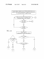
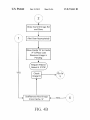
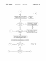
1
USOO8374461B2
(12) United States Patent
(10) Patent No.:
Humphreys et al.
(54)
(75)
(45) Date of Patent:
3,703,272 A
11/1972 Lareau
IDENTIFICATION SYSTEM
3,942,016 A
3,962,807 A
4,127,774 A
3/ 1976 SchatZ
6/1976 Pantone
11/1978 Gillen
Inventors: Geoffrey Humphreys, Baton Rouge, LA
(Us); Chad Lacour, Baton Rouge, LA
4,194,122 A
4,204,639 A
(73) Assignee: Smartplates, LLC, Baton Rouge, LA
Us)
(
_
_
Notlce:
_
_
_
Subject to any disclaimer, the term ofthis
patent is extended or adjusted under 35
5,179,579 A
1/1993 Dove et a1.
5,195,123 A
6,041,102 A
6,354,737 B1
3/1993 Clement
30000 Francs“
3/2002 Hufe et al.
6,679,847 B1 *
1/2004 Robinson et al. ........... .. 600/447
7,358,502 B1*
4/2008
Appleby et al.
.. 250/37014
7,787,687 B2*
Appl' NO; 13/180,886
8/2010 Miyano et al.
382/149
7,987,003 B2 *
2006/0098785 A1
7/2011 Hoffberg et a1. .............. .. 700/17
5/2006 Woods
2008/0084965 A1
4/2008 Ohnona et al.
'
(22)
3/1980 Mitchell et al‘
5/1980 Barber etal.
7,627,370 B2 * 12/2009 Marks ,,,,,,,,,,,,,,,,,,,,, ,, 600/544
U_s_c_ 15403) by 29 days_
(21)
Feb. 12, 2013
DIGITAL RADIOGRAPHY PLATE
(US)
(*)
US 8,374,461 B2
FOREIGN PATENT DOCUMENTS
Filed:
Jul. 12, 2011
(65)
EP
Prior Publication Data
Us 2012/0039520 A1
1 649 812 A1
400%
OTHER PUBLICATIONS
Feb 16’ 2012
GendeX Dental Systems, “Additional Mount Information”, Imaging
Software: ViXWin Platinum User Manual, 2009, pp. 8-4-8-9.
Related U_s_ Application Data
(60)
GendeX Dental Systems, “Accelerated Work?ow”, ViXWin Platinum
Brochure, 2000, pp. 5.
Provisional application No. 61/363,538, ?led on Jul.
12, 2010.
* cited by examiner
(51)
Int. Cl.
Primary Examiner * Yosef Kassa
(52)
G06K 9/32
(2006-01)
U.S. Cl. ...... .. 382/291; 382/128; 382/295; 382/296;
128/922
(74) Attorney, Agent, or Firm * Saliwanchik, Lloyd &
Eisenschenk
(58)
Field of Classi?cation Search ................ .. 382/128,
382/130’ 131: 132’ 291: 295: 296; 128/920:
_
_
_
128/922
see apphcanon ?le for complete searCh hlstOI'Y-
ABSTRACT
identi?cation,
sorting, and positioning of radiographic
images. According to one embodiment of the subject inven
_
(56)
(57)
Systems, devices, and methods for facilitating the automatic
tion, phosphorous plates for intra-oral imaging are encoded
References Clted
for automated image set identi?cation, location, and orienta
U.S. PATENT DOCUMENTS
tion. In certain embodiments, a processing system is able to
process encoded identi?ers on the phosphorous plates With
456,396 A
1,358,990 A
1,396,415 A
7/1891 Blakemore
11/1920 Scannell
11/1921 Fried
1,632,154 A
6/1927 Torpin
1,917,433 A
3,619,611 A
7/1933 Cressler
11/1971 Hall
digitized images depicted on the phosphorous plates to auto
matically identify, sort, and rotate the digitized images into
proper orientations for user vieWing.
31 Claims, 6 Drawing Sheets
Practice Mgmt. System has a Patient selected and is in 5
Waiting" state where Radiographtc Images are expected for
a “To Be Determined" image temptete.
Detey and Check iet Receipt of New/
Pending Image or User Request
YES
trnage set Atteetiy
o - em
NO
US. Patent
Feb. 12, 2013
59.5“0QEcBE.o2m3p0?m5Eo
3mi.E@uh3w:62om>c5E3tam»823oua
o$31.m2n:vg
@Q259235mH.a36n25:.%;3
B8Hma23b¢wm
US 8,374,461 B2
Sheet 1 0f 6
IiH0%28>5oz62om?m-e>m
<:23OH.H0x3;Exoz6252? wH:5095c2&5oz0252: UH.x0oz:3625%E>OIm QHemsqcomX.55>5oz02> >$m> .1 H95>N0%X>6$5002> >Em> , @HIo202HozWWI.E5QE.:W
HH}
09;25:?VS;xi0oz02X+> E .X
H_iamm29028oz>.asé8?>-§
.UEH
US. Patent
Feb. 12, 2013
Sheet 3 0f6
US 8,374,461 B2
Subordinate PSPs (continued)
Plate lD
Template =
Position =
Set Desc.
Rotation
A7
A
7
Adult FMX
O
No
No
8.7
B
7
PEDO FMX
0
No
No
Purple ‘
PFlVlX-7
F-7
F
7
7 Vert. BWX
90“ CW
No
No
Orange
7BWX—7
A-8
A
8
Adult FMX
O
No
No
Red
AFlVlX-S
8-8
B
8
PEDO FMX
O
No
No
A4)
A
9
Adult FMX
90° CW
No
No
Red
AFlVlX—9
A-1O
A
10
Adult FMX
90" CW
No
No
Red
1 AFlVlX'lO
A-11
A
11
Adult FMX
90" CW
No
No
Red
» AFMX-ll
A-12
A
12
Adult FMX
90" CW
No
No
Red
: AFMX-lZ
AB
A
13
Adult FMX
90" CW
No
No
Red
‘ AFMX-lS
A~l4
A
14
Adult FMX
90" CW
No
No
Red
AFMX-14
‘
Flip Mirror
Color
Visual lD
Red
AFMX-7
Purple ‘
PFIVlX-8
A115
A
15
Adult FMX
90° CW
N0
N0
Red
AFMX-lS
A—16
A
16
Adult FMX
90“ CW
No
No
Red
AFMX-16
A-17
A
17
Adult FMX
90" CW
No
No
Red
AFIVlX-17
A-18
A
18
Adult FMX
90" CW
No
No
Red
AFlVlX—18
A-19
A
19
Adult FMX
90° CW
No
No
Red
AFIVIX-19
A-ZO
A
20
Adult FMX
90° CW
No
No
Red
AFMX-ZO
FIG. 3
US. Patent
Feb. 12, 2013
Sheet 4 0f6
US 8,374,461 B2
Practice Mgmt. System has a Patient selected and is in a
“waiting” state where Radiograpnic Images are expected for
a “To Be Determined” image template.
V
V
FIG. 4A
Delay and Check for Receipt of New/
I
< >
Pending Image or User Request
YES
Read Plate ID
(barcode and confirm with OCR)
s This a Master
Plate?
Any Images
in Cache
YES
NO
“A”?
l/ G? t°\‘
,1!\
\\V
[blotch
2
\\\ft’///
US. Patent
Feb. 12, 2013
Sheet 5 0f6
US 8,374,461 B2
Close Current Image Set
and Save
a 1
J
AIertUserAppropriately
Move Cache “B” to Cache
“A” & Place Last
Received Image in
Pending
YES
Check
Image ID
Get/Remove Next Image
From Cache “A”
FIG. 4B
/Go to \,
\
5
I
US. Patent
Feb. 12, 2013
Sheet 6 0f6
US 8,374,461 B2
/
/
//
I
I
/
NO
I
Open New Image Set and Display
Template Dictated by Plate ID
Template
Library
Place Image in Temporary
Cache (Holding) Area “A”
Place Image in Temporary
Cache (Holding) Area “B”
with Yellow Warning Border
YES
I
Place Image in the Location Within
the Template Dictated by Plate ID
V
I Orient the Plate Image Dictated by the Plate ID I
US 8,374,461 B2
1
2
DIGITAL RADIOGRAPHY PLATE
IDENTIFICATION SYSTEM
of sequence) will cause mis-assignment and mal-rotation of
the image by the computer program.
To address this and other problems associated with PSPs
described above, what is needed is a system for processing
PSPs that does not require user-established assignments for
CROSS-REFERENCE TO A RELATED
APPLICATION
templates or image loading sequences/rotations. Current
This application claims the bene?t of Us. provisional
application Ser. No. 61/363,538, ?led Jul. 12, 2010, which is
technology does not provide PSPs or PSP imaging systems
that incorporate unique identi?ers such as marks, alphanu
incorporated herein by reference in its entirety.
meric codes, bar codes, graphics, radio frequency identi?ers,
BACKGROUND OF INVENTION
or coloring to (a) indicate to the dental technician/practitioner
the proper plate set and plate to be used to obtain a speci?c
image or set of images of desired oral locations and/ or (b) to
In the ?eld of dentistry, various types of intra-oral sensors
exist that are used for capturing images of the inside of teeth
enable the computing system (either integral with or opera
tively connected to an imaging system) to automatically and
and surrounding anatomy (e.g., bone structure) by for
properly identify, place, and orient intra-oral images obtained
example, exposing the anatomy and sensors to X-ray radia
tion. Such imaging techniques are well known, using such
intra-oral sensors as, for example, X-ray sensitive ?lm, X-ray
from PSPs within their intended “image set” or template
without additional human intervention.
sensitive phosphor plates, or X-ray sensitive digital imaging
sensors such as a corded charge-coupled device (CCD) sen
BRIEF SUMMARY
20
The subject invention provides innovative PSP identi?ca
tion methods and systems. In particular embodiments, the
invention provides a PSP intra-oral image identi?cation/cod
ing system that provides for human identi?cation of the cor
sor.
When taking intra-oral images of patients, images are often
captured on media such as radiographic ?lm or photostimu
lable phosphorplates (also known as Phosphor Storage Plates
25
rect PSPs to be used for speci?c intra-locations as well as
digitally extract the images from the media. The digitized
computer system or image management component recog
images are then often transmitted to a receiving computer
system where the images can be viewed either individually or
30
nizable identi?ers enabling images to be (a) automatically
associated with an “image set” (or template), (b) placed in the
proper location within the template and (c) properly rotated,
?ipped, etc. for proper orientation (as required).
or PSPs), which are then placed into a scanning apparatus to
along with other associated oral images within a speci?c
viewing template. In the case where a multiple image set is
being processed, the radiologist or dental technician must
carefully and manually track the PSPs being used for the
various images being acquired and, after placing the plates in
the scanning apparatus, manually select an image template
(initially), manually place each scanned image into the cor
A system of the invention can comprise: (a) a set or sets of
scannable image media each coded with (i) unique, visual,
and humanly recognizable radiolucent markings and (ii)
unique processing system recognizable identi?ers; and (b) a
35
rect location within the selected template, and then manually
direct the system to orient the image in one fashion or another
so that it will be viewed from the proper perspective.
This process can include as many as twenty (20) images
within a single template, often involves a signi?cant amount
processing system able to associate the identi?ers with
extracted images from the scannable image medium and to
process the extracted images based on the identi?ers to facili
tate automatic association with an image set, (and/or tem
40
plate) automatic identi?cation of the image location, and
automatic orientation (via rotation and/or ?ipping) of the
of time, and can be prone to user error at one point or another
image if needed. A graphical user interface can be included
in the process since all PSPs look (and are identi?ed) exactly
alike to both the human and the machine(s) involved in the
with the system(s) of the invention to automatically display
process. Technical review of associated images is often nec
essary to identify the area of the oral cavity that is represented
processed digital images correctly and still enable user-inter
action and/or image adjustments as these types of systems
45
by the image(s) in order to identify the intended template and
the proper location within the template that the image(s)
is/are to be placed for display/viewing. Because of these
limitations with current technology, the manual tracking,
identifying, organizing and orienting of dental images for
often do now.
The algorithms and methods utilized to instruct users and
the associated processing system in the present invention are
50
particularly advantageous in that they not only enable the
subject digital radiographic plate identi?cation system to pro
vide time savings when obtaining intraoral images but also
storage and display require a large amount of time for the
radiolo gist/technician and/or dentist, resulting in lost produc
enable automatic and real-time classi?cation and orientation
tivity and delay in servicing patient needs.
In particular, the subject invention does not require the user
to establish templates or loading sequences or image orien
To address some of the issues noted above, a computer
program is available that requires a great deal of initial time
and effort by the user to establish templates for sets of oral
images (e.g. creating a mount in which the user must assign a
number for each tooth image to be associated with the
mount). In addition, the computer program requires the user
to ?rst establish a speci?c sequence of images and their rota
of the digitized intra-oral radiography images.
55
is subject to user error when uploading images outside pre
established parameters. Accordingly, another advantage of
the invention is that the order of obtaining and scanning
identi?ed images/plates has no effect on the outcome of the
60
tions before image upload. Only after pre-established mount
assignments, loading sequences, and rotational information
mount assignments and loading sequences, user error in load
ing images (such as loading an image at the wrong time or out
?nal template and associated images. Furthermore, the algo
rithms and methods described herein are designed to accom
modate any practitioner’s standards and conventions where
imaging perspectives are concerned. For example, one dental
has been provided by the user can the computer program
provide some form of automated image processing. Because
processing of images is based on speci?c pre-established
tations prior to use which is an inef?cient use of resources and
practice may utilize a “standar ” orientation set of templates
65
and/or multi-image views while another may subscribe to
what is known in the industry as the “military” orientationi
both (and any other) can be dynamically con?gured within
US 8,374,461 B2
3
4
the methods described for an image processing system asso
the ability to read, extract, digitize, and transmit both the
ciated with this technology without altering the programming
digital image as well as the coded machine-readable identi
of the system.
One method of implementing the invention can include
?ers, and a processing system able to associates respective
coded machine readable identi?ers with extracted digital
placing recognizable identi?ers on scannable image media
and entering the media’s corresponding identi?er-related
images transmitted to the processing system as a “data set”
and to process the images based on the identi?ers to automati
information (e.g., media ID, template ID, position ID, and
cally select and associate with which image set (or template)
orientation index) into a table within an associated image
processing/management (computer) system’s database,
the extracted images are to be grouped and/or displayed as
well as identify and/or place the image in the correct location
wherein the processing system, upon receiving such an image
and to also orient the image properly.
from an internal or external media scanning apparatus, dis
In certain embodiments, the processing system is provided
within the scanning apparatus. In alternative embodiments,
the processing system is separate from that of the scanning
apparatus (e.g., the processing system is provided in a com
cerns the image’s ID and, along with the system’s database
information automatically (a) identi?es the proper image set
(and causes the associated template to be displayed if not
already done so), (b) identi?es the image location within the
puter system separate from the scanning apparatus).
set (and causes the image to be moved to that location in the
template), and (c) identi?es any orientation maneuvers the
image might require for user viewing on the scanning device
or processing system monitor (and causes the orientation to
occur). In certain embodiments, the recognizable identi?ers
20
are pre-established as opposed to established by the user. In
alternate embodiments, the recognizable identi?ers are estab
lished by the user.
According to one embodiment of the invention, oral x-ray
images are acquired from a patient utilizing PSPs (or a set of
?ers, combines the individual images with their identi?ers,
and creates a combined/concurrent data stream or ?le con
taining these image/identi?er pairs and transmits the entire,
25
PSPs) that are identi?ed in one or more locations with speci?c
radiolucent identi?ers intended for humans and/or radio
paque identi?ers within the image ?eld of the PSPs intended
for computing systems and the PSP is later placed into a
scanning device. The scanning device reads, extracts, and
transmits a digital version of the image (said image including
the radiopaque computer readable identi?er) from the PSP to
a connected (wireless or wired) computing/processing sys
tem. When the processing system receives an image from
such a media scanning apparatus, it discerns the image’s ID
from within the image itself (e. g., via Optical Character Rec
30
35
PSP that is coded with human and/ or machine-readable iden
device extracts and transmits a digital version of the image on
the PSP along with the coded identi?er to a computer system
40
?es the image location within the set and automatically
and further processes the image. Based on the coded identi?er
associated with the image, the processing system identi?es
45
tion, the processing system will automatically and properly
orient the image 90° in the clockwise direction before or after
50
In an alternate embodiment, the system comprises: scan
nable image media, each coded with human-readable and
(radiolucent or radiopaque) machine-readable identi?ers, a
scanning apparatus having the ability to read, extract, digi
55
received digital images transmitted to the processing system
60
According to the subject invention, the association of the
coded identi?ers with images are pre-established as opposed
to being established by a user. Alternatively, the association of
which image is associated with which coded identi?er can be
established by the user.
In certain embodiments, the coding system for the scan
nable image media is based on colors, numbers, alphabet
symbols or combination of these identi?ers that are easily
identi?ed by a user and/or machine. In a preferred embodi
as well as identify and/or place the image in the correct
location and to also orient the image properly.
nable image media, each coded with human-readable and
machine-readable identi?ers, a scanning apparatus having
image location within the image set. In addition, the process
ing system utilizes the coded identi?er associated with the
image to ascertain whether the image needs to be modi?ed for
proper orientation. For example, if an image’s coded identi
?er indicates to the image processing system the need for 90°
clockwise rotation, the image processing system will auto
matically rotate the image 90° in the clockwise direction for
letters, graphic images, barcodes, magnetic strip, or any other
matically select and associate with which image set (or tem
plate) the extracted images are to be grouped and/ or displayed
In an alternate embodiment, the system comprises: scan
with which image set the image is to be grouped as well as the
correct orientation and user viewing.
tize, and transmit both the digital image as well as the coded
machine-readable identi?ers, and a processing system able to
associate respective coded machine readable identi?ers with
and to process the images based on their identi?ers to auto
capable of processing said images containing coded identi?
ers. The image processing system within thin the computer
system associates the coded identi?er with the digital image
causes the image to be moved to that location in the template,
placing the image in the proper location within its associated
template for user viewing.
Where the processing system is operably connected to a
scanning apparatus, an image is acquired from a patient onto
ti?ers and is placed into a scanning device. The scanning
proper image set and causes the associated template to be
and (c) identi?es any orientation maneuvers the image might
require for user viewing on the scanning device or processing
system monitor and automatically causes the orientation to
occur. For example, if the identi?er indicates (via the associ
ated database information) the need for 90° clockwise rota
separately identi?ed image set to a processing/viewing sys
tem. The processing/viewing system utilizes the identi?ca
tion transmitted with the image set (and/or intemally with
each image within the set) in conjunction with an identi?ca
tion table within the processing/viewing system to automati
cally save and/or display the image set and associate each
acquired image within the transferred ?le with the appropri
ate image set, image location, and orientation for user view
ing on the processing system.
ognition) and along with the system’s PSP-ID-related data
base information, the processing system (a) identi?es the
automatically displayed if not already displayed, (b) identi
In a method of use where the processing system is located
within or integral with the scanning apparatus, one embodi
ment utilizes encoded scannable image media provided with
human and/or machine-readable identi?ers and the scanning
apparatus reads a predetermined series of scannable image
media, extracts the images and reads their associated identi
65
ment, the scannable image media are phosphorous plates,
wherein the Master plate for a set of phosphorous plate
images is coded with an easily identi?able visual indicator,
such as a color.
US 8,374,461 B2
6
5
In other embodiments, radiolucent, sterile plastic ?lm
PSP from that set based on the speci?c image to be obtained.
sleeves (also referred to herein as “sleeves” or “envelopes”)
are provided that are a part of the coding/identi?cation sys
Once the appropriate oral image is obtained from the patient,
the PSP is placed into a radiographic plate reader device/
system. The radio graphic plate reader device/ system includes
tem. For example, the subject invention provides PSP sterile
sleeves that include color and/or radiolucent graphical cod
ings that indicate targeted oral locations to further assist in the
or is connected to a processing, system that is able to elec
tronically and automatically: (1) associate the PSP’s coded
image with the proper template; (2) following identi?cation
of the proper template in step (1), place the PSP image in the
practitioner’s identi?cation of the proper PSP to be used to
acquire an oral image without affecting the quality of the
image obtained from the PSP.
correct location within the proper template; and (3) determine
whether the image is in a proper orientation and, if necessary,
rotate the image into the proper orientation.
BRIEF DESCRIPTION OF DRAWINGS
Once properly identi?ed and oriented, the images and/or
image set are/ is then automatically uploaded into an existing,
connected computer system for viewing and associating with
a speci?c patient. The subject invention also allows for the use
FIG. 1 is an illustration of an example “master and subor
dinate” coding system that can be used to identify several sets
of PSPs and the images contained within these sets for use in
of solo or wild-card PSPs for individual image capture and
transfer without being af?liated with a speci?c template or set
accordance with one or more embodiments of the invention.
FIG. 2 is an illustration of another example coding system
where each plate has a speci?c location and orientation within
a single/individual Oral Image Template for use in accor
dance with one or more embodiments of the invention.
of images such that the practitioner can manually place and
orient an image as desired.
20
the set.
FIG. 4 is a schematic ?ow diagram of a method for auto
speci?es the image set, location, and orientation, but is also
coded with a same standard recognizable identi?er (such as
by color and/or radiolucent image) to visually indicate with
25
matically identifying an image set, providing an image loca
tion within the image set, and orienting the image in accor
dance with the subject invention.
DETAILED DISCLOSURE
30
The subject invention provides novel methods, products,
and/or systems for intra-oral image PSP identi?cation/cod
ing/processing. The method/ system provides identi?ers for
human assistance in plate set and individual plate selection
35
?ers enabling an included or attached image management
process or system to perform automatic “image set” identi?
40
ing systems.
In an exemplary embodiment, an intra-oral image is cap
tured using a radiation source (e.g., an X-ray tube) and a
45
radiation toward the oral structure of interest. Some of the
X-ray radiation passes through the oral structure and exposes
the scannable image medium, capturing an image of the oral
cent identifying characters for, and/or graphical representa
50
structure on the image medium. In accordance with various
embodiments, the scannable image medium may include a
radiographic ?lm, a photosensitive ?lm, or a photostimulable
phosphorous plate, all of which are well known in the art.
Other scannable media may be possible as well Preferably,
55
the scannable medium is a PSP.
the user in identifying the correct PSP set and/or PSPs to be
used for speci?c images within one or more image sets.
According to the subject invention, the scannable image
medium includes one or more coded identi?ers that provide
information regarding the image that is captured on the scan
nable image medium. In a preferred embodiment, the coded
which contain a coded identi?er (such as one or more alpha
numeric characters and/ or a bar-code) that is associated with
60
(a) an image set; (b) an image location within the speci?c set;
and/or (c) a speci?c orientation for that image when used
plate set based on the set of images desired, and then select a
identi?ers provide information regarding the image set,
image location, and/or proper rotation of the image captured
on the scannable image medium. According to the subject
invention, the coded identi?er can be machine-readable,
within this set. Each set of PSPs represents speci?c dental
x-ray common image sets (i.e. full mouth sets, horizontal
bitewing sets, vertical bitewing sets, etc.). In practical use, a
dental technician/practitioner would select an appropriate
scannable image medium (e.g., a photostimulable phosphor
storage plate or PSP). The X-ray tube emits a dose of X-ray
In other related embodiments of the technology, radiolu
In certain embodiments, the recognizable identi?ers are
pre-established as opposed to established by the user. In alter
nate embodiments, the recognizable identi?ers are estab
lished by the user.
In a speci?c example, a set of PSPs are provided, each of
template selection, image locating, and orientation of
uploaded digital radiographic images, eliminating much of
the human intervention and time currently required by exist
associated radiolucent sterile sleeves are coded with a recog
tions indicating, targeted oral locations are placed on the
PSPs and/or corresponding sterile “sleeves” to further assist
One advantage of the subject system and methodology is
its ability to be integrated into existing/ standard phosphorous
plate readers (e.g., Scan X or DenOptix) that, without modi
?cation, can capture both the plate’s image and the plate’s
speci?c identi?cation (ID) code/markings and transfer the
captured ID information and images to existing Dental Prac
tice Management Systems and/or imaging and patient-data
storage systems (e. g., Dentrix). Another useful feature of the
Further, the subject technology enables automatic and correct
?ed location if required.
In certain related embodiments, speci?c PSPs and/or their
nizable identi?er. In one embodiment, radiolucent sterile
sleeves are color coded to assist the user in identifying the
correct PSP set and/or PSPs to be used for speci?c image sets.
which image set all of the images are to be grouped, further
ensuring that the template will be identi?ed before any other
plate images are processed and/ or transferred.
subject technology is that it requires little or no modi?cation
to such existing image readers or transfer device/software.
that does not exist today, as well as machine-readable identi
cation and display of the proper/associated image viewing
template, automatic image placement within the displayed
template, and automatic image orientation within the speci
In related embodiments, a “master” image plate within
each plate set will not only contain a coded identi?er that
FIG. 3 is an example Full Mouth X-Ray Image Template
identifying an image set, providing image locations within
the image set, and visual orientation of various images within
visual, and/or tactile. According to the subject invention, the
65
coded identi?er can be a color, number, alphabet letter, bar
code, electronic signal, magnetic strip, or any other identi?ers
that are easily read by a human user and/or machine.
US 8,374,461 B2
7
8
According to the subject invention, the association of the
coded identi?ers with images are pre-established as opposed
identify the particular image within the image set as well as
the proper rotation of the image. The versatility of the subject
invention allows combinations of templates, template posi
to being established by a user. Alternatively, the association of
which image with which coded identi?er can be established
by the user.
As illustrated in FIG. 1, a representative selection of colors
and alpha-numeric coded identi?ers can be used to identify
the images depicted on each PSP for a set of dental images to
be acquired from a patient using a “Master” and “Subordi
nate” PSP method. In this embodiment, the “Master” coding
tions, and orientations/rotations that can be programmed to ?t
a practitioner’s method and perspective of obtaining oral
images.
According to one embodiment of the invention, the scan
system dictates the appropriate identi?cation and display of
ning device comprises a laser scanner along with the media
ID reader. In certain embodiments, the scanning device fur
ther includes a digital processing and network transmitting
apparatus which is operationally connected to the laser scan
the proper image viewing template for a set of PSPs and the
ner and the media ID reader. The laser scanner is capable of
“Subordinate” coding system speci?es the appropriate image
scanning an image from a scannable image medium (e.g., a
radiographic ?lm or a PSP) to extract a digital image. The
placement within the image viewing template as well as the
appropriate image orientation within the speci?ed location
laser scanner may be con?gured to scan a photostimulable
within the template. This identi?cation method reduces the
number of uniquely identi?ed PSPs by re-using “subordi
phosphor plate or a radiographic ?lm in accordance with
various embodiments. Also, the standard coded media ID
nate” PSPs in various sets dictated by the accompanying
reader is capable of reading an encoded identi?er on a scan
“master” PSP.
For example, as illustrated in FIG. 1, a Master PSP for a full
nable image medium. The standard identi?er reader may be
20
set of adult mouth images (“Adult FMX”isee FIG. 3) is
identi?ed using the color Red and having a coded number 1.
Data regarding the image position and image rotation posi
tion of an associated Subordinate PSP is provided by an
alphabet letter. For example, a PSP coded with a letter “A”
and associated with a Red plate coded with a number 1 would
be read by a computing system to mean position 2 in the set of
Adult FMX images illustrated in FIG. 3 and that no rotation
25
The scanning apparatus is operationally connected to a com
30
resent “clockwise” and “counterclockwise,” respectively.
The terms “?ip” and “mirror” represent rotations of the image
about a central axis that bisects the image, where the image
skilled artisan, can process communicated image and/or iden
ti?cation data by applying algorithm operations of the subject
35
computer, a laptop, and/or a portable palm device.
The digitized image(s) and encoded identi?ers from the
scanning apparatus are transmitted to a processing system for
40
can be folded in half over the axis. The symbol “0” represents
no rotation of the image.
In one embodiment, a scanning apparatus is provided that
apparatus.
45
55
in that they enable the subject digital radiography plate iden
matic and real-time classi?cation and orientation of digitized
60
identi?er of any scannable image medium. The encoded iden
intra-oral radiography images. Such algorithms/logic (also
referred to herein as instructions or software) can be provided
as a computer program product, tangibly embodied in an
ti?er is associated with a particular image set, image position,
and/ or image rotation position. A ?rst plurality of scannable
information carrier (e.g., in a machine-readable storage
device or in a propagated signal, for execution by a program
image media may have a same standard encoded identi?er to
specify a set of images to be grouped together. A second
interface means known to the skilled artisan (i.e., keyboard,
interactive graphical monitors, mouse, etc.). In one embodi
ment, the computer system further comprises means for stor
ing and means for outputting processed data. The computer
system can be general purpose or application speci?c.
The algorithms/logic utilized to instruct the computing
system in the present invention are particularly advantageous
ti?cation system to provide real-time results as well as auto
medium is encoded with a machine-readable coded identi?er
encoded identi?er on each of the scannable image media in
the ?rst plurality of scannable image media may be used to
In addition to processing the digitized data images, the
computer system can also be responsible for maintenance of
acquired digital image data as well as the maintenance of the
radiographic image identi?cation system itself. The com
puter system can also detect and act upon user input via user
50
is capable of scanning the image media to digitally extract a
captured image from the image media. Furthermore, in accor
dance with a related embodiment, each scannable image
and the scanning apparatus is capable of reading the coded
image processing. In one embodiment, the processing system
is located within the scanning apparatus. In alternate embodi
ments, the processing system is located within a computer
system that is separate from but connected to the scanning
tation without utilizing a master/ subordinate method. For
example, a PSP identi?ed as “D-4” will always be placed in
the FourVertical Bitewing template in position four with a 90
degree rotation. The abbreviation “CW” and “CCW” repre
sent “clockwise” and “counterclockwise,” respectively. The
terms “?ip” and “mirror” represent rotations of the image
about a central axis that bisects the image, where the image
invention. Preferably, the digital instrumentation is a micro
processor digital signal processor (DSP), personal desktop
can be folded in half over the axis. The symbol “0” represents
no rotation of the image.
As illustrated in FIG. 2, a representative selection of colors
and alpha-numeric coded identi?ers can be used on each PSP
to identify that PSP’s speci?c template, position, and orien
The processing system includes any digital instrumenta
tion capable of processing digitized image data and/or
encoded identi?ers read by the scanning apparatus of the
invention. Such digital instrumentation, as understood by the
Red plate having a coded number 1 would be read by a
computing system to mean position 9 in the set ofAdult FMX
images and that a 90° rotation in image position would be
necessary for proper image viewing within the Adult FMX
image set. Additional orientation maneuvers such as “?ip”
and “mirror” can also be assigned to each individual image
within a template. The abbreviation “CW” and “CCW” rep
medium into the scanning apparatus to have both the exposed
image and the standard coded identi?er read by the laser
scanner and the standard coded identi?er reader, respectively.
puting system via a network (wired or wireless).
would be necessary for the image in this particular template.
A PSP with the letter “H” following and/ or associated with a
con?gured as an optical reader, a magnetic reader, or a bar
code reader in accordance with various embodiments. Other
types of readers may be possible as well, in accordance with
various embodiments. A user places a scannable image
65
mable processing system). Such instructions canbe written in
any form of programming language, including compiled or
interpreted languages, and they can be deployed in any form,
US 8,374,461 B2
10
ing processing of the “Master Plate,” each image in cache “A”
is placed within a location template dictated by the identi?
cation code/marking and the image is properly oriented in
including as a stand-alone program or as a module, compo
nent, subroutine, or other unit suitable for use in a computing
system. The machine-readable storage devices include, by
way of example, diskettes, CD-ROM disks, DVD-ROM
accordance with identi?cation code/marking. This process is
repeated until all of the images for an image set have been
entered. Once an image set is completed (all images have
been entered into slots for an image set), the program product
disks, Zip drives, portable storage devices, non-volatile
memory, semiconductor memory devices (e. g., EPROM,
EEPROM), ?ash memory devices, magnetic disks such as
internal hard disks and removable disks, magneto-optical
disks, or any other computer-readable storage medium,
wherein the computer program code is loaded into and
closes the current image set and saves it. The user is alerted of
executed by the computing system. Optionally, the opera
this action and any pending images that could not be con
?rmed to be part of the image set (such as those placed in
cache “B”) are placed in cache “A” to await further process
tional algorithms of the subject invention can be pro grammed
ing.
directly onto the CPU using any appropriate programming
The systems and methods described herein can be imple
mented in digital electronic circuitry or in computer hard
ware, ?rmware, software, or in combinations thereof. Prefer
10
language and/or human user interface device or method.
In one embodiment, as illustrated in FIG. 4, a program
ably, the computing system comprises a central processing
unit (CPU) having suf?cient processing power to perform
algorithm operations in accordance with the subject inven
product is provided that includes instructions to perform
operations that support automatic identi?cation of an image
set, provision of an image location within the image set, and
orientation of the image that includes image (radiographic
and/ or digitized) entry. Prior to processing an image, a patient
identi?er has been selected and is awaiting images for an
image set. When establishing an image set of images, the ?rst
step generally includes assessing whether a new or pending
image is present or if a user request to stop the process has
been entered. Should a user stop request be entered, the cur
tion.
20
operations in accordance with the subject invention. The
memory capacity of the invention can support loading a com
puter program code via a computer-readable storage medium,
25
the memory capacity can support directly programming the
CPU to perform the operational algorithms of the subject
30
regarding the closing and saving of the image set and
requested to associate the image set with the patient identi?er
by selecting a patient identi?er.
Should a new or pending image be present, the associated
identi?er from the phosphorous plate is read and con?rmed.
Should the identi?er indicate the image is a “Master Plate,”
wherein the program contains the source code to perform the
operational algorithms of the subject invention. Optionally,
rent image set is closed and saved. The last received image is
placed in a cache for pending images and all other images that
have already been processed are placed into a temporary
cache (holding area) for an already established image set
associated with the patient identi?er. The user is then alerted
In certain embodiments, the computer system comprises a
memory capacity suf?ciently large to perform algorithm
35
invention. A standard bus con?guration can transmit data
between the CPU, memory, ports and any communication
devices.
In addition, as understood by the skilled artisan, the
memory capacity of the computing system can be expanded
with additional hardware and with saving data directly onto
external media including, for example, without limitation,
diskettes, Zip drives, non-volatile memory and CD-ROMs.
the program product will assess if an image set has already
Communication devices such as wireless interfaces, cable
been established with processed images already associated
with the Master Plate (image set).
modems, satellite links, microwave relays, cable relays, ?ber
If an image set has already been established, any images
that have been placed in a temporary cache (holding area) “A”
40
system via a network. Networks available for transmission of
clinical data include, but are not limited to, local area net
are retrieved and assessed to verify whether they are a part of
the image set. Should an image not be a part of the image set,
works, intranets and the open intemet. A browser interface
can be incorporated into communications software to view
the transmitted data.
it is placed in a temporary cache (holding area) “B” with an
alert or waming indicating the image is not associated with an
image set. Should an image be con?rmed as being a part of the
Advantageously, a browser or network interface is incor
image set, the image is placed within a location template
dictated by the identi?er and the image is properly oriented in
accordance with the identi?er. This process is repeated until
all of the images for an images set have been entered. Once an
50
It should be understood that the examples and embodi
slots for an image set), the program product closes the current
ments described herein are for illustrative purposes only and
image set and saves it The user is alerted of this action and any
that various modi?cations or changes in light thereof will be
55
We claim:
If an image set has not been established for the Master
1. A method for automatically processing and orienting
dental radiographic images comprising the steps of:
60
is not a “Master Plate,” the program product will assess if an
image set has already been established with processed images
to await processing of the associated “Master Plate.” Follow
a) encoding each of a set of scannable image media with
identi?ers regarding image location and orientation;
b) reading the scannable image media with a scanning
Should the identi?cation code/marking indicate the image
already associated with the image set. Should an image set
have been established, the image will be placed in cache “A”
suggested to persons skilled in the art and are to be included
within the spirit and purview of this application.
Plate, an image set and display template dictated by the iden
ti?cation code/marking is established. The display template is
selected from a template library that is preferably stored in a
database.
porated into the computing system to allow the user to view
the processed image data in a graphical user interface device,
for example, a monitor. The results of algorithm operations of
the subject invention can be displayed in the form of the
interactive graphics.
image set is completed (all images have been entered into
pending images that could not be con?rmed to be part of the
image set (such as those placed in cache “B”) are placed in
cache “A” to await further processing.
optic relays, and traditional telephonic modems can transfer
digital image data from a scanning apparatus to a computing
65
apparatus to produce a set of digital data images and
transmitting the set of digital data images to a processing
system; and
c) processing the set of digital data images and their iden
ti?ers within the processing system: to determine an
US 8,374,461 B2
11
12
image location for each digital data image Within the
to group together each scannable image medium in the set of
?rst set of digital data images; to ascertain Whether any
scannable image media; and Wherein the processing system
digital data image in the set of digital data images needs
to be properly oriented; and to automatically and prop
erly orient any digital data image in the set of digital
further comprises the ability to associate the set of digital data
images With the set of scannable image media based upon the
same standard identi?er.
images ascertained to need proper orientation.
2. The method of claim 1, Wherein the scannable image
15. The system of claim 13, Wherein the identi?er is a
visual and/or machine-recognizable.
media further comprises a same standard identi?er that
16. The system of claim 15, Wherein the identi?er is
groups together each scannable image medium in the set of
selected from the group consisting of: colors, numbers, alpha
bet letters, symbols, barcodes, electronic signals and mag
netic strips.
17. The system of claim 13, Wherein the scannable image
media pis selected from the group consisting of photostimu
scannable image media; and processing the set of digital data
images With the same standard identi?er to associate the set of
digital data images With the set of scannable image media.
3. The method of claim 1, Wherein the identi?er is a visual
and/ or machine-recognizable identi?cation.
4. The method of claim 3, Wherein the identi?er selected
lable phosphor plate, radiographic ?lm, and photosensitive
?lm.
18. The system f claim 13, Wherein the processing system
is located Within the scanning apparatus.
19. The system of claim 13, Wherein the processing system
from the group consisting of: colors, numbers, alphabet let
ters, symbols, barcodes, electronic signals, and magnetic
strips.
5. The method of claim 1 Wherein the scannable image
media is selected from the group consisting of: photostimu
is located With a computer system.
20
20. The system of claim 13, further comprising ?lm holders
for each scannable image medium of the set of scannable
image media, Wherein each ?lm holder includes a visual
identi?er.
21. The system of claim 20, Wherein the visual identi?er is
25
selected from the group consisting of colors, numbers, alpha
bet letters, symbols, and graphical representations of oral
lable phosphor plate, radiographic ?lm, and photosensitive
?lm.
6. The method of claim 1, Wherein the processing system is
located Within the scanning apparatus.
7. The method of claim 6, further comprising the step of
transmitting the processed set of digital data images to a
computer system.
locations.
22. The system of claim 13, further comprising a graphical
8. The method of claim 7, Wherein the computer system can
further perform any one or more of the folloWing steps: store
the processed set of digital data images; output the processed
set of digital data images; maintain the processed set of digital
data images; and detect and act upon user commands to
manipulate the set or sets of digital data images.
9. The method of claim 1, Wherein the processing system is
separate from the scanning apparatus and is located Within a
user interface.
30
product comprising instructions to cause the processing sys
tem to: associate a set of digital data images With a set of
35
scannable image media; determine an image location for each
digital data image Within the ?rst set of digital data images;
ascertain Whether any digital data image in the set of digital
data images needs to be properly oriented; and automatically
and properly orient any digital data image in the set of digital
40
images ascertained to need proper orientation.
24. The method of claim 1, Wherein a template is provided
for the set of image locations and Wherein, based on the
computer system.
10. The method of claim 9, Wherein the computer system
can further perform any one or more of the following steps:
store the processed set of digital data images; output the
processed set of digital data images; maintain the processed
set of digital data images; and detect and act upon user com
mands to manipulate the set of digital data images.
11. The method of claim 1, further comprising the step of
providing encoding ?lm holders for each scannable image
medium of the set of scannable image media, Wherein each
45
12. The method of claim 11, Wherein the visual identi?er is
selected from the group consisting of colors, numbers, alpha
bet letters, symbols, and graphical representations of oral
50
radiographic images, said system comprising:
a) a set of scannable image media encoded With identi?ers
regarding image location and orientation;
b) a scanning apparatus With the ability to read the scan
and transmit the set of digital data images to a processing
system; and
c) a processing system With the ability to determine an
60
?rst set of digital data images; to ascertain Whether any
image media is also encoded With a same standard identi?er
29. The system of claim 28, Wherein the identi?er reader is
selected from the group consisting of an optical reader, a
magnetic reader, and a bar code reader.
30. The system of claim 19, further comprising a means for
transferring digital image data from the scanning apparatus to
the computer system.
3 1. The system of claim 3 0, Wherein the transferring means
is selected from the group consisting of: Wireless interfaces,
digital data image in the set of digital data images needs
to be properly oriented; and to automatically and prop
erly orient any digital data image in the set of digital
images ascertained to need proper orientation.
14. The system of claim 13, Wherein the set of scannable
26. The method of claim 5, Wherein the scannable image
media is a photostimul able phosphor plate and the scanning
apparatus is a photostimulable phosphor plate reader.
27. The system of claim 13, Wherein the scannable image
media is a photostimulable phosphor plate and the scanning
apparatus is a photostimulable phosphor plate reader.
28. The system of claim 13, Wherein the scanning appara
tus comprises an identi?er reader.
55
nable image media, produce a set of digital data images,
image location for each digital data image Within the
identi?ers, the processing system ?rst determines With Which
template each digital image is to be associated.
25. The method of claim 1, Wherein the digital data images
are intra-oral images.
?lm holder includes a visual identi?er.
locations.
13. A system to automatically process and orient dental
23. A computer program product, tangibly embodied in an
information carrier, for automatically processing and orient
ing dental radiographic images in a processing system, the
cable modems, satellite links, microwave relays, cable relays,
?ber optic relays, and traditional telephonic modems.
65
UNITED STATES PATENT AND TRADEMARK OFFICE
CERTIFICATE OF CORRECTION
PATENT No.
: 8,374,461 B2
APPLICATION NO.
: 13/180886
DATED
INVENTOR(S)
: February 12, 2013
: Geoffrey Humphreys et a1.
Page 1 of 1
It is certified that error appears in the above-identi?ed patent and that said Letters Patent is hereby corrected as shown below:
In the Specifications:
Column 1
Lines 15-16, “by for example” should read --by, for example,Column 2
Line 25, “intra-locations” should read --intra-oral locationsColumn 8
Line 36, “(DSP), personal” should read --(DSP), a personal-
In the Claims:
Column 12
In Claim 17, Line 13, “media pis selected” should read --media is selectedColumn 12
In Claim 17, Lines 13-14, “consisting of photostimulable” should read
--consisting of: photostimulableColumn 12
In Claim 18, Line 16, “system f claim” should read --system of claimColumn 12
In Claim 26, Line 48, “photostim able” should read --photostimulable-
Signed and Sealed this
Fourteenth Day of May, 2013
Teresa Stanek Rea
Acting Director 0fthe United States Patent and Trademark O?ice