1
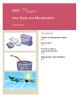
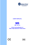
USER MANUAL
Ampliquality
CHLAM-T
Code 03-10A
Code 03-10R
Kit for the detection of
Chlamydia trachomatis
0123
03-10A-50(8033622781530)-EN.doc
1.
PRODUCT INFORMATION ......................................................................................3
2.
KIT CONTENTS........................................................................................................4
3.
STORAGE AND STABILITY OF THE REAGENTS .................................................5
4.
PRECAUTIONS FOR USE .......................................................................................6
5.
SAFETY RULES.......................................................................................................7
5.1.
General safety rules................................................................................................7
5.2.
Safety rules about the kit .......................................................................................8
6.
MATERIALS REQUIRED, BUT NOT PROVIDED....................................................9
6.1.
Reagents..................................................................................................................9
6.2.
Instruments .............................................................................................................9
6.3.
Materials ..................................................................................................................9
7.
PREPARATION OF THE REAGENTS ...................................................................10
8.
INTRODUCTION.....................................................................................................11
9.
TEST PRINCIPLE...................................................................................................12
10.
PRODUCT DESCRIPTION.....................................................................................13
11.
COLLECTION, MANIPULATION AND PRE-TREATMENT OF THE SAMPLES...14
12.
PROTOCOL............................................................................................................15
12.1.
DNA extraction...................................................................................................15
12.2.
DNA amplification..............................................................................................15
12.2.1. TST DNA amplification
15
st
12.2.2. Direct amplification (1 amplification) of C. trachomatis DNA:
15
12.3.
Visualization of the amplification products.....................................................16
12.3.1. Agarose gel electrophoresis
16
12.3.2. Sample Loading
17
12.4.
Interpretation of results: TST DNA amplification and C. trachomatis first
amplification.....................................................................................................................18
Page 1
03-10A-50(8033622781530)-EN.doc
12.5.
2nd C. trachomatis DNA amplification (nested PCR) ......................................19
12.6.
Interpretation of results: 2nd amplification of Chlamydia trachomatis DNA.20
13.
TROUBLESHOOTING............................................................................................21
14.
DEVICE LIMITS ......................................................................................................23
15.
DEVICE PERFORMANCES ...................................................................................23
15.1.
Specificity ..........................................................................................................23
15.2.
Sensitivity ..........................................................................................................23
16.
16.1.
REFERENCES........................................................................................................24
Useful websites .................................................................................................24
Page 2
03-10A-50(8033622781530)-EN.doc
1. PRODUCT INFORMATION
CHLAM-T:
Kit for the detection of Chlamydia trachomatis by DNA amplification in the
cryptic plasmid.
The kit contains the reagents for DNA amplification and visualization by
agarose gel electrophoresis, the internal control of sample amplificability, and
positive control.
Code
03-10A-25
03-10A-50
Product
CHLAM-T
CHLAM-T
Pkg
25 tests
50 tests
CHLAM-T - amplification reagents
Kit for the detection of Chlamydia trachomatis by DNA amplification in the
cryptic plasmid.
The kit contains the reagents for DNA amplification, the internal control of
sample amplifability, and the positive control.
Code
03-10R-25
03-10R-50
Product
CHLAM-T- amplification reagents
CHLAM-T- amplification reagents
Pkg
25 tests
50 tests
Page 3
03-10A-50(8033622781530)-EN.doc
2. KIT CONTENTS
Attention: the different contents correspond to different kits.
(legend: X = component included in the kit; 0 = component not included in the kit)
BOX P
code
03-10A
code
03-10R
STORE AT – 20°C
DESCRIPTION
LABEL
COLOUR
OF TUBE
(T)
OR LID
25 tests
50 tests
8 tests
X
X
Single dose premixes for
CHLAM-T direct amplification
Colourless
(T)
25
50
8
X
X
Single dose premixes for
CHLAM-T nested amplification
Green (T)
25
50
8
X
X
Single dose premixes for
TST (thiosulfate
sulfurtransferase –rhodanese)
amplification
Blue (T)
25
50
8
X
X
Thermostable Taq DNA
polymerase
Red
1 x 50μL
1 x 100μL
1 x 20μL
AB TAQ
5 U/μL
SMALL BAG
code
03-10A
code
03-10R
STORE AT – 20°C
X
X
DESCRIPTION
Plasmid DNA containing part
of Chlamidia trachomatis
genome
LABEL
CHLAM-T
positive
control
COLOUR
OF TUBE
(T)
OR LID
25 tests
50 tests
8 tests
Blue
1 x 30 μL
1 x 50 μL
1 x 10 μL
BOX F
code
03-10A
code
03-10R
STORE AT +2°/ +8°C
X
0
DESCRIPTION
Loading Buffer for
Electrophoresis
X
0
Ethidium Bromide solution
(2,5 mg/mL)
X
0
DNA Molecular Weight Marker
LABEL
COLOUR
OF TUBE
(T)
OR LID
25 tests
50 tests
8 tests
6X Blue
Blue
1 X250μL
1 X450μL
1 X100μL
Ethidium
Bromide
TOXIC
R 23 68
S 36/37 45
Red
1 X170μL
1 X250μL
1 X100μL
MW Marker
Yellow
1 X200μL
1 X400μL
1 X70μL
Page 4
03-10A-50(8033622781530)-EN.doc
BOX A
code
03-10A
code
03-10R
STORE AT +15°/ +25°C
DESCRIPTION
LABEL
COLOUR
OF TUBE
(T)
OR LID
25 tests
50 tests
8 tests
X
0
Agarose for Molecular Biology
AGAROSE
1X20g
1X40g
1X6g
X
0
Electrophoresis buffer
TRIS-Acetate-EDTA, pH 8,00
50X TAE
1X60mL
1X125mL
1X30mL
3. STORAGE AND STABILITY OF THE REAGENTS
Each component of the kit must be stored according to the directions
indicated on the label of the single boxes.
In particular:
Box P
Small bag
Box F
Box A
store at -20°C
store at -20°C
store at a +2°− +8°C
store at +15°− +25°C
When stored at the recommended temperature, all test reagents are stable
until their expiration date.
Page 5
03-10A-50(8033622781530)-EN.doc
4. PRECAUTIONS FOR USE
•
The kit must be handled by qualified investigators, who are educated and
trained in molecular biology techniques applied to diagnostics;
•
Before starting the kit procedure, read carefully and completely the
instruction manual;
•
Keep the product away from heat sources;
•
Do not use any part of the kit past the expiration date;
•
In case of any doubts or questions about the storage conditions or box
integrity,
contact
AB
ANALITICA’s
technical
support
at:
[email protected];
In the amplification of nucleic acids, the investigator has to take the
following special precautions:
•
Use filter-tips;
•
Store the biological samples, the purified DNA, the reference DNA
included in the kit, and all the amplification products in different places
from where amplification reagents are stored;
•
Organize the work space into different pre- and post-PCR units; do not
share instruments and consumables (pipettes, tips, tubes, etc.) between
them;
•
Change gloves frequently;
•
Wash the bench surfaces with 5% sodium hypochloride;
•
The PCR premixes are thawed at room temperature before use; Add the
Taq DNA polymerase and purified DNA very quickly at room temperature
or in an ice-bath;
Page 6
03-10A-50(8033622781530)-EN.doc
5. SAFETY RULES
5.1. General safety rules
•
Wear disposable gloves to handle the reagents and the clinical samples
and wash your hands at the end of work;
•
Do not pipette with the mouth;
•
Since, no known diagnostic method can assure the absence of infective
agents. It is a good rule to consider every clinical sample as potentially
infectious and handle it as such;
•
All the devices that directly touch the clinical samples must be
considered as contaminated and disposed as such. In case of accidental
spilling of the samples, clean up with 10% Sodium Hypochloride. The
materials used to clean up must be disposed of in special containers for
contaminated products;
•
Clinical samples, materials, and contaminated products should be
disposed of after decontamination by:
immersion in a solution of 5% Sodium Hypochloride (1 volume of
Sodium Hypochloride solution for every 10 volumes of contaminated
fluid) for 30 min.
OR
autoclave at 121°C for at least 2 hours (NOTE: do not autoclave
solutions containing Sodium Hypochloride!!)
Page 7
03-10A-50(8033622781530)-EN.doc
5.2.
Safety rules about the kit
The risks for the use of this kit are related to the single components:
Dangerous components:
ETHIDIUM BROMIDE (included in the 03-10A kit)
3,8-diamino-1-ethyl-6-phenylphenantridiumbromide <2%
Description of risk:
T (Toxic)
RISK SENTENCES AND S SENTENCES
ETHIDIUM BROMIDE
R 23 and R 68
S 36/37 45
Toxic for inhalation.
Risk of irreversible effects.
Wear laboratory coat and disposable gloves.
In case of accident or discomfort, seek for medical
assistance and show the package label.
R and S sentences refer to the concentrated product, as provided in the kit.
In particular for Ethidium Bromide, until it is diluted in the agarose gel.
When handling concentrated Ethidium Bromide, use a chemical dispensing
fume cabinet. Always wear disposable gloves and laboratory coat when
handling diluted Ethidium Bromide solutions as well.
The product must not be disposed of in the common waste. It must not go in
the drain system. For disposal, follow local laws.
In case of accidental spilling of Ethidium Bromide, clean with Sodium
hypochloride and water.
Material Safety data sheets of products are available upon request.
Page 8
03-10A-50(8033622781530)-EN.doc
6. MATERIALS REQUIRED, BUT NOT PROVIDED
6.1.
•
•
•
•
Reagents
Reagents for DNA extraction;
Sterile DNase and RNase free water;
Distilled water;
Reagents for DNA visualization by agarose gel electrophoresis (necessary
for 03-10R kit).
6.2.
Instruments
• Laminar flow cabinet (use is recommended while adding TAQ polymerase
to the amplification premix to avoid contamination; it would be
recommended to use another laminar flow cabinet to add the extracted
DNA);
• Micropipettes (range: 0,2-2 µL; 0,5-10 µL; 2-20 µL);
• Thermalcycler;
• Microcentrifuge (max 12.000-14.000 rpm);
• Balance;
• Magnetic heating stirrer or microwave;
• Chemical cabinet (use is recommended in handling Ethidium Bromide);
• Horizontal electrophoresis chamber for agarose minigel;
• Power supply (50-150 V);
• UV Transilluminator;
• Photo camera or image analyzer.
6.3.
•
•
•
•
•
Materials
Disposable gloves;
Disposable sterile filter-tips (range: 0,2-2 µL; 0,5-10 µL; 2-20 µL;);
Graduated cylinders (1 L) for TAE dilution;
Pyrex bottle or Becker for agarose gel preparation;
Parafilm.
Page 9
03-10A-50(8033622781530)-EN.doc
7. PREPARATION OF THE REAGENTS
Preparation of 1 L of 1X TAE buffer:
Mix 20 mL of 50X TAE (included in the 03-10A kit) with 980 mL of distilled
water.
Page 10
03-10A-50(8033622781530)-EN.doc
8. INTRODUCTION
Chlamydia trachomatis is the ethyologic agent of several common sexually
transmitted bacterial pathologies.
Chlamydia genera encompasses low-dimension microorganisms, that
reproduce exclusively in the cytoplasm of the host cell, in
the vacuole. It is a bacterium with obliged endo-cellular
parasitism (Fig. 1), due to its incapability to produce ATP
and other necessary biosynthetic cofactors.
Its morphogenetic cycle consists of two alternate cellular
cycles: the elementary body and the reticular body.
Fig. 1:
Cytoplasmic inclusion of
Chlamydia trachomatis
The elementary body is the infective form and, for its survival capacity outside
the eukaryotic cell, can be compared to a spore cell. Once it penetrates the
eukaryotic cell, the elementary body develops after 10 hours from the
infection. Then, the reticular body starts to reproduce up to cell death.
Chlamydia trachomatis encloses 15 distinct serotypes: A,B,C serotypes are
associated to trachoma (conjunctive infection); L1, L2, and L3 serotypes are
associated to lymphogranuloma venereum; D to K serotypes are ubiquitous,
generally associated to sexually-transmitted diseases; they infect the
epithelial cells and cause infection in the pharynx, respiratory apparatus,
urethra, and uterine tubes (also called Fallopian tubes).
Each serotype has a plasmid (about ten copies per cell) that is believed
to contain the essential genes for microorganism survival in the
infected cell.
Generally, C. trachomatis infection has a clinically latent form but can be the
cause of ectopic pregnancy, female sterility, and male infertility. Newborns
exposed to C. trachomatis infection can develop a conjunctivitis or an acute
pneumonia that can lead to a chronic form.
Most of the C. trachomatis infections are asymptomatic and it is generally
difficult to distinguish an evident C. trachomatis infection from other sexuallytransmitted diseases; therefore it is important to have a quick, rapid, and
reliable method for C. trachomatis detection.
Recent studies suggest that there is a correlation between the exposure
to different C. trachomatis serotypes and an increased risk of cervix
cancer (Antilla et al. JAMA 2001).
Page 11
03-10A-50(8033622781530)-EN.doc
Even if HPV infection from oncogenic strains is the first cause of cervix
cancer, the researchers are now focusing on C. trachomatis.
It is not clear if this oncogenic risk is associated to specific microorganism’s
serotypes (Koskela et al., 2000).
Considering the high incidence (4%) of C. trachomatis infection in male
donors, guidelines for the prevention of this pathogen’s transmission by
artificial insemination have been recently proposed.
The American Society for Reproductive Medicine and the European
Society for Human Reproduction and Embryology affirm that tests for C.
trachomatis identification should always be performed to screen urethral,
urine, and seminal samples (American Society for Reproductive Medicine
1998, Baratt et al.,1998).
9. TEST PRINCIPLE
PCR (Polymerase Chain Reaction) was the first method of DNA amplification
described in literature. (Saiki RK et al., 1985). It can be defined as an in vitro
amplification reaction of a specific part of DNA (target sequence) by a
thermo-stable DNA polymerase.
In the reaction, three segments of nucleic acid are involved: the double strand
DNA template to be amplified (target DNA) and two single-strand
oligonucleotides “primers” that anneal in a specific way to the template DNA.
The DNA polymerase begins the synthesis process at the region marked by
the primers and synthesizes new double stranded DNA molecules, identical
to the original double stranded target DNA region, by facilitating the binding
and joining of the complementary nucleotides that are free in solution
(dNTPs). After several cycles, we can get millions of DNA molecules which
correspond to the target sequence.
The sensitivity of this test makes it particularly suitable to the application in
laboratory diagnostic.
Besides, the amplification reaction can be executed from a wide range of
biological samples, and since PCR is able to amplify very small DNA
segments, the starting DNA can also be partially degraded.
Page 12
03-10A-50(8033622781530)-EN.doc
10. PRODUCT DESCRIPTION
The method proposed in this kit consists of the amplification of the C.
trachomatis cryptic plasmid DNA.
The plasmid is highly conserved in all sierotypes of C. trachomatis, but
absent in other species.
The method can evaluate the suitability of extracted DNA, by amplification of
TST (thiosulfate sulfurtransferase rhodanese) gene in the region 22q13.1
(amplification control). A negative result in the TST gene amplification
indicates either the presence of inhibitors of the amplification reaction in the
extracted DNA or that the DNA is highly degraded. This method helps the
operator to recognize possible false negative results for C. trachomatis.
The kit also provides a positive control for the amplification. When the
amplification of the positive control is successful, this guarantees that the
reaction worked correctly. The positive control is not dangerous for the
operator, because it is plasmid DNA containing only a part of C. trachomatis
cryptic plasmid.
The kit is in premix format: all the reagents for the amplification are pre-mixed
and aliquoted in single dose test-tubes, which Taq polymerase and the
extracted DNA will be added to. This premix format reduces the manipulation
steps in pre-amplification, saves considerable time for the operator, avoids
repeated freezing/thawing of reagents (that could alter the product
performances) and, above all, this format reduces significantly the risk of
contamination and the risk of obtaining false positive results. For this reason,
it is always recommended to use all the proper amplification controls.
Page 13
03-10A-50(8033622781530)-EN.doc
11. COLLECTION, MANIPULATION AND PRETREATMENT OF THE SAMPLES
The samples used in the laboratories for the determination of C. trachomatis
infection, are cytological, like cell cultures, urethral swabs and cervix-vaginal
swabs, sperm, urine, etc..
In the case of cervix and vaginal swabs, sample collection is performed by
using a little swab or a little sterile brush (cyto-brush). The collected cells are
diluted in a suitable transport media (1X PBS, physiological solution or other
medium).
The sample can be stored at +2 / +8°C for a max of 48 hrs.; after that, it is
better to proceed with nucleic acid extraction.
If it is not possible to extract the sample within a short period of time, the
specimen must be stored at -20°C.
When sample cellularity is too low, it could be necessary, before starting DNA
extraction, to concentrate the sample by centrifuging repeatedly. For sample
resuspension use sterile PBS, which can also be useful to remove mucus and
red blood cells, if present.
Seminal liquid is collected in a sterile container and stored at +4°C for some
hours (maximum above night). Alternatively, it is frozen by adding a specific
medium, which compatible with the DNA extraction method used in the
molecular analysis.
C. trachomatis detection from urine samples is usually performed from the
first urination. It is necessary to discard the first part of urine flux and to
collect only the second one in a sterile container.
Page 14
03-10A-50(8033622781530)-EN.doc
12. PROTOCOL
12.1.
DNA extraction
Any extraction method can be used, provided that it extracts pure and integral
C. trachomatis DNA.
AB ANALITICA tested successfully methods based on spin columns (e.g.
QIAmp DNA mini kit - QIAGEN) and methods based on matrixs (e.g. INstaGene Matrix – BIO-RAD).
It is necessary to remember that the volumes of the extracted DNA, indicated
in paragraph 12.2 for TST and C. trachomatis amplification, are referred to
the extracts obtained with AB ANALTICA methods. When using an alternative
extraction method, it will be necessary to adjust the volume of the
amplification mix by adding water if the volume of the DNA solution for
amplification is less than 20 μL.
12.2.
DNA amplification
12.2.1.
TST DNA amplification
For each sample add to each premix tube (blue tube):
0,5 µL AB Taq
10 µL extracted DNA
12.2.2. Direct amplification (1st amplification) of C. trachomatis
DNA:
For each sample add to each premixed colourless tube:
0,5 µL AB Taq
10 µL extracted DNA
Page 15
03-10A-50(8033622781530)-EN.doc
It is important to include in each experiment a negative control (add 10 μL of
distilled H2O instead of DNA to the mix) and a positive control (10 μL of the
positive control included in the kit).
Centrifuge briefly, put the tubes (TST and CHLAM-T) into the thermalcycler
as programmed below:
1 cycle
30 cycles
1 cycle
95°C
5 min
95°C
45 sec
56°C
45 sec
72°C
45 sec
72°C
5 min
Amplification products length:
CHLAM-T:
TST:
12.3.
594 bp.
202 bp.
Visualization of the amplification products
12.3.1.
Agarose gel electrophoresis
Prepare a 3% agarose gel:
Weight 1.5 g of agarose and pour it into 50 mL of 1x TAE.
Leave the solution on a magnetic stirring heater or in a microwave, until the
solution becomes clear.
Allow the gel to cool for a few minutes and then add 10 µL of Ethidium
Bromide solution.
NOTICE: Ethidium Bromide is a strong mutagenic agent; always wear
gloves and preferably work under a chemical safety cabinet during the
handling of this reagent or gels containing it.
Pour the gel into the appropriate gel casting tray, with the comb placed in and
allow the gel to cool at room temperature or in a fridge until the gel becomes
solid.
Page 16
03-10A-50(8033622781530)-EN.doc
When the gel is solidified, remove the comb carefully (pay attention not to
damage the gel wells) and transfer the tray into the electrophoresis chamber.
Pour the appropriate amount of 1x TAE buffer, so that it covers the gel
completely (about 1-2 mm over the gel surface).
12.3.2.
Sample Loading
Mix in a tube or directly on a parafilm layer:
2 μL
10 μL
of 6X Blue*
of amplification product or DNA Molecular Weight Marker
(MW*marker)
Load the mixture in the gel wells; switch on the power supply and set the
voltage between 80-100 V.
Run the gel for about 40 min, then place the gel on an UV transilluminator
and analyze the results by comparing the size of the amplification products
with the reference Molecular Weight Marker.
* = 6X Blue and PM Marker are included in 03-10A kit; for the use of another
loading buffer or of another Molecular Weight Marker, follow the indications
specified by the supplier.
DNA Molecular Weight Marker (MW Marker, included in 03-10A kit)
Fragment sizes: 501-489, 404, 331, 242, 190, 147, 111, 110, 67, 34x2, 26 bp.
The 501-489 bp bands usually are not clearly distinguishable and appear as
an unique band; the 26 and 34 bp bands are sometimes too small to be seen
in a 3% agarose gel (because of their low molecular weight).
NOTICE: UV rays are dangerous for skin and, above all, eyes: always
wear gloves and safety glass or use the protection screen of the UV
transilluminator.
Page 17
03-10A-50(8033622781530)-EN.doc
12.4.
Interpretation of results: TST DNA amplification and C.
trachomatis first amplification
The controls should show the following results:
CONTROL
RESULT
INTERPRETATION
Positive control
POSITIVE
The PCR amplification was performed
correctly
Negative control
NEGATIVE
Absence of contamination
Then, for the interpretation of the band pattern, follow the table below:
RESULT
INTERPRETATION
Absence of the TST gene band
Sample not suitable for amplification
(repeat the DNA extraction)
Presence of the TST gene band
Amplifiable sample
Absence of Chlamydia trachomatis Proceed
with
the
second
band
amplification to evaluate the real
negativity of the sample
Presence of the TST gene band
Amplifiable and positive sample
Presence of Chlamydia trachomatis for Chlamydia trachomatis
band
Page 18
03-10A-50(8033622781530)-EN.doc
12.5.
2nd C. trachomatis DNA amplification (nested
PCR)
Add to each premixed green tube, containing 48.5 μL of amplification mix:
0,5 µL AB Taq
1 µL
First amplification product
It is important to use in this step the negative and the positive controls
included in the Chlamydia trachomatis direct amplification. When it seems
useful, it is possible to include also a negative control for the nested
amplification, by adding 1 µL of sterile water to the nested mix, instead of the
direct amplification product.
Centrifuge briefly, then put the tubes into the thermalcycler as programmed
below:
1 cycle
35 cycles
1 cycle
95°C
5 min
94°C
45 sec
55°C
45 sec
72°C
45 sec
72°C
10 min
Amplification fragment length: 201 bp.
At the end of the amplification program, visualize the nested amplification
products by a 3% agarose gel electrophoresis.
Weight 1.5 g of agarose and pour it into 50 mL of 1X TAE.
Follow the instructions indicated at paragraph 12.3.
Page 19
03-10A-50(8033622781530)-EN.doc
12.6.
Interpretation of results: 2nd amplification of Chlamydia
trachomatis DNA
The controls should show the following results:
CONTROL
RESULT
Positive control
POSITIVE
Negative Control
NEGATIVE
Negative control of
Nested amplification
NEGATIVE
INTERPRETATION
The first and the second
amplification
was
performed
correctly
Absence of contamination in the
first amplification
Absence of contamination in the
second amplification
Then, for the interpretation of the band pattern, follow the table below:
RESULT
INTERPRETATION
Absence of Chlamydia trachomatis band in Negative sample for
the first and second amplification
Chlamydia trachomatis
Presence of Chlamydia trachomatis band in Positive sample for
the second amplification
Chlamydia trachomatis
Fig. 2:
3% agarose gel electrophoresis of the amplification products.
1.
2 – 4.
5 – 7.
DNA Molecular Weight Marker.
Direct PCR products.
Nested PCR products.
All samples are positive for C. trachomatis.
Page 20
03-10A-50(8033622781530)-EN.doc
13. TROUBLESHOOTING
No amplification products or positive control DNA band
• TAQ polymerase was not correctly added to the premix
-
Use pipettes and tips with suitable volumes (pipette range 0,2 - 2 μL);
Check visually that TAQ polymerase diffuse in the premix: this is easy
because the enzyme is in dissolved glycerol that has a higher density;
- Alternatively, check visually the drop of TAQ polymerase put on the
tube wall, then centrifuge briefly.
• The thermalcycler was not programmed correctly.
-
Check the conformity of the thermalcycler program and the
temperature profile in the instruction manual.
• The kit does not work properly
-
Store the premix, enzymes and reference DNA at -20°C;
Avoid repeated freezing/thawing of the premix and the regents.
No amplification bands for either TST or C. trachomatis in the tested
sample, but a good band for the Positive controls
• Problems during the extraction step
-
Be sure that the extraction kit is adequate and that you follow correctly
all the instructions;
- Consult the troubleshooting section of the extraction kit;
- Repeat the DNA extraction starting from a new sample.
The amplification was inhibited
-
Dilute the starting sample with distilled water and TE;
Repeat the DNA extraction from a smaller amount of clinical sample;
Use an adequate extraction system.
Page 21
03-10A-50(8033622781530)-EN.doc
Aspecific products or extrabands present after the visualization of the
amplified product in agarose gel
The thermalcycler makes temperature changes too slowly
-
Execute a thermalcycler revision.
The preparation of the amplification reaction took too long time at room
temperature.
Accelerate the work time at room temperature;
- Work on ice.
• The extracted product was not purified
-
Use an extraction system, which purifies the sample well.
The starting sample contained degraded DNA
-
Repeat the extraction step using another starting clinical sample;
Be sure that the sample had been collected and stored in appropriate
way.
For any further problems contact AB ANALITICA’s technical support at:
[email protected], fax (+39) 049-8709510, or tel. (+39) 049-761698.
Page 22
03-10A-50(8033622781530)-EN.doc
14. DEVICE LIMITS
The kit can have reduced performances if:
•
the clinical sample is not suitable for this analysis (incorrect transport
media or incorrect sample storage);
•
the DNA is not amplifiable because of the presence of inhibitors of the
amplification reaction or due to an inadequate extraction system;
•
the kit was not stored at the suggested temperature.
15. DEVICE PERFORMANCES
15.1.
Specificity
Primer sequence alignment in the most important databanks shows the
absence of unspecific alignment and guarantees the amplification of C.
trachomatis. Cross reaction with genomic DNA or other pathogenic
microorganism nucleic acids have not been revealed.
15.2.
Sensitivity
It has been shown that a unique amplification step, using nested primers, has
a detection limit of about 1 IFU (inclusion forming unit) after visualization by
agarose gel electrophoresis.
Each IFU contains about 10 cryptic plasmids.
The addition of another amplification step increases the sensitivity by at least
a factor of 10.
Page 23
03-10A-50(8033622781530)-EN.doc
16.
REFERENCES
American Society for Reproductive Medicine. Fertil Steril 4(3), 1S-13S, 1998.
Antilla T, Saikku P, Koskela P et al. JAMA 285(1), 47-51, 2001.
Baratt C, Englert Y, Gottlieb C and Jouannet P. Hum Reprod 13(2), VII-IX,
1998.
Koskela P, Antilla T, Bjorge T et al. Int J Cancer 85, 35-39, 2000.
Saiki RK, Scharf S, Faloona F et al. Science 230, 1350-1354, 1985.
16.1.
Useful websites
http://www.drorlandella.it/chlamydia.htm
Page 24
03-10A-50(8033622781530)-EN.doc
AB ANALITICA srl
Via Svizzera 16 - 35127 PADOVA, (ITALY)
Tel +39 049 761698 - Fax +39 049 8709510
e-mail: info@abanalitica