1
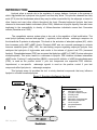
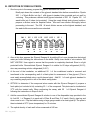
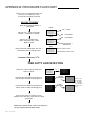
96 well Adipocyte Lipolysis Assay Kit for
Detection of Both Free Glycerol and NonEsterified Fatty Acids
500 point assay kit
Cat# LIP-3RB
INSTRUCTION MANUAL
ZBM0048.01
STORAGE CONDITIONS
Reagents & Buffers:
4°C
Vehicle & Controls: -20°C
ALL ZEN-BIO INC PRODUCTS ARE FOR RESEARCH USE ONLY. NOT APPROVED FOR HUMAN OR
VETERINARY USE OR FOR USE IN DIAGNOSTIC OR CLINICAL PROCEDURES.
LIMITED PRODUCT WARRANTY
This warranty limits our liability to replacement of this product. No other warranties of any kind, expressed or
implied, including without limitation, implied warranties of merchantability or fitness for a particular purpose, are
provided by Zen-Bio, Inc. Zen-Bio, Inc. shall have no liability for any direct, indirect, consequential, or incidental
damages arising out of the use, the results of use, or the inability to use this product.
ORDERING INFORMATION AND TECHNICAL SERVICES
Zen-Bio, Inc.
3200 Chapel Hill-Nelson Blvd., Suite 104
PO Box 13888
Research Triangle Park, NC 27709
Telephone
(919) 547-0692
Facsimile (FAX)
(919) 547-0693
Toll Free
1-866-ADIPOSE
Electronic mail (e-mail)
World Wide Web
REV. Aug 2009
(866)-234-7673
[email protected]
http://www.zenbio.com
INTRODUCTION
Lipolysis plays a central role in the regulation of energy balance. Lipolysis is the process in
which triglycerides are hydrolyzed into glycerol and free fatty acids. This process releases free fatty
acids (FFA) into the bloodstream where they may be either re-esterified by the adipocyte or travel to
other tissues and exert other effects throughout the body. Elevated adipocyte lipolysis has been
observed in obese and diabetic individuals (Arner 1996). Alterations in lipolytic capacity have also been
implicated in the susceptibility to obesity of African-American individuals versus their Caucasian
cohorts (Danadian et al. 2001).
The sympathetic nervous system plays a key role in the regulation of lipid mobilization. The
main lipolytic pathway involves beta-agonists ( -agonists), which activate -adrenergic receptors via
the intracellular Gs proteins in adipocytes. This leads to the activation of adenylate cyclase (AC), which
then increases cyclic AMP (cAMP) levels. Elevated cAMP acts as a second messenger to activate
hormone sensitive lipase (HSL). HSL, the rate-limiting enzyme regulating adipocyte lipolysis, then
catalyzes the hydrolysis of triglycerides and results in the release of glycerol and FFA (increased
lipolysis). Phosphodiesterases (PDE) are enzymes that hydrolyze cAMP to 5’-AMP (5 prime adenosine
monophosphate). This action results in a decrease in lipolysis. PDE inhibitors increase intracellular
cAMP levels. 3-isobutyl-1-methylxanthine (IBMX), a non-specific inhibitor of cAMP phosphodiesterases
(PDE), is used as the positive control if your test compounds are suspected PDE inhibitors.
Isoproterenol, a non-specific
-adrenergic agonist is used as the positive control if your test
compounds affect lipolysis via -adrenergic receptors.
This lipolysis assay kit provides the tool to study chemical compounds that may influence
lipolysis in cultured human adipocytes.
Figure 1. Overview of adipocyte lipolysis
ABBREVIATIONS:
AC
adenylate cyclase
AR
adrenergic receptors
Gs
G protein coupled receptor
FFA
free fatty acids
PKA
protein kinase
AMP adenosine monophosphate
ATP
adenosine triphosphate
IR
insulin receptor
PDE
phosphodiesterase
TG
triglyceride
REV. Aug 2009
ITEMS INCLUDED IN THE KIT
ITEM
DESCRIPTION
Cap
Color
---
UNIT
QTY
STORAGE
BOTTLE
1
4°C
---
BOTTLE
PURPLE
1 ml /
VIAL
1
5
4°C
-20°C
BLUE
10 l /
VIAL
5
-20°C
AMBER
5
4°C
PINK
100 l /
VIAL
50ML
25ML
Reconstitute using 50 ml FFA Diluent A.
Discard remainder after 10 days
Reconstitute using 25 ml FFA Diluent B per
bottle. Discard remainder after 10 days
YELLOW
BOTTLE
1
1
1
4°C
4°C
4°C
PINK
BOTTLE
1
4°C
Glycerol @ 1mM [see page 6 for dilution
instructions]
ORANGE
50 l /
VIAL
5
-20°C
40ML
BOTTLE
1
4°C
LIP-2/3 Assay
Buffer
Wash Buffer
Vehicle
500 ml
Positive control
Isoproterenol, 10 mM in DMSO. Dilute to 1 M
FFA Standard
FFA Diluent A
FFA Diluent B
FFA Reagent A
FFA Reagent B
Glycerol
Standard
LIP-2/3 Wash Buffer, 250 ml
0.1% DMSO in LIP-2/3 Assay Buffer
in Assay Buffer before use! (i.e.1 l in 10 ml
Assay Buffer)
1mM Stock. See page 5 for standard curve
preparation
YELLOW
Glycerol Reagent 40-ml- Reconstitute with 40 ml deionized water
A
prior to use.
Other equipment/reagents required but not provided with the kit:
Blank 96 well plates
Multi-channel Pipet , single channel pipet and pipet tips
Plate reader with a filter of 540 nm
Incubator at 37oC
Large gauge needle
Cultured human adipocytes
Tubes for diluting glycerol standards
REV. Aug 2009
PRINCIPLES OF THE ASSAYS
Detection of Free Glycerol
Assessing lipolytic activity by the measurement of glycerol released into the medium. Glycerol
released to the medium is phosphorylated by adenosine triphosphate (ATP) forming glycerol-1phosphate (G-1-P) and adenosine-5’-diphosphate (ADP) in the reaction catalyzed by glycerol kinase.
G-1-P is then oxidized by glycerol phosphate oxidase to dihydroxyacetone phosphate (DAP) and
hydrogen peroxide (H2O2). A quinoneimine dye is produced by the peroxidase catalyzed coupling of 4aminoantipyrine (4-AAP) and sodium N-ethytl-N-(3-sulfopropyl)m-anisidine (ESPA) with H2O2, which
shows an absorbance maximum at 540nm.
The increase in absorbance at 540nm is directly
proportional to glycerol concentration of the sample.
GLYCEROL + ATP
G-1-P + O2
G-1-P + ADP
DAP + H2O2
H2O2 +4-AAP + ESPA
Quinoneimine dye + H2O
Detection of Non-Esterified Fatty Acids (Free Fatty Acids; FFA)
Assessment of lipolytic activity can also be detected through a coupled reaction to measure
non-Esterified fatty acids (NEFA) released by adipocytes. The initial step, carried out by acyl-CoA
synthetase (ACS), produces fatty acyl-CoA thiol esters from the NEFA, ATP, Mg, and CoA in the
reaction. The acyl-CoA derivatives react
with oxygen in the presence of acyl-CoA
oxidase (ACOD) to produce hydrogen
peroxide.
Hydrogen peroxide in the
presence of peroxidase (POD) allows the
oxidative condensation of 3-methyl-Nethyl-N-( -hydroxyethyl)-aniline with 4aminoantipyrine which forms a purple
product that absorbs light at 550nm. This
allows the concentration of NEFA to be
determined from the optical density measured at 540 - 550nm.
NOTE:
3 fatty acid molecules are released per triglyceride molecule resulting in a 3:1 fatty acid to
glycerol concentration.
REV. Aug 2009
A. DETECTION OF NON-ESTERIFIED FATTY ACIDS
1. Prepare the standard curve using the FFA STANDARD SOLUTION as follows:
Briefly spin down the contents of the free fatty acid standard tube before reconstitution.
Standards are: 0, 1.4, 4.1, 12.3, 37, 111, and 333 M fatty acid. Prepare as follows:
The kit standard solution is the 1.0 mM standard. Pipette 60 l of Dilution Buffer (Assay
Buffer) into 6 tubes (not provided). Pipette 30 l of the FFA Standard Stock into a tube
labeled 333 µM. Prepare a dilution series as depicted below. Mix each new dilution
thoroughly before proceeding to the next. The Assay Buffer alone serves as the zero
standard.
30 l
30 l 30 l 30 l 30 l
30 l
Std
FFA
Std
333
M
111
M
37
M
12.3
M
4.1
1.4
M
M
2. Add 50ml FFA Diluent A to the FFA Reagent A bottle and gently invert. DO NOT VORTEX!
Store any remaining solution at 2-8°C; it is stable for 10 days after reconstitution refrigerated (28°C).
3. At the end of the incubation, 30
l of the conditioned media is removed and transferred to the
corresponding well of a blank plate for assessment of non-esterified fatty acids. [This is most
easily accomplished using a multi-channel pipet.] Add 30 l of each standard to empty wells.
4. Add the reconstituted FFA Reagent A to one of the disposable trays provided in the kit. Add 100
l of FFA Reagent A to each well. Gently shake the plate to ensure mixing. Place in a 37 oC
incubator for 10 minutes.
5. Add 25 ml FFA Diluent B to the FFA Reagent bottle and gently invert. Store any remaining
solution at 2-8°C; it is stable for 10 days after reconstitution refrigerated (2-8°C).
6. Add the reconstituted FFA Reagent B to another disposable tray. Add 50
l of FFA Reagent B
to each well. Gently shake the plate to ensure mixing. Place in a 37 oC incubator for 10 minutes.
7. Allow the plate to equilibrate to room temperature for 5 minutes. During this time, ensure that
there are no bubbles in the solution mixture. Use a large gauge needle or clean pipet tip to pop
any bubbles as this will result in inaccurate absorbance readings.
8. The optical density of each well is then measured at 540 nm.
REV. Aug 2009
B. DETECTION OF FREE GLYCEROL
1. One hour prior to the assay, prepare the glycerol standards as follows:
Briefly spin down the contents of the glycerol standard tube before reconstitution. Pipette
200 l of Wash Buffer into the 1 mM glycerol standard tube provided and mix well by
vortexing. This produces a diluted stock glycerol standard of 200 M. Pipette 125 l of
wash buffer into 6 tubes (not provided). Using the newly diluted stock glycerol solution,
prepare a dilution series as depicted below. Mix each new dilution thoroughly before
proceeding to the next. The 200 M stock dilution serves as the highest standard, and
the wash buffer serves as the zero standard.
200 l
Wash
Buffer
125 l
125 l 125 l 125 l 125 l
125 l
Std
200
M
100
M
50
M
25
M
12.5
M
6.25
M
3.125
M
2. Also at this time prepare the Glycerol Reagent A by adding 40 ml room temperature deionized
water per bottle following the instructions on the bottle. Gently invert bottle to mix contents. DO
NOT VORTEX! Use a pipet to ensure that the powder is completely dissolved. Store in a light
protected bottle. Reconstituted Glycerol Reagent A is stable for 60 days refrigerated (2-8°C);
store any remaining solution refrigerated (2-8°C)
3. At the end of the incubation, an additional 50
l of the conditioned media is removed and
transferred to the corresponding well of a blank plate for assessment of free glycerol. [This is
most easily accomplished using a multi-channel pipet. Add 50 l of each glycerol standard to
any remaining empty wells in one of the blank assay plates.
4. OPTION: to determine if the compound alone reacts with the Glycerol Reagent A, prepare a
fresh plate (not included in kit) containing 50 l of the compound. This plate can be incubated at
37oC with the treated cells. When performing the assay, add 50
following the instructions in Steps 5 and 6.
l of Glycerol Reagent A
5. Add the reconstituted Glycerol Reagent A solution to one of the disposable trays provided in the
kit. Add 50 l of Reagent A to each well of Plate B and Plate C (if used). Gently, pipet up and
down once to mix. Pop the bubbles using a large gauge needle or a clean pipet tip. The plate is
then incubated at 25oC (room temperature) for 15 minutes.
6. The optical density of each well is then measured at 540 nm.
REV. Aug 2009
FATTY ACID STANDARD CURVE
Generate standard curve: see example below
[DO NOT use this standard curve to generate your data. This is an example.]
Subtract the OD value of the 0 M standard from all OD values including the standard curve. . Note:
1mM standard is commonly omitted from analysis due to lack of linearity between 333 M and 1mM.
Optionally, a 4-parameter fit may be used to calculate standard curve.
M
std
333
111
37
12.3
4.1
1.4
0
OD
OD - zero
0.68
0.244
0.104
0.063
0.05
0.046
0.044
0.636
0.2
0.06
0.019
0.006
0.002
0
y = 0.0019x - 0.0045
R2 = 0.9995
Data are expressed as M free fatty acids released.
OPTION: express data as Fold induction over appropriate vehicle
Fold induction = M free fatty acids SAMPLE
M free fatty acids VEHICLE
The R2 value should be equal or greater then 0.98 for the standard curve to be valid. Any R2
values below 0.98, must have the standard curve run again.
REV. Aug 2009
GLYCEROL STANDARD CURVE
Generate standard curve: see example below
[DO NOT use this standard curve to generate your data. This is an example.]
Subtract the OD value of the 0 M standard from all OD values including the standard curve.
y = observed O.D. minus the blank
x = concentration of glycerol in M
To calculate x for each y, (i.e. to change the observed O.D. into glycerol concentration) use the
following equation:
y=(slope) times (x) plus intercept
y=mx+b so x=(y-b)/m
x=(y – 0.0075)/0.003 where 0.003= slope of the line and 0.0075= y intercept. Be careful to enter the
proper sign for the y intercept value as it may be a negative number.
Any OD values greater than the highest standard (200 M) ) should be suspect. The compound
should be re-assayed using a lower dose of the compound at treatment OR a dilute solution of the
condition medium at the time of the assay.
The R2 value should be equal or greater then 0.98 for the standard curve to be valid. Any R2
values below 0.98, must have the standard curve run again.
Data are expressed as M glycerol released.
OPTION: express data as Fold induction over appropriate vehicle
Fold induction = M glycerol SAMPLE
M glycerol VEHICLE
REV. Aug 2009
APPENDIX A: PLATE LAYOUT
.
A
B
C
D
E
F
G
H
1
2
3
4
5
6
7
8
9
10
11
12
REV. Aug 2009
APPENDIX B: PROCEDURE FLOWCHART
Remove 150 l of the shipping medium and
place in your incubator for 5-7 days
(3-5 days for international customers)
ON DAY OF ASSAY
Make all test compound dilutions in
Assay Buffer.
Plate A
120 l media
OOOOOOOOOOOO
OOOOOOOOOOOO
OOOOOOOOOOOO
OOOOOOOOOOOO
OOOOOOOOOOOO
Remove 120 l media from all wells.
Add 200 l Wash Buffer to all wells.
200 l Wash Buffer
Plate A
Remove 120 l media & Wash
Buffer. Add another 200 l Wash
Buffer to all wells.
200 l Wash Buffer
OOOOOOOOOOOO
OOOOOOOOOOOO
OOOOOOOOOOOO
OOOOOOOOOOOO
OOOOOOOOOOOO
Add another 200 l
Wash Buffer
Plate A
OOOOOOOOOOOO
OOOOOOOOOOOO
OOOOOOOOOOOO
OOOOOOOOOOOO
OOOOOOOOOOOO
Remove all media & Wash Buffer. Add 100 l
treatments/controls to 3 wells at a time.
Remove 3 wells at a time
Add treatments 3 wells at a time
Incubate 3-5 hours at 37oC.
FREE FATTY ACID DETECTION
Plate A 30
Assay Plate
Remove 30 l/well conditioned media from
Plate A to Plate B.
OOOOOOOOOOOO
OOOOOOOOOOOO
OOOOOOOOOOOO
OOOOOOOOOOOO
OOOOOOOOOOOO
Reconstitute FFA Reagent A using Diluent A.
Add 100 l/well. Incubate 10 minutes @ 37°C.
OOOOOOOOOOOO
OOOOOOOOOOOO
OOOOOOOOOOOO
OOOOOOOOOOOO
OOOOOOOOOOOO
Reconstitute FFA Reagent B using Diluent B.
Add 50 l/well. Incubate 10 minutes @ 37°C.
OOOOOOOOOOOO
OOOOOOOOOOOO
OOOOOOOOOOOO
OOOOOOOOOOOO
OOOOOOOOOOOO
Place at room temp. for 5 minutes. Pop any
bubbles in each well using a clean pipet tip or large
gauge needle.
Measure the optical density of each well at 540 nm
using a spectrophotometer plate reader.
REV. Aug 2009
l
OOOOOOOOOOOO
OOOOOOOOOOOO
OOOOOOOOOOOO
OOOOOOOOOOOO
OOOOOOOOOOOO
100 l/well
FFA Reagent A
50 l/well
FFA Reagent B
OOO
OOO
OOO
OOO
OOO
OOO
OOO
OOO
OOO
OOO
Plate C may
be necessary
for the assay
of standards
if al 96 wells
of Plate A are
used.
FREE GLYCEROL DETECTION
One hour prior to assay, reconstitute
Glycerol Reagent A and prepare standards.
Keep all at room temp.
Remove 50 l/well conditioned media
from Plate A to a blank assay plate. Add
50 l standards to empty wells.
Add 50 l/well reconstituted Glycerol Reagent A to
a blank assay plate (including the glycerol
standards at 50 l/well and optional plate without
cells).
OOOOOOOOOOOO
OOOOOOOOOOOO
OOOOOOOOOOOO
OOOOOOOOOOOO
OOOOOOOOOOOO
OOOOOOOOOOOO
OOOOOOOOOOOO
OOOOOOOOOOOO
OOOOOOOOOOOO
OOOOOOOOOOOO
OOOOOOOOOOOO
OOOOOOOOOOOO
OOOOOOOOOOOO
OOOOOOOOOOOO
OOOOOOOOOOOO
50 l
GLYCEROL
REAGENT A
OOO
OOO
OOO
OOO
OOO
Plate C may be necessary for the
assay of glycerol standards if al 96
wells of Plate A are used.
o
Incubate at 25 C (room temperature)
for 15 minutes. Pop the bubbles in
each well.
Measure the optical density of each well
at 540 nm using a spectrophotometer
plate reader.
REFERENCES
1. Arner P (1996) Diabetes Rev 4(4):450-463.
2. Botion LM & Green A. Diabetes (1999) 48:1691-1697
3. Brasaemle DL, Dolios G, Shapiro L, Wang R. (2004) J Biol Chem 279(45): 46835-42.
4. Cooper DMF, Schlegel W, Lin MC, Rodbell M. (1979) J Biol Chem 254(18):8927-8931.
5. Dyck DJ Can J Appl Physiol (2000) 25(6):495-523.
6. Kordik CP & Reitz AB. J Medicinal Chem (1999) 42(2):181-201.
7. Rieusset J, Chambrier C, Bouzakri K, Dussere E, Auwerx J, Riou J-P, Laville M, Vidal H.
Diabetologia (2001) 44:544-554.
8. Robidoux J, Martin TL, Collins S. (2004) Ann Rev Chem 253: 7570-7578.
9. Scriba D, Aprath-Husmann I, Blum WF, Hauner H. Eur J Endocrinol (2000) 143:439-445
10. Snyder PB Emerging Therapeutic Targets (1999) 3(4): 587-599.
11. Tansey JT, Sztalryd C, Hlavin EM, Kimmel AR, Londos C. (2004) IUBMB Life 56(7): 379-85.
REV. Aug 2009