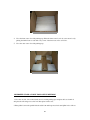
1
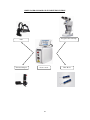
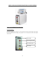
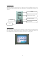
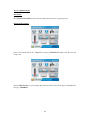
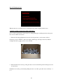
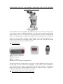
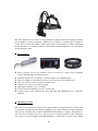
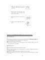
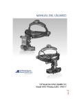
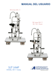
5 step slit lamp is used for laser delivery. Laser adaptor which is connected with slit lamp used to
deliver the laser beam to the required spot size on the retina. This Laser adaptor consists of set of
lenses with reflecting mirror to deliver high power laser beam on retina. Spot size of the beam is
controlled by rotating the adaptor and it will be varied from 50 to 1000 micron.
The height of the instrument table and of the examiners chair has to be adjusted; so that the
surgeon can operate the instrument conveniently. The height of the patients chair is adjusted to
suit his stature. The patient should rest his chin and fore head firmly against the head rest. Move
the chin rest, by turning the control knob until the eyes are level with the black marker at the side
of the head rest. The sliding plate on the table must always be kept clean, to ensure free
movement of the slit lamp.
The transformer fixed underneath the tabletop is designed for connection to A.C mains supplies of
either 110 or 220 volts. Its output for operation of the main lamp is 12 volts. The joystick lever
enables the entire slit lamp to be moved easily in all directions in a horizontal plane. The cross
slide moves parallel to itself during the adjustments. Displace the slit lamp, with the joystick lever
held firmly and slightly inclined towards the examiner, until the image of the slit appears sharply
at the depth of the eye, which is to be observed. The horizontal motion of the cross-slide can be
locked by tightening the knob. Rotate the joystick to adjust the height of the light to the level of
the eye.
The microscope can be locked on the pivoting axis while the illumination unit can be freely
rotated about the common axis. The illumination and the microscope can be rotated together or
separately with any fixed angle between them, if the knobs are loosened. If you wish to move the
instrument with or without the table, it is advisable to lock all movable parts by tightening screws.
The ability to focus each eye piece separately not only enables the examiners refraction to be
corrected, but also compensates for any asymmetrical course of the two microscope pencils in the
examined eye, especially during Fundus observation. A satisfactory binocular and stereoscopic
examination of the slit image on the Fundus is usually possible only when an accurate setting of
each eye piece has been made.
Maintenance:
The room condition should be maintained as
Operating condition
Use only the type of power source that indicated on label
Storage condition
+10C to +40C
-40C to +70C
Relative air humidity
10% to 90%
Transportation humidity
10% to 90%
Air pressure
Warning:
600 – 1300 mbar
Connect the Equipment to properly grounded power outlets.
Unplug the Equipment before servicing / cleaning.
Confirm whether the AC power cord meets the relevant local safety standards.
23