Download 1-Auto Refractometer
Transcript
1-Auto Refractometer
astigmatism
An automated refractor, or autorefractor, is a
computer-controlled machine used during an eye
examination to provide an objective measurement
of a person's refractive error and prescription
for glasses or contact lenses. This is achieved
by measuring how light is changed as it enters a
person's eye.
The automated refraction technique is quick,
simple and painless. The patient takes a seat and
places their chin on a rest. One eye at a time,
they look into the machine at a picture inside. The
picture moves in and out of focus and the machine
takes readings to determine when the image is
on the retina. Several readings are taken which
the machine averages to form a prescription. No
feedback is required from the patient during this
process.
Within seconds an approximate measurement of a
person's prescription can be made by the machine
and printed out.
In some offices this is used to provide the starting
point for the optometrist in subjective refraction
tests. Here, lenses are switched in and out of a
phoropter and the patient is asked "which looks
better" while looking at a chart. This feedback
refines the prescription to one which provides the
patient with the best vision.
Automated refraction is particularly useful when
dealing with non-communicative people such as
young children or those with disabilities.
for any device it has serial number & markka &
model
2-Lensmeter
●
●
●
●
●
●
●
●
●
●
●
●
Automatic measurement and data storag
e
Automatic measurement for progressive lenses
Measurement of UV-permeabilit
y
Measurement of pupilary distanc
e
Contact lens mod
e
Backlit color touch scree
n
Extended measuring range -80 to +80 diopto r
Calculation of resulting pris
m
Ergonomic design and operatio
n
Feature selection via touch screen, to enable nergonomic operatio
Touch Screen with adjustable contras
t
3-slit lam
p
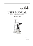
The slit lamp is an instrument consisting of a high-intensity light source
that can be focused to shine a thin sheet of light into the eye. It is used
in conjunction with abiomicroscope. The lamp facilitates an examination
of the anterior segment, or frontal structures and posterior segment, of
the human eye, which includes the eyelid, sclera,conjunctiva, iris, natural
crystalline lens, and cornea. The binocular slit-lamp examination provides
stereoscopic magnified view of the eye structures in detail, enabling
anatomical diagnoses to be made for a variety of eye conditions.
While a patient is seated in the examination chair, he rests his chin and
forehead on a support to steady the head. Using the biomicroscope, the
ophthalmologist oroptometrist then proceeds to examine the patient's eye.
A fine strip of paper, stained with fluorescein, a fluorescent dye, may be
touched to the side of the eye; this stains the tear film on the surface of the
eye to aid examination. The dye is naturally rinsed out of the eye by tears.
A subsequent test may involve placing drops in the eye in order to dilate
the pupils. The drops take about 15 to 20 minutes to work, after which the
examination is repeated, allowing the back of the eye to be examined.
Patients will experience some light sensitivity for a few hours after this
exam, and the dilating drops may also cause increased pressure in
the eye, leading to nausea and pain. Patients who experience serious
symptoms are advised to seek medical attention immediately.
Adults need no special preparation for the test; however children may need
some preparation, depending on age, previous experiences, and level of
trust.
The slit lamp exam may detect many diseases of the eye, including:
● Cataract
● Conjunctivitis
● Corneal injury such as corneal ulcer or corneal swelling
● Diabetic retinopathy
● Fuchs' dystrophy
● Keratoconus (Fleischer ring)
● Macular degeneration
● Presbyopia
● Retinal detachment
● Retinal vessel occlusion
● Retinitis pigmentosa
● Sjögren's syndrome
● Uveitis
● Wilson's disease (Kayser-Fleischer ring)
5-projector
5-air puff
An air puff type tonometer having a source for projecting a light beam
toward the cornea of an eye to be tested, an alignment sensor for
receiving the light beam after reflection from the cornea and providing a
corresponding received light signal, a device for detecting optical alignment
based on the received light signal, a device for directing an air puff, a
device for providing an applanation signal corresponding to the applanation
state of the cornea when the cornea is applanated by an air puff upon
the cornea, an A/D converter for converting the applanation signal in
accordance with a preset comparison reference value, and a device for
setting the comparison reference value based on quantity of light received
by the alignment sensor.
Puff tonometry or "non-contact" tonometry is definitely still used, especially
in optometric practices. It is a good screening test but can sometimes
overestimate pressures. Not as accurate as goldman tonometry but very
sensitive in picking up pressure problems. Glaucoma, however takes much
more than pressure to diagnose and up to 30% of people with glaucoma
can have normal pressures. By the way the test will not hurt the eye and
glaucoma can occur at any age but much more common as you get older.
1-audiometer
An audiometer is a machine used for evaluating hearing loss. The
invention of this machine is generally credited to Dr. Harvey Fletcher
ofBrigham Young University. Audiometers are standard equipment at
ENT clinics and in audiology centers. They usually consist of an embedded
hardware unit connected to a pair of headphones and a feedback button,
sometimes controlled by a standard PC. Audiometer requirements and
the test procedure are specified in IEC 60645, ISO 8253, and ANSI S3.6
standards. Audiometer were originally invented byAlexander Graham Bell
An alternative to hardware audiometers are software audiometers, which
are available in many different configurations.
Screening PC-based audiometers use a standard computer and can be
run by anybody in their home to test their hearing, although their accuracy
is not as high due to lack of a standard for calibration. Some of these
audiometers are even available on a handheld Windows driven device.
2-Tympanometer
http://www.afhcan.org/manuals/PUB-114%20Rev%20B%20v.pdf
hospital design
www2.doh.gov.ph/BHFS/planning_and_design.pdf
1-fetal dopplar
Invented in 1958 by Dr. Edward H. Hon a Doppler fetal monitor or
Doppler fetal heart rate monitor is a hand-held ultrasound transducer
used to detect the heart beat of a fetus for prenatal care. It uses the
Doppler effect to provide an audible simulation of the heart beat. Some
models also display the heart rate in beats per minute. Use of this monitor
is sometimes known asDoppler auscultation. A common name for the
device is simply "Doppler;" plural is "Dopplers."
A Doppler fetal monitor provides information about the fetus similar to the
information a fetal stethoscope provides. One advantage of the Doppler
fetal monitor over an acoustic (not electronic) fetal stethoscope is the audio
output, which allows people other than the user to listen to the heartbeat.
One disadvantage is the greater complexity and cost, and lower reliability,
of an electronic device.[citation needed]
Originally intended for use by health care professionals, this device is
becoming popular for personal use.
Fetal heart rates
Starting at week 5 the fetal heart will accelerate at a rate of 3.3 beats per
day for the next month.
The fetal heart begins to beat at approximately the same rate as the
mothers, which is 80 to 85 bpm. Below illustrates the approximate fetal
heart rate for weeks 5 to 9, assuming a starting rate of 80
● Week 5 starts at 80 and ends at 103 bpm
● Week 6 starts at 103 and ends at 126 bpm
● Week 7 starts at 126 and ends at 149 bpm
● Week 8 starts at 149 and ends at 172 bpm
● At week 9 the fetal heartbeat tends to beat within a range of 155 to
195 bpm.
The fetal heart rate will begin to decrease and generally will fall within
the range of 120 to 160 bpm by week 12
EEG
http://en.wikipedia.org/wiki/Electroencephalography
EMG
http://en.wikipedia.org/wiki/Electromyography
autoclave
An autoclave is a device to sterilize equipment and supplies by subjecting
them to high pressure steam at 121 °C or more, typically for 15 to 20
minutes depending on the size of the load and the contents. It was invented
by Charles Chamberland in 1879, although a precursor known as the
steam digester was created by Denis Papin in 1679. The name comes from
Greek auto, ultimately meaning self, and Latin clavis meaning key — a selflocking device
Syringe Pump
a mechanical device that moves fluid or gas by pressure or suction shoe | a
low-cut shoe without fastenings
Syringe pump is designed to deliver drug at a predetermined rate
and speed. In the recent years, pharmaceutical companies have
developed more and more concentrated and effective medicine.
Hence these medicines are required to be injected very slowly as
well as continuously. Syringe pumps are particularly helpful under
such circumstances as they are programmed to do deliver drug
through the vein at a determined rate.
Syringe pump generally consist of a drum that is attached to a
piston. The piston is operated by a motor through a drive screw
or worm gear which helps in pushing the plunger of syringe in
or out resulting in a smooth flow. The syringe is engaged on a
clamp on the frame and the plunger of the syringe is displaced
by movement of drum. Most of the syringe pump can work
with different syringes of different diameter, but the diameter
has to be entered in beginning to make sure correct volume is
dispensed. These guidelines should be read from manufacturer
guidelines, and made sure whether syringes with different
diameter can be used. The user can set the parameters such as
flow rate, dispense volume and syringe diameter.
Syringe pumps can deliver drugs even in very small doses, 0 .1ml
per hour to 200 ml per hour. The accuracy is mentioned by the
manufacturer in their user manual and will ideally be mentioned
in terms of flow rate i.e. over the entire time of delivering the
drug, the variation won’t be more than the prescribed limit.
These pumps have a smooth delivery over the entire period.
Some syringe pumps accuracy might appear in terms of
displacement of piston. These pumps will deliver the determined
dose over the time, but the rate of flow might not be smooth.
More than one syringe pump is also used if there is a need to
deliver more than one drug at same time.
The most popular use of syringe drivers is in palliative care, to continuously
administer analgesics (painkillers), anti emetics (medication to suppress
nausea and vomiting) and other drugs. This prevents periods during which
medication levels in the blood are too high or too low, and avoids the use of
multiple tablets (especially in people who have difficulty swallowing). As the
medication is administered subcutaneously, the area for administration is
practically limitless, although edema may interfere with the action of some
drugs.
infusion pump
Infusion pumps are devices that are used to deliver therapeutic
fluids which can be either medication or nutrients at a
predetermined rate. Over the years there has been lot of
research on these pumps resulting in several varieties of pumps
giving more and more control on the volume and time of fluid to
be delivered. The most common used method of delivering these
fluids is intravenous, but in particular requirements therapist
might prefer epidural or subcutaneous. There are various types
of infusion pump designed with a view to serve different purpose
of therapy but it is important that the pumps selected can deliver
the fluids at desired rate and volume. Overall they can be divided
in two sub groups i.e. electronically operated infusion device and
Gravity operated infusion device. Electronic operated infusion
device has a microchip that controls the delivery of fluid while
gravity controlled device have regulator that operates as clamp to
vary the flow rate.
Types of infusion
● Continuous infusion usually consists of small pulses of infusion, usually
between 500 nanoliters and 10000 microliters, depending on the pump's
design, with the rate of these pulses depending on the programmed
infusion speed.
● Intermittent infusion has a "high" infusion rate, alternating with a low
programmable infusion rate to keep the cannula open. The timings are
programmable. This mode is often used to administer antibiotics, or other
drugs that can irritate a blood vessel.
● Patient-controlled is infusion on-demand, usually with a preprogrammed
ceiling to avoid intoxication. The rate is controlled by a pressure pad or
button that can be activated by the patient. It is the method of choice
for patient-controlled analgesia (PCA), in which repeated small doses
of opioid analgesics are delivered, with the device coded to stop
administration before a dose that may cause hazardous respiratory
depression is reached.
● Total parenteral nutrition usually requires an infusion curve similar to
normal mealtimes.
Some pumps offer modes in which the amounts can be scaled or
controlled based on the time of day. This allows for circadian cycles which
may be required for certain types of medication.
monitors
1.BLOOD PRESSURE MONITORS
Blood Pressure - The force applied per unit area is called
pressure. Similarly, blood pressure is defined as the force of
blood on per unit area of blood vessel. Blood pressure is usually
measured in the right arm in the brachial artery and is measured
in millimeter of mercury. The variability in the blood pressure
leads to two readings: the systolic BP and diastolic BP. Systolic
pressure is the pressure exerted by blood on the blood vessel
which occurs when the heart contracts or expands and is the
peak pressure. Diastolic pressure is the pressure reading that
occurs when the heart is relaxing and thus is a lower reading.
Blood pressure is measured by invasive and non-invasive
methods.
The
invasive
method
requires
intra-arterial
catheterization and is the most accurate method, but this
method is not done routinely due to its invasive nature. Hence
the standard practice of monitoring the blood pressure is noninvasive nature and all the blood pressure monitors discussed
hereafter are non – invasive.
Blood Pressure Monitors : Till late 1990’s mercury filled
sphygmomanometer were maximally used for measuring
the blood pressure even in intensive care settings. It is
still the obvious choice for measuring BP in office practice.
The older versions of mercury filled sphygmomanometer
would with breakage lead to mercury spills that are toxic
and can be hazardous. Thereafter modern mercury filled
sphygmomanometers have been introduced in market which do
not spill mercury in case of breakage.
Further advancements have brought aneroid monitors and
electronic monitors, which again are non-invasive. Finger
monitors and wrist monitors are also new in the Blood pressure
monitors category, but all have their own pros and cons.
Mercury Sphygmomanometer – It has been one of the oldest
blood pressure monitor, and includes a cuff that can be inflated
or deflated. A mechanical bulb assists in inflating bulb while
deflating is done through a valve. The reading is taken through
mercury filled in glass tube.
Aneroid Blood Pressure Monitors – Aneroid blood pressure
monitor includes a cuff, mechanical bulb, deflating valve
and a round monitor. The basic difference in mercury
sphygmomanometer and aneroid manometer is the round
monitor that replaces the mercury column. This monitor has a
round dial covered with glass and a metal spring that gives the
reading.
Both aneroid and mercury BP apparatus require a stethoscope for
reading the BP.
Digital Blood Pressure Monitors – This model again contains cuff
but have semi automatic and fully automatic models. The semi
automatic models has to be inflated manually and deflation is
automatic while the fully automatic model has is inflated as well
as deflated automatically Digital blood pressure monitors come
with an inbuilt LCD screen to show the readings and hence a
stethoscope is not required. The concern in this device is to hold
the body at a specific position or irregular heart rate that can
change the reading. These models are used in Neonatal Intensive
Care Units and Pediatric Intensive Care Units.
Cuff requirements: Selection of right cuff size for measuring
blood pressure e is always important. A smaller cuff size would
lead to falsely elevated BP and a larger cuff would lead to falsely
lower BP. The ideal cuff should cover 2/3 rd the length of the arm
and should be 20% wider than the diameter of the limb. The cuff
should encircle atleast 75% of the arm and bladder should overlie
the artery.
Wrist
and
Finger
Blood
pressure
Monitors
–
Wrist
blood
pressure monitor can be used around the wrist and the button
automatically inflates the cuff. The reading is than shown on the
LCD panel.
Finger blood pressure monitor can be used by in inserting the
index finger in the adjustable cuff. The automatic cuff fits to size
and inflates and shows the reading LCD panel.
The disadvantage with Wrist and Finger blood pressure monitor is
its sensitiveness to body movement and temperature.
Choice of blood pressure monitor: The choice of Blood pressure
Monitor often depends on the accuracy, ease of use, and the
price, but it is necessary to have a proper understanding of any
instrument that is used. Also it is important to understand the
limitations of each device as well as having a proper training to
use it.
2.MULTIPARAMETER MONITOR
The multiparameter monitors are designed to give number of
information on one screen and hence provides multiple
information that is needed to understand the patient condition.
It
has emerged as a monitor to offer flexible solution for varying
critical care need.
They provide a comprehensive understanding of patient by giving
a
broader monitoring scope. These monitors provide reading such
as
heart rate, central venous pressure, non-invasive blood pressure,
ECG, SpO2, PaCO2 and invasive blood pressure and temperature
. The
monitor has alarm where the parameters can be set and theecar
giver will be alerted for change beyond the set parameter.
Mechanical Ventilato
r
A machine that helps in the process of breathing mechanically by
helping in movement of air into and out of the lungs is called as
mechanical ventilator. It helps to sustain breathing in a patient
who is unable to breathe properly (has respiratory failure).
Ventilators are of 2 types: positive pressure ventilators and
negative pressure ventilators. Negative pressure ventilators (iron
lung machines are no longer used now).
Absolute
indication for
intubation
and
mechanical
ventilation
are:
●
Emergency
●
Apnea and
severe
irregularity of
spontaneous
breathing
●
Severe
respiratory
failure
●
Ineffectiveness
of oxygen
therapy by
mask, CPAP or
non- invasive
ventilation
Advantages
of
mechanical
ventilation:
●
●
●
●
Correct ventilation of both lungs and progressive
improvement of lung pathology
Improvement of oxygenation and reduction of hypercapnia
Reduction of respiratory fatigue and oxygen consumption
Protection of airways and efficacious bronchosuctioning if
tracheal intubation is performed.
Modes of ventilation: Mechanical ventilation using positive
pressure can need airway invasion using an endotracheal
tube or, as is being more frequently seen, non-invasion of
airways can be performed (use of facial and nasal masks).
CPAP, Pressure or Volume Support Ventilation) or ventilation
can be totally or partially controlled (Volume and Pressure
Controlled Ventilation, Synchronized Intermittent Mandatory
Ventilation). Each model has precise indications which allow
better application on the one hand, while on the other avoid side
effects.
Continuous Positive Airway Pressure (CPAP): CPAP is
a mode of ventilation, which enables the elevation of endexpiratory pressure to levels above atmospheric pressure to
increase total lung volume, and functional residual capacity, thus
favoring improved oxygenation.
Pressure Support Ventilation (PSV): Pressure support
ventilation (PSV) is designed to support spontaneous breaths
during inspiratory phase. It is primarily designed to assist
spontaneous breathing and therefore the patient should have an
intact respiratory drive. Cycles are pressure limited and there is
no pre-set tidal volume.
Volume Support Ventilation (VSV): The ventilator, breath
by breath, adapts inspiratory pressure support to changes
in the mechanical properties of the lung and the thorax in
order to ensure that the lowest possible pressure is used to
deliver pre-set tidal and minute volume that remain constant.
Controlled Mechanical Ventilation (CMV): This mode of
ventilation controls the patient's respiratory activity completely.
Introduction of gases into the lung, inspiration, is obtained
using positive pressure, which pushes gases into the lung.
SPECIAL MODES OF VENTILATION:
1. High Frequency Ventilation – HFV: The most fundamental
difference between high frequency ventilation (HFV) and
intermittent positive pressure ventilation (IPPV) is that with HFV
the tidal volume (Vt) required is approximately 1-3 ml/kg body
weight, compared with 6-10 ml/kg with intermittent positive
pressure ventilation (IPPV). The increase in ventilation rate to
frequencies of 60 b.p.m. or more in HFV is obviously mandatory if
even comparable minute volume ventilation is to result.
http://www.pediatriconcall.com/fordoctor/
diseasesandcondition/PEDIATRIC_EMERGENCIES/
mechanical_ventilation.asp
Incubators
1.RADIANT HEAT WARMER /
INTENSIVE CARE WARMER
Radiant Warmer, is a body warming device to provide heat to
the body. This device helps to maintain the body temperature of
the baby and limit the metabolism rate. Heat has a tendency to
flow in the heat gradient direction that is from high temperature
to low temperature. The heat loss in some newborn babies is
rapid; hence body warmers provide an artificial support to keep
the body temperature constant. In certain areas with very cold
climate, babies are kept on Radiant Warmer for couple of hours
immediately after birth to ensure the baby is stabilized after
birth.
Radiant Warmers consists of an open tray (where the baby
is kept) and the artificial heating is provided by a heating
mechanism mounted overhead. The heating mechanism consists
of quartz which produces the desired heat and a reflecting
mechanism to divert the heat at the baby tray. The skin
temperature of the baby can be monitored by a temperature
measuring knob that is kept continuously attached to the body.
The variation in the skin temperature can be seen on a small LCD
panel which continuously shows the body temperature. Radiant
warmers are equipped with alarm to indicate the change in
temperature and hence attract attention of medical professional
attending the baby. The heat generated can be controlled
manually by a knob as well as automatically depending on the
Radiant Heat Warmer.
Radiant Warmers can be manual or automatic (servo system
– heater output is determined automatically based on skin
temperature. The skin temperature is set at 36.5 degree
Celsius) depending on the mechanism that the manufacturer
employs for temperature control. The heat generated and the
temperature of the skin can be individually seen but the basic
difference between these two models will be the regulation of
temperature. The automatic model increases the heat output in
small predetermined steps to reach at the desired temperature
of the body. The device may seem simple to handle, but it is
always recommended to have a proper training and read the
manufacturers guidelines for person handling this equipments.
It is necessary to regularly clean and disinfect the instrument.
2.INCUBATOR
Incubators are device that provides sufficient warmth to the
body to
maintain a desired temperature. Premature babies have very
s fat les
around them and loose heat rapidly to the surrounding
environment. The incubator plays an important role in
maintaining the small environment of desired temperature which
minimizes the heat loss. Once the heat loss is reduced, the
nutrition given to premature babies will be utilized in organ
development and weight gain.
Incubators consist of the baby tray that is enclosed in a box
like
structure to provide a fix warm environment. The box is generall
y made
of fibre glass or acrylic which is transparent and the heating
mechanism is placed below the tray. The heat generated by
heating
mechanism is not used directly to heat the body. This heatused
is to
warm the air mixture which is then circulated in the closed
environment around the baby. The temperature of the air as
as the
well
baby is indicated on panels and the temperature control can be
automatic as well as manual based on the incubators. Incubators
are armed with alarms to derive attention for temperature
change. Incubators are available with single wall and double wall,
and the selection can depend on the environment temperature in
which it is to be used.
There is no specific time frame for which a baby has to be kept
in an Incubator, and the choice varies on case to case basis
depending on how premature the baby and the weight of baby.
Hence the time for which a baby is kept in an incubator can vary
from couple of days to weeks and requires handling by trained
professionals. Hence the universal rule that a baby should be
kept in an incubator only as along as it is needed in the best time
frame. Precaution should be taken while removing the baby from
the incubator, so as not to move the baby immediately from a
comfortable warm environment to a cold environment resulting in
a high temperature gradient. Hence when the baby starts gaining
weight, it is a practice to gradually reduce the temperature of the
incubator.
The cost and the heat lost can be determining factor between
selection of warmer and incubators. When it comes to critical
care, such as low birth weight, incubators have been preferred
while radiant warmers have been preferred to prevent heat
loss in normal newborn babies. It is recommended to clean and
disinfect the instrument regularly to avoid infection. Also it is
necessary to have a proper training and read the manufacturers
guidelines for person handling these equipments.
3.TRANSPORT INCUBATOR
The purpose of transport incubator is same to an incubator
used in Intensive Care Unit. It is used to provide a desired
warm environment to a neonate to reduce in heat loss to the
environment. There are situations when a neonate or baby has
to
be transferred via air or road from one place to other either in
search of an intensive care unit or to a different hospital for a
different therapy itself. As these neonates or baby are already
weak by nature, and when they are ill their condition becomes
even more delicate. Under this circumstance, the most
challenging part of critical care is to transfer the baby without
exposing them to temperature gradient of the environment. This
is
achieved by transferring the baby in a transport incubator which
maintains a small environment of required temperature
.
The basic mechanism is same of transport incubator is same as
the
intensive care incubator. A special attention is given to makeesur
that the device is relatively light and small which helps in
movement of device. A baby tray is present on a trolley which is
covered by a fibre glass or acrylic structure to provide a closed
environment for the baby. Openings are provided so that medical
staff
can access the baby whenever required and separate openings
are
provided to insert oxygen tube etc. Temperature required can be
set and the skin temperature of baby as well as environment
temperature is displayed in screen provided. The airflow can be
regulated by a knob and these systems have alarm to derive
attention in undesired circumstances such as rise in temperature.
The major difference from an intensive care incubator is the
presence of an external portable power source (mounted on the
transport incubator trolley) which makes the instrument work
when the instrument is moved. These instruments can also work
on electricity derived from ambulance.
BY Eng.Mohamed Salah
Thanks..


