1
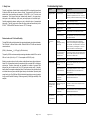
Introduction
C ontents
Introduction. . . . . . . . . . . . . . . . . . . . . . . . . . . . . . . . . . . . . . . . . . . . . . . . . . . . . . . . 2
Storage and Stability.. . . . . . . . . . . . . . . . . . . . . . . . . . . . . . . . . . . . . . . . . . . . . . . . 2
Applications. . . . . . . . . . . . . . . . . . . . . . . . . . . . . . . . . . . . . . . . . . . . . . . . . . . . . . . 2
Kit Contents. . . . . . . . . . . . . . . . . . . . . . . . . . . . . . . . . . . . . . . . . . . . . . . . . . . . . . . 3
Before Starting. . . . . . . . . . . . . . . . . . . . . . . . . . . . . . . . . . . . . . . . . . . . . . . . . . . . . 3
A. SE Blood DNA Kit Protocol (for up to 300ìl whole Blood). . . . . . . . . . . . . . . . . . 4
B. SE Blood DNA Kit Protocol (for 0.3-1 ml whole Blood) . . . . . . . . . . . . . . . . . . . . 6
C. Buffy Coat. . . . . . . . . . . . . . . . . . . . . . . . . . . . . . . . . . . . . . . . . . . . . . . . . . . . . . 7
The E.Z.N.A.® SE Blood DNA Kit provides a rapid and easy method for the isolation of
genomic DNA from 1 ìl-1ml fresh, frozen, and anticoagulated whole blood. The kit
allows single or multiple, simultaneous processing of samples in less than 30 minutes.
Normally, up to 300 ìl of whole blood can be used in a single experiment. There is no
need for phenol-chloroform extractions, and time-consuming steps such as CsCl
gradient ultracentrifugation, and precipitation with isopropanol or ethanol, are
eliminated. DNA purified using the E.Z.N.A.® SE Blood DNA method is ready for
applications such as PCR*, Southern blotting, and restriction digestion.
The E.Z.N.A.® SE Blood DNA Kit uses the reversible nucleic acid-binding properties
of Omega Bio-Tek’s HiBind® matrix, combined with the speed of mini-column spin
technology. A specially formulated buffer system allows genomic DNA up to 60 kb to
bind to the matrix. Samples are first lysed under denaturing conditions and then applied
to the HiBind® DNA spin columns to which DNA binds, while cellular debris, hemoglobin,
and other proteins are effectively washed away. High quality DNA is finally eluted in
sterile deionized water or low salt buffer.
Determination of DNA Yield and Quality. . . . . . . . . . . . . . . . . . . . . . . . . . . . . . . . . 7
Troubleshooting Guide. . . . . . . . . . . . . . . . . . . . . . . . . . . . . . . . . . . . . . . . . . . . . . . 8
Storage and Stability
All components of the E.Z.N.A.® SE Blood DNA Kit should be stored at 22o C-25o C,
expect that RNase A should be stored at -20o C. Under these conditions, DNA has
successfully been purified and used for PCR after 24 months of storage. Under cool
ambient conditions, a precipitate may form in Buffer BL. In case of such an event, heat
the bottle at 37oC to dissolve.
Expiration Date: All E.Z.N.A.® SE Blood DNA Kit components are guaranteed for at
least 24 months from the date of purchase when stored at 22-25oC
Applications
The E.Z.N.A.® SE Blood DNA Kit don’t require Proteinase K or OB Protease to digest
samples. This kits is suitable for isolation of genomic DNA from whole blood, purified
leukocytes and some animal cells. Some samples may need to add Proteinase K to
increase the yield. At this conditions, you may choose E.Z.N.A.® Blood DNA Kit
(D3392). For recovery of DNA from dried blood spots use the E.Z.N.A.® Forensic DNA
Kits (D3591) or order Buffer TL (TL-100, 100 ml) to use with the Blood DNA Kit. For
isolation of DNA or Viral DNA from blood or other body fluids, use the E.Z.N.A.® Blood
DNA Kit.
*
The P C R process is covered by U .S . P atents 4,683,195 and 4,683,202
(and international equivalents) owned by H offm ann-LaR oche, Inc.
P age 2 of 8
A. SE Blood DNA Kit Protocol (Up to 300ul whole blood)
Kit Contents
Product Number
D3471-00
D3471-01
D3471-02
Purification times
5 Preps
50 Preps
200 Preps
HiBind DNA columns
5
50
200
2 ml Collection Tubes*
5
50
200
Buffer ERL (10x)
3 ml
25 ml
70 ml
Buffer WTL
2 ml
20 ml
60 ml
Buffer BL
2 ml
20 ml
60 ml
DNA Wash Buffer Concentrate
2 ml
20 ml
3 x 20 ml
RNase A
20 ìl
150 ìl
500 ìl
Buffer HB
3 ml
30 ml
110 ml
Elution Buffer
2 ml
15 ml
60 ml
User Manual
1
1
1
®
Materials to be Provided by User
!
Tabletop microcentrifuge and nuclease-free 2 ml tubes
!
Water bath - set to 65oC
!
Incubator or heat block - preset to 37oC
!
Absolute Ethanol (96%-100%)
!
Optional: Proteinase K (20mg/ml)
1.
Transfer the blood sample to a 2 ml nuclease-free micro-centrifuge tube. Add 3
volume of 1 x Buffer ERL and mix by inverting the tube a few times. Incubate
5 minutes at room temperature. Invert the tube several times during the
incubation.
NOTE: Buffer ERL is supplied as a 10 x concentrate and must be diluted with
ddH2O according to bottle label or ‘before use’ on Page 3.
2.
Centrifuge at 14,000 x g for 30 seconds at room temperature. Remove and
discard as much supernatant as possible without disturbing the visible white pellet.
Leave about 10ìl of residue liquid in the tube.
Note: If some red blood cells or cell debris are still visible along with the white
blood cell pellet, resuspend the white blood cell pellet and mix with 2 volumes 1
x Buffer ERL. Incubate at room temperature for 2 minutes. Then pellet the white
blood cells by repeating Step 2.
3.
Vortex to resuspend cells in residue liquid and add 240 ìl Buffer WTL to the
Buffer BL contains a chaotropic salt. Use gloves and
protective eyeware when handling this solution.
Before Starting
tube. Pipet up and down or vortex at maxi speed for 30 seconds to lyse the cells.
The solution should become very viscous. If cell clumps are visible after mixing,
IMPORTANT
1
2
Dilute 10 x Buffer ERL as follows and store diluted 1 x
Buffer ERL at room temperature.
D3471-00
Add 27 ml ddH2O
D3471-01
Add 225 ml ddH2O in a new bottle
D3471-02
Dilute with 630 ml ddH2O in a new bottle
incubate the solution at 37oC until the clumps cannot be seen.
4.
(Optional for maximum yield) Add 3 ìl Proteinase K (20mg/ml) to the cell
lysate and vortex to mix well. Incubate at 55oC for 10 -30 minutes.
5.
Add 2 ìl RNase A solution to the cell lysate and mix well. Incubate the
mixture at room temperature for 10 minutes.
6.
Add 250ìl Buffer BL and mix throughly by vortexing, Incubate at 65o C for
10 min. Vortex to mix twice during incubation.
7.
Add 250 ìl absolute ethanol to the lysate and mix completely by vortexing.
If precipitation can be seen at this point, break the precipitation by pipetting up and
down 10 times.
DNA Wash Buffer Concentrate must be diluted with
absolute ethanol as follows and store at room temperature:
D3471-00
Add 8 ml absolute ethanol (96-100%)
D3471-01
Add 80 ml absolute ethanol to bottle
D3471-02
Add 80 ml absolute ethanol to each bottle
All centrifugation steps must be carried out at room temperature.
Page 3 of 8
P age 4 of 8
8.
Insert a HiBind® DNA column in a 2 ml collection tube (provided). Load the
lysate from Step 7 (including any precipitation) into the column and
centrifuge at $8,000 x g for 1 min to bind DNA. Discard flow-through and reuse the collection tube.
9.
Insert the column back into the 2 ml collection tube and wash by adding 500ìl
HB Buffer. Centrifuge at $8,000 x g for 1 min. Discard flow-through liquid and
re-use the 2 ml collection tube.
10.
Insert the column back into the 2 ml collection tube and wash by adding 650 ìl
of DNA Wash Buffer diluted with ethanol. Centrifuge at $8,000 x g for 1 min.
Discard flow-through liquid and re-use the collection tube in the next step.
N ote: D N A W ash B uffer is provided as a concentrate and m ust be diluted with absolute
B. SE Blood DNA Kit Protocol for 400ìl -1 ml whole Blood
Materials Supplied by User
!
Microcentrifuge capable of at least 13,000 x g
!
Table top centrifuge capable of 2000 x g with rotor for 15 ml tubes
!
Water bath or heat block preset at 37EC
!
Nuclease-free 1.5 ml microcentrifuge tube
!
Optional: Proteinase K (20mg/ml)
1.
Add up to 1 ml whole blood sample into a 15 ml tube. Add 3 volume of 1 X
Buffer ERL and mix throughly by inverting the tube a few times. Incubate 5
minutes at room temperature. Invert the tube several times during the incubation.
NOTE: Buffer ERL is supplied as a 10 x concentrate and must be diluted with
ddH2O according to bottle label or Page 3 before use.
ethanol as indicated on the bottle label or P age 3. If refrigerated, the diluted w ash buffer
m ust be brought to room tem perature before use. R efrigeration is NO T recom m ended.
11.
Insert the column back into the 2 ml collection tube and wash by adding another
650 ìl of DNA Wash Buffer diluted with ethanol. Centrifuge at $8,000 x g for
1min. Discard flow-through liquid and re-use the collection tube in the next step.
12.
Insert the column back into the empty 2 ml collection tube, centrifuge the empty
column at maximum speed ($13,000 x g) for 2 min to dry the column
membrane.
2.
Centrifuge at 2,000 x g for 5 minutes at room temperature. Remove and
discard as much supernatant as possible without disturbing the visible white pellet.
Leave about 20ìl of residue liquid in the tube.
Note: If some red blood cells or cell debris are still visible along with the white
blood cell pellet, resuspend the white blood cell pellet and mix with 2 volumes 1
x Buffer ERL. Incubate at room temperature for 2 minutes. Then pellet the white
blood cells by repeating Step 2.
3.
Vortex to resuspend cells in residue liquid and then add 230 ìl Buffer WTL.
Pipet up and down or vortex at maxi speed for 30 seconds to lyse the cells. The
solution should become very viscous. If cell clumps are visible after mixing,
incubate the solution at 37EC until the clumps cannot be seen.
4.
Optional for maximum yield: Add 3 ìl Proteinase K (20mg/ml) to the cell
lysate and vortex to mix well. Incubate at 55oC for 30 minutes.
This step is critical for rem oval of trace ethanol that m ight otherw ise interfere w ith
dow nstream applications.
13.
Place the column into a nuclease-free 2 ml microfuge tube and add 50-100
ìl of preheated (70o C) Elution Buffer. Allow column to set for 5 min at room
temperature. (For higher yields, incubate 5 min at 60oC rather than at room
temperature.)
14.
To elute DNA from the column, centrifuge at 10,000 x g for 1 min. Retain
flow-through containing the DNA.
5.
Add 2ìl RNase A solution to the cell lysate and mix well. Incubate the mixture
at room temperature for 10 minutes.
15.
Place column into a second 1.5 ml tube and repeat elution step with another
50-100 ìl of preheated Elution Buffer.
Note: Each elution typically yields 60%-70% of the DNA bound to the column.
Thus two elutions generally yield >90%. However, increasing elution volume
reduces the concentration of the final product. To obtain DNA at higher
concentrations, elution can be carried out using two of 50 ìl Elution Buffer.
Volumes lower than 50 ìl greatly reduce yields. Alternatively, use the first
eluate to perform the second elution.
6.
Add 250ìl Buffer BL and mix throughly by vortexing. Incubate at 65oC for 10
min. Vortex to mix twice during incubation.
7.
Add 250 ìl absolute ethanol to the lysate and mix immediately by vortexing.
If precipitation can be seen at this point, break the precipitation by pipetting up and
down 10 times.
8.
Following Protocol A step 8-15 on page 5.
Page 5 of 8
P age 6 of 8
Troubleshooting Guide
C. Buffy Coat
The buffy coat fraction of whole blood is enriched with WBC, and usually gives at least
5-fold more DNA than the same volume of blood. To prepare buffy coat from fresh
whole blood, simply centrifuge the sample at 3,000-4,000 x g for 10 min at room
temperature. Three layers should be obtained with plasma in the upper layer,
leukocytes in the middle layer (buffy coat), and erythrocytes in the bottom layer.
Carefully aspirate the plasma, making sure not to disturb the layer of concentrated
leukocytes. The buffy coat can be drawn off with a pipette and used directly in the
E.Z.N.A.® SE Blood DNA Protocol or frozen at -70oC for storage.
P roblem
P ossible C ause
S uggestions
C logged C olum n
Incom plete lysis
A dd the correct volum e of B uffer BL and
incubate for specified tim e at 70 o C . It m ay be
necessary to extend incubation tim e by 10 m in.
S am ple too large
R educe the sam ple volum e.
S am ple too viscous
Increase the volum e of all buffer proportionally
C logged colum n
S ee above
P oor elution
R epeat elution or increase elution volum e
Im proper washing
D N A W ash B uffer C oncentrate m ust be diluted
with absolute ethanol
E xtended
centrifugation during
elution step.
R esin from the colum n m ay be present in
eluate. A void centrifugation at speeds higher
than specified. The m aterial can be rem oved
from the eluate by centrifugation — it will not
interfere with P C R or restriction digests.
P oor cell lysis due to
incom plete m ixing
with B uffer W TL
R epeat the procedure, this tim e m aking sure to
vortex the W BC pellet with E RL residue in the
tube before m ix B uffer W TL
H em oglobin rem ains
R epeat the R B C lysis step with step 1-3
im proper m ixing with
B uffer B L.
M ix thoroughly with B uffer B L prior to loading
H iB ind™ colum n.
A bsolute ethanol not
added to B uffer B L.
B efore applying sam ple to colum n, an aliquot
of B uffer B L/ethanol m ust be added. S ee
protocol above.
N o ethanol added to
W ash B uffer
C oncentrate.
D ilute W ash B uffer with the indicated volum e of
absolute ethanol before use.
im proper m ixing with
B uffer B L.
B uffer B L is viscous and the sam ple m ust be
N o ethanol added to
D ilute W ash B uffer with the indicated volum e of
W ash B uffer
absolute ethanol before use.
S am ple too large.
R educe sam ple volum e and follow directions
Low D N A Yield
Determination of Yield and Quality
The total DNA yield can be determined by a spectrophotometer using deionized water,
Tris-HCl buffer, or Elution Buffer as blank. Dilute the DNA in TE buffer and calculate
concentration as:
Low A 2 6 0 /A 2 8 0
ratio
[DNA] = (Absorbance260) x (0.05 ìg/ìl) x (Dilution factor)
The quality of DNA can be assessed by measuring absorbance at both 260 nm and at
280 nm. A ratio of (A260/A280) of 1.7-1.9 corresponds to 85%-95% purity.
Blood is a complex mixture of cells, proteins, metabolites and many other substances.
About 50% of human blood volume is comprised of cells, more than 99% of which are
erythrocytes. Therefore, these cells are unsuitable for isolation of genomic DNA
because they don’t contain nuclei. Only 0.3% leukocytes contain nuclei. Normally,
healthy blood, for example contains fewer than 107 leukocytes per 1ml, while blood from
an infected donor may have a tenfold higher leukocyte concentration. Expected yields
range from 4 ìg to 12 ìg DNA per 250 ìl whole blood, depending on source of sample,
its age, and the method of storage. Yields are generally 5-fold higher with Buffy Coat
samples.
N o D N A E luted
W ashing Leaves
C olored Residue
In C olum n
E luted M aterial
H as R ed/B rown
C olor
Page 7 of 8
P age 8 of 8
vortexed thoroughly.