1
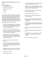
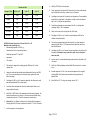
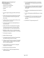
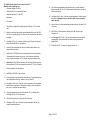
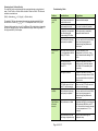
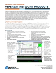
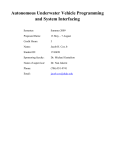
Contents
Introduction
Introduction.. . . . . . . . . . . . . . . . . . . . . . . . . . . . . . . . . . . . . . . . . . . . . . . . . . . . 2
The E.Z.N.A.™ SQ Blood DNA Kit is designed for isolating high molecular
weight genomic DNA from fresh, frozen, and anti-coagulated whole blood. The
method can also be used for preparation of genomic DNA from buffy coat, bone
marrow or cultured cells. The kit allows single or multiple, simultaneous
processing of samples in under 90 minutes. There is no need for
phenol/chloroform extractions, and time-consuming steps such as CsCl
gradient ultracentrifugation are eliminated.
Principle. . . . . . . . . . . . . . . . . . . . . . . . . . . . . . . . . . . . . . . . . . . . . . . . . . . . . . . 2
Storage and Stability. . . . . . . . . . . . . . . . . . . . . . . . . . . . . . . . . . . . . . . . . . . . . 2
DNA Yield from Various Samples.. . . . . . . . . . . . . . . . . . . . . . . . . . . . . . . . . . . 4
Kit Contents. . . . . . . . . . . . . . . . . . . . . . . . . . . . . . . . . . . . . . . . . . . . . . . . . . . . 3
DNA purified using the E.Z.N.A.™ SQ Blood DNA method is ready for
applications such as PCR*, Southern blotting, and restriction digestion.
Before Starting. . . . . . . . . . . . . . . . . . . . . . . . . . . . . . . . . . . . . . . . . . . . . . . . . . 3
Principle
A. Protocol for 100 ìL whole blood.. . . . . . . . . . . . . . . . . . . . . . . . . . . . . . . . . . 5
B. Protocol for 200 ìL whole blood. . . . . . . . . . . . . . . . . . . . . . . . . . . . . . . . . . 7
C. Protocol for 300 ìL whole blood. . . . . . . . . . . . . . . . . . . . . . . . . . . . . . . . . . 9
D. Protocol for 600 ìL whole blood. . . . . . . . . . . . . . . . . . . . . . . . . . . . . . . . . 11
E. Protocol for 800 ìL whole blood. . . . . . . . . . . . . . . . . . . . . . . . . . . . . . . . . 13
F. Protocol for 1 mL whole blood. . . . . . . . . . . . . . . . . . . . . . . . . . . . . . . . . . 15
G. Protocol for 2 mL whole blood. . . . . . . . . . . . . . . . . . . . . . . . . . . . . . . . . . 17
H. Protocol for 3 mL whole blood. . . . . . . . . . . . . . . . . . . . . . . . . . . . . . . . . . 19
I. Protocol for 4 mL whole blood. . . . . . . . . . . . . . . . . . . . . . . . . . . . . . . . . . . 21
J. Protocol for 5 mL whole blood. . . . . . . . . . . . . . . . . . . . . . . . . . . . . . . . . . . 23
K. Protocol for 6mL whole blood. . . . . . . . . . . . . . . . . . . . . . . . . . . . . . . . . . . 25
L. Protocol for 12 whole blood. . . . . . . . . . . . . . . . . . . . . . . . . . . . . . . . . . . . 27
M . Protocol for Buffy coat. . . . . . . . . . . . . . . . . . . . . . . . . . . . . . . . . . . . . . . . 29
N. Protocol for Cultured cells 0.5-1 x 10⁶.. . . . . . . . . . . . . . . . . . . . . . . . . . . . 31
O. Protocol for Cultured cells 3-5 x 10⁶ . . . . . . . . . . . . . . . . . . . . . . . . . . . . . . 33
P. Protocol for Cultured cells 3-5 x 10⁷ . . . . . . . . . . . . . . . . . . . . . . . . . . . . . . 35
Q. Protocol for 50 ìL Clotted Blood.. . . . . . . . . . . . . . . . . . . . . . . . . . . . . . . . 37
R Protocol for 1mL Clotted Blood. . . . . . . . . . . . . . . . . . . . . . . . . . . . . . . . . . 39
S. Protocol for Large volume of Clotted Blood(>1mL). . . . . . . . . . . . . . . . . . . 41
E.Z.N.A.® SQ Blood DNA Kit uses a highly efficient solution based system to
provide a convenient, fast, reliable and non-toxic method to isolate high
molecular weight genomic DNA from whole blood or buffy coat. Red blood
cells are first lysed with ERL buffer, followed by lysis of the white blood cells
and their nuclei in the WTL Buffer. Cellular proteins are removed by
precipitation and high molecular weight genomic DNA will remain in solution.
High quality genomic DNA is then purified by isopropanol precipitation.
Storage and Stability
All components of the E.Z.N.A.® SQ Blood DNA Kit should be stored at 22oC25o C. Under cool ambient conditions, a precipitate may form in the Buffer WTL.
In case of such an event, heat the bottle at 55o C to dissolve.
Expiration Date: All E.Z.N.A.® SQ Blood DNA Kit components are guaranteed
for at least 24 months from the date of purchase when stored at 22-25oC
Determination of Yield and Quality. . . . . . . . . . . . . . . . . . . . . . . . . . . . . . . . . . 43
Troubleshooting Guide. . . . . . . . . . . . . . . . . . . . . . . . . . . . . . . . . . . . . . . . . . . 44
Revised April 2008
*
T he P C R process is covered by U .S . P atents 4,683,195 and 4,683,202
(and international equivalents) owned by H offm ann-LaR oche, Inc.
Page 2 of 45
Kit Contents
Product
D5032-00
D5032-01
D5032-02
D5032-03
Total Blood Volume
10 mL
50 mL
150 mL
300 mL
ERL Buffer (10 x)
5 mL
25 mL
80 mL
2 x 80 mL
Buffer WTL
10 mL
50 mL
150 mL
300 mL
PCP Buffer
4 mL
20 mL
60 mL
120 mL
EB Buffer
5 mL
20 mL
60 mL
RNase A
50 ìL
250 ìL
1
1
User Manual
DNA Yields From Various Starting Materials
Species and Material
Amount of Starting
material
Typical Yield
Human Whole Blood
(Yield varies depending on
the quantity of white blood
cells present)
50 ìL
0.3-0.6 ìg
100 ìL
1-5 ìg
200 ìL
3-10 ìg
125 mL
300 ìL
5-15 ìg
750 ìL
1.5 mL
500 ìL
7-23 ìg
1
1
600 ìL
10-30 ìg
800 ìL
12-35 ìg
1 mL
15-48 ìg
2 mL
30-90 ìg
3 mL
50-150 ìg
4 mL
65-200 ìg
5 mL
100-300 ìg
10 mL
150-600 ìg
12 mL
200-700 ìg
from 300 ìL blood
5-15 ìg
from 2 mL blood
25-75 ìg
50 ìL
0.2-0.6 ìg
100 ìL
0.5-1.0 ìg
200 ìL
2 -5 ìg
300 ìL
4–7 ìg
Before Starting
ERL Buffer Concentrate must be diluted with
ddH2O as follows before use
IMPORTANT
D5032-00
D5032-01
D5032-02
D5032-03
Add
Add
Add
Add
45 mL water/bottle
225 mL water/bottle
720 mL water/bottle
720 mL water/bottle
Buffy Coat
NOTE: The procedures below have been optimized for use with FRESH
or FROZEN blood samples. Anti-coagulated blood, or Buffy Coat can
also be used. However, DNA yield will be reduced with time of storage.
In addition, leukocytes or cultured cells may be used with this
procedure.
Mouse Whole Blood
Cultured Cells
2 x 10⁶ cells
10-15 ìg
Guideline for Sample and Volume of Reagents
The SQ Blood DNA system is a solution based system and the protocol can be
easily modified based on the sample volume. User can change the protocol
based on the following guideline:
Page 4 of 45
Sample volume
Reagent Name
Reagent volume
1x
ERL Buffer
3x
1x
WTL Buffer
1x
1x
PCP Buffer
0.33 x
1x
Isopropanol
1x
1x
70% ethanol
1x
1x
EB Buffer
0.35 x *
* For #300µl blood volume. Volume of EB Buffer used can be adjusted depends
on the desired final concentration.
A. DNA Purification Protocol for 100ìL whole blood
Materials to be supplied by user
•
Microcentrifuge capable of 14,000 x g
•
Nuclease-free 1.5 mL microcentrifuge tubes
•
Water Bath preset at 37EC and 65EC
•
Isopropanol
•
70% ethanol
1.
Add 100ìL whole blood (or bone marrow) to a nuclease-free 1.5 mL
microcentrifuge tube containing 300ìL ERL Buffer. Mix by inverting the tube
a few times. Incubate 5 minutes at room temperature. Invert the tube
several times during the incubation.
4.
Add 100ìL WTL Buffer to the tube containing the re-suspended cells. Pipet
up and down to lyse the cells. The solution should become very viscous. If
cell clumps are visible after mixing, incubate the solution at 37EC until the
clumps cannot be seen.
5.
(Optional) Add 0.5ìL RNase A solution to the cell lysate. Mix the sample by
inverting the tube 20-25 times. Incubate the mixture at 37EC for 5-10
minutes.
6.
Cool the sample to room temperature.
7.
Add 33ìL PCP Buffer to the cell lysate.
8.
Vortex vigorously at high speed for 30 seconds to mix. Some protein clumps
may be visible after vortexing. Incubate in ice for 5 minutes.
9.
Centrifuge at max speed for 3 minutes at room temperature. The
precipitated protein will form a tight, dark brown pellet. If the pellet is not
tight or visible, incubate the tube in ice for 5 minutes and repeat Step 9.
10.
Transfer the supernatant to a new nuclease-free 1.5 mL centrifuge tube
containing 100 ìL of 100% isopropanol.
11.
Gently mix the solution by inverting the tube 30-40 times.
12.
Centrifuge at 14,000 x g for 1 minute at room temperature. DNA will be
visible as a small white pellet.
13.
Pour out the supernatant and drain the tube briefly on a clean absorbent
paper towel. Add 100ìL of 70% ethanol and invert the tube few times to
wash the DNA pellet.
14.
Centrifuge at 14,000 x g for 2 minutes at room temperature. Carefully pour
off the ethanol. Pellet may be very loose at this point, so pour slowly
and watch the pellet.
15.
Invert the tube on a clean absorbent paper towel and air dry the pellet for
10-15 minutes.
16.
Add 35 ìL of DNA rehydration solution (Buffer EB) and vortex for 1 minute
to mix.
17.
Incubate sample at 65oC for 10 min. Some samples may need to incubate at
65o C for 1 hour to rehydrate DNA.
18.
Store DNA at 2-8oC. For long-term storage, store at -20EC.
NOTE: ERL Buffer is supplied as a 10x concentrate and must be diluted
with ddH2O according to bottle label or Page 4 above before use.
2.
Centrifuge at 14,000 x g for 30 seconds at room temperature. Remove and
discard as much supernatant as possible without disturbing the visible white
pellet. Leave about 10ìL of residue liquid in the tube. If the blood sample
has been frozen, repeat Steps 1-2 until the pellet is white.
Note: If some red blood cells or cell debris are still visible along with the
white blood cell pellet, resuspend the white blood cell pellet and mix with 2
volumes ERL Buffer. Incubate 2 min at room temperature. Then pellet the
white blood cells by repeating Step 2.
3.
Vortex the tube vigorously until the white blood cells are completely resuspended.
Page 6 of 45
B. DNA Purification Protocol for 200ìL whole blood
Materials to be supplied by user
•
Microcentrifuge capable of 14,000 x g
•
Nuclease-free 1.5 mL microcentrifuge tubes
•
Water Bath preset at 37EC and 65EC
•
Isopropanol
•
70% ethanol
1.
Add 200ìL whole blood (or bone marrow) to a nuclease-free 1.5 mL
microcentrifuge tube containing 600ìL ERL Buffer. Mix by inverting the tube
a few times. Incubate 5 minutes at room temperature. Invert the tube
several times during incubation.
precipitated protein will form a tight, dark brown pellet. If the pellet is not
tight or visible, incubate the tube in ice for 5 minutes and repeat Step 9.
10.
Transfer the supernatant to a new nuclease-free 1.5 mL centrifuge tube
containing 200ìL of 100% isopropanol.
11.
Gently mix the solution by inverting the tube 30-40 times.
12.
Centrifuge at 14,000 x g for 1 minute at room temperature. DNA will be
visible as a small white pellet.
13.
Pour out the supernatant and drain the tube briefly on a clean absorbent
paper towel. Add 200ìL of 70% ethanol and invert the tube a few times to
wash the DNA pellet.
14.
Centrifuge at 14,000 x g for 2 minutes at room temperature. Carefully pour
off the ethanol. Pellet may be very loose at this point, so pour slowly
and watch the pellet.
15.
Invert the tube on a clean adsorbent paper towel and air dry the pellet for
10-15 minutes.
16.
Add 65 ìL of DNA rehydration solution (Buffer EB) and vortex for 1 minute
to mix.
17.
Incubate sample at 65oC for 10 min. Some sample may need to incubate at
65o C for 1 hour to rehydrate DNA..
18.
Store DNA at 2-8oC. For long-term storage, store at -20EC.
NOTE: ERL Buffer is supplied as a 10x concentrate and must be diluted
with ddH2O according to bottle label or Page 4 above before use.
2.
Centrifuge at 14,000 x g for 30 seconds at room temperature. Remove and
discard as much supernatant as possible without disturbing the visible white
pellet. Leave about 15ìL of residue liquid in the tube. If the blood sample
has been frozen, repeat Steps 1-2 until the pellet is white.
Note: If some red blood cells or cell debris are still visible along with the
white blood cell pellet, resuspend the white blood cell pellet and mix with 2
volumes ERL Buffer. Incubate 2 min at room temperature. Then pellet the
white blood cells by repeating Step 2.
3.
Vortex the tube vigorously until the white blood cells are completely resuspended.
4.
Add 200ìL WTL Buffer to the tube containing the re-suspended cells. Pipet
up and down to lyse the cells. The solution should become very viscous. If
cell clumps are visible after mixing, incubate the solution at 37EC until the
clumps cannot be seen.
5.
(Optional) Add 1 ìL RNase A solution to the cell lysate. Mix the sample by
inverting the tube 20-25 times. Incubate the mixture at 37EC for 5-10
minutes.
6.
Cool the sample to room temperature.
7.
Add 67ìL PCP Buffer to the cell lysate.
8.
Vortex vigorously at high speed for 30 seconds to mix. Some protein clumps
may be visible after vortexing. Incubate in ice for 5 minutes.
9.
Centrifuge at max speed for 3 minutes at room temperature. The
Page 8 of 45
C. DNA Purification Protocol for 300ìL whole blood
9.
Centrifuge at max speed for 3 minutes at room temperature. The
precipitated protein will form a tight, dark brown pellet. If the pellet is not
tight or visible, incubate the tube in ice for 5 minutes and repeat Step 9.
10.
Transfer the supernatant to a new nuclease-free 1.5 mL centrifuge tube
containing 300ìL of 100% isopropanol.
11.
Gently mix the solution by inverting the tube 30-40 times.
12.
Centrifuge at 14,000 x g for 1 minute at room temperature. DNA will be
visible as a small white pellet.
13.
Pour out the supernatant and drain the tube briefly on a clean absorbent
paper towel. Add 300ìL of 70% ethanol and invert the tube few times to
wash the DNA pellet.
14.
Centrifuge at 14,000 x g for 2 minutes at room temperature. Carefully pour
off the ethanol. Pellet may be very loose at this point, so pour slowly
and watch the pellet.
15.
Invert the tube on a clean adsorbent paper towel and air dry the pellet for
10-15 minutes.
16.
Add 100 ìL of DNA rehydration solution (Buffer EB) and vortex for 1 minute
to mix.
17.
Incubate sample at 65oC for 10 min. Some samples may need to incubate at
65oC for 1-2 hour to rehydrate DNA..
18.
Store DNA at 2-8oC. For long-term storage, store at -20EC.
Materials to be supplied by user
•
Microcentrifuge capable of 14,000 x g
•
Nuclease-free 1.5 mL microcentrifuge tubes
•
Water Bath preset at 37EC and 65EC
•
Isopropanol
•
70% ethanol
1.
Add 300ìL whole blood (or bone marrow) to a nuclease-free 1.5 mL
microcentrifuge tube containing 900ìL ERL Buffer. Mix by inverting the tube
a few times. Incubate 5 minutes at room temperature. Invert the tube
several times during incubation.
NOTE: ERL Buffer is supplied as a 10x concentrate and must be diluted
with ddH2O according to bottle label or Page 4 above before use.
2.
Centrifuge at 14,000 x g for 30 seconds at room temperature. Remove and
discard as much supernatant as possible without disturbing the visible white
pellet. Leave about 15ìL of residue liquid in the tube. If the blood sample
has been frozen, repeat Steps 1-2 until the pellet is white.
Note: If some red blood cells or cell debris are still visible along with the
white blood cell pellet, resuspend the white blood cell pellet and mix with 2
volumes ERL Buffer. Incubate 2 min at room temperature. Then pellet the
white blood cells by repeating Step 2.
3.
Vortex the tube vigorously until the white blood cells are completely resuspended.
4.
Add 300ìL WTL Buffer to the tube containing the re-suspended cells. Pipet
up and down to lyse the cells. The solution should become very viscous. If
the cell clumps are visible after mixing, incubate the solution at 37EC until
the clumps cannot be seen.
5.
(Optional) Add 1.5 ìL RNase A solution to the cell lysate. Mix the sample
by inverting the tube 20-25 times. Incubate the mixture at 37EC for 5-10
minutes.
6.
Cool the sample to room temperature.
7.
Add 100ìL PCP Buffer to the cell lysate.
8.
Vortex vigorously at high speed for 30 seconds to mix. Some protein clumps
may be visible after vortexing. Incubate in ice for 5 minutes.
Page 10 of 45
D. DNA Purification Protocol for 600ìL whole blood
9.
Add 200ìL PCP Buffer to the cell lysate.
Materials to be supplied by user
•
Microcentrifuge capable of 14,000 x g
10.
Vortex vigorously at high speed for 30 seconds to mix. Some protein clumps
may be visible after vortexing. Incubate in ice for 5 minutes.
•
Nuclease-free 2.0 mL microcentrifuge tubes
11.
•
Water Bath preset at 37EC and 65EC
•
Isopropanol
Centrifuge at max speed for 3 minutes at room temperature. The
precipitated protein will form a tight, dark brown pellet. If the pellet is not
tight or visible, incubate the tube in ice for 5 minutes and repeat Step 11.
•
70% ethanol
12.
Transfer the supernatant to a new nuclease-free 2.0 mL centrifuge tube
containing 600ìL of 100% isopropanol.
1.
Add 600ìL whole blood (or bone marrow) to a nuclease-free 2.0 mL
microcentrifuge tube containing 1.2mL ERL Buffer. Mix by inverting the tube
a few times. Incubate 5 minutes at room temperature. Invert the tube
several times during incubation.
13.
Gently mix the solution by inverting the tube 30-40 times.
14.
Centrifuge at 14,000 x g for 1 minute at room temperature. DNA will be
visible as a small white pellet.
NOTE: ERL Buffer is supplied as a 10x concentrate and must be diluted
with ddH2O according to bottle label or Page 4 above before use.
15.
Centrifuge at 14,000 x g for 30 seconds at room temperature. Remove and
discard supernatant containing lysed red blood cells. Leave behind any
supernatant that appears to contain non-lysed red blood cells..
Pour of the supernatant and drain the tube briefly on a clean absorbent
paper towel. Add 600ìL of 70% ethanol and invert the tube a few times to
wash the DNA pellet.
16.
Centrifuge at 14,000 x g for 2 minutes at room temperature. Carefully pour
off the ethanol. Pellet may be very loose at this point, so pour slowly
and watch the pellet.
17.
Invert the tube on a clean adsorbent paper towel and air dry the pellet for
10-15 minutes.
18.
Add 100 ìL of DNA rehydration solution (Buffer EB) and vortex for 1 minute
to mix.
19.
Incubate sample at 65oC for 10 min. Some samples may need to incubate at
65oC for 1-2 hour to rehydrate DNA..
20.
Store DNA at 2-8o C. For long-term storage, store at -20EC.
2.
3.
Vortex the tube vigorously until the cell pellet is completely resuspended.
4.
Add 1.2 mL ERL buffer to the tube and mix well by inverting the tube a few
times. Incubate at room temperature for 2 minutes.
5.
Centrifuge at 14000 x g for 30 seconds at room temperature. Remove and
discard as much supernatant as possible without disturbing the visible white
pellet. Leave about 15-20ìL of residue liquid in the tube. If the blood sample
has been frozen, repeat Steps 1-5 until the pellet is white.
Note: If some red blood cells or cell debris are still visible along with the
white blood cell pellet, resuspend the white blood cell pellet and mix with 2
volumes ERL Buffer. Incubate 2 min at room temperature. Then pellet the
white blood cells by repeating Step 2.
6.
Add 600ìL WTL Buffer to the tube containing the re-suspended cells. Pipet
up and down to lyse the cells. The solution should become very viscous. If
cell clumps are visible after mixing, incubate the solution at 37EC until the
clumps cannot be seen.
7.
(Optional) Add 3 ìL RNase A solution to the cell lysate. Mix the sample by
inverting the tube 20-25 times. Incubate the mixture at 37EC for 5-10
minutes.
8.
Cool the sample to room temperature.
Page 12 of 45
E. DNA Purification Protocol for 800 ìL whole blood
27.
Centrifuge at 2,000 x g or higher for 5 minutes at room temperature. The
precipitated protein will form a tight, dark brown pellet. If the pellet is not
tight or visible, repeat Step 8, incubate the tube in ice for 5 minutes, and
then repeat Step 9.
28.
Transfer the supernatant to a new nuclease-free 15 mL centrifuge tube
containing 800 ìL of 100% isopropanol. Do not transfer the protein pellet.
29.
Gently mix the solution by inverting the tube 40-50 times.
30.
Centrifuge at 2,000 x g for 3 minutes at room temperature. DNA will be
visible as a small white pellet.
31.
Pour out the supernatant and drain the tube briefly on an absorbent paper
towel. Add 800 ìL of 70% ethanol and invert the tube a few times to wash
the DNA pellet.
32.
Centrifuge at 2,000 x g for 2 minutes at room temperature. Carefully pour
out the ethanol. Pellet may be very loose at this point, so pour slowly
and be careful not to pour out the pellet.
Centrifuge at 2,000 x g for 5 minutes at room temperature. Remove and
discard as much supernatant as possible without disturbing the visible white
pellet. Leave about 50 ìL of residue liquid in the tube. If the blood sample
has been frozen, repeat Steps 1-2 until the pellet is white.
33.
Invert the tube on an absorbent paper towel and air dry the pellet for 10-15
minutes.
34.
Add 100 ìL of Buffer EB and vortex for 1 minute to mix.
Note: If some red blood cells or cell debris are still visible along with the
white blood cell pellet, resuspend the white blood cell pellet and mix with 2
volumes ERL Buffer. Incubate 2 min at room temperature. Then pellet the
white blood cells by repeating Step 2.
35.
Incubate sample at 65oC for 1 hour to rehydrate DNA. Gently shake tube
several times during incubation to disperse the DNA. Some samples may
need to incubate at room temperature overnight to rehydrate DNA.
36.
Store DNA at 2-8oC. For long-term storage, store at -20EC.
Materials to be supplied by user
•
Centrifuge capable of 2,000 x g
•
Nuclease-free 15 mL centrifuge tubes
•
Water baths preset at 37EC and 65EC
•
Paper towels
•
Isopropanol (100%)
•
70% ethanol
19.
Add 800 ìL whole blood (or bone marrow) to a nuclease-free 15 mL
centrifuge tube containing 2.4 mL ERL Buffer. Mix by inverting the tube a
few times. Incubate 5 minutes at room temperature. Invert the tube several
times during the incubation.
NOTE: ERL Buffer is supplied as a 10x concentrate and must be diluted
with ddH2O according to bottle label or Page 4 above before use.
20.
21.
Vortex the tube vigorously until the white blood cells are completely
resuspended.
22.
Add 800ìL WTL Buffer to the tube containing the resuspended cells. Pipet
up and down to lyse the cells. The solution should become very viscous. If
cell clumps are visible after mixing, incubate the solution at 37EC until the
clumps cannot be seen.
23.
(Optional) Add 4 ìL RNase A solution to the cell lysate. Mix the sample by
inverting the tube 20-25 times. Incubate the mixture at 37EC for
approximately 10 minutes.
24.
Cool the sample to room temperature.
25.
Add 270 ìL PCP Buffer to the cell lysate.
26.
Vortex vigorously at high speed for 30 seconds to mix. Some protein clumps
may be visible after vortexing.
Page 14 of 45
F. DNA Purification Protocol for 1 mL Whole Blood
precipitated protein will form a tight, dark brown pellet. If the pellet is not
tight or visible, repeat Step 8, incubate the tube in ice for 5 minutes, and
then repeat Step 9.
Materials to be supplied by user
•
Centrifuge capable of 14,000 x g
10.
Transfer the supernatant to a new nuclease-free 15 mL centrifuge tube
containing 1 mL of 100% isopropanol. Do not transfer the protein pellet.
Paper towels
11.
Gently mix the solution by inverting the tube 40-50 times.
•
Isopropanol (100%)
12.
•
70% ethanol
Centrifuge at 2,000 x g for 3 minutes at room temperature. DNA will be
visible as a small white pellet.
1.
Add 1 mL whole blood (or bone marrow) to a nuclease-free 15 mL centrifuge
tube containing 3 mL ERL Buffer. Mix by inverting the tube a few times.
Incubate 5 minutes at room temperature. Invert the tube several times
during incubation.
13.
Pour out the supernatant and drain the tube briefly on an absorbent paper
towel. Add 1 mL of 70% ethanol and invert the tube a few times to wash the
DNA pellet.
14.
Centrifuge at 2,000 x g for 2 minutes at room temperature. Carefully pour
out the ethanol. Pellet may be very loose at this point, so pour slowly
and be careful not to pour out the pellet.
15.
Invert the tube on a clean adsorbent paper towel and air dry the pellet for
10-15 minutes.
16.
Add 100 ìL of Buffer EB and vortex for 1 minute to mix.
17.
Incubate sample at 65oC for -2 hour to rehydrate DNA. Gently shake tube
several times during incubation to disperse DNA. Some samples may need
to incubate at room temperature overnight to rehydrate DNA.
18.
Store DNA at 2-8oC. For long-term storage, store at -20EC.
•
Nuclease-free 1.5 mL microcentrifuge tubes
•
Water baths preset at 37EC and 65EC
•
NOTE: ERL Buffer is supplied as a 10x concentrate and must be diluted
with ddH2O according to bottle label or Page 4 above before use.
2.
Centrifuge at 2,000 x g for 5 minutes at room temperature. Remove and
discard as much supernatant as possible without disturbing the visible white
pellet. Leave about 100 ìL of residue liquid in the tube. If the blood sample
has been frozen, repeat Steps 1-2 until the pellet is white.
Note: If some red blood cells or cell debris are still visible along with the
white blood cell pellet, resuspend the white blood cell pellet and mix with 2
volumes ERL Buffer. Incubate 2 min at room temperature. Then pellet the
white blood cells by repeating Step 2.
3.
Vortex the tube vigorously until the white blood cells are completely
resuspended.
4.
Add 1 mL WTL Buffer to the tube containing the resuspended cells. Pipet up
and down to lyse the cells. The solution should become very viscous. If cell
clumps are visible after mixing, incubate the solution at 37EC until the
clumps cannot be seen.
5.
(Optional) Add 5 ìL RNase A solution to the cell lysate. Mix the sample by
inverting the tube 20-25 times. Incubate the mixture at 37EC for
approximately 10 minutes.
6.
Cool the sample to room temperature.
7.
Add 335 ìL PCP Buffer to the cell lysate.
8.
Vortex vigorously at high speed for 30 seconds to mix. Some protein clumps
may be visible after vortexing.
9.
Centrifuge at 2,000 x g or higher for 5 minutes at room temperature. The
Page 16 of 45
G. DNA Purification Protocol for 2 mL Whole Blood
Materials to be supplied by user
•
Centrifuge capable of 2,000 x g
•
Nuclease-free 15 mL centrifuge tubes
•
Water baths preset at 37EC and 65EC
•
Paper towels
•
Isopropanol (100%)
•
70% ethanol
1.
Add 2 mL whole blood (or bone marrow) to a nuclease-free 15 mL
microcentrifuge tube containing 6 mL ERL Buffer. Mix by inverting the tube
a few times. Incubate 5 minutes at room temperature. Invert the tube
several times during incubation.
NOTE: ERL Buffer is supplied as a 10x concentrate and must be diluted
with ddH2O according to bottle label or Page 4 above before use.
2.
Centrifuge at 2,000 x g for 5 minutes at room temperature. Remove and
discard as much supernatant as possible without disturbing the visible white
pellet. Leave about 150 ìL of residue liquid in the tube. If the blood sample
has been frozen, repeat Steps 1-2 until the pellet is white.
Note: If some red blood cells or cell debris are still visible along with the
white blood cell pellet, resuspend the white blood cell pellet and mix with 2
volumes ERL Buffer. Incubate 2 min at room temperature. Then pellet the
white blood cells by repeating Step 2.
3.
Vortex the tube vigorously until the white blood cells are completely
resuspended.
4.
Add 2 mL WTL Buffer to the tube containing the resuspended cells. Pipet up
and down to lyse the cells. The solution should become very viscous. If cell
clumps are visible after mixing, incubate the solution at 37EC until the
clumps cannot be seen.
5.
(Optional) Add 10 ìL RNase A solution to the cell lysate. Mix the sample
by inverting the tube 20-25 times. Incubate the mixture at 37EC for
approximately 10 minutes.
6.
Cool the sample to room temperature.
7.
Add 670 ìL PCP Buffer to the cell lysate.
8.
Vortex vigorously at high speed for 30 seconds to mix. Some protein clumps
may be visible after vortexing.
Centrifuge at 2,000 x g or higher for 5 minutes at room temperature. The
precipitated protein will form a tight, dark brown pellet. If the pellet is not
9.
tight or visible, repeat Step 8, incubate the tube in ice for 5 minutes, and
then repeat Step 9.
10.
Transfer the supernatant to a new nuclease-free 15 mL centrifuge tube
containing 2 mL of 100% isopropanol. Do not transfer the protein pellet.
11.
Gently mix the solution by inverting the tube 40-50 times.
12.
Centrifuge at 2,000 x g for 3 minutes at room temperature. DNA will be
visible as a small white pellet.
13.
Pour out the supernatant and drain the tube briefly on an absorbent paper
towel. Add 2 mL of 70% ethanol and invert the tube few times to wash the
DNA pellet.
14.
Centrifuge at 2,000 x g for 2 minutes at room temperature. Carefully pour
out the ethanol. Pellet may be very loose at this point, so pour slowly
and be careful not to pour out the pellet.
15.
Invert the tube on an adsorbent paper towel and air dry the pellet for 10-15
minutes.
16.
Add 200 ìL of Buffer EB and vortex for 1 minute to mix.
17.
Incubate sample at 65oC for 1 hour and room temperature for overnight to
rehydrate DNA. Gently shake tube several times during incubation to
disperse DNA.
18.
Store DNA at 2-8oC. For long-term storage, store at -20EC.
Page 18 of 45
H. DNA Purification Protocol for 3 mL Whole Blood
Materials to be supplied by user
•
Microcentrifuge capable of 14,000 x g
•
Nuclease-free 15 mL centrifuge tubes
•
Water baths preset at 37EC and 65EC
•
Paper towels
•
Isopropanol
•
70% ethanol
1.
Add 3 mL whole blood (or bone marrow) to a nuclease-free 15 mL
centrifuge tube containing 9 mL ERL Buffer. Mix by inverting the tube a few
times. Incubate 5 minutes at room temperature. Invert the tube several
times during incubation.
NOTE: ERL Buffer is supplied as a 10x concentrate and must be diluted
with ddH2 O according to bottle label or Page 4 above before use.
2.
Centrifuge at 2,000 x g for 5 minutes at room temperature. Remove and
discard as much supernatant as possible without disturbing the visible white
pellet. Leave about 150-200 ìL of residue liquid in the tube. If the blood
sample has been frozen, repeat Steps 1-2 until the pellet is white.
NOTE: ERL Buffer is supplied as a 10x concentrate and must be diluted
with ddH2 O according to bottle label or Page 4 above before use.
3.
Vortex the tube vigorously until the cell pellet is completely resuspended.
4.
Add 3 mL WTL Buffer to the tube containing the resuspended cells. Pipet up
and down to lyse the cells. The solution should become very viscous. If cell
clumps are visible after mixing, incubate the solution at 37EC until the
clumps cannot be seen.
5.
(Optional) Add 15 ìL RNase A solution to the cell lysate. Mix the sample
by inverting the tube 20-25 times. Incubate the mixture at 37EC for
approximately 10 minutes.
6.
Cool the sample to room temperature.
7.
Add 1 mL PCP Buffer to the cell lysate.
8.
Vortex vigorously at high speed for 30 seconds to mix. Some protein clumps
may be visible after vortexing.
9.
Centrifuge at 2,000 x g for 5 minutes at room temperature. The precipitated
protein will form a tight, dark brown pellet. If the pellet is not tight or visible,
repeat Step 8, incubate the tube in ice for 5 minutes, and then repeat Step 9
10.
Transfer the supernatant to a new nuclease-free 15 mL centrifuge tube
containing 3 mL of 100% isopropanol. Do not transfer the protein pellet.
11.
Gently mix the solution by inverting the tube 40-50 times.
12.
Centrifuge at 2,000 x g for 3 minutes at room temperature. DNA will be
visible as a small white pellet.
13.
Pour out the supernatant and drain the tube briefly on an absorbent paper
towel. Add 3 mL of 70% ethanol and invert the tube a few times to wash the
DNA pellet.
14.
Centrifuge at 2,000 x g for 2 minutes at room temperature. Carefully pour
out the ethanol. Pellet may be very loose at this point, so pour slowly
and be careful not to pour out the pellet.
15.
Invert the tube on an adsorbent paper towel and air dry the pellet for 10-15
minutes.
16.
Add 250 ìL of Buffer EB and vortex for 1 minute to mix.
17.
Incubate sample at 65o C for 1 hour and room temperature for overnight to
rehydrate DNA. Gently shake tube several times during incubation to
disperse DNA.
18.
Store DNA at 2-8o C. For long-term storage, store at -20EC.
Page 20 of 45
I. DNA Purification Protocol for 4 mL Whole Blood
precipitated protein will form a tight, dark brown pellet. If the pellet is not
tight or visible, repeat Step 8, incubate the tube in ice for 5 minutes, and
then repeat Step 9.
Materials to be supplied by user
•
Centrifuge capable of 2,000 x g
10.
Transfer the supernatant to a new nuclease-free 15 mL centrifuge tube
containing 4 mL of 100% isopropanol. Do not transfer the protein pellet.
11.
Gently mix the solution by inverting the tube 40-50 times.
12.
Centrifuge at 2,000 x g for 3 minutes at room temperature. DNA will be
visible as a small white pellet.
13.
Pour out the supernatant and drain the tube briefly on an absorbent paper
towel. Add 4 mL of 70% ethanol and invert the tube few times to wash the
DNA pellet.
14.
Centrifuge at 2,000 x g for 2 minutes at room temperature. Carefully pour
out the ethanol. Pellet may be very loose at this point, so pour slowly
and be careful not to pour out the pellet.
Invert the tube on an adsorbent paper towel and air dry the pellet for 10-15
minutes.
•
Nuclease-free 15 mL centrifuge tubes
•
Water baths preset at 37EC and 65EC
•
Paper towels
•
Isopropanol (100%)
•
70% ethanol
1.
Add 4 mL whole blood (or bone marrow) to a nuclease-free 15 mL
microcentrifuge tube containing 12 mL ERL Buffer. Mix by inverting the tube
a few times. Incubate 5 minutes at room temperature. Invert the tube
several times during incubation.
NOTE: ERL Buffer is supplied as a 10x concentrate and must be diluted
with ddH2 O according to bottle label or Page 4 above before use.
15.
2.
Centrifuge at 2,000 x g for 5 minutes at room temperature. Remove and
discard as much supernatant as possible without disturbing the visible white
pellet. Leave about 300 ìL of residue liquid in the tube. If the blood sample
has been frozen, repeat Steps 1-2 until the pellet is white.
Note: If some red blood cells or cell debris are still visible along with the
white blood cell pellet, resuspend the white blood cell pellet and mix with 2
volumes ERL Buffer. Incubate 2 min at room temperature. Then pellet the
white blood cells by repeating Step 2.
3.
Vortex the tube vigorously until the white blood cells are completely resuspended.
4.
Add 4 mL WTL Buffer to the tube containing the re-suspended cells. Pipet
up and down to lyse the cells. The solution should become very viscous. If
cell clumps are visible after mixing, incubate the solution at 37EC until the
clumps cannot be seen.
5.
(Optional) Add 20 ìL RNase A solution to the cell lysate. Mix the sample
by inverting the tube 20-25 times. Incubate the mixture at 37EC for
approximately 10 minutes.
6.
Cool the sample to room temperature.
7.
Add 1.35 mL PCP Buffer to the cell lysate.
8.
Vortex vigorously at high speed for 30 seconds to mix. Some protein clumps
may be visible after vortexing.
9.
Centrifuge at 2,000 x g or higher for 5 minutes at room temperature. The
16.
Add 400 ìL of Buffer EB and vortex for 1 minute to mix.
17.
Incubate sample at 65o C for 1 hour and overnight at room temperature to
rehydrate DNA. Gently shake tube several times during incubation to
disperse DNA.
18.
Store DNA at 2-8o C. For long-term storage, store at -20EC.
Page 22 of 45
J. DNA Purification Protocol for 5 mL Whole Blood
9.
Centrifuge at 2,000 x g or higher for 5 minutes at room temperature. The
precipitated protein will form a tight, dark brown pellet. If the pellet is not tight
or visible, repeat Step 8, incubate the tube in ice for 5 minutes, and then
repeat Step 9.
10.
Transfer the supernatant to a new nuclease-free 15 mL centrifuge tube
containing 5 mL of 100% isopropanol. Do not transfer the protein pellet.
11.
Gently mix the solution by inverting the tube 40-50 times.
12.
Centrifuge at 2,000 x g for 3 minutes at room temperature. DNA will be
visible as a small white pellet.
13.
Pour out the supernatant and drain the tube briefly on an absorbent paper
towel. Add 5 mL of 70% ethanol and invert the tube few times to wash the
DNA pellet.
14.
Centrifuge at 2,000 x g for 2 minutes at room temperature. Carefully pour out
the ethanol. Pellet may be very loose at this point, so pour slowly and
be careful
Invert the tube on an adsorbent paper towel and air dry the pellet for 10-15
minutes.
Materials to be supplied by user
•
Centrifuge capable of 2,000 x g
•
Nuclease-free 15 mL centrifuge tubes
•
Water baths preset at 37EC and 65EC
•
Paper towels
•
Isopropanol (100%)
•
70% ethanol
1.
Add 5 mL whole blood (or bone marrow) to a nuclease-free 15 mL
microcentrifuge tube containing 15 mL ERL Buffer. Mix by inverting the tube
a few times. Incubate 5 minutes at room temperature. Invert the tube several
times during incubation.
NOTE: ERL Buffer is supplied as a 10x concentrate and must be diluted with
ddH2 O according to bottle label or Page 4 above before use.
15.
2.
Centrifuge at 2,000 x g for 5 minutes at room temperature. Remove and
discard as much supernatant as possible without disturbing the visible white
pellet. Leave about 350 ìL of residue liquid in the tube. If the blood sample
has been frozen, repeat Steps 1-2 until the pellet is white.
Note: If some red blood cells or cell debris are still visible along with the
white blood cell pellet, resuspend the white blood cell pellet and mix with 2
volumes ERL Buffer. Incubate 2 min at room temperature. Then pellet the
white blood cells by repeating Step 2.
3.
Vortex the tube vigorously until the white blood cells are completely resuspended.
4.
Add 5 mL WTL Buffer to the tube containing the re-suspended cells. Pipet up
and down to lyse the cells. The solution should become very viscous. If cell
clumps are visible after mixing, incubate the solution at 37EC until the clumps
cannot be seen.
5.
(Optional) Add 25 ìL RNase A solution to the cell lysate. Mix the sample by
inverting the tube 20-25 times. Incubate the mixture at 37EC for
approximately 10 minutes.
6.
Cool the sample to room temperature.
7.
Add 1.70 mL PCP Buffer to the cell lysate.
8.
Vortex vigorously at high speed for 30 seconds to mix. Some protein clumps
may be visible after vortexing.
16.
Add 500 ìL of Buffer EB and vortex for 1 minute to mix.
17.
Incubate sample at 65o C for 1 hour and overnight at room temperature to
rehydrate DNA. Gently shake tube several times during incubation to
disperse DNA.
18.
Store DNA at 2-8o C. For long-term storage, store at -20EC.
Page 24 of 45
K. DNA Purification Protocol for 6 mL Whole Blood
precipitated protein will form a tight, dark brown pellet. If the pellet is not tight
or visible, repeat Step 8, incubate the tube in ice for 5 minutes, and then
repeat Step 9.
Materials to be supplied by user
•
Centrifuge capable of 2,000 x g
•
Nuclease-free 15 mL centrifuge tubes
•
Water baths preset at 37EC and 65EC
•
Paper towels
•
Isopropanol (100%)
•
70% ethanol
1.
Add 6 mL whole blood (or bone marrow) to a nuclease-free 50 mL
microcentrifuge tube containing 18 mL ERL Buffer. Mix by inverting the tube
a few times. Incubate 5 minutes at room temperature. Invert the tube several
times during incubation.
10.
Transfer the supernatant to a new nuclease-free 15 mL centrifuge tube
containing 6 mL of 100% isopropanol. Do not transfer the protein pellet.
11.
Gently mix the solution by inverting the tube 40-50 times.
12.
Centrifuge at 2,000 x g for 3 minutes at room temperature. DNA will be
visible as a small white pellet.
13.
Pour out the supernatant and drain the tube briefly on an absorbent paper
towel. Add 6 mL of 70% ethanol and invert the tube few times to wash the
DNA pellet.
14.
Centrifuge at 2,000 x g for 2 minutes at room temperature. Carefully pour out
the ethanol. Pellet may be very loose at this point, so pour slowly
15.
Invert the tube on an adsorbent paper towel and air dry the pellet for 10-15
minutes.
16.
Add 600 ìL of Buffer EB and vortex for 1 minute to mix.
17.
Incubate sample at 65o C for 1 hour and overnight at room temperature to
rehydrate DNA. Gently shake tube several times during incubation to
disperse DNA.
18.
Store DNA at 2-8o C. For long-term storage, store at -20EC.
NOTE: ERL Buffer is supplied as a 10x concentrate and must be diluted with
ddH2 O according to bottle label or instruction on Page 4 above before use.
2.
Centrifuge at 2,000 x g for 5 minutes at room temperature. Remove and
discard as much supernatant as possible without disturbing the visible white
pellet. Leave about 400 ìL of residue liquid in the tube. If the blood sample
has been frozen, repeat Steps 1-2 until the pellet is white.
Note: If some red blood cells or cell debris are still visible along with the
white blood cell pellet, resuspend the white blood cell pellet and mix with 2
volumes ERL Buffer. Incubate 2 min at room temperature. Then pellet the
white blood cells by repeating Step 2.
3.
Vortex the tube vigorously until the white blood cells are completely resuspended.
4.
Add 6 mL WTL Buffer to the tube containing the re-suspended cells. Pipet up
and down to lyse the cells. The solution should become very viscous. If cell
clumps are visible after mixing, incubate the solution at 37EC until the clumps
cannot be seen.
5.
(Optional) Add 30 ìL RNase A solution to the cell lysate. Mix the sample by
inverting the tube 20-25 times. Incubate the mixture at 37EC for
approximately 10 minutes.
6.
Cool the sample to room temperature.
7.
Add 2.0 mL PCP Buffer to the cell lysate.
8.
Vortex vigorously at high speed for 30 seconds to mix. Some protein clumps
may be visible after vortexing.
9.
Centrifuge at 2,000 x g or higher for 5 minutes at room temperature. The
Page 26 of 45
L. DNA Purification Protocol for 12 mL Whole Blood
9.
Centrifuge at 2,000 x g or higher for 10 minutes at room temperature. The
precipitated protein will form a tight, dark brown pellet. If the pellet is not tight
or visible, repeat Step 8, incubate the tube in ice for 5 minutes, and then
repeat Step 9.
10.
Transfer the supernatant to a new nuclease-free 50 mL centrifuge tube
containing 12 mL of 100% isopropanol. Do not transfer the protein pellet.
11.
Gently mix the solution by inverting the tube 40-50 times.
12.
Centrifuge at 2,000 x g for 5 minutes at room temperature. DNA will be
visible as a small white pellet.
13.
Pour out the supernatant and drain the tube briefly on an absorbent paper
towel. Add 12 mL of 70% ethanol and invert the tube 5 times to wash the
DNA pellet.
14.
Centrifuge at 2,000 x g for 5 minutes at room temperature. Carefully pour out
the ethanol. Pellet may be very loose at this point, so pour slowly and
be careful not to pour out the pellet.
Materials to be supplied by user
•
Centrifuge capable of 2,000 x g
•
Nuclease-free 50 mL centrifuge tubes
•
Water baths preset at 37EC and 65EC
•
Paper towels
•
Isopropanol (100%)
•
70% ethanol
1.
Add 12 mL whole blood (or bone marrow) to a nuclease-free 50 mL
microcentrifuge tube containing 36 mL ERL Buffer. Mix by inverting the tube
5 times. Incubate 5 minutes at room temperature. Invert the tube once every
minute during incubation.
NOTE: ERL Buffer is supplied as a 10x concentrate and must be diluted with
ddH2 O according to bottle label before use.
2.
Centrifuge at 2,000 x g for 5 minutes at room temperature. Remove and
discard as much supernatant as possible without disturbing the visible white
pellet. Leave about 350 ìL of residue liquid in the tube. If the blood sample
has been frozen, repeat Steps 1-2 until the pellet is white.
Note: If some red blood cells or cell debris are still visible along with the
white blood cell pellet, resuspend the white blood cell pellet and mix with 2
volumes ERL Buffer. Incubate 2 min at room temperature. Then pellet the
white blood cells by repeating Step 2.
3.
Note: If the resulting pellets are loose, centrifuge at higher speed or with
prolonged time.
15.
Invert the tube on an adsorbent paper towel and air dry the pellet for 10-15
minutes.
16.
Add 1mL of Buffer EB and vortex for 1 minute to mix.
17.
Incubate sample at 65o C for 1 hour and room temperature for overnight to
rehydrate DNA. Gently shake tube several times during incubation to
disperse DNA.
18.
Store DNA at 2-8o C. For long-term storage, store at -20EC.
Vortex the tube vigorously until the white blood cells are completely
resuspended.
4.
Add 12 mL WTL Buffer to the tube containing the resuspended cells. Vortex
for 2 minutes to lyse the cells. The solution should become very viscous. If
cell clumps are visible after mixing, incubate the solution at 37EC until the
clumps cannot be seen.
5.
(Optional) Add 50 ìL RNase A solution to the cell lysate. Mix the sample by
inverting the tube 20-25 times. Incubate the mixture at 37EC for
approximately 10 minutes.
6.
Cool the sample to room temperature.
7.
Add 4.0 mL PCP Buffer to the cell lysate.
8.
Vortex vigorously at high speed for 30 seconds to mix. Some protein clumps
may be visible after vortexing.
Page 28 of 45
M. DNA Purification Protocol for Buffy Coat (Prepared from 1-1.5 mL whole
blood)
8.
Vortex vigorously at high speed for 30 seconds to mix. Some protein clumps
may be visible after vortexing. Incubate in ice for 5 minutes.
Materials to be supplied by user
•
Microcentrifuge capable of 14,000 x g
9.
Centrifuge at max speed for 3 minutes at room temperature. The precipitated
protein will form a tight, dark brown pellet. If the pellet is not tight or visible,
incubate the tube in ice for 5 minutes and repeat Step 9.
10.
Transfer the supernatant to a new nuclease-free 2.0 mL centrifuge tube
containing 1.3 mL of 100% isopropanol.
11.
Gently mix the solution by inverting the tube 30-40 times.
12.
Centrifuge at 14,000 x g for 1 minute at room temperature. DNA will be
visible as a small white pellet.
13.
Pour out the supernatant and drain the tube briefly on a clean absorbent
paper towel. Add 1.3 mL of 70% ethanol and invert the tube a few times to
wash the DNA pellet.
14.
Centrifuge at 14,000 x g for 2 minutes at room temperature. Carefully pour off
the ethanol. Pellet may be very loose at this point, so pour slowly and
watch the pellet.
15.
Invert the tube on a clean adsorbent paper towel and air dry the pellet for 1015 minutes.
16.
Add 500 ìL of DNA rehydration solution (Buffer EB) and vortex for 1 minute
to mix.
17.
Incubate sample at 65o C for 1 hour and room temperature for overnight to
rehydrate DNA. Gently shake tube several times during incubation to
disperse DNA.
18.
Store DNA at 2-8o C. For long-term storage, store at -20EC.
•
Nuclease-free 2.0 mL microcentrifuge tubes
•
Water Bath preset at 37EC and 65EC
•
Isopropanol
•
70% ethanol
The buffy coat fraction of whole blood is enriched with WBC, and usually gives at
least 5-fold more DNA than the same volume of blood. To prepare buffy coat from
fresh whole blood, simply centrifuge the sample at 3,000-4,000 x g for 10 min at
room temperature. Three layers should be obtained, with plasma in the upper
layer, leucocytes in the middle layer (buffy coat), and erythrocytes in bottom layer.
Carefully aspirate the plasma, making sure not to disturb the layer of concentrated
leukocytes. The buffy coat can be drawn off with a pipette and used directly in the
E.Z.N.A.® Blood DNA Protocol, or frozen at -70o C for storage.
1.
2.
Add 75-120 µl buffy coat preparation (prepared from 1.5 mL whole blood) to a
nuclease-free 2 mL microcentrifuge tube containing 400ìL ERL Buffer. Mix
by inverting the tube a few times. Incubate 5 minutes at room temperature.
Invert the tube several times during incubation.
Centrifuge at 14,000 x g for 30 seconds at room temperature. Remove and
discard as much supernatant as possible without disturbing the visible white
pellet. Leave about 15ìL of residue liquid in the tube. If the blood sample has
been frozen, repeat Steps 1-2 until the pellet is white.
Note: If some red blood cells or cell debris are still visible along with the
white blood cell pellet, resuspend the white blood cell pellet and mix with 2
volumes ERL Buffer. Incubate 2 min at room temperature. Then pellet the
white blood cells by repeating Step 2.
3.
Vortex the tube vigorously until the white blood cells are completely
resuspended.
4.
Add 1.3 mL WTL Buffer to the tube containing the resuspended cells. Pipet
up and down to lyse the cells. The solution should become very viscous. If
cell clumps are visible after mixing, incubate the solution at 37EC until the
clumps cannot be seen.
5.
(Optional) Add 5 ìL RNase A solution to the cell lysate. Mix the sample by
inverting the tube 20-25 times. Incubate the mixture at 37EC for 5-10 minutes.
6.
Cool the sample to room temperature.
7.
Add 433ìL PCP Buffer to the cell lysate.
Page 30 of 45
Number of Cells
8.
Add 50ìL PCP Buffer to the cell lysate.
9.
Vortex vigorously at high speed for 30 seconds to mix. Some protein clumps
may be visible after vortexing. Incubate in ice for 5 minutes.
10.
Centrifuge at max speed for 3 minutes at room temperature. The precipitated
protein will form a tight pellet. If the pellet is not tight or visible, incubate the
tube in ice for 5 minutes and repeat Step 9.
11.
Transfer the supernatant to a new nuclease-free 2.0 mL centrifuge tube
containing 150ìL of 100% isopropanol.
Reagent Cells
0.5-1.0 x 10⁶
3-5 x 10⁶
3-5 x 10⁷
WTL
150µL
600µL
6.0 mL
Rnase A
1µL
3 µL
30 µL
PCP
50µL
200 µL
2.0 mL
Isopropanol
150µL
600 µL
6.0 mL
70 % EtOH
150µL
600 µL
6.0 mL
12.
Gently mix the solution by inverting the tube 30-40 times.
Buffer EB
25µL
100µL
500µL
13.
Centrifuge at 14,000 x g for 1 minute at room temperature. DNA will be
visible as a small white pellet.
14.
Pour out the supernatant and drain the tube briefly on a clean absorbent
paper towel. Add 150ìL of 70% ethanol and invert the tube a few times to
wash the DNA pellet.
15.
Centrifuge at 14,000 x g for 2 minutes at room temperature. Carefully pour off
the ethanol. Pellet may be very loose at this point, so pour slowly and
watch the pellet.
16.
Invert the tube on a clean adsorbent paper towel and air dry the pellet for 1015 minutes.
N. DNA Purification Protocol for Cultured Cells 0.5-1 x 10⁶
Materials to be supplied by user
•
Microcentrifuge capable of 14,000 x g
•
Nuclease-free 2.0 mL microcentrifuge tubes
•
Water Bath preset at 37EC and 65EC
•
Isopropanol
•
70% ethanol
1.
This protocol is designed for isolating genomic DNA from 0.5-1 million
cultured cells.
17.
Add 25 ìL of DNA rehydration solution (Buffer EB) and vortex for 1 minutes
to mix.
2.
Harvest the cells and transfer them with salt balanced buffer (such as PBS)
to a 2.0 mL microcentrifuge tube. For adherent cells, trypsinize the cells
before harvesting.
18.
Incubate sample at 65o C for 1 hour and room temperature for overnight to
rehydrate DNA. Gently shake tube several times during incubation to
disperse DNA.
3.
Centrifuge at 14,000 x g for 10 seconds to pellet the cells. Remove the cells
and leave behind about 25 ìL residue liquid.
19.
Store DNA at 2-8o C. For long-term storage, store at -20EC.
4.
Vortex the cells to resuspend the cells in the residue liquid. Make no cell
clumps visible at this point.
5.
Add 150ìL of WTL Buffer to the resuspended cells and mix by pipetting. The
solution should become very viscous. If cell clumps are visible after mixing,
incubate the solution at 37EC until the clumps are cannot be seen.
6.
(Optional) Add 1 ìL RNase A solution to the cell lysate. Mix the sample by
inverting the tube 20-25 times. Incubate the mixture at 37EC for 5-10 minutes.
7.
Cool the sample to room temperature.
Page 32 of 45
P. DNA Purification Protocol for Cultured Cells 3-5 x 10⁶
Materials to be supplied by user
•
Microcentrifuge capable of 14,000 x g
14.
Pour out the supernatant and drain the tube briefly on a clean absorbent
paper towel. Add 600 ìL of 70% ethanol and invert the tube a few times to
wash the DNA pellet.
15.
Centrifuge at 14,000 x g for 2 minutes at room temperature. Carefully pour off
the ethanol. Pellet may be very loose at this point, so pour slowly and
watch the pellet.
•
Nuclease-free 2.0 mL microcentrifuge tubes
•
Water Bath preset at 37EC and 65EC
•
Isopropanol
•
70% ethanol
16.
Invert the tube on a clean adsorbent paper towel and air dry the pellet for 1015 minutes.
1.
This protocol is designed for isolating genomic DNA from 3-5 x 10⁶ cultured
cells.
17.
Add 100 ìL of DNA rehydration solution (Buffer EB) and vortex for 1 minutes
to mix.
2.
Harvest the cells and transfer them with salt balanced buffer (such as PBS)
to a 2.0 mL microcentrifuge tube. For adherent cells, trypsinize the cells
before harvesting.
18.
Incubate sample at 65o C for 1 hour and room temperature for overnight to
rehydrate DNA. Gently shake tube several times during incubation to
disperse DNA.
3.
Centrifuge at 14,000 x g for 10 seconds to pellet the cells. Remove the cells
and leave behind about 25 ìL residue liquid.
19.
Store DNA at 2-8o C. For long-term storage, store at -20EC
4.
Vortex the cells to resuspend the cells in the residue liquid. Make no cell
clumps visible at this point.
5.
Add 600 ìL of WTL Buffer to the re-suspended cells and mix by pipetting.
The solution should become very viscous. If cell clumps are visible after
mixing, incubate the solution at 37EC until the clumps are cannot be seen.
6.
(Optional) Add 3 ìL RNase A solution to the cell lysate. Mix the sample by
inverting the tube 20-25 times. Incubate the mixture at 37EC for 5-10 minutes.
7.
Cool the sample to room temperature.
8.
Add 200ìL PCP Buffer to the cell lysate.
9.
Vortex vigorously at high speed for 30 seconds to mix. Some protein clumps
may be visible after vortexing. Incubate in ice for 5 minutes.
10.
Centrifuge at max speed for 3 minutes at room temperature. The precipitated
protein will form a tight pellet. If the pellet is not tight or visible, incubate the
tube in ice for 5 minutes and repeat Step 9.
11.
Transfer the supernatant to a new nuclease-free 2.0 mL centrifuge tube
containing 600 ìL of 100% isopropanol.
12.
Gently mix the solution by inverting the tube 30-40 times.
13.
Centrifuge at 14,000 x g for 1 minute at room temperature. DNA will be
visible as a small white pellet.
Page 34 of 45
Q. DNA Purification Protocol for Cultured Cells 3-5 107
Materials to be supplied by user
•
Centrifuge capable of 2,000 x g
•
Nuclease-free 15 mL microcentrifuge tubes
•
Water Bath preset at 37EC and 65EC
•
Isopropanol
•
70% ethanol
1.
This protocol is designed for isolating genomic DNA from 3-5 107 cultured
cells.
2.
Harvest the cells and transfer them with salt balanced buffer (such as PBS)
to a15 mL microcentrifuge tube. For adherent cells, trypsinize the cells before
harvesting.
3.
14.
Pour out the supernatant and drain the tube briefly on a clean absorbent
paper towel. Add 6000 ìL of 70% ethanol and invert the tube a few times to
wash the DNA pellet.
15.
Centrifuge at 2000 x g for 10 minutes at room temperature. Carefully pour off
the ethanol. Pellet may be very loose at this point, so pour slowly and
watch the pellet.
16.
Invert the tube on a clean adsorbent paper towel and air dry the pellet for 1015 minutes.
17.
Add 1000 ìL of DNA rehydration solution (Buffer EB) and vortex for 2
minutes to mix.
18.
Incubate sample at 65o C for 1 hour and room temperature for overnight to
rehydrate DNA. Gently shake tube several times during incubation to
disperse DNA.
19.
Store DNA at 2-8o C. For long-term storage, store at -20E C
Centrifuge at 500 x g for 3 minutes to pellet the cells. Remove the cells and
leave behind about 2500 ìL residue liquid.
4.
Vortex the cells to resuspend the cells in the residue liquid. Make no cell
clumps visible at this point.
5.
Add 6000 ìL of WTL Buffer to the re-suspended cells and mix by pipetting.
The solution should become very viscous. If cell clumps are visible after
mixing, incubate the solution at 37EC until the clumps are cannot be seen.
6.
(Optional) Add 30 ìL RNase A solution to the cell lysate. Mix the sample by
inverting the tube 20-25 times. Incubate the mixture at 37EC for 5-10 minutes.
7.
Cool the sample to room temperature.
8.
Add 2000ìL PCP Buffer to the cell lysate.
9.
Vortex vigorously at high speed for 30 seconds to mix. Some protein clumps
may be visible after vortexing. Incubate in ice for 5 minutes.
10.
Centrifuge at 2000 x g for 10 minutes at room temperature. The precipitated
protein will form a tight pellet. If the pellet is not tight or visible, incubate the
tube in ice for 5 minutes and repeat Step 9.
11.
Transfer the supernatant to a new nuclease-free 2.0 mL centrifuge tube
containing 6000 ìL of 100% isopropanol.
12.
Gently mix the solution by inverting the tube 30-40 times.
13.
Centrifuge at 2000 x g for 10 minutes at room temperature. DNA will be
visible as a small white pellet.
Page 36 of 45
R: DNA Purification from 50ìL Clotted Blood
1
Transfer the 50ìL blood include any liquid residual into a 1.5 mL centrifuge
tube.
17
Incubate sample at 65o C for 1 hour and room temperature for overnight to
rehydrate DNA. Gently shake tube several times during incubation to
disperse DNA.
2
Add 550ìL WTL Buffer and pipet up an down few times to mix.
18
Store DNA at 2-8o C. For long-term storage, store at -20EC.
3
Add 3 ìL Proeinase K solution (25mg/mL) and mix by inverting 20 times.
4
Incubate at 55EC for 1 hour to overnight until clots has dissolved.
5
Place the tube on ice for 1 minute.
6
Add 3 ìL RNase A to the cell lysate and invert 10 time to mix throughly.
Incubate the tube at 37EC for 5 minutes.
7
Place the tube on ice for 1 minute.
8
Add 200 ìL PCP buffer to the cell lysate.
9
Vortex vigorously at high speed for 30 seconds to mix. Some protein clumps
may be visible after vortexing. Incubate in ice for 5 minutes.
10
Centrifuge at max speed for 3 minutes at room temperature. The precipitated
protein will form a tight, dark brown pellet. If the pellet is not tight or visible,
incubate the tube in ice for 5 minutes and repeat this step.
11
Transfer the supernatant to a new nuclease-free 2.0 mL centrifuge tube
containing 600ìL of 100% isopropanol. If the DNA yield is expected to be
lower than 2ìg, add 2ìL of glycogen (20mg/mL) per sample.
12
Gently mix the solution by inverting the tube 30-40 times. Centrifuge at
14,000 x g for 1 minute at room temperature. DNA will be visible as a small
white pellet.
13
Pour of the supernatant and drain the tube briefly on a clean absorbent paper
towel. Add 600ìL of 70% ethanol and invert the tube a few times to wash the
DNA pellet.
14
Centrifuge at 14,000 x g for 2 minutes at room temperature. Carefully pour off
the ethanol. Pellet may be very loose at this point, so pour slowly and
watch the pellet.
15
Invert the tube on a clean adsorbent paper towel and air dry the pellet for 1015 minutes.
16
Add 20 ìL of DNA rehydration solution (Buffer EB) and vortex for 1 minute to
mix.
Page 38 of 45
S: DNA Purification from 1mL Clotted Blood
1
Transfer 1 mL clotted blood include any liquid residual into a 50 mL
centrifuge tube.
rehydrate DNA. Gently shake tube several times during incubation to
disperse DNA.
18
2
Add 11 mL WTL Buffer and pipet up an down few times to mix.
3
Add 50 ìL Proteinase K solution (25mg/mL) and mix by inverting 20 times.
4
Incubate at 55EC for 3 hour to overnight until clots has dissolved.
5
Place the tube on ice for 1-2 minute.
6
Add 50 ìL RNase A to the cell lysate and invert 10 time to mix throughly.
Incubate the tube at 37EC for 5 minutes.
7
Place the tube on ice for 1-2 minute.
8
Add 4 mL PCP buffer to the cell lysate.
9
Vortex vigorously at high speed for 30 seconds to mix. Some protein clumps
may be visible after vortexing. Incubate in ice for 10 minutes.
10
Centrifuge at 2000 x g for 10 minutes at room temperature. The precipitated
protein will form a tight, dark brown pellet. If the pellet is not tight or visible,
incubate the tube in ice for 5 minutes and repeat this step.
11
Transfer the supernatant to a new 50 mL centrifuge tube containing12 mL of
100% isopropanol. Add 20ìL of glycogen (20mg/mL) per sample.
12
Gently mix the solution by inverting the tube 30-40 times. Centrifuge at 2000
x g for 5 minute at room temperature. DNA will be visible as a small white
pellet.
13
Pour of the supernatant and drain the tube briefly on a clean absorbent paper
towel. Add 12mL of 70% ethanol and invert the tube a 5 times to wash the
DNA pellet.
14
Centrifuge at 2000 x g for 2 minutes at room temperature. Carefully pour off
the ethanol. Pellet may be very loose at this point, so pour slowly and
watch the pellet.
15
Invert the tube on a clean adsorbent paper towel and air dry the pellet for 1015 minutes.
16
Add 400 ìL of DNA rehydration solution (Buffer EB) and vortex for 1 minute
to mix.
17
Incubate sample at 65o C for 1 hour and room temperature for overnight to
Store DNA at 2-8o C. For long-term storage, store at -20EC.
Page 40 of 45
Q : DNA Purification from large volume (>1mL) Clotted Blood
1.
Transfer the clotted blood include any liquid residual into a 50 mL centrifuge
tube.
2.
Homogenize the sample with a rotor-stator homogenizer until the sample is
uniformly homogenous
3.
Add 3 volume of 1 x ERL buffer and mix by invert the tube 5-7 times.
4.
Incubate at RT for 5 minutes.
5.
Centrifuge at 2000 x g for 5 minutes
6.
Discard the supernatant and leave the tube inverted on a clean absorbent
paper for 2 minutes. Make sure that the pellet remain in the tube.
7.
Add 5mL WTL Buffer and 50 ìL Proteinase K solution (25mg/mL), close the
cap and vortex immediately until the pellet is completely homogenized.
17.
Discard the supernatant and invert the tube onto a clean absorbent paper for
5 minutes, make sure the DNA pellet remains inside the tube.
18.
Air dry the DNA pellet until all liquid are evaporated. Do not over dry the
pellet because it is very difficult to dissolve the over-dried DNA pellet.
19.
Add 1 mL EB Buffer or TE Buffer, vortex 5 seconds at lower speed. Dissolve
the DNA by incubating 1 hour at 65C and overnight at room temperature.
Note: When processing multiple samples, vortex each tube immediately after
addition of WTL Buffer/ Proteinase K . Do not wait until buffer has been
added
to all samples before vortexing. Although the pellet can be easily
homogenized with 3-4 pulses of high-speed vortexing, however, traces of
pellet with a jelly-like consistency (often barely visible) may remain. If these
traces are seen, vortex sample for another 30 seconds.
8.
Incubate the tube at 65EC for 30 minutes in a water bath or heating block.
9.
After the lysate cool to room temperature, add 1.7 mL PCP buffer. Mix by
vortexing for 15 seconds.
10.
Incubate at ice for 10 minutes.
11.
Centrifuge at 2000-4000 x g for 10 minutes to pellet the protein.
12.
Transfer the supernatant to a new 50 mL tube, add 4.75mL of isopropanol
and mix gently by invert the tube 20-30 times. DNA precipitate will become
visible as threads or a clump.
13.
Centrifuge at 2000 -4000 x g for 10 minutes to pellet the DNA.
14.
Discard the supernatant and briefly invert the tube onto a clean absorbent
paper, make sure the DNA pellet remains inside the tube.
15.
Add 5 mL 70% ethanol and vortex for 10 seconds.
16.
Centrifuge at 2000-4000 x g for 5-10 minutes.
Page 42 of 45
Determination of Yield and Quality
The total DNA yield can be determined by a spectrophotometer using deionized
water, Tris-HCl buffer, or Elution Buffer as blank. Dilute the DNA in TE buffer and
calculate concentration as:
[DNA] = (Absorbance260 ) x (0.05 ìg/ìL) x (Dilution factor)
The quality of DNA can be assessed by measuring absorbance at both 260 nm
and at 280 nm. A ratio of (A 260/A280 ) of 1.7-1.9 corresponds to 85%-95% purity.
Troubleshooting Guide
Problem
Possible Cause
Suggestions
Low DNA
yield
Blood Sample contains too
few white blood cells
Draw new blood samples
Blood sample is too old.
Try to use fresh blood if possible.
Incompletely re-suspended
white blood cell pellet
before adding WTL buffer.
Vortex vigorously to completely
resuspend white blood cell.
DNA pellet was lost during
isopropanol precipitation
Be very careful not to lose the DNA
when removing isopropanol or
ethanol during precipitation and
wash steps.
The sample was not cooled
to room temperature before
adding PCP buffer
Cool the sample to room
temperature or chill on ice for at
least 5 minutes before adding PCP
buffer.
Poor cell lysis due to
incomplete mixing with
Buffer WTL
Repeat the procedure, this time
making sere to vortex the sample
with Buffer WTL immediately and
completely.
Hemoglobin remains
Repeat the procedure, this time
making sure enough volume of ERL
is used and white blood cell pellet is
white in color .
PCP Buffer was not mixed
with WTL buffer throughly.
Make sure that PCP buffer and cell
lysate is mixed throughly.
No DNA
DNA pellet was lost during
isopropanol precipitation
Be very careful not to lose the DNA
when removing isopropanol or
ethanol during precipitation and
wash steps.
DNA Pellet is
difficult to
dissolve
DNA pellet was over dried
Rehydrate the DNA by incubating the
DNA pellet with EB Buffer at 65EC
for 1 hour and then leave the sample
at room temperature or 4EC for
overnight.
DNA pellet was not mixed
well during rehydration step.
Shake a few times during the
rehydration step.
Expected yields range from 4 ìg to 12 ìg DNA per 250 ìL whole blood, depending
on source of sample, its age, and the method of storage. Yields are generally 5fold higher with Buffy Coat samples.
Low A260 /A280
ratio
Page 44 of 45
Page 45 of 45