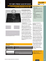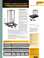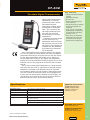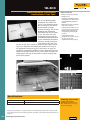
1
DIAGNOSTICO POR IMAGENES RADIACION ONCOLOGICA SEGURIDAD RADIOLOGICA CATALOGO DE PRODUCTOS 2011 ETYC S.A. Av. Rivadavia 2358 3º Piso Oficina 2 C1034ACP - Ciudad de Buenos Aires - Argentina Tel / Fax: 54 11 4953 3040 Líneas Rotativas E-mail: [email protected] [email protected] [email protected] Web: www.etyc.com.ar Diagnostic Imaging Product Catalog 2011 TNT 12000 X-Ray Test Device 35080M/199XRAY kVp Divider and Medical ScopeMeter 07-QRX Wireless QA RADCHEX CR QC Productivity Device Fluke Biomedical. Better products. More choices. One company. 76-424-4156 Nested CT Dose Phantom Kit for Pediatric/Adult Head and Body Radiation Oncology Product Catalog 2011 Providing solutions, not just products Today, biomeds, physicists, RSO’s, other medical personnel must meet increasing regulatory pressures, higher quality standards, and rapid technological growth, while performing their work faster and more efficiently than ever. Fluke Biomedical provides a diverse range of software and hardware tools to meet today’s challenges. Service Fluke Biomedical is dedicated to providing the best service within the healthcare industry. Equipped with the bestcredentialed facilities, onsite experts, and full asset-management capabilities, Fluke Biomedical’s service team is always on call to take care of its customers. Fluke Biomedical’s world-class staff leads the industry in post- and pre-sale support, including helping customers choose the best products and accessories for their needs, technical support, product calibration, and repairs. Regulatory compliance Fluke Biomedical’s benchmark quality operates to the most rigorous standards in the industry, including compliance with ISO 9001:2000, ISO 13485:2003, FDA/QSR as applicable, and NRC/ Part 50, Appendix B/Part 21 and adheres to ISO 17025:2005, ANSI Z540, Mammography MQSA and CNSC. Many of the Fluke Biomedical products are CE-marked and CSA-certified. In addition, the Global Calibration Laboratory holds its NVLAP Lab Code 200566-0 certification and is traceable to both the NIST & PTB. Legacy You may be familiar with some of our legacy brand names, including: •Metron • Victoreen® • Nuclear Associates •DNI Nevada • Keithley •Bio-Tek Instruments Fluke Biomedical has taken the best elements and products of these former brands and have incorporated them into the Fluke Biomedical culture and product line available today. Our newest catalog Thank you for requesting our Radiation Oncology catalog. Within these pages, you will find solutions to manage your quality assurance and maintain a safe, regulatory-compliant facility in the radiation oncology physics field. If you are interested in receiving catalogs or information about any of Fluke Biomedical’s other product-lines, please visit www.flukebiomedical.com/catalog. Catalogs are available for the following product lines: • Biomedical Test •Radiation Safety • Diagnostic Imaging QA •Service About Fluke Biomedical Fluke Biomedical leads the world in the manufacture of biomedical test and simulation products, including standalone electrical safety testers to fully integrated and automated performance testing and documentation systems. Fluke Biomedical also provides some of the most trusted and accurate radiation safety, medical imaging, and oncology quality-assurance solutions for regulatory compliance. About Fluke Corporation Fluke Biomedical is a division of Fluke Corporation. Fluke Corporation is the world leader in the manufacture, distribution, and service of electronic test tools and software and is a wholly owned subsidiary of Danaher Corporation (NYSE:DHR). Diagnostic Imaging Product Catalog Contact us: Contents Diagnostic X-Ray Quality Assurance Instruments TNT 12000 X-Ray Test Device.................................................................................................................... 3 35080M/199XRAY Non-Invasive kVp Divider and Medical Scopemeter........................................... 6 10100AT, 10500AT, and 10500AMT TRIAD™ TnT X-Ray Field Service/Calibration/QA Kit....... 9 10500EXL TRIAD Toolkit for Excel........................................................................................................... 15 8000 NERO® mAx X-Ray Test Device...................................................................................................... 17 8000mAx NERO mAx Toolkit for Excel .................................................................................................. 21 4000M+ X-Ray Test Device.......................................................................................................................23 4000EXL 4000 Toolkit for Excel...............................................................................................................25 07-CRXW and 07-QRX Wireless CR RADCHEX and QA RADCHEX...................................................29 07-MAS5 mAs-5 Meter............................................................................................................................... 31 06-526 RAD-CHECK® Plus Dosimeter ....................................................................................................34 06-526-5240 RAD-CHECK MICRO-R......................................................................................................35 18-526 Series Service and Quality Control Kits...................................................................................37 07-453 Digital X-Ray Pulse Counter/Timer...........................................................................................38 07-487 Dual-Range Digital mAs Meter...................................................................................................39 07-469 Wave Precision High-Voltage Divider ......................................................................................40 18-116 VeriLUM® Color Dual Mode Pod.................................................................................................. 41 07-621 Precision Photometer ..................................................................................................................42 07-661-7662 Collimator/Beam Alignment Test Tool .........................................................................43 07-644 Grid Alignment Test Tool ...........................................................................................................43 07-591 Focal Spot Test Tool......................................................................................................................44 07-501 to 07-555 Test Patterns..............................................................................................................45 07-503 to 07-551 Star Patterns...............................................................................................................46 07-456 High-Purity Aluminum Step Wedges........................................................................................46 07-451 X-Ray Output Detector ................................................................................................................48 07-608 Screen/Film Contact Mesh .........................................................................................................48 07-706 Patient Phantom/Penetrometer System....................................................................................48 07-622 Multipurpose Focal Spot/HVL Test Stand................................................................................50 07-623 Mammography Focal Spot Measurement Test Stand ...........................................................50 07-533 Radiopaque Rulers........................................................................................................................50 0 Radiography/Fluoroscopy Phantoms and Accessories 57-4 Series CLEAR-Pb® Compensation Filters......................................................................................52 07-600QC/07-600QCN Quality Control Kit . ..........................................................................................55 07-680 NEMA® Cardiology/Fluoroscopy Phantom ..............................................................................56 07-649 CDRH Fluoroscopic Phantom.......................................................................................................60 07-601 Fluoroscopic System Resolution Test Tools ............................................................................ 61 07-800 Series Flex Film Cassettes ........................................................................................................ 61 07-600 Fluoroscopic Beam Alignment Device .....................................................................................62 07-678+ DXR Direct X-Ray Ruler ...........................................................................................................62 76-710 Digital Subtraction Angiography (DSA) Phantom .................................................................63 07-652 CDRAD Contrast Detail Digital and Conventional Radiography Phantom.........................65 07-605-7777 CR/DR DIN Test Tool ........................................................................................................67 07-647 R/F QC Phantom ...........................................................................................................................68 07-643 Contrast Imaging Phantom..........................................................................................................69 07-653 Fluoroscopic Imaging Test Phantom ........................................................................................70 76-2 Series Diagnostic X-Ray Phantoms .............................................................................................. 71 76-025 CDRH Dental Image Quality Test Tool......................................................................................73 76-606DX ATOM MAX Diagnostic Head Phantom ...............................................................................75 07-645 Fluoro-Test ™ Tool..........................................................................................................................77 Fluke Biomedical 6045 Cochran Road Cleveland, OH 44139-3303 U.S.A. Fluke Biomedical Europe Science Park Eindhoven 5110 5692EC Son, The Netherlands For more information, contact us In the U.S.A. (800) 850-4608 or Fax (440) 349-2307 In Europe/M-East/Africa +31 40 267 5435 or Fax +31 40 267 5436 From other countries +1 (440) 248-9300 or Fax +1 (440) 349-2307 Email: [email protected] Web access: www.flukebiomedical.com Service Center/Repair/ Calibration US Fluke Biomedical 6045 Cochran Road Cleveland OH 44139-3303 Tel: 440-498-2560 Toll free: 800-850-4608 ext 2564 Email: [email protected] Service Center/Repair/ Calibration Europe Fluke Biomedical Europe Science Park Eindhoven 5110 5692EC Son, The Netherlands Tel: +31 (40) 267 5435 Fax: +31 (40) 267 5436 Email: [email protected] 1 Diagnostic Imaging Product Catalog Contents Film Processor Quality Assurance 07-444 07-443 07-417 07-402 07-424 “The Little Genius” Scanning Densitometer............................................................................80 Handheld Deluxe Digital Clamshell Densitometer................................................................. 81 Handheld Dual-Color Sensitometer .........................................................................................82 Portable Digital Thermometer.....................................................................................................83 Digital Densitometer.....................................................................................................................84 Film Handling and Storage Mammography Quality Assurance 18-220 Mammographic Accreditation Phantom...................................................................................92 18-250 Digital Stereotactic Breast Biopsy Accreditation Phantom...................................................93 18-251-2000 Contrast and Resolution Mammography Phantom.....................................................94 18-252 Contrast Detail Phantom for Mammography ..........................................................................95 18-227 CDMAM Phantom...........................................................................................................................96 18-222 Tissue-Equivalent Mammography Phantom ..........................................................................98 18-216 Single-Exposure High Contrast Resolution Phantom............................................................99 18-228- 229-1313 Stereotactic Biopsy Phantom.................................................... ...............................100 18-229 Triple-Modality Biopsy Training Phantom............................................................................101 18-203 Mammography Phototimer Consistency Test Tool . ............................................................103 18-224 Mammography Phantom Material ..........................................................................................103 18-303 Mammography Collimation Assessment Test Tool .............................................................104 18-210-8000 View Markers for Mammography ...............................................................................105 CT Quality Assurance 76-410-4130 AAPM CT Performance Phantom..................................................................................106 76-409 Spiral/Helical CT Lesion Detectability Phantom...................................................................108 76-424-4156 Nested CT Dose Phantom Kit for Pediatric/Adult Head and Body .......................109 CT Ion Chambers ...................................................................................................................................... 110 84-357 Interventional Triple-Modality 3D Abdominal Phantom.................................................... 113 MRI Quality Assurance 76-907 76-908 76-904 76-903 MR Uniformity/Linearity Phantom (AAPM)............................................................................ 114 MR 3D Slice Thickness/High Contrast Resolution Phantom (AAPM) .............................. 114 MRI Surface Coil Phantom......................................................................................................... 115 MRI Multipurpose Phantom ..................................................................................................... 117 Ultrasound Quality Assurance 84-349 84-317 84-340 84-342 Ultrasound Elasticity QA Phantom...........................................................................................122 Multipurpose Tissue/Cyst Ultrasound Phantom...................................................................123 General Purpose Multi-Tissue Ultrasound Phantom ..........................................................124 General Purpose Urethane Ultrasound Phantom..................................................................125 Service and Calibration Service Info............................................................................................................................ 126 Calibration Info..................................................................................................................... 126 2 Diagnostic X-Ray Quality Assurance Instruments Radiation Oncology Product Catalog Contents Linear Accelerator Quality Assurance Instruments 35040 Advanced Therapy Dosimeter......................................................................................5 37-705 VeriDose® PDMQC System.........................................................................................11 30-471 VeriDose® Solid-State Diode Detectors...................................................................12 37-720 Dual-Diode Dosimeter Patient Dose Monitor..........................................................13 37-105 mobileMOSFET Wireless Dose Verification System..............................................14 05-433 PRIMALERT® 10 Teletherapy Radiation Monitor..................................................16 37-731 Precision Isocentric QC Beam-Checker II...............................................................17 37-001 TEL-ALIGN™ Teletherapy Alignment Gauge..........................................................18 37-013 GARD™ Geometric Accuracy Radiotherapy Device..............................................19 Radiation Oncology Chambers 30-351 Farmer-Type Ionization Chamber, 0.6 cm3, waterproof......................................21 30-344 Semiflex™ Ionization Chamber, 0.125 cm3, waterproof..................................... 23 30-329 Markus™ Electron Ionization Chamber, 0.055 cm3, waterproof........................31 30-355 Extension Cable Reel for Ionization Chambers................................................... 33 Phantoms and Accessories 74-0 Series IMRT Phantoms: Thorax, Homogeneous, Advanced Pelvic 3D, and Head and Neck ................................................................................................................ 35 76-417 CT Simulation Phantom............................................................................................ 40 76-462 Electron Density Phantom........................................................................................41 37-020 Oakton® Temperature Compensated Barometers.................................................42 Service and Calibration Service Info.............................................................................................................................. 43 Calibration Info....................................................................................................................... 43 Fluke Biomedical 6045 Cochran Road Cleveland, OH 44139-3303 U.S.A. Fluke Biomedical Europe Science Park Eindhoven 5110 5692EC Son, The Netherlands For more information, contact us In the U.S.A. (800) 850-4608 or Fax (440) 349-2307 In Europe/M-East/Africa +31 40 267 5435 or Fax +31 40 267 5436 From other countries +1 (440) 248-9300 or Fax +1 (440) 349-2307 Email: [email protected] Web access: www.flukebiomedical.com Service Center/Repair/ Calibration US Fluke Biomedical 6045 Cochran Road Cleveland OH 44139-3303 Tel: 440-498-2560 Toll free: 800-850-4608 ext 2564 Email: [email protected] Service Center/Repair/ Calibration Europe Fluke Biomedical Europe Science Park Eindhoven 5110 5692EC Son, The Netherlands Tel: +31 (40) 267 5435 Fax: +31 (40) 267 5436 Email: [email protected] 3 Radiation Safety Product Catalog Contents Ion Chamber Survey Meters 451B Ion Chamber Survey Meter with Beta Slide................................................................4 451EXL 451 Assistant for Excel................................................................................................8 Other Survey Meters and Probes ASM-990 Series Advanced Survey Meter...........................................................................10 489-200WTF Wipe Test Fixture for Advanced Survey Meter.........................................13 489-110D GM Pancake Probe.................................................................................................14 Geiger-Mueller and Scintillation Probe Selection Guide.............................................15 190N Portable Neutron Survey Meter....................................................................................18 Area Monitors 190F Area Monitor/Frisker Count Rate Meter......................................................................19 1060AM Digital Smart Detector Area Monitor.................................................................... 20 05-437 PRIMALERT® 35 Area Radiation Monitor..............................................................21 05-443/444 PRIMALERT Digital Area Monitors................................................................ 22 05-450 PRIMALERT Digital Doorway Monitor................................................................... 23 Personal Dosimetry Service and Calibration Fluke Biomedical 6045 Cochran Road Cleveland, OH 44139-3303 U.S.A. Fluke Biomedical Europe Science Park Eindhoven 5110 5692EC Son, The Netherlands For more information, contact us In the U.S.A. (800) 850-4608 or Fax (440) 349-2307 In Europe/M-East/Africa +31 40 267 5435 or Fax +31 40 267 5436 From other countries +1 (440) 248-9300 or Fax +1 (440) 349-2307 Email: [email protected] Web access: www.flukebiomedical.com Service Center/Repair/ Calibration US Fluke Biomedical 6045 Cochran Road Cleveland OH 44139-3303 Tel: 440-498-2560 Toll free: 800-850-4608 ext 2564 Email: [email protected] Service Center/Repair/ Calibration Europe Fluke Biomedical Europe Science Park Eindhoven 5110 5692EC Son, The Netherlands Tel: +31 (40) 267 5435 Fax: +31 (40) 267 5436 Email: [email protected] 3 TNT 12000 X-Ray Test Device The TNT 12000 X-Ray Test Device is the newest and most versatile instrument available for measuring key x-ray imaging parameters. It sets up in seconds and measures kVp, dose, dose rate, time, and half value layer (HVL) in a single exposure. A totally-wireless ZigBee® interface enables quick and easy setup and the wireless detector can be used with the companion wireless display or a laptop computer. X-ray imaging QA, calibration, and maintenance in today’s demanding digital environment require very high productivity and compliance with local and regional regulations. The TNT 12000 delivers high productivity through 100 % wireless connectivity to either a display, which instantly responds with all values in a single exposure, or a laptop, where measured values are displayed and categorized in organized templates. Instant HVL with just a single exposure further enhances productivity. Featuring all-in-one-exposure measurements and ZigBee wireless communication combined with the rugged, reliable, and accurate design that is a Fluke trademark, the TNT 12000 is truly a new breed of non-invasive x-ray test tool. TNT 12000 is ideal for use by OEM factory and field service engineers, independent service organizations, physicists, biomedical and clinical engineers, and local and regional field inspectors of x-ray imaging equipment. Its small, lightweight design enhances portability and 100 % wireless operation ensures setup can be accomplished in seconds. Because the TNT 12000 measures all parameters with every exposure, there is no need for complicated menu selection, further enhancing user productivity. TNT 12000 always defaults to the last use when powered on, so when used often for repetitive procedures it is truly a one-button (power-on) solution. The TNT 12000 has the expanded functionality needed for modern applications and can be managed with minimum keystrokes. Users can identify and select custom measurement protocols and save them for future use. Full test automation and documentation software is available for TNT 12000, creating the advantage of accurate, repeatable testing processes. Ansur Test Automation Software is only available from Fluke Biomedical. Key features • 100 % ZigBee wireless operation between detector and hand-held display or laptop • Compact hand-held design for maximum portability and ease-of-use • Simple user interface with minimum menu routines means setup in seconds • Fluke Biomedical ruggedness provides reliable operation. It’s tough! • 40 kHz sampling rate to ensure accuracy with the most difficult applications • Global support network delivering prompt service and peace-of-mind to Fluke Biomedical customers worldwide are instantly available on the hand-held display or on the user’s laptop. The latter application places measured values into templates that are standard with the Excel software provided with TNT 12000. Because the TNT 12000 Excel package is a spreadsheet, users can customize their templates and create their own reports to send to others. Accuracy, reproducibility, and reliability are also critically important and Fluke Biomedical is the industry standard customers count on to deliver uncompromised performance. Primary end benefit Productivity and accuracy are central goals when performing any maintenance or QA process on diagnostic imaging equipment because image system uptime is critical to patient care objectives. TNT 12000 is small, portable, and wireless. It is easily transported to the imaging room, set up in seconds, and all results Diagnostic X-Ray Quality Assurance Instruments 3 TNT 12000 X-Ray Test Device Specifications kVp measurements Units kVp Average (average of peaks during a specified interval) kVp Max (highest peak during a specified interval) PPV (peak practical voltage) Ranges Radio/Fluoro modes 40 kV to 150 kV Mammo modes Mo/Mo: 22 kV to 35 kV (standard calibration) Rh/Rh: 25 kV to 49 kV (optional calibration) Mo/Rh: 22 kV to 40 kV (optional calibration) Mo/Al: 22 kV to 49 kV (optional calibration) Rh/Al: 25 kV to 49 kV (optional calibration) W/Rh: (optional calibration) W/Ag: (optional calibration) Resolution 0.1 kV Optional accessories Accuracy Radio/Fluoro modes: ± 2 % or ± 1 kV, whichever is greater 35035 mA/mAs Meter TNT 12000 Ansur Test Automation Software Plug-in Mammo modes: ± 2 % or ± 0.7 kV, whichever is greater Reproducibility ± 1 % (std of 5 readings) Filtration correction range Radio/Fluoro modes: 1 mm Al to 10 mm Al or equivalent Mammo mode: 0 mm Al to 0.4 mm Al added filtration Dose/exposure measurements Units Roentgens, grays Range 0.5 mR to 999 R 5 µGy to 999 Gy Resolution 1 µR 0.01 µGy Accuracy ±5% Reproducibility ± 0.5 % (std % of five readings) Filtration correction range Radio/Fluoro modes: 1 mm Al to 10 mm Al or equivalent Mammo mode: 0 mm Al to 0.4 mm Al added filtration kV correction ranges Radio/Fluoro modes: 40 kV to 150 kV Mammo mode: Mo/Mo: 22 kV to 35 kV Dose/exposure rate measurements Units Roentgens or grays per hour, minute, second, pulse Range 8 mR/s to 10 R/s 70 µGy/s to 100 mGy/s 130 µR/pulse to 160 mR/pulse (@ 60 PPS) 12 µGy/pulse to 1.4 mGy/pulse (@ 60 PPS) Resolution 1 µR/s 0.01 µGy/s 0.02 µR/pulse (@ 60 PPS) 0.2 nGy/pulse (@ 60 PPS) Accuracy ±5% Filtration correction range Radio/Fluoro modes: 1 mm Al to 10 mm Al or equivalent Mammo mode: 0 mm Al to 0.4 mm Al added filtration kV correction range Radio/Fluoro modes: 40 kV to 150 kV Mammo mode: Mo/Mo: 22 kV to 35 kV Exposure time—radiographic modes Units Milliseconds, pulses Range (@ stated accuracy) Milliseconds: 10 ms to 9999 ms Pulses: 1 pulse to 999 pulses Resolution Milliseconds: 0.1 ms Pulses: 1 pulse 4 Diagnostic X-Ray Quality Assurance Instruments TNT 12000 X-Ray Test Device Specifications Accuracy Optional accessories Milliseconds: 1 % or 0.5 ms Pulses: ± 1 pulse Reproducibility Milliseconds: 1 % or 0.5 ms Pulses: ± 1 pulse 3335538 TA-TNT12K, TNT 12000 with Test Automation 3340639 TA-TNT12KWD, TNT 12000 Wireless Detector with Test Automation Elapsed time—fluoro modes Range 10 sec to 9999 sec Resolution 0.1 second Accuracy 1 % or 0.5 sec Average pulse rate—pulsed fluoro Range 1 pps to 999 pps (pulses per second) Resolution 1 pps Accuracy 1 pps Average pulse width— pulsed fluoro Range 10 ms to 999 ms Resolution 0.1 ms Accuracy 1 % or 0.5 ms HVL Range Radio/Fluoro modes: 1.2 mm Al to 10 mm Al (equivalent) Mammo mode: 0.2 mm Al to 0.6 mm Al (equivalent) Resolution Radio/Fluoro modes: 0.1 mm Al (equivalent) Mammo mode: 0.01 mm Al (equivalent) Accuracy Radio/Fluoro modes: ± 10 % or 0.2 mm Al (equivalent) Mammo mode: ± 5 % or 0.05 mm Al (equivalent) Electrical specifications Battery Battery type: Lithium-ion, 3.7 V, 4000 mAh Battery charge time: Approx. 5 hr Battery discharge time: Approx. 8 hr Battery cutoff voltage: 2.75 V AC adapter Input voltage: 100 V ac to 240 V ac Input frequency: 50/60 Hz Input current: 0.5 A (rms) Output voltage: 6 V dc Environmental specifications Operating temperature 0 °C to 35 °C (32 °F to 95 °F) Storage temperature -35 °C to 50 °C (-31 °F to 122 °F) Operating humidity 20 % to 80 % RH (non-condensing) Physical specifications Display 320 × 240 Color LCD Size (WxDxH) Display: 15.2 cm x 11.4 cm x 4.45 cm (6 in × 4.5 in × 1.75 in) Detector: 15.2 cm x 11.4 cm x 4.45 cm (6 in × 4.5 in × 1.75 in) Weight Display: 0.422 kg (0.93 lb) Detector: 0.68 kg (1.5 lb) Ordering information Kit #1 TNT 12000 X-Ray Test Device Included accessories 1320005000 TNT 12000WD Wireless Detector 1330005000 TNT 12000D Wireless Display 14-445 (2) AC Adapters/Chargers 50-197 Cable, Type A to Mini B USB 50-198 Cable, Mini A to Mini B USB 1320003000 Excel Software/ User Manual on CD 1320033000 Carrying Case 90-183 ZigBee® USB Dongle TNT12QRG Quick Start Reference Guide Kit #2 3335774 TNT 12000WD Wireless Detector Included accessories 1320005000 TNT 12000WD Wireless Detector 14-445 (1) AC Adapter/Charger 50-197 Cable, Type A to Mini B USB 1320003000 Excel Software/ User Manual on CD 1320033000 Carrying Case 90-183 ZigBee® USB Dongle TNT12QRG Quick Start Reference Guide Kit #3 3335795 TNT 12000D Wireless Display Included accessories 1330005000 TNT 12000D Wireless Display 14-445 (1) AC Adapter/Charger 50-197 Cable, Type A to Mini B USB 50-198 Cable, Mini A to Mini B USB 1320003000 Excel Software/ User Manual on CD TNT12QRG Quick Start Reference Guide Diagnostic X-Ray Quality Assurance Instruments Back to Top 5 35080M/199XRAY Non-Invasive kVp Divider and Medical ScopeMeter The winning combination. The 35080M and 199XRAY are commonly used x-ray tools unmatched by traditional meters. This winning combination allows busy service engineers and biomedical personnel the ability to perform fast and accurate verification of kVp values for calibration or QA assessment. Fluke Biomedical offers this combination in a convenient kit with all the accessories you need to get started. 35080M Non-Invasive kVp Divider The 35080M Non-Invasive kVp Divider quickly and accurately measures kVp for all modalities. The unit checks both above and below table tubes and displays the direct kVp values on either the 35050AT Dosimeter, the 199XRAY Medical ScopeMeter, or optional Excel Add-in software. The 35080M is highly portable and eliminates the need for bulky and heavy high-voltage divider tanks. In fact, it’s so compact in size that it fits into a shirt pocket. A patented* wide-range filter pack is included with the 35080M and provides accurate readings for the range of 50 kVp to 150 kVp. Four optional filter packs are available for use with the 35080M for CT, mammographic, and mobile applications. 199XRAY Medical Scopemeter The 199XRAY Medical ScopeMeter has all the normal oscilloscope functions, as well as the speed, performance, and analysis power for more-demanding applications. This high-performance oscilloscope offers the functionality of top-end bench instruments. With up to 200 MHz bandwidth, 2.5 GS/s real-time sampling, and a deep memory of 27,500 points per input, the 199XRAY is ideal for engineers who need the full capabilities of a high-performance oscilloscope in a handheld, battery-powered instrument. In addition, the 199XRAY is specially designed for use with x-ray systems. This ScopeMeter displays kVp waveforms and direct kVp values simultaneously on an easy-to-read screen, eliminating time-wasting calculations of scope traces to derive kVp values. 6 Diagnostic X-Ray Quality Assurance Instruments Key features 35080M •New miniaturized configuration for convenient transport to the job site •Fast/easy non-invasive kVp values for calibration/QA •Non-invasive technology eliminates the hazards of high-voltage cables and the need for bulky divider tanks •Auto ON/OFF when connected/disconnected •Optional filter packs enable testing in all modalities •Rh/Rh measurement capability when 35080M is used with Cadmium K-Edge and Linear Mammo Filter Pack Pair •Convenient storage/ carrying case 199XRAY • ScopeMeter displays kVp wave forms and direct kVp values simultaneously on an easy-to-read screen •No more time spent calculating scope traces to derive kVp values •Full medical oscilloscope scope functionality with color display ScopeMeter triggers on • standard interlaced and high-resolution, noninterlaced video systems. Triggers on all lines nonselectively or select an individual video line—up to 2800 lines per frame •mAs measurement calculates current over time •Smart averaging capabilities •Extended vertical offset •Selectable persistence mode •Extended video triggering •FlukeView® for Windows® for documenting, enhancing, waveform analysis, and archiving results 35080M/199XRAY Non-Invasive kVp Divider and Medical ScopeMeter Specifications 35080M Range 50 kVp to 150 kVp, using only the wide-range radiographic filter pack (37617). Range and versatility are extended with the use of special optional filter packs. Accuracy ± 2 % of reading in the range of 50 kVp to 150 kVp, exclusive of linearity, filtration, and gain effects. Linearity corrections automatically applied when using 35080M with either 35050AT Dosimeter or 199XRAY. Response time 150 µs (10 % to 90 %) Calibration Internally generated signal provides a calibration check Minimum time for valid reading 1 ms, three-phase; one line cycle, single-phase Tube current Wide dynamic range from 4 mA to 3000 mA (three-phase), 2 mA to 1500 mA (single phase). Generator settings will vary in waveform and distance. Less than ± 1 kVp effect for wide-range radiographic filter pack covering 50 kVp to 150 kVp. Specialty filter packs may have different characteristics. Environmental Temperature range: 0 °C to 35 °C Relative humidity: 20 % to 80 % Storage temperature: -35 °C to 50 °C Orientation Long axis of the Model 35080M Non-invasive kVp Divider oriented perpendicular to axis of x-ray tube to eliminate heel effect. Power requirements 9 V battery, 60 hours operation Dimensions (LxWxH) 6 cm x 9 cm x 21 cm (2.38 in x 3.5 in x 8.25 in) Weight 0.68 kg (1.5 lb) MA190 Accessory Kit The MA190 accessory set enables interconnection of the 199XRAY for use in the field of medical imaging and video systems. The kit is included with the 199XRAY as a standard accessory. The accessory kit includes the following: •FlukeView for Windows software •50 Ω BNC feedthrough terminator, in insulated enclosure, to maintain proper termination of test connections during measurement •50 Ω BNC terminator with 10:1 signal attenuation, to keep test terminal properly loaded while getting optimum signal amplitude to benefit from the instrument’s extended offset range •1 Ω current shunt for current measurements, in insulated enclosure •Safety-designed BNC cable, 1.5 m (5 ft), with plastic connectors for safe connection to test terminals even when not at ground potential •Insulated BNC (f) to 4 mm banana-plug adapter •Dual 4 mm banana receptacles (1 red, 1 black) Diagnostic X-Ray Quality Assurance Instruments 7 35080M/199XRAY Non-Invasive kVp Divider and Medical ScopeMeter Specifications Optional accessories 199XRAY Field applications Bandwidth Dual input: 200, 100 or 60 MHz Real-time sampling rate Up to 2.5 GS/s Trigger types Connect-and-View ™ automatic triggering and a full range of manual trigger modes Extended video triggering Along with its triggering capability for standard, interlaced TV signals, the instrument also triggers on high-resolution, noninterlaced video systems. The ScopeMeter 199XRAY will trigger on all lines (nonselective), or can select an individual video line from systems with up to 2800 lines per frame. Persistence Digital persistence for analyzing complex dynamic waveforms, similar to an analog scope. Selectable persistence Persistence mode with selectable decay time helps to find anomalies in the wave shape and optimizes the display for color information when working with composite color video. Display Fast-display update rate for seeing dynamic behavior instantaneously 33551 CT filter pack 37351 Linear mammo filter pack 37355 Cadmium K-Edge filter pack 37946 Mobile filter pack 38237 Low range filter pack Automatic capture and replay of 100 screens Maximum record length 27,500 points-per-input record length using ScopeRecord mode Trend analysis TrendPlot paperless chart recorder for trend analysis up to 22 days Independently floating isolated inputs Up to 1,000 V Waveform compare Waveform reference for visual comparisons and automatic pass/fail testing of waveforms Vpwm Vpwm function for motor drive and frequency inverter applications mA mAs measurement calculates current over time. Using the cursors, you can now measure directly the amount of radiation produced by x-ray systems, or the total amount of charge applied to a system. Smart averaging Smart averaging gives the averaged waveform over successive acquisitions, reducing noise in the displayed waveform. Thanks to smart averaging, you can now also see an incidental curve of a different wave shape with no effect on the averaged curve. This allows you to see the averaged curve of a sequence of video lines, for example, while still seeing the incidental flyback line flash by. The oscilloscope gives an immediate response when the signal makes large changes. Extended offset Vertical offset is now extended to a maximum of 16 divisions, allowing vertical zoom-in for study of small details of the signal. Electrical safety 1000 V CAT II and 600 V CAT III safety certified Power requirements Rechargeable NiMH battery pack, four hours operation Dimensions (LxWxH) 25.6 cm x 16.9 cm x 6.4 cm (10.1 in x 6.6 in x 2.5 in) Weight 2.0 kg (4.4 lb) 35080M Non-Invasive kVp Divider 199XRAY Medical ScopeMeter MA190 Medical ScopeMeter Accessory Kit 37617 Wide-range filter pack 121002900 Carrying Case 199XRAY FlukeView® for Windows® 8 Included accessories 35080M/199XRAY Documenting Transfer waveforms, screens, and measurement data from the ScopeMeter to a PC. Print or import the data into your report. Enhancing Add user text to individual ScopeMeter settings, providing guidance to the operator when recalling a setup. Archiving Create a library of waveforms with your comments for easy reference and comparison. Store complete replay cycles for analysis of waveform changes. Store complete memory content of the ScopeMeter on your PC for backup purposes. Waveform comparison Store reference waveforms, add operator instructions, and send both to the ScopeMeter for waveform comparison and “Pass/Fail” testing. Analysis Use cursors, perform spectrum analysis, or export data to other analysis programs. Diagnostic X-Ray Quality Assurance Instruments MA190 Medical ScopeMeter Accessory Kit 35080M 37617 Wide-range filter pack 1210029000 Carrying case Ordering information 35080M/199XRAY kVp Divider and Medical ScopeMeter Kit 199XRAY Medical ScopeMeter with kVp capabilities 35080M Non-Invasive kVp Divider Back to Top 10100AT, 10500AT and 10500AMT TRIAD™ TnT X-Ray Field Service/ Calibration/QA Kit Key features •Bright display with direct readout in user-selected units The TRIAD™ TnT X-Ray Field Service/Calibration/ QA Kit is a full-function, x-ray dosimeter kit that performs fast, highlysensitive measurements. It is ideal for government compliance testing, troubleshooting, repair of diagnostic x-ray equipment, installation and setup of new equipment, preventive maintenance, radiographic QA measurements, and measurements required for JCAHO accreditation. The kit performs measurements for all modalities: radiographic, fluoroscopic, mammographic (MQSA), CT, cine and dental. The TRIAD TnT Kit comes in three popular configurations: • 10100AT is the base-level dosimeter kit and features the 35050AT dosimeter, a technologically advanced, microprocessorcontrolled, x-ray radiation dosimeter. The kit also includes ion chambers and test stand, triaxial/coaxial cable, ac adapter, HVL filter set, RS-232 interface cable with adapters, customization software, instruction manual CD, and lightweight carrying case. • 10500AT includes all of the components from the 10100AT, in addition to the 35080M non-invasive kVp divider and 37617 wide-range filter pack (50 kVp to 150 kVp), for quick and accurate kV measurement. • 10500AMT is equipped with all the components from the 10500AT, as well as the 35035 mA/mAs meter and CA-23 universal test-lead kit, allowing engineers to accurately measure mAs and fluoroscopic mA for diagnostic, radiographic, and fluoroscopic imaging equipment. Diagnostic X-Ray Quality Assurance Instruments •Image intensifier measure- ments at 0.1 µR and 0.1 µR/s resolution; cine in µR/frame •Expanded kVp and exposure-time measurement capabilities •Simplified controls include autoreset, autoranging, automatic offset and drift compensation, automatic power-down, and automatic pressure and temperature correction Optional TRIAD toolkit for • Excel for remote operation, waveform capture, and calibration Multiple self-checking • features to reduce testing time •Battery-powered with auto power-down feature to extend battery life Automatic temperature and • pressure correction for faster operation in any environment •Timesaving scroll functionality •Recognizes and ignores spurious background signals •Very low dose rate: 20 nGy/s at a 1 nGy/s resolution •Broader range of dental unit kV and time measurement Back to Top 9 10100AT, 10500AT and 10500AMT TRIAD™ TnT X-Ray Field Service/ Calibration/QA Kit Specifications 10100AT TRIAD TnT Kit Exposure and exposure rate accuracy Basic accuracy of 35050AT ± 1 % of reading ± 2 range resolution steps over range of 18 °C to 28 °C and ± 2 % of reading ± 2 range resolution steps over the full operating temperature range of 0 °C to 50 °C Note: A 3 % NIST-traceable calibration is provided with each system and includes effects of 35050AT, 96035B, and 96020C. Exposure time measurement Exposure time accuracy ± 0.1 % of reading; ± 0.2 msec Maximum exposure time 6.5 s Measurement resolution 0.2 ms Measurement modes kVp/Dose/Time Single-shot (“all-in-one” exposure), direct-beam measurement of exposure, kVp, and time; autoranging across three-decade ranges; auto reset between exposures; display updates after each exposure kVp/Rate Simultaneous measurement of kVp and exposure rate Full sensitivity dose Autoranging across five decades of sensitive ranges; automatic drift and offset compensation; automatic postexposure display hold Full sensitivity rate Measurement range covers a span from low-level image intensifier measurements to unattenuated, direct beams; automatic offset compensation and nonlinear filtering. Autoranging provides five decades of sensitivity ranges. Display updates once per second Very low dose rate (VLDR) This mode is only for making very low dose rate measurements. Nonlinear digital filtering and autoranging provide five decades of sensitivity ranges. Display updates once per second. In this mode, automatic current offset and drift compensation are disabled. As a result, the system can display very low dose rates. Power requirements Battery life ~30 hours with six AA alkaline batteries; automatic power-down after user-selected period of unattended operation (5 min to 255 min); AC adapter supplied with each 35050AT Note: When the ac adapter is in use, the auto power-down feature is disabled, providing continuous operation. User selections for ion chamber, units, kV filter pack, temperature, pressure, and frame are stored in nonvolatile memory before automatic turnoff; eliminates manual reselection at power-up Bias voltage supply Fixed electronic bias (~300 V); bias voltage removed from triaxial input connector at instrument turnoff Customization Allows user to modify contents of nonvolatile memory, including ion chamber and kV filter pack conversion factors, temperature and pressure units, radiation units, and power down interval. A field customization software program is included for use with an IBM®-PC or compatible. Connections 35080M Interface Male, two lug BNC Computer interface RS-232, using RJ-45 connector; 9,600 baud 8-bit, 1 stop, no parity, xon/xoff; enables fully-programmable operation and waveform display from a PC with optional Excel add-in; powered when connected to computer Ion chamber input Triax, BNC; collector and guard positive-biased relative to ion chamber body and dosimeter chassis Power 2.1 mm dc power jack, power input for an unregulated 9 V, 200 mA adapter with a center negative, 2.1 mm plug General information Display Two-line, 20-character alphanumeric PLED (polymer LED), with 0.5 cm character height; indicates all ion chamber/kV filter pack identification information, numerical measurement results, battery level, calibration date and other information Weight 6.4 kg (14 lb) Dimensions (LxWxH) 46 cm x 33 cm x 15 cm (18 in x 13 in x 6 in) Diagnostic Ionization Chambers (96035B and 96020C) Energy range 96035B: 30 kVp to 150 kVp; 20 kVp to 50 kVp for mammographic 96020C: 30 kVp to 150 kVp Nominal sensitivity 96035B: 2.00 R/C x 108 R/C (1.75 Gy/C x 106 Gy/C) at 22 °C and 1013 hPa 2.21 R/C x 108 R/C (1.94 Gy/C x 106 Gy/C) at 22 °C and 1013 hPa (flat response suitable for conventional diagnostic radiography and mammography) 96020C: 2.08 R/C x 107 R/C (1.82 Gy/C x 105 Gy/C) at 22 °C and 1013 hPa (optimized for low-level image intensifier and cine measurements) Construction 96035B: Graphite-coated acrylic, parallel-plate, air-vented 96020C: Composite graphite-filled thermoplastic; parallel-plate, air-vented Volume 96035B: 15 cm3 96020C: 150 cm3 10 Diagnostic X-Ray Quality Assurance Instruments Back to Top 10100AT, 10500AT and 10500AMT TRIAD™ TnT X-Ray Field Service/ Calibration/QA Kit Specifications 10100AT TRIAD TnT Kit (continued) Ion Chamber Units Effective Range *** Resolution Step Size 15 cc R 100 µ to 20 1µ R/s 100 µ to 20 1µ R/m 5 m to 1200 50 µ R/h 100 m 72 k 1m R/f ** 2 µ to 333 m 0.02 µ Gy 1 µ to 175 m 0.01 µ Gy/s 1 µ 174 m 0.01 µ Gy/m 50 µ to 10.5 0.5 µ Gy/h 1 m to 630 0.01 m Gy/f ** 0.02 µ to 2.9 m 0.2 n R 10 µ to 2 0.1 µ R/s 10 to 2 0.1 µ R/m 0.5 m to 120 5µ R/h 10 m to 7.2 k 0.1 m R/f ** 0.2 µ to 33 m 0.002 µ Gy 0.1 µ to 17.5 m 0.001 µ Gy/s 0.1 µ to 17.5 m 0.001 µ Gy/m 5 µ to 1050 m .05 µ Gy/h 0.1 m to 63 0.001 m Gy/f ** 0.002 µ to 290 µ 0.02 n R/s 2 µ to 2* 0.1 µ R/m 0.1 m to 120* 5µ R/h 2 m to 7.2 k* 0.1 m R/f ** 0.04 µ to 33 m* 0.002 µ Gy/s 0.02 µ to 17.5 m* 0.001 µ Gy/m 1 µ to 1050 m* 0.05 µ Gy/h 0.02 m to 63* 0.001 m Gy/f ** 0.4 n to 290 µ* 0.02 n C 1 p to 100 n 0.01 p A 1 p to 100 n 0.01 p 150 cc 150 cc VLDR Electrical Units Values for ion chambers are calculated using nominal sensitivities: 15 cc: 2.4 x 10⁸ R/C, 150 cc: 2.4 x 10⁷ R/C *Very Low Dose Rate effective range at 5 % resolution steps. **At 60 f/s (1 to 120 frames/selectable). ***IEC 61674 effective range at 1 % resolution steps 96020C and 96035B Diagnostic Ion Chambers Energy range 96020C: 30 kVp to 150 kVp 96035B: 30 kVp to 150 kVp for diagnostic measurements; 20 kVp to 50 kVp for mammographic measurements Nominal volume 96020C: 150 cm3; 11.3 cm diameter by 1.5 cm thick active volume 96035B: 15 cm3; 3.96 cm diameter by 1.22 cm thick active volume Nominal sensitivity 96020C and 96035B •Very low leakage and low noise • Rugged mechanical construction • Ionization chambers are supplied with triaxial BNC connectors 96020C and 96035B Diagnostic Ion Chambers The 96020C and 96035B Diagnostic Ion Chambers are vented-volume, parallel-plate air ionization chambers with side-mounted BNC triaxial connectors. The 96020C Ion Chamber has a nominal volume of 150 cm3, and the 96035B has a nominal volume of 15 cm3. Both ion chambers have a fullyguarded, centrally-located collector plate that provides superior collection efficiency. The patented* 96035B has a dual-energy range that enables both diagnostic and mammographic measurements. They are accomplished using the other side as the entrance window. *Patent numbers 4,843,619, 4,916,727 and 5,508,526. 96020C: H60: 2.08 R/C x 107 R/C at 22 °C and 760 mmHg (optimized for low-level image intensifier and cine measurements) 96035B: L100: 2.0 R/C x 108 R/C at 22 °C and 760 mmHg MV30 (PTB Mammo Point): 2.21 R/C x 108 R/C at 22 °C and 760 mmHg (flat energy response suitable for conventional diagnostic radiography and mammography) Leakage current < 10 fA under normal bias conditions (300 V) Collection efficiency 96020C: 95 % at 2,000 R/min 96035B: 95 % at 5,000 R/min Diagnostic X-Ray Quality Assurance Instruments Back to Top 11 10100AT, 10500AT and 10500AMT TRIAD™ TnT X-Ray Field Service/ Calibration/QA Kit Specifications 96020C and 96035B Diagnostic Ion Chambers (continued) Wall material 96020C: Composite graphite-filled thermoplastic 96035B: Graphite-coated acrylic (methyl-methacrylate) Window material 96020C: 0.76 mm thick, graphite-coated polycarbonate 96035B: Both entrance windows are made of 0.25 mm graphite-coated polycarbonate Window density 96020C: 91 mg/cm2 96035B: 32 mg/cm Active window area 2 96020C: 100 cm2, centered within the chamber body 96035B: Each side of the chamber has a circular active window region centered 7.1 mm further from the BNC connector than the center of the chamber body; active window regions have an area of 12.32 cm2 Collector plate 96020C: 0.8 mm thick graphite-coated acrylic plate, 10.8 cm in diameter; 2.16 cm x 2.85 cm guard region electrically isolated from collector area 96035B: 0.25 mm thick, centrally mounted, graphite-coated, polycarbonate plate, 3.18 cm, ± 0.01 cm in diameter; 1.27 cm x 0.89 cm guard region is electrically isolated from the collector area Connector Side-mounted, triaxial, two-lug BNC connector Calibration 96020C Standard Calibration: Standard calibration performed at H60 (NIST defined as 60 kVp, first HVL of 6.0 mm Al, homogeneity coefficient of 94) 96035B Standard Calibration: Standard calibration performed at one diagnostic and one mammographic beam quality; calibration factors normalized to 22 °C and 760 mmHg Diagnostic Unattenuated Beam: Calibration on diagnostic side of chamber is performed at M80 (NIST defined as 80 kVp, first HVL of 2.97 mm Al, homogeneity coefficient of 57) Mammographic Beam: Calibration on mammographic side performed at Mo/Mo28 (NIST defined as 28 kVp, first HVL of 0.332 mm Al, homogeneity coefficient of 74.3) or MV30 (PTB defined as 30 kVp, first HVL of 0.337 mm Al) 35080M Non-Invasive kVp Divider Range 50 kVp to150 kVp, using only the wide-range radiographic filter pack (37617); range and versatility extended with use of special optional filter packs Accuracy ± 2 % of reading in the range of 50 kVp to 150 kVp, exclusive of linearity, filtration, and gain effects; linearity corrections automatically applied when using 35080M Non-invasive kVp Divider with either the 35050AT Dosimeter or the 199XRAY Medical Scopemeter Response time 150 µs (10 % to 90 %) Calibration Internally generated signal provides calibration check Minimum time for valid reading 1 ms, 3-phase; one line cycle, single-phase Tube current Wide Dynamic Range: From 4 mA to 3000 mA (3-phase), 2 mA to 1500 mA (single-phase) Note: Generator settings will vary in waveform and distance. Less than ± 1 kV effect for wide-range radiographic filter pack, covering 50 kVp to 150 kVp. Specialty filter packs may have different characteristics 12 Diagnostic X-Ray Quality Assurance Instruments Key features 35080M •New pocket-size configuration •Non-invasive technology eliminates the hazards of high-voltage cables and need for bulky divider tanks •Auto ON/OFF when connected/disconnected •Optional filter packs enable testing in all modalities Rh/Rh measurement • capability when 35080M Non-Invasive kVp Divider is used with cadmium K-Edge and linear mammo filter pack pair 35080M Non-Invasive kVp Divider The 35080M Non-Invasive kVp Divider quickly and accurately measures kV for all modalities. The unit checks both above and below table tubes, and the direct kV values are displayed on either the 35050AT Dosimeter or the 199XRAY Medical ScopeMeter. Derived kV can also be calculated using a storage oscilloscope. The 35080M Non-Invasive kVp Divider is highly portable and eliminates the need for bulky and heavy high-voltage divider tanks—so compact in size that it fits into a shirt pocket. A patented* wide-range filter pack is included with the 35080M Non-Invasive kVp Divider and provides accurate readings for the range of 50 kVp to 150 kVp. Four optional filter packs are available for use with the 35080M Non-Invasive kVp Divider for CT, mammographic, and mobile applications. 10100AT, 10500AT and 10500AMT TRIAD™ TnT X-Ray Field Service/ Calibration/QA Kit Specifications 35080M Non-Invasive kVp Divider (continued) Environmental requirements Temperature range: 0 °C to 35 °C Relative humidity: 20 % to 80 % Storage temperature: -35 °C to 50 °C Orientation Long axis of the 35080M Non-invasive kVp Divider oriented perpendicular to axis of x-ray tube to eliminate heel effect Power requirements 9 V battery, 50 hours operation; battery-check function connects battery to output terminals for voltage measurement Dimensions (LxWxH) 6 cm x 9 cm x 21 cm (2.375 in x 3.5 in x 8.25 in) Weight 0.68 kg (1.5 lb) 35035 mA/mAs Meter Controls 1) Power mA/mAs switch, 2) Reset switch, 3) Range switch: 200 mA/mAs, 2000 mA/mAs and 20 mA range settings, 4) ac/dc switch Accuracy 1 % of reading ± two least significant digits for all ranges Environmental requirements Temperature range: 5 °C to 35 °C Relative humidity: 0 % to 80 % Storage temperature: -20 °C to 50 °C Display Liquid crystal display (LCD), 3.5 digit, .5 in H (13 mm) Input Two banana jacks Power requirements 9 V alkaline battery with easy replacement Dimensions (LxWxH) 15 cm x 5.0 cm x 8.75 cm (2 in x 2 in x 3.50 in) Weight 0.35 kg (0.78 lb) Please refer to charts at the right for Miliamp, current and signal Input limit specifications for the 35035 mA/mAs Meter Milliamp seconds (mAS) Range Resolution Input impedance* 200 mAs 0.1 mAs 10 Ω 2000 mAs 1.0 mAs 1Ω *Does not include fuse resistance. Also, does not include effect of bridge rectifier present when unit is set for ac specifications Current (mA) Range Resolution Input impedance* 20 mA 0.01 mA 100 Ω 200 mA 0.1 mA 10 Ω 2000 mA 1 mA 1Ω *Does not include fuse resistance. Also, does not include effect of bridge rectifier present when unit is set for ac specifications Signal input limits Function Range Max input limit mA OFF Input shorted; 2.0 A maximum (fuse protected) 20 250 mA for 30 s* 200 1.0 A for 30 s* mAs 2000 2.0 A maximum (fuse protected) 200 1.0 A for 30 s* 2000 2.0 A maximum (fuse protected) *Limits set by power dissipation rating of shunt resistors Diagnostic X-Ray Quality Assurance Instruments 13 10100AT, 10500AT and 10500AMT TRIAD™ TnT X-Ray Field Service/ Calibration/QA Kit 35035 Digital mA/mAs Meter The 35035 Digital mA/mAs Meter is a versatile instrument for x-ray service engineers, field service engineers, and biomedical engineers to measure mAs and fluoroscopic mA accurately for diagnostic, radiographic, and fluoroscopic imaging equipment. The 35035 Digital mA/mAs Meter operates with an easilyreplaceable 9-volt alkaline battery for extra convenience and portability. Included accessories 10100AT 35050AT Dosimeter 96035B 15 cm3 Ion Chamber 96020C 150 cm3 Ion Chamber 38208 Coax/triaxial cable, 6 m (20 ft) 37594 Programming kit (37594), includes customization software on CD, two- meter RS-232 interface cable and adapters 37581 Test stand (37581), ion chamber stem, HVL filter tray 37688 HVL filter set (37668) 35050ATCD User/service manual 37500D Kit carrying case 10500AT Components in 10100AT Kit plus 35080M Non-Invasive kVp Divider 37617 Wide-range filter pack (50 kVp to 150 kVp) 10500AMT Components in 10500AT Kit plus 35035 mA/mAs Meter A-23 Universal Test Lead Kit Ordering information Optional accessories All Kits 500-100 CT Ion Chamber 3.2 cm3 500-200 CT Ion Chamber High Sensitivity, 10 cm3, for multislice CT 07-434 Ultra-high purity HVL Attenuators (for mammo set of six) 10500EXL TRIAD toolkit for Excel 38617 USB to RS-232 adapter 199XRAY Medical ScopeMeter with kVp capabilities (includes the MA190 Medical ScopeMeter accessory kit) 37946 Mobile filter pack (50 kVp to 135 kVp) ± 2 % accuracy 33551 CT filter pack (70 kVp to 140 kVp) ± 2 % accuracy kVp) ± 2 % accuracy 38237 Low Range Filter Pack (30 kVp to 90 kVp) ± 2 % accuracy 10100AT TRIAD TnT Dosimeter Kit 10500AT TRIAD TnT X-Ray Field Kit Service/Calibration/ QA Kit 10500AMT TRIAD TnT X-Ray Field Kit Service/Calibration/ QA Kit Available ac adapters 9 V, 200 mA (specify with order) 14-106 USA and Japan 14-107 Europe 14-108 UK 14-109 Australia 10500AT and 10500AMT Kits 37355/37351 Mammographic filter pack pair includes: Cadmium k-edge mammo filter pack (27.5 kVp to 29.5 kVp) ± 0.5 kV accuracy, linear mammo filter pack (22 kVp to 40 kVp) ± 1.0 kV accuracy Note: Mammo filter packs are designed for molybdenum anode, beryllium window generators. 14 Diagnostic X-Ray Quality Assurance Instruments Back to Top 10500EXL TRIAD™ Toolkit for Excel Key features •Automatically collects measurement results and places them in an Excel worksheet •Automatically captures kV waveforms and charts them in an Excel worksheet •Templates are provided for QA tests on radiographic, fluoroscopic and mammographic x-ray machines •Templates may be modified to perform user-specific tests and generate customized reports Allows complete remote • control of the TRIAD •Automatically downloads Model 35050A and 35050AT Dosimeter configuration settings •Compatible with Windows® 2000 and above, and Microsoft® Excel 97, 2000 The TRIAD™ Toolkit for Excel is a complete software package for the TRIAD that includes an Excel Add-In, called TRIAD Tool, and Excel templates used to evaluate the performance of radiographic, mammographic, and fluoroscopic x-ray machines. The TRIAD Tool collects measured results from the 35050A and 35050AT Dosimeter and places the data in the active Excel worksheet. In addition, the TRIAD Tool may be used to acquire and graph kV waveforms from the TRIAD as well as remotely control the TRIAD Dosimeter. Diagnostic X-Ray Quality Assurance Instruments 15 10500EXL TRIAD™ Toolkit for Excel Specifications System requirements Windows 2000 and above Microsoft Excel 97, 2000 One serial port (COM1 through COM4) Controls The TRIAD Tools menu provides an interface to remotely control the TRIAD, select filter packs, chambers, units, retrieve kV waveforms, open templates, and change the options. A description of each control follows: Connect/disconnect Connects or disconnects the TRIAD to the communications port. Connect instructs the TRIAD Tool to read configuration information such as the filter packs, chambers, and units. Options Several options can be specified including COM port, temperature, pressure, and frame rate. Measure Sets the TRIAD up for a single exposure using the selected filter pack, chamber, and unit Auto reset Informs the TRIAD Tool to automatically reset the TRIAD for another exposure, allowing a series of exposures to be made without used intervention. At the time Auto Reset is clicked, the TRIAD will be set up for an exposure. Filter packs Selects the filter pack to use for kVp measurements Chambers Selects the ion chamber to use for exposure measurements Units Selects the unit to use for exposure measurements Download waveform The Waveform button is enabled after an exposure is made and before the TRIAD is set up for another exposure. When the Waveform button is pressed, a dialog box prompts for the start and end times for the waveform chart, allowing the user to specify any portion of the kV waveform. The default start and end times are for the complete waveform. The waveform data is then placed in the active cell in the active workbook Help Opens the TRIAD Toolkit for Excel Instruction Manual Templates Three templates are also provided with the TRIAD Toolkit for Excel: a radiographic template, a mammographic template and a fluoroscopic template. Each template includes a help worksheet with detailed instructions for its use. The TRIAD radiographic template Is used to perform the following radiographic tests: reproducibility, kVp accuracy, timer accuracy, linearity, and beam quality The TRIAD mammographic template May be used to perform mammography tests required for ACR and MQSA. Measured data from the 35050A Dosimeter may be automatically collected in the following worksheets: kVp accuracy, kVp reproducibility, Beam quality, Breast entrance exposure, AEC reproducibility, average glandular dose, and radiation output rate The TRIAD fluoroscopic template Is used to perform the following fluoroscopic tests: kVp accuracy, beam quality, and fluoro exposure rate These templates are easy to use and can be modified to fit the user’s needs. Ordering information 10500EXL TRIAD Toolkit for Excel 16 Diagnostic X-Ray Quality Assurance Instruments Back to Top 8000 NERO® mAx X-Ray Test Device Key features •Non-invasive evaluation of radiation outputs The NERO® mAx, Noninvasive NERO mAx X-Ray Test Device, tests the spectrum of x-ray machines on the market today. Offering evaluation of pulsed fluoro, cine, computed tomography (CT), portable, mammographic, dental, radiographic, fluoroscopic, low-, medium-, and high-frequency machines with a single device, the NERO mAx is a benchmark instrument for quality and accuracy. This fifth generation quality assurance tool features 100 kHz sampling speed and direct mA/ mAs measurements. The NERO mAx’s innovative Easy Flow Menu (EFM) system and flexible soft keys provide an intuitive, userfriendly operating environment for quick, accurate, and easy measurements. All measurement modes and functions are displayed on the NERO mAx’s super-bright LCD and are controlled by the five soft keys directly below the display and three hard keys to the right. Seven user-selectable measurement modes and three systemcontrol modes are available and clearly displayed on the control console screen for easy access and selection. Applications The NERO mAx consists of the control console, detector, detector cable, two filter cards, mAs leads, Excel Add-in, ac adapter, HVL plates, instruction manual, and carrying case. The compact control console houses the rechargeable battery, super-bright easy-to-read backlit display, eight control buttons, and the sophisticated electronics necessary for accurate, reproducible measurements. Connectors for power input, RS-232, printer, scope output and the NERO mAx detector are located on the control console’s rear panel. The NERO mAx detector contains sensors for simultaneously measuring kV, exposure or rate and mA or mAs. Solid-state detectors are used to measure kV. An ion chamber, located in the top of the detector, is used for exposure/rate measurements. Connectors for external ion chambers and the NERO mAx detector interface are located on the rear panel of the detector. The front panel has a keyed opening for the NERO mAx filter cards and a connector for mAs leads. The filter cards contain the various filters needed to accurately measure kilovoltage. Each filter card is coded so that the NERO mAx “knows” which filter is in use and its position. The NERO mAx also verifies that the filter card is valid for the selected measurement mode. The two filter cards are keyed so they may only be inserted properly. The W/Al filter card and the Mammo filter card are clearly labeled to the x-ray tube targets for which they are calibrated. Diagnostic X-Ray Quality Assurance Instruments •100 kHz sampling speed captures data from the most difficult machines 0.5 • kV or 1 % accuracy from 22 kV to 160 kV • Measures kVp average, kV effective, kV peak, time, exposure or rate, mA or mAs, HVL, exposure/frame, and mAs/ frame • Displays R or Gy • Excel Add-in includes MQSA, Rad, and Fluoro templates • RS-232 computer interface • Enhanced dental capabilities External chambers External ion chambers for CT, mammographic, image intensifier tube, and special radiographic applications are available. Chamber calibration factors can be stored in the NERO mAx for direct readout of measurements. The Excel Add-in acquires measured data and waveforms directly into an Excel spread sheet to maximize flexibility for report generation. 17 8000 NERO® mAx X-Ray Test Device Real-time CT Easy Flow menu, Mode Select screen. Easy Flow menu, Radio screen. High-frequency mammography Easy Flow menu, Mammo screen. Easy Flow menu, Pulsed Fluoro display. Easy Flow menu, AMSE screen. Pulsed fluoro/AMSE Easy Flow menu, CT EXP screen. Easy Flow menu, EXP screen. Easy Flow menu, HVL screen. 18 Diagnostic X-Ray Quality Assurance Instruments 8000 NERO® mAx X-Ray Test Device Specifications NERO mAx operating modes Radio mode Radio mode is used to make measurements on tungsten target, aluminum-filtered radiographic x-ray generators. Selections are available for % kV peak for the type of generator being tested. For example, Zero Crossing, Single Phase Pulse, 75 %, 80 %, or 90 % of kVp modes are available for accurate exposure measurements on difficult x-ray machines. Radio mode simultaneously measures: kVp Avg, Exposure, kV Eff, Exposure time, kV Peak, kV Peak, and mAs. Mammo mode Mammo mode is used to make measurements on mammographic x-ray generators. Mammo mode simultaneously measures: kVp Avg, Exposure, kV Eff, Exposure time, kV Peak, and mAs. Fluoro mode Fluoro mode is used to make measurements on fluoroscopic x-ray generators. Fluoro mode supports both continuous fluoro and pulsed fluoro measurements. In the continuous fluoro mode, the NERO mAx measures: kVp Avg, kV Eff, exposure rate (R/min), mA, and kV Peak In the pulsed fluoro mode, the NERO mAx measures: kVp Avg, kV Eff, Exposure rate (R/min and mR/pulse), mAs/pulse, and kV Peak. AMSE mode AMSE mode is used for Automated Measurement of Sequential Exposures. This mode is used to measure the output of CINE generators. In AMSE mode, the NERO mAx measures: kVp Avg, Exposure rate (mR/frame), kV Eff, mAs/frame, kV Peak, and Time/frame (ms/frame) CT exposure mode CT Exposure mode is used to make CT exposure measurements using the 6000-100 CT ion chamber. The CT probe must be connected to the NERO mAx detector’s external ion chamber input in this mode. Exposure mode Exposure mode is used to make exposure and rate measurements using the NERO mAx’s internal ion chamber or an external ion chamber. HVL mode In the HVL mode, the NERO mAx calculates half value layer based upon a series of exposure or rate measurements made with varying thicknesses of aluminum absorbers placed in the x-ray beam. A minimum of two exposures are required and up to ten exposures may be used. Calibrate mode Calibrate mode is used to enter and store calibration factors for ion chambers used with the NERO mAx. Setup mode Setup mode is used to set the instrument’s parameters such as the real time clock, temperature and pressure. Unit ID Displays the NERO mAx’s serial number, firmware part number and level. Optional external chamber accessories 6000-528 30 cm3; Energy response: within 7 % from 30 kVp to 150 kVp (15 keV to 65 keV); Cable: 4.5 m (15 ft); Chamber dimensions: 10.2 cm x 10.2 cm x 1.4 cm (4 in x 4 in x 0.54 in thick) 6000-529 3.3 cm3; Energy response: within 5 % from 0.2 mm to 5.0 mm Al HVL (16 kVp to 90 kVp); Cable: 4.5 m (15 ft); Chamber dimensions: 4 cm Ø x 1.5 cm thick. This chamber meets the needs of the MQSA for an external transparent chamber. 6000-530B 150 cm3; Energy response: ± 10 % from 1.8 mm to 10 mm Al HVL; Cable: 3 m (10 ft); Chamber dimensions: 15.9 cm x 20.6 cm x 1.6 cm (6.26 in x 8 in x 0.63 in) 6000-532B 400 cm3; Energy response: ± 5 % from 32 keV to 662 keV; Cable 3 m (10 ft); Chamber dimensions: 15.9 cm x 20.6 cm x 1.6 cm (6.26 in x 8 in x 0.63 in) 6000-100 and 500-100 CT 3.2 cm3; Energy response: ± 5 % from 1 mm to 10 mm Al HVL; Cable: 0.9 m (3 ft); Sensitive length: 10 cm (4 in); chamber inside Ø: 6.4 mm (0.25 in) 6000-200 and 500-200 CT 10 cm3, for multislice CT; Energy response: ± 5 % from 1 mm to 10 mm Al HVL; Cable: 0.9 m (3 ft); Sensitive length: 10 cm (4 in); Chamber inside Ø: 11.44 mm (0.45 in) Diagnostic X-Ray Quality Assurance Instruments 19 8000 NERO® mAx X-Ray Test Device Specifications Optional accessories Kilovoltage (Measured during the first 480 ms of exposure) Measured quantity kVp Avg, kV Eff, kV Peak Accuracy 0.5 kV or ± 1% Reproducibility 0.5 kV or ± 1% Range Target/Filter Range Filtration W/Al 30 kV to 60 kV 50 kV to 100 kV 80 kV to 160 kV 1.2 mm of Al Mo/Mo 22 kV to 35 kV 30 μ of Mo Mo/Rh 22 kV to 40 kV 25 μ of Rh Mo/Al 22 kV to 49 kV 1 mm of Al Rh/Rh 25 kV to 49 kV 25 μ of Rh Rh/Al 25 kV to 49 kV 1 mm of Al All calibrations performed with NIST traceable calibration beam. Analyze/display cycle time Radio and mammo Three seconds for 0.1 second exposure, ine second for each 32 ms of exposure time Fluoro and AMSE 15 seconds for all exposures Time Radio mode Measured during entire exposure at 90 %, 80 %, 75 % rise/fall of waveform, zero crossing, or pulse count Mammo mode Measured during entire exposure at 90 % rise/fall of waveform Accuracy 1 ms Resolution 0.1 ms Range All diagnostic exposures from 1 ms to 60 seconds Exposure and rate (Measured during entire exposure with automatic energy, temperature, and pressure correction) Measured quantity Roentgens or grays Accuracy ±5% Reproducibility Radio and mammo modes: ± 2 % or 2 mR Resolution 0.1 mR Range All diagnostic exposure and rate measurements from 1 mR to 9999 R Fluoro rate 0.1 R/min to 999 R/min mAs and mA (Measured invasively during entire exposure) Accuracy 2% Reproducibility ± 1 % or 0.2 mAs Range 0.1 mAs to 9999 mAs, 0 to 1000 mA HVL Accuracy ±5% Range 0.1 to 99.9 mmAl Physical Display Super-bright 240 x 60 pixel, super-twist LCD with cold cathode fluorescent backlight Detectors Ion chamber and solid-state (kV detectors) Ion chamber volume 45 cc nominal Window area/density 38 mg/cm2 Polycarbonate HVL set 2.30 mm, 1.0 mm, 0.3 mm Al Power requirements Size Weight 20 12 V dc 1 A external supply. Rechargeable internal batteries supply more than 4 hours of continuous service with overnight charge Volume: 0.065 m3 (3960 in3) Console: 22.86 x 23.17 x 8.26 cm (9.00 x 9.12 x 3.25 in) Detector: 16.66 x 9.4 x 6.55 cm (6.56 x 3.70 x 2.58 in) Filter cards: 6.1 x 15.88 x 0.8 cm (2.4 x 6.25 x 0.31 in) Shipping: 10.43 kg (23 lb) Console: 2.067 kg (4.56 lb) Detector (with card): 0.747 kg (1.65 lb) Filter cards: 0.090 kg and 0.094 kg (2.9 oz and 3.2 oz) Diagnostic X-Ray Quality Assurance Instruments 07-434 Ultra-High Purity HVL Attenuators for mammo, set of 6 External chamber accessories 6000-528 Radiographic Ion Chamber 6000-529 Mammographic Ion Chamber 6000-529-95 Probe Holder for BRH2 test stand 6000-530B Image Intensifier Ion Chamber 6000-532B Scatter Ion Chamber 6000-100 and 500-100 CT Ion Chamber 6000-200 and 500-200 CT High Sensitivity Ion Chamber Available ac adapters for (specify with order) 14-328 110 V ac, 12 V dc, 1000 mA, USA and Japan 14-401 230 V ac, 12 V dc, 1000 mA, Europe 14-414 230 V ac, 12 V dc, 1000 mA, UK 14-414 and14-416 adapter 230 V ac, 12 V dc, 1000 mA, Australia Included accessories 8000-100-5 Control Console 8000-101-5 Detector 105-252 Detector Cable 105-253 and 105-254 mAs Leads 8000MAX Excel Add-in 38667 HVL Plates 8000-200-1 Instruction Manual 8000-70 Carrying Case Ordering information 8000 NERO mAx X-Ray Test Device Back to Top 8000mAx NERO® mAx Toolkit for Excel Key features •Automatically collects measurement results and places them in an Excel worksheet •Automatically captures kV waveforms and charts them in an Excel worksheet •Templates are provided for QA tests on radiographic, fluoroscopic, and mammographic x-ray machines •Templates may be modified to perform user-specific tests and generate customized reports Allows complete remote • control of the NERO mAx •Complete online help speeds learning •Compatible with Microsoft® Windows® 2000 and above, and Excel 97, 2000 The NERO® mAx Toolkit for Excel is a complete software package for the NERO mAx that includes an Excel Add-In, called NERO mAx Add-In and Excel templates that may be used to evaluate the performance of radiographic, mammographic and fluoroscopic x-ray machines. The NERO mAx Add-In collects measured results from the NERO mAx and places the data in the cells of the active Excel worksheet, starting at the active worksheet cell. The NERO mAx Add-In also may be used to acquire and graph radiation and kV waveforms from the NERO mAx as well as remotely control the NERO mAx. Diagnostic X-Ray Quality Assurance Instruments 21 8000mAx NERO® mAx Toolkit for Excel Specifications System requirements Controls The NERO mAx menu provides an interface for the user to remotely control the NERO mAx and retrieve radiation and kV waveforms. A description of each menu option follows: Select mode Selects the NERO mAx measurement mode. This allows the user to select the correct measurement mode for the template in use. For instance, the radio mode is selected when using the radiographic template, the mammo mode is selected when using the mammographic template and the fluoro mode is selected when using the fluoroscopic template. Retrieve rad waveform Retrieves the radiation waveforms from the NERO mAx. When this is selected, a dialog box opens and the user may select either all of the waveform or a portion of the waveform to be charted. If a portion of the waveform is desired, the user prompted for start and end times (in milliseconds) of the waveform window. Retrieve kV waveform Retrieves the kV waveform from the NERO mAx. When this is selected, a dialog box opens and the user may select either all of the waveform or a portion to be charted. If a portion of the waveform is desired, the user is prompted for start and end times (in milliseconds) of the waveform window. Select com port Allows the user to choose serial communication port COM1—COM4 for 8000 NERO mAx connection. Windows 2000 and above Microsoft Excel 97, 2000 One serial port (COM1 through COM4) Templates Three templates are also provided with the NERO mAx. Toolkit for Excel: a radiographic template, a mammographic template and a fluoroscopic template. Each template includes a help worksheet with detailed instructions for its use. NERO mAx radiographic template Used to perform the following radiographic tests: reproducibility, kVp accuracy, timer accuracy, linearity, and beam quality NERO mAx mammographic template Used to perform mammography tests required for ACR and MQSA. Measured data from the Model 8000 NERO mAx may be automatically collected in the following worksheets: kVp accuracy, kVp reproducibility, beam quality, breast entrance exposure, AEC reproducibility, average glandular dose, and radiation output rate NERO mAx fluoroscopic template Used to perform the following fluoroscopic tests: kVp accuracy, beam quality, and fluoro exposure rate These templates are easy to use and can be modified to fit the user’s needs. Ordering information 8000mAx NERO mAx Toolkit for Excel 22 Diagnostic X-Ray Quality Assurance Instruments Back to Top 4000M+ X-Ray Test Device The 4000M+ X-Ray Test Device does it all. Simply place the instrument in the x-ray beam, make one exposure, and it serially displays kVp Maximum, kVp Average, kVp Effective, dose, and time. The Model 4000M+ then automatically resets for the next exposure. A CsI photodiode pair provides the kVp measurements through five user-selectable filter pairs. This ensures optimum accuracy over the entire diagnostic range with minimum filtration dependence. Exposure measurements are made with a parallel plate ionization chamber located above the filter wheel. Exposure time is measured with quartz crystal accuracy. Plus, a variety of external ion chambers may be connected for even greater flexibility. Key features •Measures kVp maximum, kVp average, kVp effective, dose and time in one exposure •Compact, lightweight design •Displays R or Gy •External ion chambers for Mammo, CT, image intensifier and phototiming measurements •Automatic exposure reset for hands-off operation Rechargeable Ni-Cd batteries • provide more than six hours of continuous service •RS-232 computer interface •Storage scope output for realtime waveform display •Reversible display for fluoro measurements Specifications Kilovoltage Accuracy 1 kV Mo/Mo (22 kVp to 35 kVp) (Mammo generators w/30 μ Mo) Range 38 mg/cm2 Polycarbonate W/Al tubes: 27 kVp to 155 kVp Mo/Mo tubes: 21 kVp to 50 kVp Time Measured during entire exposure at 90 % rise/fall of waveform Accuracy Within 2 % or 2 ms, whichever is greater Range 1 ms to 10 seconds Exposure Measured during entire exposure at 90 % rise/fall of waveform Accuracy ±5% Range 10 mR to 10 R Fluoroscopic Measured over one second intervals during fluoro exposure Accuracy ±5% Range 0.5 to 200 R/min Detectors kV CsI/photodiode pair measures x-ray transmission through differential attenuators Time Computed from kV waveform stored in memory against quartz crystal time base Exposure Parallel plate ionization chamber Volume 36 cm3 Window Calibration 38 mg/cm2, 18.9 cm2 polycarbonate Reference to a NIST traceable voltage divider and a calibrated exposure monitor during irradiation Diagnostic X-Ray Quality Assurance Instruments 23 4000M+ X-Ray Test Device Specifications Optional accessories Physical Display 16 character dot-matrix LCD Controls Model 4000+ Five rocker switches On/Off: Power switch Radio/Fluoro: Select radiographic or fluoro operation High/Low: Select for sensitivity Roll: Roll thru data Exposure/All: Select exposure only for external ion chamber Mo/Mo or W/Al: Select anode/filter of x-ray tube Connectors Power: 9 V dc, 500 mA Scope: BNC for oscilloscope connection RS-232: DB-9 connector configured as DCE. BNC and banana plug for external Ion chamber Power requirements 9 V dc 500 mA external supply. Rechargeable internal Ni-Cd batteries supply more than six hours of continuous service with overnight charge Dimensions (WxDxH) 21.5 cm x 23 cm x 7.6 cm (8.5 in x 9 in x 3 in) Weight Approximately 1.59 kg (3.5 lb) HVL set Aluminum filters: 2.3 mm, 1.0 mm, and 0.3 mm Optional external chamber accessories 24 6000-528 30 cm3; Energy response: within 7 % from 30 kVp to 150 kVp (15 keV to 65 keV); Cable: 4.5 m (15 ft); Chamber dimensions (HxWxT): 10.2 cm x 10.2 cm x 1.4 cm (4 in x 4 in x 0.54 in) 6000-529 3.3 cm3; Energy response: within 5 % from 0.2 mm to 5.0 mm Al HVL (16 kVp to 90 kVp); Cable: 4.5 m (15 ft); Chamber dimensions: 4 cm Ø x 1.5 cm thick. This chamber meets the needs of the MQSA for an external transparent chamber. 6000-530B 150 cm3; Energy response: ± 10 % from 1.8 mm to 10 mm Al HVL; Cable: 3 m (10 ft); Chamber dimensions (HxWxT): 15.9 cm x 20.6 cm x 1.6 cm (6.26 in x 8 x 0.63 in) 6000-532B 400 cm3; Energy response: ± 5 % from 32 keV to 662 keV; Cable 3 m (10 ft); Chamber dimensions (HxWxT): 15.9 cm x 20.6 cm x 1.6 cm (6.26 in x 8 in x 0.63 in) 6000-100 and 500-100 CT 3.2 cm3; Energy response: ± 5 % from 1 mm to 10 mm Al HVL; Cable: 0.9 m (3 ft); Sensitive length: 10 cm (4 in); chamber inside Ø: 6.4 mm (0.25 in) 6000-200 and 500-200 CT 10 cm3, for multislice CT; Energy response: ± 5 % from 1 mm to 10 mm Al HVL; Cable: 0.9 m (3 ft); Sensitive length: 10 cm (4 in); Chamber inside Ø: 11.44 mm (0.45 in) Diagnostic X-Ray Quality Assurance Instruments 4000EXL 4000 Toolkit for Excel 07-434 Ultra-High Purity HVL Attenuators for mammo, set of 6 4000-69 Carrying Case 190004 RS-232 Cable 7.6 m (25 ft), 9-pin to 9-pin External chamber accessories 6000-528 Radiographic Ion Chamber 6000-529 Mammographic Ion Chamber 6000-529-95 Probe Holder for BRH2 test stand 6000-530B Image Intensifier Ion Chamber 6000-532B Scatter Ion Chamber 6000-100 and 500-100 CT Ion Chamber 6000-200 and 500-200 CT High Sensitivity Ion Chamber Available ac adapters for (specify with order) 14-301 110 V ac, 9 V dc, 500 mA, USA and Japan 14-399 230 V ac, 9 V dc, 500 mA, Europe 14-415 230 V ac, 9 V dc, 500 mA, UK 14-415 and 14-416 adapter 230 V ac, 9 V dc, 500 mA, Australia Ordering information 4000M+ X-Ray Test Device Back to Top 4000EXL 4000 Toolkit for Excel Key features •Automatically collects measurement results and places them in an Excel worksheet •Captures radiation and kV waveforms and charts them in an Excel worksheet •Templates are provided for QA tests on radiographic, fluoroscopic, and mammographic x-ray machines •Templates may be modified to perform user-specific tests and generate customized reports Allows complete remote • control of the 4000M+ NERO •Complete online help speeds learning •Compatible with Microsoft® Windows® 2000 and above, and Excel 95, 97, 2000 •Automatically detects the presence of 4000M+ NERO The 4000 Toolkit for Excel is a complete software package for the 4000M+ NERO® that includes an Excel Add-In, called 4000 Add-In and Excel templates that may be used to evaluate the performance of radiographic, mammographic and fluoroscopic x-ray machines. The 4000 Add-In collects measured results from the 4000M+ NERO and places the data in the cells of the active Excel worksheet, starting at the active worksheet cell. The 4000 Add-In also may be used to acquire and graph radiation and kV waveforms from the 4000M+ NERO as well as remotely control the 4000M+ NERO. Diagnostic X-Ray Quality Assurance Instruments 25 4000EXL 4000 Toolkit for Excel Specifications System requirements Controls The 4000 menu provides an interface for the user to remotely control the 4000M+ NERO and retrieve radiation and kV waveforms. A description of each menu option follows: Remote control Selects the 4000M+ NERO measurement mode and measurement options. This allows the user to select the correct measurement mode for the template in use. For instance, the radio mode is selected when using the radiographic template, the Mo/Mo target filter is selected when using the mammographic template and the fluoro mode is selected when using the fluoroscopic template. Retrieve rad waveform Retrieves the radiation waveform data from the 4000M+NERO and charts it in a new Excel chart. Retrieve kV waveform Retrieves the kV waveform data from the 4000M+ NERO and charts it in a new Excel chart. Windows 2000 and above Microsoft Excel 97, 2000 One serial port (COM1 through COM4) Templates Three templates are also provided with the 4000 Toolkit for Excel: a radiographic template, a mammographic template and a fluoroscopic template. Each template includes a help worksheet with detailed instructions for its use. 4000 radiographic template Used to perform the following radiographic tests: Reproducibility, kVp accuracy, Timer accuracy, Linearity, and Beam quality 4000 mammographic template used to perform mammography tests required for ACR and MQSA. Measured data from the Model 4000M+ NERO may be automatically collected in the following worksheets: kVp accuracy, kVp reproducibility, Beam quality, Breast entrance exposure, AEC reproducibility, Average glandular dose, and Radiation output rate NERO mAx fluoroscopic template Used to perform the following fluoroscopic tests: kVp accuracy, Beam quality, and Fluoro exposure rate These templates are easy to use and can be modified to fit the user’s needs. Ordering information 4000EXL 4000 Toolkit for Excel 26 Diagnostic X-Ray Quality Assurance Instruments Back to Top 07-CRXW and 07-QRX Wireless CR RADCHEX and QA RADCHEX The wireless 07-CRXW CR RADCHEX and 07-QRX QA RADCHEX are factory radiationcalibrated, NISTtraceable light meters that can be used to calibrate (balance) CR plate readers (also radiation-calibrated light meters) in the field. The CR plate reader in the field will be calibrated and traceable to the Fluke Biomedical factory radiation-calibrated and traceable x-ray-produced light exposure. Both 07-CRXW and 07-QRX have the same x-ray energy response as a CR system (x-ray-to-light conversion efficiency is the same for various beam conditions). This enables them to be used as accurate and precise replacements for the plate reader’s light measurement value (exposure index value). Most importantly, the 07-CRXW and 07-QRX can save valuable time when calibrating or accessing CR readers and AEC used with multiple x-ray systems. These x-ray systems may have different filtration and beam characteristics even when located in the same department or imaging center. Balancing system performance and dose is an important QA requirement best satisfied with either the 07-CRXW or 07-QRX. Applications The wireless 07-CRXW is ideal for use by service engineers to initially calibrate and troubleshoot the CR plate reader, AEC, and density selector settings. Physicists use 07-CRXW to assess the performance of CR-AEC for compliance to clinical system speed objectives and patient dose. Radiology managers can use 07-CRXW to assist in the establishment of technique charts and training to determine ALARA techniques for various exam types. QA personnel can use 07-CRXW to periodically document the performance of the CR system and to compare CR to film/screen systems regarding desired ALARA objectives. The 07-CRXW uses Bluetooth® to communicate with a laptop computer so that the CRLU (CR Light Units), EI (Exposure Index) and estimated mR values measured by the electronic cassette are automatically recorded and displayed on the laptop screen. Key features •Calibrates computed radiog- raphy (CR) plate readers and automatic exposure control (AEC) •Assesses ongoing performance of CR plate reader, AEC, and automatic programmed radiography (APR) •Sets and maintains desired clinical system speed (dose) of the CR system •Calibrates CR plate readers in the field to be traceable to a factory radiation-produced light condition •Links radiation exposure (mR) to the front of the plate accurately and predictably to a CR light measurement value (CRLU) •Provides a reliable and reproducible method of accurately maintaining a CR manufacturers’ specific factory calibration •Provides three different tube-head filtration choices for users who desire a nonfiltered beam condition for field plate reader calibration •Software selections of multiple beam conditions for different CR manufacturers •Ideal tools for service engineers, physicists, and quality assurance personnel Benefits Using the 07-CRXW to calibrate x-ray system AEC and CR plate readers instead of a dosimeter can improve productivity significantly. A full system AEC and CR plate reader calibration process can take as much as six hours. Employing the 07-CRXW the process can be completed in under two hours. A substantial productivity gain for service, biomedical or physics professionals. Essentially, the wireless 07-QRX performs all of the functions of the 07-CRXW, however CRLU and speed numbers (representing the relative system speed of the CR system compared to a film/screen system) are displayed on a LCD readout built into the electronic cassette. A pressure sensitive On/Off switch activates the 07-QRX and is used to reset the meter between exposures. The values may be manually entered into the software program on a laptop or PC if desired but a laptop is not required to use the 07-QRX. The 07-CRXW and 07-QRX are designed to work with all major brands of CR equipment. The 07-QRX QA RADCHEX is ideally suited for fast and easy daily checks of AEC and CR system exposure continuity. Data obtained from daily checks can be used for trend analysis of individual systems as well as a way to monitor the balanced performance of CR systems throughout the healthcare enterprise. Diagnostic X-Ray Quality Assurance Instruments 29 07-CRXW Wireless CR RADCHEX Specifications X-ray energy dependence Simulates relative light output of photostimulatable phosphor plate (PSP) within ± 3 % over kVp range of 60 kVp to 120 kVp and a patient equivalent thickness range of 5 cm to 35 cm (within specified operating rates) Digital range Computed radiography light units: CRLU (AEC#); 0 to 500 CRLU (AEC#); 0 to 5000 (07-QRX) Minimum CRLU rate 1.5/sec (approx. 0.15 mR/sec entrance exposure rate), 7/sec (approx. 0.7 mR/sec entrance exposure rate) (07-QRX) Maximum CRLU rate 2500/sec (approx. 250 mR/sec entrance exposure rate), 25000/sec (approx. 2500 mR/sec entrance exposure rate) (07-QRX) Power on/off Manual switch Controls Wireless communications with computer software; Bluetooth wireless communications (07-CRX only) Functions Measures CRLU (AEC#); converts CRLU to CR manufacturers specific CR plate reader light exposure index value (EI); user selectable; calculates cassette input exposure values for various x-ray beam conditions (exposure in mR plus backscatter) Power requirements Built-in NiMH rechargeable battery pack (9.6 V) Typical battery life between charging 5 hours, 20 hours (07-QRX) X-ray beam filter 1.5 mm copper (B152-110); 6 in x 6 in complete with velcro straps to attach to x-ray tube collimator housing Environmental requirements Operating temperature: 15 ºC to 35 ºC (59 ºF to 95 °F) General information Electronic cassette dimensions (WxDxH) 30 cm x 24 cm x 1.3 cm (12 in x 10 in x 0.5 in) Weight 1.8 kg (3.9 lb) Computer software CD-ROM containing Microsoft® Excel program Computer requirements Computer capable of running Windows® 98 or higher with Microsoft Excel, Computer not required to operate (07-QRX). Software is provided with (07-QRX) to manually enter values if desired Optional accessories 07-AEC6 For film/screen applications to assess and calibrate automatic exposure control (AEC) – radiographic and mammographic systems 07-AEC6M For film/screen applications to assess and calibrate automatic exposure control (AEC) – mammographic systems Ordering information 07-CRXW Wireless CR RADCHEX, including PC-based Excel documentation software 07-QRX Wireless QA RADCHEX, including PC-based Excel documentation software 30 Diagnostic X-Ray Quality Assurance Instruments Back to Top 07-MAS5 mAs Meter The 07-MAS5 mAs Meter simultaneously provide mAs, exposure time, and mA, as well as mA waveform information. This intelligent meter displays three 50-milliseconds mA waveform samples so radiographic and mammographic pre-heat circuits can be analyzed and adjusted without using an oscilloscope. Based on feedback from field service engineers, the 07-MAS5 also features a button that causes the meter to ignore the first 10-milliseconds of the mA waveform. Featuring a microcontroller to analyze the digital mA waveform, the 07-MAS5 mAs Meter accurately display the values essential for analyzing and calibrating radiographic and mammographic equipment. Key features •An intelligent meter that measures mAs, exposure time, and mA all at the same time •Provides mA waveform information •At a button press, the 07-MAS5 meter ignores the first 10 milliseconds of exposure •AC and DC inputs •A diagnostic power-up sequence to indicate operational status •Auto LCD update •Optional manual reset •Automatic power-down when meter is not used for more than five minutes •Displays when an exposure is detected •Low battery indication The four line LCD displays the following: Line 1 m As (average tube current (mA) times mA waveform exposure time) Line 2 Exposure time (mA waveform exposure time in seconds) Line 3 mA (average tube current (mA) over the entire mA waveform) Line 4 Three sample mA waveform values: 1st waveform value represents the average mA for the 1st 50 milliseconds of exposure 2nd waveform value represents the average mA for the 2nd 50 milliseconds of exposure 3rd waveform value represents the average mA for the 3rd 50 milliseconds of exposure Specifications Reset Auto LCD update; optional manual reset Dynamic range 10 mA to 2000 mA; 0.1 mAs to 999.9 mAs; 1 ms to 6.535 sec Accuracy mAs: ± 0.1 mAs or 1 % (whichever is greater) mA: ± 1.0 mA or 0.5 % (whichever is greater) Time: ± 1 ms or 1 % (whichever is greater) Operating temperature 15 °C to 35 °C (59 °F to 95 °F) Power requirements One 9 V battery Typical battery life > 40 hours Size 10.16 cm x 16.51 cm x 0.84 cm (4 in x 6.5 in x 0.33 in) Weight 0.28 kg (0.625 lb) Diagnostic X-Ray Quality Assurance Instruments Ordering information 07-MAS5 mAs Meter Back to Top 31 06-526 RAD-CHECK® Plus Dosimeter Key features •Entrance skin exposure measurements (ESE) The 06-526 RAD-CHECK Plus uses proven technology specifically designed to provide you with the ultimate in versatility and cost-effective operation. Accurate, lightweight, portable; this industry-standard dosimeter enables you to gain the critical edge in your QC program. Battery operation and builtin detector virtually eliminate setup time. Just place the meter or external ion chamber on x-ray table; collimate, shoot, and read the result. Precision ion chamber and digital display ensure accuracy plus easy readability. Specifications Ranges 0.001 R to 2 R, 0.01 R/min to 20 R/min Internal chamber 30 cc volume, energy response ± 5 % from 15 keV to 65 keV (30 kVp to 150 kVp filtered). 20.5 cm2 (5.1 cm Ø) effective measurement area. Center of chamber 1.03 cm below top of chamber Standard calibration At 75 kVp with 4 mm Al filtration at 22 °C and one atmosphere Reproducibility Within 2 % short-term over 100 mR to 2 R range (1 mGy to 20 mGy) Electrometer drift 0.5 to 1 mR/min typical; 6 mR/min maximum (5 µGy to 10 µGy; 60 µGy/min maximum) Maximum exposure rate Minimum 90 % collection efficiency at 20 R/sec Automatic reset Resets display to zero; can also be reset manually Operating temperature 10 °C to 40 °C Relative humidity 0 to 90 %, non-condensing Display 3.5 in x 0.5 in LCD, low battery indicator Controls Auto or manual reset selector. Display zero reset button. Dose or dose-rate output selector. Integral or remote ion chamber selector. On/off switch Power 9 V alkaline battery, > 100 hours operation (50 hours in manual reset mode) Dimensions (WxDxH) 15.25 cm x 15.9 cm x 7 cm (6 in x 6.25 in x 2.75 in) Weight 0.51 kg (1.125 lb) Optional external chamber accessories 34 6000-528 30 cm3; Energy response: within 7 % from 30 kVp to 150 kVp (15 keV to 65 keV); Cable: 4.5 m (15 ft); Chamber dimensions: 10.2 cm x 10.2 cm x 1.4 cm (4 in x 4 in x 0.54 in thick) 6000-529 3.3 cm3; Energy response: within 5 % from 0.2 mm to 5.0 mm Al HVL (16 kVp to 90 kVp); Cable: 4.5 m (15 ft); Chamber dimensions: 4 cm Ø x 1.5 cm thick. This chamber meets the needs of the MQSA for an external transparent chamber. 6000-530B 150 cm3; Energy response: ± 10 % from 1.8 mm to 10 mm Al HVL; Cable: 3 m (10 ft); Chamber dimensions: 15.9 cm x 20.6 cm x 1.6 cm (6.26 in x 8 in x 0.63 in) 6000-100 3.2 cm3; Energy response: ± 5 % from 1 mm to 10 mm Al HVL; Cable: 0.9 m (3 ft); Sensitive length: 10 cm (4 in); Chamber inside Ø: 6.4 mm (0.25 in) Diagnostic X-Ray Quality Assurance Instruments •Fluoroscopy exposure measurements •Exposure checks; radiographic (mR/mAs) Beam quality; half value • layer (HVL) •mAs reciprocity; mA station checks, plus many others, depending on the remote external chambers used •The American College of Radiology (ACR) recommends this type of product in their quality-assurance program •Fast and easy use •Battery operation and built-in detector eliminate setup time •Measures dose up to 2 R; dose rate up to 20 R/min •Energy response is ± 5 % from 30 kVp to 150 kVp for the RAD-CHECK PLUS internal chamber Optional remote chambers • for mammographic and cine imaging systems •Extremely compact 15 cm x 15.9 cm x 7 cm (6 in x 6.25 in x 2.75 in); weighs only 0.51 kg (1.125 lb) Optional accessories 6000-528 Radiographic Ion Chamber 6000-529 Mammographic Ion Chamber 6000-100 CT Ion Chamber 6000-530B Image Intensifier Ion Chamber 89-525 Carrying Case, holds RAD-CHECK Plus and accessories Ordering information 06-526 RAD-CHECK Plus Dosimter 06-526-2200 RAD-CHECK Plus Dosimeter, SI Units Back to Top 06-526-5240 RAD-CHECK® MICRO-R Key features •Entrance skin exposure measurements (ESE) This state-of-the-art electrometer is designed for measuring dose and rate under high and low dose-rate conditions. It is excellent for cardiac cath and fluoroscopy and the perfect choice for tight budgets. RAD-CHECK® MICRO-R technology gives you the ability to measure dose and rate in fluoroscopy with the accuracy and reliability of equipment that costs two or three times more. With the RAD-CHECK MICRO-R, measurements are easy to perform and highly accurate. Incorporate RAD-CHECK MICRO-R into your routine QC program for fluoroscopy now, and accurately measure what your patient exposures actually are from fluoroscopically-guided procedures. This precision electrometer also features a tiltstand for convenient adjustment of display visibility. •Fluoroscopy exposure examinations •Exposure checks; radiographic (mR/mAs) Beam quality; half value layer • (HVL) •mAs reciprocity; mA station checks, plus many others depending on the remote external chambers used •The American College of Radiology (ACR) recommends this type of product in their quality-assurance program •Fast and easy use •Dual-range for high and low dose-rate fluoroscopy for use with our •Optimized 100 cm3 Image Intensifier Ion Chamber (06-524-3000) •Portable, no ac power cords Specifications Ranges Low: 0.01 mR to 19.99 mR; 0.1 R/min to 199.9 R/min High: 0.01 R to 19.99 R; 0.1 R/min to 1999 R/min Standard calibration At 75 kVp with 4 mm Al filtration at 22 °C and one atmosphere using 06-524-3000 chamber Reproducibility Within 2 % short-term over 100 mR to 2 R range (1 mGy to 20 mGy) Maximum exposure rate Minimum 90 % collection at 20 R/sec Electrometer drift Low range: 1 mR/min typical; 6 mR/min maximum High range: 10 µR/min typical; 60 µR/min maximum Manual reset Resets display to zero Operating temperature 10 °C to 40 °C (50 °F to 104 °F) Relative humidity 0 % to 90 % Display 3.5 in x 0.5 in LCD, low battery indicator Optional accessories Control Reset button, dose or dose rate output selector, high or low range selector, on/off switch Power requirements 9 V alkaline battery, > 50 hour life 89-525 Carrying Case: holds RAD-CHECK MICRO-R and accessories Dimensions (WxDxH) 15.25 cm x 6.25 cm x 7 cm (6 in x 6.25 in x 2.75 in) Weight 0.51 kg (1.125 lb) Ordering information 06-526-5240 RAD-CHECK MICRO-R (Must have 06-524-3000 chamber and must be calibrated at the same time) Diagnostic X-Ray Quality Assurance Instruments Back to Top 35 18-526 Series Service and Quality Control Kits 18-526-3000 Cardiac Cath/ Special Procedures QC Kit Contains the essential noninvasive test tools for special procedures. 18-526-1000 Mammography QC Kit Everything you need to make compliance with ACR and MQSA regulations easy. Key features •Each kit contains the essen- tial instruments that service personnel, physicists, and QC technicians rely on to check and calibrate today’s most vital equipment • Every instrument selected for inclusion in our Service/QC kits was selected for reliability, accuracy, and ease of use • Each Service/QC kit includes an easy-to-carry, durable, insulated carrying/storage case to keep your equipment safe wherever you go • Optional mAs meter available Included accessories 89-426 Carrying Case 18-526-3000 06-526-5240 RAD-CHECK® MICRO-R 07-494 Wide-Range Digital kVp Meter 07-453 Digital X-Ray Pulse Counter/Timer 06-524-3000 Image Intensifier Ionization Chamber, 100 cm3 18-526-1000 18-526-2000 Radiography/ Fluoroscopy QC Kit All the test devices necessary to perform QC in radiographic and fluoroscopic suites. 06-526 RAD-CHECK PLUS 07-492 Mammographic Digital kVp Meter 07-453 Digital X-Ray PulseCounter/Timer 6000-529 Mammographic Ion Chamber, 3.3 cm3 18-526-2000 06-526 RAD-CHECK PLUS 07-494 Wide-Range Digital kVp Meter 07-453 Digital X-Ray PulseCounter/Timer 6000-528 Radiographic Ion Chamber, 30 cm3 18-526-4000 18-526-4000 Dental QC Kit Service and QC are easier and more cost effective than ever with this comprehensive kit. 06-526 RAD-CHECK PLUS 07-479 Dental Digital kVp Meter 07-453 Digital X-Ray PulseCounter/Timer 07-453-2000 Remote Sensor for use with 07-453 6000-528 Radiographic Ion Chamber, 30 cm3 Ordering information 18-526-3000 Cardiac Cath/ Special Procedures QC Kit 18-526-1000 Mammography QC Kit 18-526-2000 Radiography/ Fluoroscopy QC Kit 18-526-4000 Dental QC Kit Diagnostic X-Ray Quality Assurance Instruments Back to Top 37 07-453 Digital X-Ray Pulse Counter/Timer Poor or inconsistent quality of x-ray images is caused by an inaccurate generator timer. This results in repeat examinations, which cost time and money. A poorly maintained system is also hazardous to the patient. When a malfunction in the timer occurs, the patient may receive unnecessary radiation doses. Regular monitoring of x-ray systems and timers is an essential part of a good qualityassurance program. 07-453 Digital X-Ray Pulse Counter/Timer is a non-invasive, solid-state instrument to measure the exposure time of either ac or dc x-rays as well as the duration of radiation output produced by a wide variety of medical and dental x-ray systems. A sensitive x-ray detector in the instrument allows direct measurement of exposure from the x-ray head. Pulses produced by half-wave and full-wave x-rays are measured as 60 or 120 pulses per second. For dc, capacitor discharge and three-phase x-rays, 07-453 measures the exposure time in milliseconds. When testing x-ray timers and controls, the time of relay contact closure is measured using the ac input feature. An output connector on the side of 07-453 allows the user to view a radiation output waveform on an oscilloscope to diagnose and troubleshoot problems with x-ray generators. Key features •Measures duration of radiation output produced by x-ray generators • Measures ac or dc x-rays • Gives direct readings (time or pulses) Can be used for medical or • dental x-ray systems • Designed specifically to allow service personnel to accurately and easily assess the performance of x-ray generators, timers, and controls • Saves time and money by reducing repeat examinations Easy-to-read digital display • Automatic reset; holds a read• ing until the next exposure • Battery-operated, lightweight; fits easily into tool box or pocket • Output connector (included) allows a radiation output waveform to be viewed on an oscilloscope For • added operator convenience, the remote sensor is available as an option • The optional remote sensor can be used when the user has the unit in their hand. The remote sensor can also be used when placement of the Digital X-Ray Pulse Counter/ Timer in the beam is questionable, such as in a Panorex dental x-ray unit Specifications Accuracy AC input ± 1 count, dc input 2 %, ± 1 count, X-ray detection ± 1 count Sensitivity AC input: 90 V ac minimum X-ray input: 50 kVp, 5 mA at 5 cm from top surface of case, pointed to target on case Range 9999 pulses; 9999 ms Display 0.3 inch liquid crystal Power requirements 9 V battery, alkaline or equivalent, 48 hours minimum; typically six months of normal use AC input jacks 130 V ac maximum; 90 V ac minimum; input circuit not affected by reversed polarity Output signal BNC connector for waveform analysis Connections None required for direct exposure measurement Controls/indicators Three-position switch: Pulse, Off, Milliseconds Four-digit LCD (0.4 in character) Low battery indicator Optional accessories 07-453-2000 Remote sensor with 10 ft cable 88-453 Oscilloscope leads 89-453 Carrying case Ordering information 07-453 Digital X-Ray Pulse Counter Timer “Low Batt” appears in display when battery voltage reaches 5.3 V ± 0.3 V Power-on: LED (green); oscilloscope output 38 Dimensions (LxWxH) 14.7 cm x 8 cm x 4 cm (5.8 in x 3.15 in x 1.6 in) Weight 0.21 kg (0.5 lb) Diagnostic X-Ray Quality Assurance Instruments Back to Top 07-487 Dual-Range Digital mAs Meter Key features •Accurately measure x-ray generator mAs values The 07-487 Dual-Range Digital mAs Meter allows service personnel to check and adjust the mA settings of x-ray generators. This easy-to-use instrument is calibrated directly in mAs, thus eliminating the need for the calculations typically required with more complicated and expensive equipment. The digital mAs meter is very sensitive. It can measure increments of 0.1 mAs. It has a low range of 0 mAs to 199.9 mAs; push a button and the range expands to 0 mAs to 1999 mAs. The greatest use for the 07-487 mAs meter is in calibrating the high-current, short-time station where a conventional mAs meter is precluded by tube ratings. The instrument can be used (after verifying the generator accuracy) to set all mA stations and check that phototiming error does not exceed the limits of good practice. To use, simply connect the cable to the x-ray generator and make the required exposure. The mAs reading appears instantaneously on the four-digit LCD. A display indicator warns of the need for battery replacement. •Meets today’s QC needs for accuracy and dependability •Used for calibration of high current and phototimer accuracy •Calibrated directly in mAs; no calculations required •Handheld, battery-operated, and lightweight Specifications Ranges 0 mAs to 199.9 mAs (“+” overrange indicator above 160 mAs). Also 0 mAs to 1999 mAs (“+” overrange indicator above 1600 mAs) Accuracy ± 2 % of reading Input 25 mA to 1000 mA Drift Zero Operating temperature 15 °C to 30 °C (50 °F to 100 °F) Controls POWER (on/off), RANGE (highlow), and RESET Power requirements Single 9 V alkaline battery; typical life 80 hours Input jack Uses two banana jacks Dimensions (WxDxH) 8.9 cm x 16.8 cm x 3.5 cm (3.50 in x 6.63 in x 1.38 in) Weight 0.2 kg (0.44 lb) Included accessories 177002 24 inch Cable with banana plugs and insulated alligator clips on opposite ends Ordering information 07-487 Dual-Range Digital mAs Meter Diagnostic X-Ray Quality Assurance Instruments Back to Top 39 07-469 Wave Precision High-Voltage Divider Key features •Replacement for GE divider 07-469 Wave Precision HighVoltage Divider provides two ranges that allows it to be used with a variety of readout devices. One range of 07-469 is 10,000:1, when operated into a 1 MΩ load, such as the direct input of an oscilloscope. The other mode of operation provides the 1000:1 ratio into a 10 MΩ load. In this mode, it can be used as a replacement for the GE divider when connected in a similar manner. It can also be connected to 10 MΩ input impedance dc digital voltmeters or 10 M 10 X scope probes. The three anode connections are individually wired for use on the latest GE CT Scanners. 07-469 is designed with the same dc resistance values as the General Electric C1515A and 46-15496681. Those voltage dividers have a frequency response valid to 1000 Hz. 07-469 performs as a frequency-compensated replacement for those dividers and is usable at high frequencies as well as short exposures. With a well-characterized rise time, 07-469 is suitable for use in radiographic, cine, pulsed, and mammographic applications. Specifications 40 •Can be used with oscilloscopes or dc digital voltmeters •Three individually-wired anode connectors for use with the latest GE CT scanners Optional accessories DC accuracy when operated into rated load impedance 1% Divider ratio Switch selectable 10,000:1 or 1,000:1 Divider resistance 100 megohm Load impedance 1 megohm @ 10,000:1 or 10 megohm @ 1,000:1 Voltage range 0 kVp to 150 kVp DC accuracy 1 % or better, 10 kV to 75 kV per side Frequency response DC to 1 kHz ± 3 %, to 100 kHz ± 5 % Insulation Oil filled, may be operated continuously HV terminals Federal standard 3 pin. 4 pin optional for cathode Output terminals BNC. Oil tight selector switch Dimensions (LxH) 25 cm x 30.5 cm (10 in x 12 in) Weight 14.5 kg (32 lb) Diagnostic X-Ray Quality Assurance Instruments 87-476 Carrying Case 07-478 High-Voltage Cable, 5 ft Ordering information 07-469 Wave Precision HighVoltage Divider, without cables 07-469-4780 The Wave Precision High-Voltage Divider, with two cables Back to Top 18-116 VeriLUM® Color Dual Mode Pod 5.2 VeriLUM is an innovative tool for ensuring consistent display monitor performance. It provides an easy and efficient way to judge whether a display system is continuing to function normally or needs adjustment or replacement. VeriLUM can be used for acceptance testing of a CRT or LCD display system. It also provides a quick visual check for the user. A SMPTE test pattern is displayed on each monitor. If the gray scale range and stability is adequate and if all the monitors have essentially the same look and feel, then the display system is ready for use. Bitmaps, DICOM® images (e.g. AAPM TG-18) can be displayed. VeriLUM makes a rapid set of measurements of the display luminance for tracking consistent performance over time. These measurements take less than 30 seconds per monitor and the history chart can be printed when hard-copy documentation is needed. VeriLUM can be used to perform gamma correction in conformance with the DICOM Part 14 Grayscale Standard Display Function or any other user-defined luminance response model. VeriLUM provides on-board gamma correction using BARCO/ Metheus, DOME, Image Systems, Matrox, and RealVision gray scale video boards. If the operating system is Microsoft Windows® 2000, XP®, ME®, or Vista and if the color video card supports downloadable gamma ramps, then VeriLUM will use that capability. VeriLUM software can be installed on as many workstations as desired; no additional licenses are required. This allows the VeriLUM luminance pod to be taken from workstation to workstation to perform measurements. The VeriLUM luminance pod supports all CRTs and all LCD panels. It uses a USB port on the PC, thus a simple extension cable (provided) allows for simple connection. Specifications Key features •A quality control tool for ensuring consistent color and grayscale video display performance •Can be used for acceptance testing •Calibrates luminance to conform with DICOM Part 14 Grayscale Standard Display Function •Supports Microsoft® Windows 2000, XP, and Vista operating systems •Each VeriLUM package includes: pod and cd-rom with version 5.2 software and user guide •Applicable to CRT and LCD displays Ordering information Minimum computer requirements Intel® PC, CD-Rom, Microsoft Windows 2000, Vista, and a video board capable of 1024 x 768 pixels with a minimum of 256 colors or a grayscale video board Calibration Traceable to a NIST source Luminance accuracy ±2% Luminance repeatability ±1% Luminance range 0.05 cd/sqm to 1000 cd/sqm Weight 0.45 kg (1 lb) Diagnostic X-Ray Quality Assurance Instruments 18-116 VeriLUM Color Dual Mode Pod Complies with European restrictions on hazardous substances (RoHS). (please specify serial or USB connector) Back to Top 41 07-621 Precision Photometer Photometers are required for medical, scientific, and laboratory applications. The 07-621 Precision Photometer utilizes a filtered sensor with spectral response tightly calibrated to the CIE photopic response. The illuminance receptors closely follow the Cosine Law relative sensitivity versus angle of illuminance. The 07-621 is a highly accurate instrument designed to measure both illuminance (the amount of light falling on a surface) in lux (lumens per m2) and luminance (the amount of light emitted from a surface) in nit (candela per m2). The 07-621 easily and quickly verifies that an x-ray collimator light and/or CT system light source used for patient alignment and localization is in accordance with regulations and guidelines. It also measures the brightness and uniformity of an x-ray view box, quickly detecting non-uniformity (which may appear as artifacts, causing misdiagnosis). This battery-operated photometer has a bright LED display and only two operating controls: “Measure” for taking a reading and “Range” to adjust the meter display to the light level being measured. Specifications Key features •The luminance of view boxes for interpretation or QC of mammography images meets or exceeds minimum levels •Ambient illuminance levels are below prescribed levels •Viewing conditions have been optimized •NIST-traceable •Performs required ACR (NITS) measurements Easy to read digital display • •Small, convenient to carry and supplied with its own carrying case •Rugged construction •Measure button: press it to get continuously updated readings. Releasing the measure button freezes the last reading for convenient reference •Range button: adjusts the measurement display for the resolution desired •LED display: visible in very dim light, as well as direct sunlight. LED displays are inherently robust in comparison to liquid crystal displays (LCDs) •The battery-powered photometer provides tens of thousands of readings Optional accessories Capabilities Illuminance measured in lux (foot-candles); or luminance measured in candela/m2 (nit) Readout display Three-digit LED, 0.25 in high Range 0.1 to 999,000 lux or nits (candela/m2), equivalent to 0.01 to 99,000 foot candles Accuracy Within 7 % of full scale range, for light sources between 2500° and 5400° Kelvin Electrical accuracy 1 % plus two digits Sensor Silicon photodiode with photometric filter Response curve shape Close match to CIE photopic response curve Power requirements Type A-76 alkaline button cells or silver oxide equivalents Dimensions (WxDxH) 7 cm x 3 cm x 10 cm (2.8 in x 1.2 in x 4 in) Weight 0.11 kg (0.25 lb) 07-634 Fiber-optic Probe, Flexible 12 in (must be calibrated with meter) 07-634-1000 Fiber-optic Probe, Rigid 1 inch (must be calibrated with meter) 07-634-1100 Rotating Illuminance Receptor (must be calibrated with meter) 89-621 Carrying Case Ordering information 07-621 Precision Photometer 07-621-6341 Precision Photometer with rotating illuminance receptor 07-621-6342 Precision Photometer with rigid 1 inch fiber-optic probe 07-621-6343 Precision Photometer with flexible 12 inch fiber-optic probe Factory recalibration available 42 Diagnostic X-Ray Quality Assurance Instruments Back to Top 07-661-7662 and 07-644 Collimator/Beam Alignment Test Tool and Grid Alignment Test Tool Need new pic 07-661-662 Collimator/Beam Alignment Test Tool 07-644 Grid Alignment Test Tool 07-661-7662 Collimator/ Beam Alignment Test Tool In radiographic quality control, it is essential to verify proper alignment of the collimator light field with the x-ray field. The 07-661-7662 readily indicates a 1 % or 2 % misalignment at a 40 in focal-film distance (FFD), but it may be used at any FFD. It consists of a flat 8 in x 10 in plate with a 14 cm x 18 cm pattern etched on its surface. It can also be used to check fluoroscopy alignment and collimation. Improper central-ray alignment will distort a radiographic image. The 07-661-7662 provides a simple means of determining if the x-ray beam is perpendicular to the image receptor and centered with respect to the light field. A steel ball is mounted in the center of a disc at each end of the 15 cm high clear plastic cylinder. When the balls are positioned over one another and at a right angle to the film, their images will appear as one if the central ray is truly perpendicular to the film. The approximate degree of improper angulation can also be determined. Specifications 07-661-7662 Dimensions beam alignment 15 cm (h) x 006.3 OD cm (5.9 in x 2.5 in) Weight beam alignment 0.24 kg (0.54 lb) Weight collimator 0.19 kg (0.41 lb) Key features 07-661-7662 •Verifies proper alignment of collimator light field to x-ray field • Verifies alignment of central ray is perpendicular to image receptor 07-644 •Checks focused grid alignment with relation to central ray Checks focused grid align• ment to the center of a film cassette 07-644 Grid Alignment Test Tool Increased patient radiation dose and reduced image contrast can result from lateral decentering or tilting of a focused grid used in a Bucky apparatus. The 07-644 Grid Alignment Test Tool is used to check whether a focused grid is aligned properly with the central ray and the center of the film cassette. The easy-to-use test tool is centered on the x-ray table and fixed in position perpendicular to the grid lines. Five exposures are made, with the x-ray beam sequentially centered on each of five holes, and the optical densities of the hole images are compared. A properly-centered and leveled grid will result in equal density changes in the hole images on either side of the central hole. Unequal density changes indicate the need for corrective action. 07-644 Lead plates Three plastic-covered, 0.062 inch thick lead plates (1) 9.125 in x 3.625 in test plate (2) 3.56 in x 2.375 in blocker plates Test plate (5) 0.375 inch test holes and (5) 0.062 in orientation holes Weight 0.68 kg (01.5 lb) Diagnostic X-Ray Quality Assurance Instruments Ordering information 07-661-7662 Collimator/Beam Alignment Test Tool 07-644 Grid Alignment Test Tool, including three lead plates Back to Top 43 07-591 Focal Spot Test Tool The 07-591 Focal Spot Test Tool provides a simple passfail test to determine if an x-ray tube focal spot has been damaged. This tool consists of a 6-inch-high stand with a thirteen-group test pattern. Each group has six bars, three of which are positioned at right angles to the adjacent set. The groups diminish in size from 0.63 line pairs/mm (2 mm) to 2.52 line pairs/mm (0.8 mm). By observing the radiograph and using the supplied chart, showing resolution vs. focal spot size, the nominal focal spot dimension (in mm) can be determined. Optional accessories 07-800-5007 Flex Film Cassette, 5 in x 7 in Specifications Dimensions (stand) (WxH) 6.35 cm x 15.24 cm (2.50 in Ø x 6 in) Weight 0.11 kg (0.25 lb) Ordering information 07-591 Focal Spot Test Tool 44 Diagnostic X-Ray Quality Assurance Instruments Back to Top 07-501 to 07-555 Test Patterns X-Ray Test Patterns for measuring resolution Fluke Biomedical offers a range of patterns for use in a variety of desired applications. The sector test patterns are 0.4°, and the group test patterns have varying numbers of line pair groups. Lead thicknesses are limited by the resolution, with a maximum thickness of 0.1 mm for test patterns up to 5 LP/mm. Radiopaque numbers indicate the resolution (in LP/mm) of each group. Shown in Model 07-501-2000 Specifications and ordering information Shown in Model 07-523 1000/2000 Model Range of resolution in LP/mm Number of groups Lead-foil thickness in mm Dimensions in mm 16 0.1 110 x 40 X-Ray Test Patterns for measuring resolution Shown in Model 07-527 Shown in Model 07-541-2000 07-501-2000 1.0 to 4.8 07-501-1000 Ultra-High Precision Pattern (same specifications as 07-501-2000) 07-521 2.0 to 10.0 15 0.05 07-525 3.15 to 16.6 15 0.03 94 x 50 07-555 5.0 to 20.0 13 0.02* 25 x 10 07-515 1.0 to 10.0 1 0.05 80 x 40 07-523-2000 0.5 to 5.0 1 0.1 157 x 50 07-523-1000 Ultra-High Precision Pattern (same specifications as 07-523-2000) 94 x 50 07-539 1.5 to 20.0 1 0.025 60 x 30 07-526 0.6 to 10.0 26 0.05 65 x 55 07-527 0.6 to 5.0 20 0.01 50 x 50 07-535 0.6 to 5.0 20 0.05 50 x 50 07-538-2000 0.6 to 5.0 20 0.1 50 x 50 07-538-1000 Ultra-High Precision Pattern (same specifications as 07-538-2000) 07-541-2000 0.6 to 3.4 07-541-1000 Ultra-High Precision Pattern (same specifications as 07-541-2000) 2 x 15 07-548 2.0 to 6.0 2 x 14 0.1 0.08 50 x 50 50 x 50 X-Ray Test Patterns for measuring resolution of image intensifiers and video systems Shown in Model 07-506 07-506 1.0 to 2.0 2x6 0.1 32 07-507 3.0 to 4.0 2x6 0.1 32 07-511 2.0 to 3.0 2x6 0.1 32 07-519 1.8 to 3.15 2x6 0.1 32 07-529 2.8 to 5.0 2x6 0.1 32 07-532 5.0 to 6.0 2x6 0.05 32 07-537 5.0 to 7.0 2x6 0.05 32 07-526 0.6 to 10.0 26 0.05 65 x 55 Ultra-High Precision Test Pattern for measuring modulation transfer function The Ultra-High Precision Test Pattern utilizes 22 groups of line pairs. Each group is indicated by the extended line above the pattern. The resolution of the individual groups can be taken from the table. Lead thickness is 0.05 mm. Pattern size is 71 mm x 44 mm. Shown in Model 07-553 Group 1 2 3 4 5 6 7 8 9 10 11 LP/mm 0.25 0.5 0.6 0.7 0.85 1 1.2 1.4 1.7 2.0 2.4 Group 12 13 14 15 16 17 18 19 20 21 22 LP/mm 2.9 3.5 4.2 5 6 7 8.5 10 8.5 7 6 Diagnostic X-Ray Quality Assurance Instruments Ordering information 07-553 Ultra-High Precision Test Pattern Back to Top 45 07-503 to 07-551, 07-456 Star Patterns High-Purity Aluminum Step Wedges Shown in 07-509-2000 Shown in 07-509-2000 Star x-ray test patterns Focal spot size can be determined by observing the regions of blurring which occur when the pattern is radiographed by an x-ray source of finite dimensions. Radiation from different areas of the focal spot will cause a periodic blurring of the pattern due to penumbra effects. Knowledge of the geometric factors, and the distance from the center of the pattern to the region where blurring occurs, will permit the calculation of the focal spot size with the same accuracy as measurements made with a pinhole camera. Specifications Ordering information 07-503-2000 07-503-1000 07-550 07-551 Dimension: 55 mm Ø For measuring focal spots from 0.1 mm to 0.3 mm Sectors: (4) 15° patterned sectors with a 0.5° angle of a single line within a sector Sectors (07-550): (4) 45° patterned sectors, for easier interpretation Sectors (07-551): (4) 15° patterned sectors with a 0.25° angle Thickness: Lead-foil thickness 0.03 mm 07-509-2000 07-509-1000 Dimension: 55 mm Ø For measuring focal spots from 1 mm and up Sectors: (4) 45° sectors with a 2° angle of a single line within a sector Thickness: Lead-foil thickness 0.05 mm 07-542-2000 Dimension: 55 mm Ø 07-542-1000 For measuring focal spots from 0.3 mm to 0.6 mm Sectors: (4) 28° patterned sectors with a 1° angle of a single line within a sector Thickness: Lead-foil thickness 0.03 mm 07-543-2000 07-543-1000 Dimension: 55 mm Ø For measuring focal spots from 0.8 mm to 1.5 mm Sectors: (4) 35° patterned sections with a 1.5° angle of a single line within a sector Thickness: Lead-foil thickness 0.03 mm 07-510-2000 07-510-1000 Dimension: 55 mm Ø For measuring focal spots from 1 mm and up Sectors: (1) 35° 360° pattern sector with a 2° angle of a single line within a sector Thickness: Lead-foil thickness 0.05 mm 07-456 11 steps Dimensions Step wedge: 2.5 in x 5.5 in x 1.375 in Each step: 0.5 in surface; 3 mm rise Weight 0.50 kg (1.10 lb) 21 steps 07-456 High-Purity Aluminum Step Wedges 46 07-456 High-Purity Aluminum Step Wedges • Built to US Federal specification GG-X-635C • Determines mAs linearity • Determines contrast vs. kVp • Used for: - Darkroom fog testing - Film and screen comparison - Technique chart development On these high-purity aluminum step wedges, evennumbered steps are identified with lead numerals. Dimensions Step wedge: 3 in x 10.3 in x 1.85 in Each step: 12 mm surface; 2.1 mm rise Weight 1.45 kg (3.20 lb) Diagnostic X-Ray Quality Assurance Instruments 07-503-2000 High-Precision Star X-Ray Test Pattern 07-503-1000 Ultra-High Precision Star X-Ray Test Pattern 07-509-2000 High-Precision Star X-Ray Test Pattern 07-509-1000 Ultra-High Precision Star X-Ray Test Pattern 07-542-2000 Precision Star X-Ray Test Pattern 07-542-1000 Ultra-High Precision Star X-Ray Test Pattern 07-543-2000 High-Precision Star X-Ray Test Pattern 07-543-1000 Ultra-High Precision Star X-Ray Patterns 07-550 Ultra-High Precision Star X-Ray Patterns 07-551 Ultra-High Precision Star X-Ray Pattern 07-510-2000 High-Precision Star X-Ray Test Pattern 07-510-1000 Ultra-High Precision Star X-Ray Test Pattern 07-456 11 Step Wedge, Type-2024 Aluminum 07-456-1100 11 Step Wedge, Type-1100 Aluminum 07-456-2100 21 Step Wedge, Type-2024 Aluminum 07-456-2111 21 Step Wedge, Type-1100 Aluminum Back to Top 07-451, 07-608 and 07-706 X-Ray Output Detector, Screen/Film Contact Mesh, and Phantom/Penetrometer System 07-451 07-451 X-Ray Output Detector The 07-451 X-Ray Output Detector offers a dynamic means of demonstrating x-ray generator performance. It is used with a storage or camera oscilloscope to display the intensity-time relationship of an x-ray beam. To use, the detector is placed in the x-ray beam, and the output cable is connected to the oscilloscope input. The resulting waveshape patterns are used to calibrate and/or diagnose malfunctions in the x-ray generator. The detector supplies a crisp 200 mV signal at standard diagnostic conditions (80 kVp, 100 mA). This extremely high output permits the simple interpretation of oscilloscope displays. Since the detector rise time is better than 1 ms, no alteration of the true x-ray output pulse shape is introduced. 07-608 Screen/Film Contact Mesh The 07-608 Film/Screen Contact Mesh test tool determines the clarity of the focused image. This device allows problems to be identified so that image clarity can be restored. It consists of a 35.6 cm x 43 cm (14 in x 17 in) copper screen, with 0.3 cm (0.125 in) mesh, embedded in durable plastic for long life use. To use, simply lay the unit over the cassette, radiograph, and develop the film. Look for screen image clarity across the film. Blurring or distortion indicates poor film/screen contact. 07-608 07-706 07-706 Patient Phantom/Penetrometer System To check the tabletop output of image-intensified fluoroscopic equipment properly, a simulated body or phantom should be placed between the x-ray output meter and the input phosphor. The 07-706 phantom protects the phosphor from the direct beam and provides the simulated attenuation needed to check the performance of image-intensifier systems. A penetrometer permits the determination of system contrast gradient under simulated operating conditions. Specifications 07-451: Shock-resistant, solid-state diode detector Power source None required Rise time Less than 1 μsec Dimensions (WxDxH) 3.175 cm x 3.175 cm x 1.27 cm (1.25 in x 1.25 in x 0.5 in) Weight 16.6 g (0.58 oz) 07-706 48 Dimensions (WxDxH) 17.8 cm x 17.8 cm x 4.2 cm (7 in x 7 in x 1.656 in) Weight 4.3 kg (9.5 lb) Diagnostic X-Ray Quality Assurance Instruments Key features •Timer calibration (single-phase, three-phase or CP units) •Loading characteristics •Rectifier malfunctions •Contactor problems •Cable or connector arcing •Shutter calibration, etc. Optional accessories 07-451 88-222 Cable, 6 m (20 ft), BNC to BNC 07-706 07-629-1000 Aluminum Blocks, two Type-1100 Al 7.125 in x 7.125 in x 0.75 in thick Included accessories 07-706 (a) Two 7 in x 7 in x 0.75 in blocks of high-purity aluminum, which represent the equivalent absorption of 26 cm of water and simulate a thick or heavy-set patient at 90 kVp. A single block is the equivalent of a child or adult chest. Aluminum simulates the scatter characteristics of the human body. (b) One 7 in x 7 in x 0.125 in lead beam-stop plate. When placed in the beam, this plate allows automatic brightness-control machines to deliver maximum output. (c) One 7 in x 7 in x 0.03125 in aluminum penetrometer plate with 0.25 in, 0.176 in, 0.125 in, 0.088 in, and 0.0625 in holes. Place this plate between the two aluminum blocks and ascertain the contrast gradient of the penetrometer on image-amplified systems. (d) Two sets of legs: one 1.25 in long and one 10.375 in long. Ordering information 07-451 X-Ray Output Detector, includes BNC Output Connector 07-706 Patient Phantom/ Penetrometer System 07-608 Screen/Film Contact Mesh Back to Top 07-622, 07-623, and 07-533 Multipurpose Test Stand, Mammography Test Stand, and Radiopaque Rulers 07-622 Multipurpose Focal Spot/HVL Test Stand The 07-622 Multipurpose Focal Spot/HVL Test Stand* can be used for half-value layer measurements and features extendible legs that provide the enlargement factors required by the NEMA® standard. It’s designed for both over-table and under-table x-ray tube measurements. Long leveling screws allow the positioning of a screen-film cassette under the base. 07-622 07-623 Mammography Focal Spot Test Stand It is particularly important to verify the size of the focal spot during acceptance testing of new mammographic equipment or when a new x-ray tube is installed. The 07-623 Mammography Focal Spot Measurement Test Stand** test stand is designed to make these important procedures easy to perform, and ensures accurate results. The test stand includes a magnification insert, alignment device, and fluorescent alignment screen. **Designed by Joel E. Gray, Ph.D., Professor Emeritus, Department of Radiology, Mayo Clinic, Rochester, MN 55905. Manufactured under licensing agreement with Mayo Foundation for Medical Education and Research. 07-533 Radiopaque Rulers All versions of the radiopaque rulers provide an anatomic landmark measurement scale on the radiographic image. Other applications where object firm distance (magnification) corrected measurement is needed. Specifications 07-623 07-533 *Designed by Joel E. Gray, Ph.D., Professor Emeritus, Department of Radiology, Mayo Clinic®, Rochester, MN 55905. Manufactured under licensing agreement with Mayo Foundation for Medical Education and Research. Optional accessories 07-622 89-622 Carrying Case 07-633 Pinhole Assembly, 0.010 mm 07-617 Pinhole Assembly, 0.075 mm 07-613 Pinhole Assembly, 0.030 mm 07-611 Pinhole Assembly, 0.100 mm 07-623 07-622 Dimensions 12 in x 12 in at base; 6.15 in x 6.15 inch at top Height Adjustable from 16.94 in to 31.38 in Weight 5 kg (11 lb) 89-622 Carrying Case 07-611 Pinhole Assembly, 0.100 mm 07-623 Dimensions 9 in x 11.50 in at base; 4 in x 6 in at top Height Adjustable from 9 in to 18 in Weight 5 kg (11 lb) Ordering information 07-533 50 Model 07-533 Dimensions 30 cm long, 100 cm long, 110 cm long, 36 cm long, 2 mm divisions 2 mm divisions 2 mm divisions 2 mm divisions 07-533-1000 Weight 0.04 kg (0.05 lb) 0.24 kg (0.5 lb) Diagnostic X-Ray Quality Assurance Instruments 07-533-1100 0.24 kg (0.5 lb) 07-533-3600 0.04 kg (0.05 lb) 07-622 Multipurpose Focal Spot/HVL Test Stand 07-623 Mammography Focal Spot Test Stand 07-533 Radiopaque Ruler 07-533-1000 Radiopaque Ruler 07-533-1100 Radiopaque Ruler 07-533-3600 Radiopaque Ruler Back to Top 57-4 Series CLEAR-Pb® Compensation Filters Lightweight CLEAR-Pb lead-plastic filter is fully transparent CLEAR-Pb Compensation Filters eliminate the problems inherent in imaging a wide range of densities on one radiograph. Because they are far superior, they replace the bulky, heavy aluminum filters that block the collimator light field. In addition, CLEAR-Pb filters are only one-fifth as heavy as aluminum filters. And, they eliminate the use of gradientspeed intensifying screens. CLEAR-Pb filters are made of lightweight plastic that is 30 % lead by weight. A unique “quick-stik” magnetic mounting system plus a filter holder that slides into the collimator tray ensure that the filter is held firmly in place. It also permits instant repositioning as the area and/or degree of filter coverage changes. Key features •Improve image quality •Filters are 30 % lead by weight •Reduce the need for multiple exposures •Ensure a more uniform image density Reduce patient exposure • by selectively attenuating the x-ray beam •Lightweight, easy to use •Mounts to any collimator •Collimator light field is never blocked Full-Spine Scoliosis Filter Tested and proven in FDA and specialized radiography studies, enables scoliosis radiography with more diagnostic detail and less radiation exposure. Single-exposure AP view of the foot: radiograph at left wastaken without a CLEAR-Pb filter. Notice “burnout” at the toesdue to increased technique needed to properly expose the dense tarsal bones. The radiograph on the right was taken with a CLEAR-Pb filter. The image density is uniform from instep to toes. Single-exposure spinal radiographs: the radiograph at left was obtained without a CLEAR-Pb filter. Notice “burnout” in cervical and thoracic areas. At right is radiograph obtained with a CLEARPb AP/PA filter. Note uniform density throughout the spinal column. 52 Radiography/Fluoroscopy Phantoms and Accessories 57-4 Series CLEAR-Pb® Compensation Filters Specifications CLEAR-PB Compensation Filters Model Filter Weight Length Width Filter holder Application required Configuration 57-429 Chest 16 oz 5.125 in 5.125 in No PA View (72 in FFD) 57-432 2 inch Wedge Lateral Decubitus at 40 in FFD 3.4 oz 6.5 in 2 in Yes (see 57-426) • A ll lateral decubitus position views • A ngiography of neck and head (use 2 filters) 57-433 3 inch Wedge Lateral Decubitus at 40 in FFD 5.5 oz 6.5 in 3 in Yes (see 57-426) Suggested for children 57-426 (Required for 57-432, 57433, 57-441) Filter Holder (set of mounting plates included) 24 oz 6.5 in 6.5 in Has two steel rails, (WxD) 1 in x 5.5 in, for positioning filters and shields. Only one F lter Holder is needed per x-ray machine. Comes with a Plexiglas cutter 57-440 Foot and Ankle Filter 3.9 oz 6.5 in 2 in No • AP foot • Podiatric equipment 57-441 2 in Wedge AP Foot at 40 in FFD 2 oz 6.5 in 2 in Yes (see 57-426) • Lateral and oblique • Axial view of calcaneus 57-414 Wall Rack 8 oz 12 in 1 in Can hold a complete filter set plus 3 to 5 extra filters. Two-sided foam tape holds the rack to the wall. 57-411 Replacement Mounting Plate 6.5 oz 9 in 9 in Attaches to filter holder with screws. Easily cut to fit collimator assembly. Set of two. 57-405* AP/PA (72 in FFD) 5 oz 6.5 in 2.5 in Yes (see 57-426) Lateral chest 57-415 AP/PA (40 in FFD) 9 oz 6.5 in 4.125 in Yes (see 57-426) Sectional, AP cervical thoracic 57-406* Lateral Cervical (72 in FFD) 3.2 oz 6.5 in 1.25 in Yes (see 57-426) • Lateral full spine (with 57-407 filter) • Lateral aortic arch 57-407* Lateral Thoracic (72 inch FFD) 2.6 oz 6.5 in 1.25 in Yes (see 57-426) • Oblique or AP esophagram (obese patients) • Lateral chest tomography • Routine lateral thoracic 57-430 Thin Buildup 2.5 oz 6.5 in 2.5 in Yes (see 57-426) 57-434 Thick Buildup 3.5 oz 6.5 in 1.25 in Yes (see 57-426) 57-437** Thin Wedge; for conventional machines 8 oz 6.5 in 4.5 in Yes (see 57-426) AP scanograms, for determination of long-leg length discrepancies 57-438** Thick Wedge; for conventional machines 13 oz 6.5 in 4.5 in Yes (see 57-426) • F ull-leg radiography (under bodyweight load) • Orthopedic angiographic • Cross table lateral hips 57-408 Adult Gonad Shield 0.5 oz 6.5 in 1 in Yes (see 57-426) 57-444 Pediatric Gonad Shield (72 inch FFD) 0.25 oz 6.5 in 1 in Yes (see 57-426) 57 -402 Basic Full-Spine Filter Set. Includes CLEAR-Pb AP/PA Filter (57-405), CLEAR-Pb Lateral Cervical Filter (57-406), Lateral Thoracic Filter (57-407), Adult Gonad Shield (57-408), Breast Shield Set (57-409), and Filter Holder (57-426). Weight: 1.4 kg (3 lb) 57-445 Deluxe Full-Spine Filter Set. Includes CLEAR-Pb AP/PA Filter (57-405), CLEAR-Pb Lateral Cervical Filter (57-406), Lateral Thoracic Filter (57-407), Thin Buildup Filter (57-430), Thick Buildup Filter (57-434), Adult Gonad Shield (57-408), Pediatric Gonad Shield (57-444), Breast Shield Set (57-409), and Filter Holder (57-426). Weight: 1.4 kg (3 lb) X-Ray Protection System. Includes Adult Gonad Shield (57-408), Breast Shield Set (57-409), Filter Holder (57-426), and Pediatric Gonad Shield (57-444). Lead-Steel Sandwich Plastic *These models manufactured under licensing agreement with the Mayo Clinic ® Mayo Foundation. **These models manufactured under licensing agreement with Alvarado Orthopedic Research Company. Magnetic Tape Lead-Plastic 57-404 Radiography/Fluoroscopy Phantoms and Accessories 53 57-4 Series CLEAR-Pb® Compensation Filters Chest X-Ray Compensation Filter For greater diagnostic detail over entire lung with fewer repeats. Reveals details that conventional x-rays usually miss. The CLEAR-Pb Chest X-Ray Filter eliminates a problem commonly encountered in chest x-rays: a portion of the lung field is usually severely underexposed because it is hidden Thick Buildup Filter (57-434): same as 57-430 filter (above), but for patients whose measurements behind the hilum. With the exceed 25 cm. CLEAR-Pb filter, you can increase the beam intensity sufficiently so that all details of the lung and the posterior mediastinal field are clearly revealed. The CLEAR-Pb filter attenuates the x-ray beam while protecting the rest of the field from overexposure. You get clear diagnostic detail of the lungs, heart and spine. Lateral Decubitus X-Ray Compensation Filter Excellent diagnostic detail in double-contrast barium enema examinations. To obtain the optimum diagnostic detail, the AP/PA Wedge Filter (57-405) should be used when performing fullspine examinations. First the Breast Shields (57-409) are placed on the filter holder. The AP/PA Wedge Filter (57-405) is placed on top of the Breast Shields. The Gonad Shield (57-408) is placed below the AP/PA Wedge Filter. Buildup filters are used to provide additional filtration in the cervical area to compensate for the added exposure that may be needed in the lumbar area. Buildup filters are placed on the AP/PA Wedge Filter. Reference 1. Gray J.E., Hoffman, A.D., Peterson H.A., Mayo Clinic: “Dose Reduction in Radiography for Scoliosis.” J. Bone Joint Surg., (January, 1983), 5-12. 2. “ Patient Exposure Reduction During Scoliosis Radiography,” FDA Publication: 85-8251, August, 1985. Request Reprint No. 361B. 3. A merican Academy of Orthopedic Surgeons Bulletin 32:1, January, 1984. 4. Downey, E.F., Jr., Butler, P. “Less Radiation and Better Images: a New Scoliosis Radiography System,” Milit. Med., 149 (September, 1984), 526-528. 5. Butler, P.F., Thomas, A.W., Thompson, W.E., Wollerton, M.A., Rachlin, J.A., “Simple Methods to Reduce Patient Exposure During Scoliosis Radiography.” Radiologic Technology, 57:5 (1986). 6. Petersen, T.D., Rohr, W. “Improved Assessment of Lower Extremity Alignment Utilizing New Radiographic Techniques,’’ Clinical Orthopedics, (June, 1987). 7. Feczko PJ, et al. “Compensation Filtration for Decubitus Radiography During Double-Contrast Barium Enema Examinations,” Radiology, 149:3 (December, 1983), 848-850. 54 Radiography/Fluoroscopy Phantoms and Accessories Optional accessories Lead-Plastic Filters 57-409 Breast Shields provides protection to the radiosensitive breast and lung parenchyma adjacent to the spine. Fully adjustable; may be used on the Filter Holder Assembly (57-426) with or without a compensation filter. Consists of two 3 in x 3 in steel/lead shields with magnetic tape on one side. 57-408 Adult Gonad Shield Shamrock-shaped insert has three overlapping lead circles (each 0.50 inch Ø) cemented to clear plastic. 57-444 Pediatric Gonad Shield For imaging children and the sacroiliac joints of adults. Overlapping lead circles are 0.25 inch Ø. 57-430 Thin Buildup Filter Used with AP/PA filters for patients with measurements from 14 cm to 25 cm. Provides additional filtration in the cervical area to compensate for the added exposure that may be needed in the thoracic/lumbar area. Included accessories 57-402 57-405 57-406 57-407 57-408 57-409 57-426 AP/PA Filter Lateral Cervical Filter Lateral Thoracic Filter Adult Gonad Shield Breast Shield Set Filter Holder 57-445 57-405 57-406 57-407 57-430 57-434 57-408 57-444 57-409 57-426 AP/PA Filter Lateral Cervical Filter Lateral Thoracic Filter Thin Buildup Filter Thick Buildup Filter Adult Gonad Shield Pediatric Gonad Shield Breast Shield Set Filter Holder 57-404 57-408 57-409 57-426 57-444 Adult Gonad Shield Breast Shield Set Filter Holder Pediatric Gonad Shield Ordering information 57-429 Chest Filter 57-432 2-inch Wedge Lateral Decubitus at 40-inch FFD 57-433 3-inch Wedge Lateral Decubitus at 40-inch FFD 57-426 Filter Holder (set of mounting plates included) 57-440 Foot and Ankle Filter 57-441 2-inch Wedge AP Foot at 40-inch FFD 57-414 Wall Rack 57-411 Replacement Mounting Plate 57-405 AP/PA (72-inch FFD) 57-415 AP/PA (40-inch FFD) 57-406 Lateral Cervical (72-inch FFD) 57-407 Lateral Thoracic (72inch FFD) 57-430 Thin Buildup 57-434 Thick Buildup 57-437 Thin Wedge for conventional machines 57-438 Thick Wedge for conventional machines 57-408 Adult Gonad Shield 57-444 Pediatric Gonan Shield (72-inch FFD) 57-402 Basic Full-Spine Filter Set 57-445 Deluxe Full-Spine Filter Set 57-404 X-Ray Protection System Back to Top 07-600QC/07-600QCN Quality Control Kit Fluke Biomedical’s 07-600QC Quality Control Kit is ideal for performing quality-assurance inspections in conventional radiography applications. The 07-600QC is best used in conjunction with a dosimeter kit, enabling the user to measure and verify the quality of x-ray tube voltage accuracy, linearity, and reproducibility of the x-ray beam, as well as the dose rate. Originally designed to meet French regulations. The kit is ideal for use anywhere comprehensive quality assurance is needed. The 07-600QC Quality Control Kit comes in two configurations: The 07-600QC is the standard quality-control kit and includes all of the recommended phantoms and test objects for quality control to meet regulatory requirements. This kit is best paired with Fluke Biomedical’s 10500AMT TRIAD™ TnT x-ray test device, allowing users to accurately test the integrity of their x-ray tube. The 07-600QCN is also designed to work with Fluke Biomedical’s 8000 NERO® mAx x-ray test device. This kit version includes all the components of the 07-600QC, as well as several additional accessories to improve the setup and ease-of-use while testing with the 8000 NERO mAx. Both kits come in a rugged carrying case, designed to easily transport and protect all of the phantoms and test object. The carrying case also doubles as a safe storage compartment, conveniently holding all of the delicate components. Key features •Includes all of the phantoms and test objects needed for quality control in conventional radiography applications •Ideal for QC measurements of beam geometry, kilovoltage, and radiation quality, spatial resolution, automatic exposure control (AEC) function, and half-value layer (HVL) to meet regulatory requirements •Custom carrying and storage case with sturdy wheels and extendable pull handle allows users to easily transport components from site to site Included accessories 07-600QC Al plate (1 mm2 x 100 mm2) Two Al plates (2 mm2 x 100 mm2) Two Cu plates (1 mm2 x 200 mm2) Pb assembly (2 mm2 x 300 mm2 between two pieces of 2 mm PMMA) Four PMMA plates (50 mm2 x 300 mm2 with four polyethylene spacers) Leeds TOR 18FG phantom Leeds TOR Cu plate 07-661-7662 Three-piece Collimator Alignment Tool 07-533 Radiopaque Ruler 07-523-2000 Test Pattern 07-620 Magnifier Metric Tape Measure (3 m) 07-600QCN All components from the 07-600QC kit plus 37596 8000 NERO mAx stand base 37589 Stand Vertical Rod 37586 HVL Holder One stand for external ion chamber Ordering information 07-600QC Quality Control Kit 07-600QCN Quality Control Kit for Fluke Biomedical 8000 NERO mAx system Radiography/Fluoroscopy Phantoms and Accessories Back to Top 55 07-680 NEMA® Cardiology Phantom The 07-680 NEMA Cardiology Phantom was designed by collaboration with SCA&I to provide a cardiovascular fluoroscopy benchmark phantom. It is used to test systems under conditions simulating normal clinical use for fluoroscopically-guided invasive and interventional procedures. The phantom test ensemble includes: tests for imaging-field geometry, spatial resolution, low-contrast iodine detectability, working thickness range, motion unsharpness and phantom entrance dose. Applications Test objects are positioned Figure 1. The NEMA Cardiology phantom at the center of the NEMA Cardiology Phantom. This simulates the location of clinically important organs. The NEMA Cardiology Phantom, positioned with its center at the x-ray system’s isocenter, simulates clinical imaging geometry. Therefore, the geometric magnification of the test objects is similar to that of the clinical target. The receptor blur, focal spot penumbra blur and x-ray scatter are also similar in test and clinical conditions. The entrance surface of a thick phantom is closer to the x-ray tube than the entrance surface of a thin phantom. This is an additional reason why patient (phantom) dose increases with phantom thickness. •Visualized field size: A plate is placed on the entrance surface of the image receptor. The plate is fluorographed to determine the actual field of view (FOV). Congruence of irradiated and visualized fields: This test is not • needed if the shutters are fully seen in the FOV under test. (CAUTION: digitally synthesized shutters may simulate this effect without actual beam collimation.) Spatial resolution: A standard bar pattern insert is included in • the central test plate. The test plate is placed with the bars at 45° to the video lines or digital image matrix. This produces the smallest change in the moiré pattern, resulting from a small change in angle. See Figures 2 and 3. Figure 2. Spatial Resolution: the 1.4 and 1.6 line-pair/mm targets are resolved. The 1.8 and 2 targets are not resolved. 56 Radiography/Fluoroscopy Phantoms and Accessories Key features •Independent confirmation: Reassurance of an optimally working system •Quick evaluation: The machine is tested in its clinical configuration •Verification: That the system actually needs to be serviced, allowing you to save time, money and avoid more serious problems later on •Ease-of-use: Anyone with technical knowledge can do the tests to determine if corrective action is necessary •Peace of mind: To make sure that you are getting just what you paid for 07-680 NEMA® Cardiology Phantom Air cylinder Air test pin Lead test pin Aluminum cylinder Figure 3. Photograph and diagram of the central test plate. Note the resolution test plate, iodine contrast-detail targets, and working thickness range targets. •Low-contrast detectability: Four sets of holes with diameters of 4 mm, 3 mm, 2 mm, and 1 mm are filled with elemental iodine dispersed in epoxy. The relative areal concentration of iodine in the four patterns is 20, 10, 5, 2.5 mg/cm2. The test operator is required to identify the smallest visible pair of targets in each pattern. See Figure 4. •Visibility of moving structures: A rotating spoke target allows visual evaluation of motion unsharpness and the effects of temporal averaging. The device contains five steel wires of different diameters (0.022 in, 0.016 in, 0.012 in, 0.009 in and 0.005 in or 0.56 mm, 0.41 mm, 0.30 mm, 0.23 mm, 0.13 mm). Two lead dots are used to evaluate lag and recursive filtering. Rotation speed is 30 revolutions/min. The linear velocity of the outer lead dot is 200 mm/sec. The rotating disk replaces the central test plate at the isocenter. See Figure 5. •Dosimetry tools: The NEMA Cardiology Phantom entrance exposure rate is measured at a standardized position in front of the entrance surface of the phantom (25 mm). This position is considered an acceptable choice for this particular benchmarking phantom. The phantom can also be configured to generate the FDA measuring point (30 cm in front of the image receptor). See Figure 6. •Working thickness range: The ability to image structures overlaid by bone or air. Systems with inadequate single-image latitude are unable to do this in bright (air) or dark (bone) portions of the image. The NEMA Cardiology Phantom contains eight cylinders composed of different heights of air, aluminum and plastic. These cylinders are calibrated for a total 20 cm phantom thickness. A 50 mm deep air challenge target overlaps the four air cylinders. The bright side dynamic range is determined by how many of these targets are seen. A 5 mm lead challenge target overlaps the four aluminum cylinders. The dark side dynamic range is determined by counting these targets. See Figure 4. Radiography/Fluoroscopy Phantoms and Accessories Figure 4. Working thickness range. Three examples of white clipping and two examples of black clipping Figure 5. Motion target Figure 6. Example of typical dosimetry measurement Geometry. Dosimetry center is always 25 mm below bottom of phantom 57 07-680 NEMA® Cardiology Phantom How does the NEMA Cardiology Phantom actually work? The field size plate is placed on top of the phantom. A second plate with a centered radiopaque dot is placed in the base. The imaging gantry is adjusted until the cross wires intersect the approximate center of the disk. See Figure 7. Fluorographs A and B demonstrate acceptable alignment of the NEMA phantom. In fluorograph A, the spatial resolution test plate and several of the low contrast detectability targets are shown. Both lines cross at the dot. Fluorograph B shows both lines crossing at the dot. (The test plates have been removed.) Fluorographs C and D demonstrate poor alignment of the NEMA phantom. In fluorograph C, the spatial resolution test plate and several of the low contrast detectability targets are also seen in this image. The intersection of the two lines is outside the dot. Fluorograph D shows the intersection of the two lines outside the dot. (The test plates have been removed.) B Figure 7. Alignment Tools *N EMA Base with both lower (dot) and upper (cross) alignment plates in position A *N EMA Base with both lower (dot) and upper (cross) alignment plates in position. Note that the grooves on the side of each plate ensure the correct orientation of the plates C NEMA Base with lower alignment plate (dot plate) in position. Note that the leading edge of the plate fully engages the cutout in the rear leg D 58 Radiography/Fluoroscopy Phantoms and Accessories 07-680 NEMA® Cardiology Phantom Plate identification and stacking order Specifications Material Thickness tolerance PMMA plates ± 1 mm Aluminum ± 0.5 mm Type-1100 Piano wires Commercial steel These are “standard” items Lead pins ± 1 mm Lead plate ± 0.1 mm Copper plate ± 0.1 mm Iodine ±5% PC boards Comments Reagent grade tolerance is concentration in epoxy Solder-covered traces thick enough to be seen through 30 cm of PMMA Included accessories Phantom, rotating target (110 V or 220 V), test stand, alignment pins, x-ray test pattern, and carrying case Ordering information 07-680 NEMA Cardiology Phantom Radiography/Fluoroscopy Phantoms and Accessories Back to Top 59 07-649 CDRH Fluoroscopic Phantom Test Tool: Plastic thickness = 0.34 in Aluminum disk = 0.25 in 2 in dia. High contrast lines/inch 12 16 20 24 30 40 50 60 Low contrast holes in aluminum disk: Each dia. = 0.375 in Hole depths: 0.0063 in 0.0091 in 0.0126 in 0.018 in 0.025 in 0.035 in 0.049 in 0.068 in 1 in gap The Nationwide Evaluation of X-Ray Trends (NEXT*) fluoroscopy protocol has been issued to provide guidelines for quality control procedures for diagnostic fluoroscopy. In order to perform these procedures, a suitable phantom was developed: the 07-649 CDRH Fluoroscopic Phantom. In a survey of fluoroscopic facilities for the NEXT program, it was determined that a substantial proportion of facilities could not visualize low-contrast test objects; this strongly suggests image quality problems. Measurements for this survey were performed using the 07-649 CDRH Fluoroscopic Phantom. In addition to air kerma rate (free in air) measurements, imaging performance was assessed using the Fluoroscopic Image Quality Test Object (included with phantom). The phantom also contains a lead stop plate and copper attenuation plate. By using the 07-649 CDRH Fluoroscopic Phantom, doses at fluoroscopy can be reduced, and fluoroscopic image quality can be improved. Specifications This patient-equivalent phantom of uniform thickness consists of a 7 inch thick acrylic block, one Fluoroscopic Image Quality Test Object, one lead stop plate and one copper attenuation plate. Base (2) type-1100 aluminum plates, each 2.3 mm thick Phantom (4) lead beads embedded on top, to be used as collimation orientation points Lead stop plate This 3.2 mm (0.125 in) plate simulates maximum attenuation, and can be used to measure the maximum air kerma rate (free in air) Copper attenuation plate This 1.6 mm (0.06 in) copper filter simulates the presence of a 2 mm thick layer of barium sulfate, and can be used to measure the air kerma rate (free in air). Fluoroscopic image quality test object This is comprised of eight low-contrast test holes (each 0.375 in Ø, and ranging in depth from 0.0063 in to 0.068 in) and eight wire meshes (ranging from 12 to 60 lines per inch). The test object is used for the assessment of spatial resolution, and can easily be taken on and off the phantom. Dimensions (LxWxH) 17. 8 cm x 17.8 cm x 19.3 cm (7 in x 7 in x 8 in) It stands on two legs, approximately 4 inches off the tabletop. One leg is specially designed as a probe holder. Weight 60 9.55 kg (21 lb) Radiography/Fluoroscopy Phantoms and Accessories Key features •Conforms to Center for Devices and Radiological Health (CDRH) specifications •This phantom is now required in order to comply with QC tests recommended in the ACR’s Barium Enema QC Manual •Recommended in AAPM Report No. 60, “Instrumentation Requirements of Diagnostic Radiological Physicists” •Optimized for both under- and over-table fluoroscopic tubes Compact, and easy to use • *T he Conference of Radiation Control Program Directors (CRCPD), the professional organization of state and local radiation control agencies, along with the Food and Drug Administration (FDA) of the federal government, conducts the Nationwide Evaluation of X-Ray Trends (NEXT) survey program. Optional accessories 07-649-1169 Fluoroscopic Image Quality Test Object Included accessories Fluoroscopic image quality test object, lead stop plate, and copper attenuation plate Ordering information 07-649 CDRH Fluoroscopic Phantom Back to Top 07-601 and 07-800 Series Fluoroscopic System Resolution Test Tools and Flex Film Cassettes 07-601 Fluoroscopic System Resolution Test Tools The Fluke Biomedical Flouroscopic Resolution Tools are 7.5-inch square, plastic plates that each have a 7-inch square area containing eight groups of copper or brass mesh screening in the following mesh-size ranges: 16 to 60 lines/inch, 30 to 100 lines/ inch or 60 to 150 lines/inch. The screens are arranged in an irregular rotation to permit discrete visualization of groups. They can also be used to optimize television system focus as well as mirror optics and image intensifier settings. 07-800 Series Flex Film Cassettes The 07-800 Series Flex Film Cassettes are flexible vinyl x-ray film holders that provide unsurpassed detail and resolution. Unlike conventional cassettes, Flex Film Cassettes contain no screen, so you get direct exposure of the x-ray film and a better image. Flex Film Cassettes offer an ideal combination of firmness and flexibility for a variety of medical and industrial applications; that’s why they are the most widely used flexible film cassettes in the industry. Flex Film Cassettes are the best choice for QC testing of imaging equipment. They are ideal for use with such test tools as: the Mini CT QC Phantom, all X-Ray Test Patterns, and all Focal Spot Imaging Test Tools, as well as many others. Specifications 07-601 Dimensions (WxDxH) 19 cm x 19 cm x 0.3 cm (7.5 in x 7.5 in x 0.35 in) Weight 0.225 kg (0.5 lb) 07-800 Series Weight Less than 1 lb Radiography/Fluoroscopy Phantoms and Accessories Key features 07-601 •For resolution checks of fluoroscopic imaging systems 07-800 •Convenient to use: an align- ment grid is printed on one side Easy-to-load: they fit easily • around contoured items •Durable: use them again and again •Resistant to moisture and dirt: they’re easy to clean •Available in custom sizes: cassettes have been manufactured in sizes up to 68 inches long. Metric sizes are also available on special order Ordering information 07-601 Fluoroscopic Resolution Tool, 16-60 mesh 07-619 Fluoroscopic Resolution Tool, 30-100 mesh 07-618 Fluoroscopic Resolution Tool, 60-150 mesh 07-601-1414 Fluoroscopic Resolution Tool, 16-60 mesh, 14 in x 14 in 07-800-5007 Flex Film Cassette, 5 in x 7 in 07-800-8010 Flex Film Cassette, 8 in x 10 in 07-800-8012 Flex Film Cassette, 8 in x 12 in 07-800-1012 Flex Film Cassette, 10 in x 12 in 07-800-1417 Flex Film Cassette, 14 in x 17 in Back to Top 61 07-600 and 07-678+ Fluoroscopic Beam Alignment Device and DXR Direct X-Ray Ruler 07-600 Fluoroscopic Beam Alignment Device In misaligned fluoroscopic image intensifier systems, the portion of the field that falls outside the visible area of the image receptor does not contribute to the useful fluoroscopic image and can result in unnecessary exposure to the patient. If corrective measures are required, the 07-600 Fluoroscopic Beam Alignment Device will provide a measurement of optimum beam alignment. It consists of an aluminum plate with four sliding brass strips set in recessed channels. The strips define the visible area of the image receptor and are adjustable with respect to the center of the measurement plate. A transparent plastic overlay on the aluminum plate prevents the vertical displacement of the brass strips. Holes drilled at 0.5 inch intervals through the center of each channel are filled with high density plugs. The visibility of the plugs in the fluoroscopic image permits their use as a means of centering the device. Key features 07-600 •Reduces exposure to the patient 07-678+ •Fully automatic •10 ms exposure time •Auto power on •Auto power off •6 years to 8 years battery life 07-678+ DXR Direct X-Ray Ruler The 07-678+ DXR, Direct X-Ray Ruler, represents the latest in today’s technology for alignment of the light and radiation field. The 07-678+ DXR is extremely easy to use as it is powered on by simply exposing the meter. Features include auto reset and auto power off. There is no need to adjust the light field to a square phantom before making an exposure. No time is wasted waiting for films to be developed. The pocket-sized DXR gives an objective, reproducible and immediate read-out. Specifications Ordering information 07-600 Dimensions (WxDxH) 23 cm x 23 cm x 1.6 cm (9 in x 9 in x 0.625 in) Weight 2.27 kg (5 lb) 07-678+ Range (Mammo) 07-600 Fluoroscopic Beam Alignment Device 07-678+ Direct X-Ray Ruler 30 kVp, 50 kVp, 70 kVp, 100 kVp >100 mA, >200 mA, >100 mA, >100 mA 62 Dimensions (WxDxH) 15 mm x 30 mm x 145 mm (0.59 in x 1.18 in x 5.71 in) Weight 75 gr (2.6 oz) Radiography/Fluoroscopy Phantoms and Accessories Back to Top 76-710 Digital Subtraction Angiography (DSA) Phantom* This 76-710 Digital Subtraction Angiography (DSA) Phantom† conforms to the recommendation in Report No. 15 by the American Association of Physicists in Medicine (AAPM)—Digital Radiology/Fluorography Task Group of the Diagnostic X-Ray Imaging Committee. Dramatic improvement in the quality of the subtracted image due to improved phantom stability and increased homogeneity of bone material in bone blocks. This phantom eliminates occurrence of mis-registration artifacts caused by inadvertent movement of the phantom components during image acquisition Key features •Conforms to Report #15 by the American Association of Physicists in Medicine (AAPM) •Checks contrast range, resolution, linearity, uniformity, amplifier dynamic range, registration accuracy and subtraction effectiveness •Provides easy-to-interpret results •Quantitatively measures highand low-contrast spatial resolution •Retaining hasps ensure a tight fit between the step blocks, for reduced motion artifacts •Specially-designed “stop” on the end of the slot blocks improves the positional accuracy of the insert material during image acquisition, and reduces the number of DSA frames that must be acquired The U-block provides a very • sturdy support when entrance exposures are being measured with a dosimeter ion chamber Two artery blocks in two • concentrations of iodine: 15 mg/ml and 150 mg/ml, for increased clinical relevance •A 300 mg/ml iodine artery block is available as an option * Designed by Joel E. Gray, Ph.D., Professor Emeritus, Mayo Graduate School of Medicine and Jerome P. Taubel, R.T., Department of Diagnostic Radiology, Mayo Clinic® and Foundation. Manufactured under licensing agreement with Mayo Foundation for Medical Education and Research. † T his phantom conforms to the recommendation in Report #15 by the American Association of Physicists in Medicine (AAPM)-Digital Radiography/Fluoroscopy Task Group of the Diagnostic X-Ray Imaging Committee. Radiography/Fluoroscopy Phantoms and Accessories 63 76-710 Digital Subtraction Angiography (DSA) Phantom Optional accessories Possible DSA phantom configurations Bone Block Registration Plate Slot Block for use with Slot Block Inserts Folded Step Block with Retaining Hasp U-Block to support phantom when using with dosimeter probe 76-705 Artery Block, with 15 mg per ML venous concentration 76-705-1150 Artery Block (from 76-700-1150 phantom), with 150 mg per ML arterial concentration 76-705-1300 Artery Block (from 76-700-1300 phantom), with 300 mg per ML arterial concentration 76-711 Step Wedge 76-712 Slot Block 76-713 Bone Block 76-714 Blank Insert 76-715 Low-Contrast Artery Insert 76-716 Low-Contrast Iodine Line Pair Insert 76-717 High-Contrast Resolution Pattern Insert, does not include test pattern(s) 76-718 Registration Plate 76-719 Linearity Insert Optional high-contrast resolution test patterns 07-527 High-Precision Test Pattern, 0.01 mm thick 07-538-1000 High-Precision Test Pattern, 0.1 mm thick 07-538-2000 Ultra-High Precision Test Pattern, 0.1 mm thick Unfolded Step Block with Retaining Hasp removed Registration Plate Specifications Physical Weight 13.9 kg (30.7 lb) Included accessories Registration Plate, 150 mg/ml Artery Block, Bone Block, U-Block Base, Slot Block, 15 mg/ml Artery Block, Step Block, Retaining Hasps Ordering information 76-710 DSA Phantom 64 Radiography/Fluoroscopy Phantoms and Accessories Back to Top 07-652 CDRAD Contrast Detail Digital and Conventional Radiography Phantom Key features • Optimized for evaluation of digital systems Most definitions of image quality in radiology are based on characterizing the physical properties of the image chain. However, medical diagnosis is not made by the image alone; observer perception greatly affects the result. Digital radiography The 07-652 CDRAD Phantom is an excellent tool for evaluating the imaging characteristics of digital radiographic systems, including stimulable phosphor computed radiography systems and teleradiography systems. One of the principle concerns with the use of digital radiography is the potential reduction in the visibility of detail due to the blurring introduced at various places within the system, such as the film digitizers, display monitors, and the sampling of the image into discrete pixels. Loss of detail is the image characteristic which can have an adverse affect on diagnosis. Resolution (bar phantom) test objects which are used to evaluate conventional x-ray imaging systems are generally not appropriate for evaluating digital systems. The 07-652 CDRAD Phantom provides a reliable and objective evaluation of the loss of detail from blurring at any point within the system. Radiography/Fluoroscopy Phantoms and Accessories • Improves diagnostic accuracy • Can also be used for conventional radiography systems Used to evaluate loss of detail in: • Film digitizers •Computed Radiography (CR) systems •Display monitors •Laser printers Used to adjust and optimize: •Image processing parameters •Viewing conditions 65 07-652 Detail Diameter (mm) CDRAD Contrast Detail Digital and Conventional Radiography Phantom Image evaluation • To evaluate the phantom image, the observer indicates the location of the second spot in each square. Correct indication proves that a contrast is actually seen. • At the transition from visible to invisible, it is difficult to decide in which corner the second spot is located, and the response equals pure chance. • The line connecting the central spots with the smallest visible diameter and contrast is called the Contrast Detail (CD) curve. Contrast Depth (mm) • For comparison of the imagContrast detail lines of monitor image () ing performance of different and the hard copy image () from the same systems, phantom images digital equipment are made under identical conditions and evaluated by the same observer at the same time. The better system will produce an image in which smaller contrasts and details are visible. This results in a shift of the CD curve to the lower left part of the image. (See graph) In • the detail (vertical) direction, the diameter of the holes increases step-wise and logarithmically from 0.3 mm to 8 mm. The image shows 15 rows of spots with increasing detail. Specifications Plexiglas® tablet Cylindrical holes of exact diameter and depth (tolerances: 0.02 mm) Radiographic image Provides information about the imaging performance of the whole system 225 squares: 15 rows and 15 columns In each square, either one or two spots (the images of the holes) are present. The first three rows show only one spot, while the other rows have two identical spots; one in the middle and one in a randomly chosen corner. (See graph) The optical densities of the spots are higher than the uniform background In the contrast (horizontal) direction, the depth of the holes increases logarithmically, and the image shows 15 columns of spots with increasing contrast 66 Performance comparison Comparison of the performance of several observers is also possible. The better performing observer produces a CD curve more to the lower left part of the image Dimensions (WxDxH) 26.4 cm x 26.4 cm x 0.76 cm (10.4 in x 10.4 in x 0.3 in thick) Weight 1.34 kg (3 lb) Radiography/Fluoroscopy Phantoms and Accessories Ordering information 07-652 CDRAD Contrast Detail Digital and Conventional Radiography Phantom Back to Top 07-605-7777 CR/DR DIN Test Tool The 07-605-7777 CR/DR DIN Test Tool is a timely and valuable solution to the image quality maintenance problem. Technologists, radiologists and physicists can easily perform quick and reliable assessments of their CR/DR systems. Today’s new image acquisition chains are considerably more complex than conventional screen/film systems. Computed Radiography (CR)/ Digital Radiography (DR) systems involve special processing for each body part. This is controlled by computers, rather than chemical processors and soft copy displays, which are calibrated using light meters rather than visual inspection. CR/DR systems also incorporate laser beams, photomultiplier tubes, network gateways and laser printers. The 07-605-7777 is designed specifically for evaluating the entire CR/DR image acquisition chain. Ideal for use as a preventive maintenance quality control test tool, the 07-605-7777 can also be used to take regularly scheduled measured data points from the image, such as line pair resolution measurements, ROIs (regions of interest) and geometry symmetry. Measurements/angle can be used to evaluate monitor, as well as printed film image quality. By performing daily quality-control checks, both before the first patient is examined and at the end of the day, equipment problems can be accurately and easily pinpointed and corrected. Equipment downtime is significantly reduced, resulting in increased patient throughput. Patients no longer need to endure repeat exams due to poor image quality. You’ll soon realize a dramatic savings in film costs when you use the 07-605-7777 as part of your QC program. In addition, radiological personnel will experience significantly less of the problems and frustrations associated with equipment maintenance and thank you for it. Key features •Quick and easily optimize images from your CR/DR system •Effectively reduces equipment downtime Incorporates the “DIN” • standard test pattern, DIN 6868/58 •Dramatically reduces repeat patient exams; thus preventing unnecessary patient exposure due to problems related to the image acquisition chain and poor image quality •Lightweight, durable •Easy-to-use, no moving parts •Cost-effective • Quickly verifies important parameters, including dynamic range, contrast resolution, homogeneity, and resolution Specifications Dimensions (WxDxH) 35.5 cm x 43.1 cm x 1.5 cm (14 in x 17 in x 0.5 in) Weight 3.2 kg (7.05 lb) Ordering information 07-605-7777 CR/DR DIN Test Tool Radiography/Fluoroscopy Phantoms and Accessories Back to Top 67 07-647 R/F QC Phantom The 07-647 R/F QC Phantom is designed to provide the diagnostic radiologic technologist with an accurate, easy-to-use tool for evaluating the image quality and performance of standard diagnostic radiographic and fluoroscopic imaging systems. For fine-tuning of radiographic and fluoroscopic imaging systems, it is recommended that the phantom be imaged at least monthly on all radiographic and fluoroscopic equipment. To attain the most accurate, up-to-date quality control information, a daily or weekly frequency is preferable. When used daily, the R/F QC Phantom will allow the technologist to quickly determine whether the equipment is functioning correctly. This easy-to-use phantom allows the user to complete the suggested protocol in approximately five minutes or less, when used on a standard R/F system. Once the phantom is imaged, simply graph the results to determine any trends that may indicate a degradation of imaging system performance, such as a steady but slow change in the fluoro kVp or in the radiographic mAs. Dimensions (WxDxH) 17.78 cm x 17.78 cm x 1.42 cm (7 in x 7 in x 0.56 in) Weight 0.5 kg (1.1 lb) Key features •Designed specifically with the radiologic technologist in mind •Provides an accurate overall evaluation of image quality consistency •Ideal for use in determining subtle degradation in imaging performance •Average test time is less than 5 minutes per unit •Verifies fluoroscopic monitor contrast and brightness adjustment •Includes pie-shaped wedges of varying mesh sizes: 20#, 30#, 40#, 60#, 80#, and 100# L/in, for evaluating high-contrast performance •Surrounding the mesh are four low-contrast “masses” of different diameters: 2 mm, 4 mm, 6 mm, and 8 mm •At one edge of the phantom is a small “density difference” patch, for a measure of contrast on the films •At the opposite edge of the phantom are two monitor adjustment squares, each having a low-contrast square insert The phantom contains a • 1 mm copper attenuator which allows it to simulate the attenuation of an average adult •At the corners of the test tool are lines for aligning the light field •QC charts are provided for plotting both the radiographic and fluoroscopic results •Along the sides of the test tool are beads 1 cm inside and outside of the lines started in the corners Ordering information 07-647 R/F QC Phantom 68 Radiography/Fluoroscopy Phantoms and Accessories Back to Top 07-643 Contrast Imaging Phantom The 07-643 Contrast Imaging Phantom is an accurate, easyto-use, indispensable tool for evaluating image quality and determining that the imaging system is operating at its full potential. It will immediately let you know if there’s a problem. Key features •Provides the ability to simul- taneously check the dynamic range of the video system •Allows users to evaluate during all fluoro modes— pulsed, non-pulsed, etc. •Provides the ability to check film range and density X-ray image Diagram Specifications Dimensions (WxDxH) Outside diameter: 23 cm (9.05 in) Thickness: 1.28 cm (0.5 in) Weight 1.26 kg (2.8 lb) Ordering information 07-643 Contrast Imaging Phantom Radiography/Fluoroscopy Phantoms and Accessories Back to Top 69 07-653 Fluoroscopic Imaging Test Phantom Ensure the optimum performance of your fluoroscopic system with the 07-653 Fluoroscopic Imaging Test Phantom. This compact, versatile, and extremely easy-to-use phantom is inventively designed to enable you to evaluate, adjust and optimize fluoro video cameras, brightness systems and image processing systems. Its proven design makes it ideal for use by x-ray service engineers. Key features Provides a test pattern enabling the precise adjustment of many critical parameters of the fluoroscopic system: •Video level, contrast, peak whites, black level •Shading or vignetting correction •Automatic brightness •Sweep linearity •Frequency response, aperture correction Substrate: uniformdensity plate Dual grey scale steps to set video level, gamma, peak whites Lead disks to set black level White dots to check resolution, preamp frequency characteristics, aperture correction Largecircles to set sweep linearity X-ray image 07-653 Two(2) absorber plates (not shown) to check operation of automatic brightness system Y X Phantom diagram Specifications Outside diameter 22.78 cm (8.97 in) Thickness 1.28 cm (0.5 in) Weight 1.86 kg (4.1 lb) Ordering information 07-653 Fluoroscopic Imaging Test Phantom 70 Radiography/Fluoroscopy Phantoms and Accessories Back to Top 76-2 Series Diagnostic X-Ray Phantoms 76-211 76-212 JCAHO requires that x-ray exposure measurements be determined for commonly used projections in all radiographic suites. In order to provide this information when using automatic exposure control (AEC) or automatic brightness control (ABC) systems, specially designed phantoms must be used. Attenuating material must be used between the source and AEC or ABC detectors. Since these detectors are energy dependent, measurement of skin entrance exposure requires the use of patient-equivalent phantoms for meaningful results. AAPM Report No. 31 recommends the use of four unique phantoms for use in diagnostic x-rays. Fluke Biomedical’s 76-2 Series phantoms meet this need. These acrylic and aluminum phantoms are patient-equivalent and are specifically designed to conform to the AAPM recommendations. Key features •Phantoms conform to AAPM recommendations contained in Report No. 31, “Standardized Methods for Measuring Diagnostic X-Ray Exposure” •Patient-equivalent acrylic and aluminum phantoms provide the necessary attenuation between the source and AEC or ABC detectors Helps you comply with JCAHO • requirements for radiographic exposure measurements •These phantoms are recommended in AAPM Report No. 60, “Instrumentation Requirements of Diagnostic Radiological Physicists” 76-213 76-214 Radiography/Fluoroscopy Phantoms and Accessories 71 76-2 Series Diagnostic X-Ray Phantoms Specifications 76-211 Dimensions (WxDxH) (4) 25 cm x 25 cm x 2.54 cm clear acrylic sheets (1) sheet of 25 cm x 25 cm x 1 mm type-1100 high-purity aluminum (1) sheet of 25 cm x 25 cm x 2 mm type-1100 high-purity aluminum Weight 8 kg (17.5 lb) 76-212 Dimensions (WxDxH) (5) sheets of 25 cm x 25 cm x 2.54 cm and (1) sheet of 25 cm x 25 cm x 5.08 cm clear acrylic to achieve a 17.78 cm thick phantom Weight 17 kg (37 lb) 76-213 The Skull Phantom has the same configuration as the 76-211, but without the air gap. Dimensions (WxDxH) Center sheet: 25 cm x 25 cm x 5.08 cm clear acrylic Weight 12 kg (26.5 lb) 76-214 Dimensions (WxDxH) (1) 25 cm x 25 cm x 1 mm piece of high-purity alloy aluminum sandwiched between (2) sheets of 25 cm x 25 cm x 2.54 cm clear acrylic Weight 4.5 kg (10 lb) 76-215 This kit contains all the components needed to make any one of the phantoms on this page Dimensions (WxDxH) (5) 25 cm x 25 cm x 2.54 cm thick acrylic sheets (1) 25 cm x 25 cm x 5.08 cm thick acrylic sheet (1) 25 cm x 25 cm x 1 mm thick aluminum sheet (1) 25 cm x 25 cm x 2 mm thick aluminum sheet (1) 7 cm x 25 cm x 4.5 mm thick aluminum sheet Spacers for a 5.08 cm air gap Weight 15.3 kg (34 lb) Included accessories 76-211 Spacers to provide a 5.08 cm air gap 76-212 In order to provide additional attenuation in the spinal region, a 7 cm x 25 cm x 4.5 mm thick piece of high-purity alloy aluminum is included. Ordering information 76-211 Chest X-Ray Phantom 76-212 Abdomen/Lumbar Spine Phantom 76-213 Skull X-Ray Phantom 76-214 Extremity X-Ray Phantom 76-215 Make-Your-OwnPhantom Modular Kit 72 Radiography/Fluoroscopy Phantoms and Accessories Back to Top 76-025 CDRH Dental Image Quality Test Tool The Nationwide Evaluation of X-Ray Trends (NEXT*) dental protocol has been issued to provide guidelines for quality control procedures for diagnostic dental radiography. In order to perform these procedures, a suitable phantom was developed: the 07-625 CDRH Dental Image Quality Test Tool. The JCAHO requires certain standards to be met regarding radiographic quality control. The 07-625 CDRH Dental Image Quality Test Tool facilitates compliance with these standards, since the standards were compiled using a prototype of this phantom. The 07-625 CDRH Dental Image Quality Test Tool is designed specifically for testing the functionality of dental x-ray units and provides a means of evaluating half-value layer, determining kVp, and assessing overall image quality. It is the only dental test tool designed with dental service personnel and inspectors in mind. The test tool will significantly improve the ability of service personnel to quickly and accurately survey the image quality of the x-ray unit. The 76-025 CDRH Dental Image Quality Test Tool can also be used as a constancy check for x-ray film processing, making it the most versatile and cost-effective dental test tool available today. Clinical imaging involves diagnosis of tooth pathology. In order to permit an accurate simulated clinical image evaluation, the test tool contains a human tooth encased in its center. The 76-025 CDRH Dental Image Quality Test Tool consists of a wooden cradle (to hold the test tool body), built-in slots (for attenuation filters), a film slot, an exposure chamber holder, and a mounting screw (for use with a tripod). The test tool comes with an aluminum step wedge that is designed for evaluating darkroom fog and consistency testing. The step wedge has two slots, one for exposing a film pack and one for evaluating darkroom fog. The film slot also ensures easy, reproducible placement of the film for consistent imaging. To use the 76-025 CDRH Dental Image Quality Test Tool, it is necessary to establish an acceptable baseline or standard for the x-ray unit performance. The test tool should be imaged using the same technical factors that were used to establish the baseline. These images, when compared to the baseline, will allow the user to determine if image quality degradation is occurring so appropriate corrective action can be taken. Key features •Designed to meet the requirements for the NEXT dental survey protocol •Conforms to Center for Devices and Radiological Health (CDRH) specifications •Provides a means of reproducible setup, ensuring a consistent test protocol •Reduces the need for repeat films •Reduces setup time •Increases patient safety •Minimizes chance of misdiagnosis •Ideal for dental service engineers and inspectors *N EXT (Nationwide Evaluation of X-Ray Trends) is a committee of the Conference of Radiation Control Program Directors (CRCPD) that oversees quality control procedures for diagnostic radiology. They issue procedure protocols and guidelines for imaging modalities. Radiography/Fluoroscopy Phantoms and Accessories 73 76-025 CDRH Dental Image Quality Test Tool Optional accessories 76-025-4000 Aluminum Step Wedge CDRH Dental Image Quality Test Tool (76-025) set up for half value layer measurements Diagram of Aluminum Step Wedge used for darkroom fog and consistency testing CDRH Dental Image Quality Test Tool (76-025) set up for dental exposure measurement protocols Specifications Materials Base: Wood Test tool: Acrylic Step wedge: Type-1100 aluminum Dimensions (WxDxH) Base: 10 cm x 20 cm x 4.9 cm (3.94 in x 7.87 in x 1.93 in) Test tool: 7.6 cm Ø x 5.5 cm long (3 cm x 2.17 cm) Step wedge: 5.1 cm x 12.7 cm x 1.3 cm Weight 0.88 kg (2.06 lb) Included accessories Four different copper wire meshes that have the following lines-per-inch ratios: 100, 120, 150, and 200 Four air steps for contrast and density measurements One human tooth encased in the phantom material One aluminum step wedge Ordering information 76-025 CDRH Dental Image Quality Test Tool 76-025-6661 Dental Image Quality Test Tool with Decayed Tooth 74 Radiography/Fluoroscopy Phantoms and Accessories Back to Top 76-606DX ATOM MAX Diagnostic Head Phantom The 76-606DX ATOM MAX Diagnostic Head Phantom is a standard of reference for diagnostic radiology of the head. The phantom is designed to assist technical and clinical staff in the selection, monitoring, training and verification of scanning parameters common to most radiological procedures requiring fine anatomical details. The 76-606DX provides a consistent tool for researchers, clinicians and technologists. It is ideal for determining optimum system settings, commissioning new equipment, monitoring system performance and training in dental x-ray, panoramic x-ray, CT, and cone beam CT procedures. The phantom includes an adjustable stand for positioning within a cone beam CT or panoramic x-ray system. The jaw of the phantom is slightly opened and front teeth are vertically aligned to replicate correct positioning with a bite guide. Please note that an actual bite guide can not be positioned in this product. The phantom is constructed of proprietary tissue equivalent materials. ATOM MAX is made of tissue simulating resins that mimic the X-ray attenuation properties of human tissue for both CT and therapy energy ranges (50 keV to 25 MeV). The 76-606DX approximates the average male human head in both size and structure. The phantom includes detailed 3D anthropomorphic anatomy including brain, bone, larynx, trachea, sinus, nasal cavities and teeth. The bones contain both cortical and trabecular separation. The teeth include distinct dentine, enamel and root structure including the nerve. The sinus cavities are fully open. Specifications Overall size (phantom only) 22.9 cm x 22.9 cm x 29.2 cm (9 in x 9 in x 11.5 in) Phantom weight 5 kg (11 lb) Stand weight 3.2 kg (7 lb) Shipping weight 13.2 kg (29 lb) Key features •Our most realistic maxillofa- cial phantom for dental cone beam CT and panoramic x-ray •Includes detailed anatomical features •Frankfurt plane identified to ensure proper alignment •Tissue equivalent from 50 keV to 25 MeV •Positioning stand with six degrees-of-freedom Carrying case included • •Four-year warranty Simple to set up and easy to use The 76-606DX stand allows accurate phantom positioning. The included stand is adjustable in the x-y-z axes as well as allowing rotations about each of these axes. The head easily screws onto the stand and locks into place. Capabilities and applications • Commission x-ray, panoramic x-ray, CT, and cone beam CT systems • Learn how to properly position head for optimal images • Test reconstruction techniques and algorithms for implant planning and maxillofacial reconstruction • Train and evaluate personnel during implementation of new equipment and techniques • Validate consistency of image quality Included accessories Positioning stand with soft sided carrying case and phantom carrying case Ordering information 76-606DX ATOM MAX Diagnostic Head Phantom Radiography/Fluoroscopy Phantoms and Accessories Back to Top 75 07-645 Fluoro-Test™ Tool* Key features •Designed to yield a quantitative assessment of fluoroscopic threshold contrast The 07-645 Fluoro-Test Tool is a fluoroscopic contrast resolution device that employs a target arrangement designed to reduce ambiguity and difficulty associated with employing a sequential array of targets with small differences in contrast between adjacent targets. With the 07-645 Fluoro-Test target plates, the user focuses on a subset of three targets at a given time. In each subset or column, large differences (approx. 3 %) are present between adjacent targets and it is easy to decide if a target is visualized or not. The threshold contrast for a plate is the lowest of the values observed for the three columns of targets, and a contrast resolution precision of 0.5 % is obtained by the sequential use of the two plates. When imaged at 80 kVp with 2 mm of Cu beam attenuation, the targets of Plate A range in contrast from 1 % to 8 % in 1 % increments. Likewise, the targets of Plate B range from 0.5 % to 7.5 %. Tables of target contrast versus kVp permit the user to determine target contrast (and therefore threshold contrast resolution) at other fluoroscopic tube potentials. Specifications 07-645 Dimensions (WxDxH) Two 6 in x 6 in x 0.25 in (6.1 mm) thick aluminum plates, with each plate containing an array of 1.1 cm targets of varying contrast Three 6 in x 6 in x 1 mm thick copper attenuator sheets Weight 1.42 kg (3.15 lb) * Manufactured under licensing agreement with UAB Research Foundation, University of Alabama at Birmingham, Alabama. he development of the 07-645 is based on the work of A. J. Wagner, G. T. Barnes and X. Wu, T “Assessing Fluoroscopic Contrast Resolution: A Practical and Quantitative Test Tool,” Medical Physics, 18 (1991), 894-899. Ordering information 07-645 Fluoro-Test Tool Radiography/Fluoroscopy Phantoms and Accessories Back to Top 77 07-444 “The Little Genius” Scanning Densitometer The 07-444 “Little Genius” Scanning Densitometer stands head and shoulders above all other densitometers when it comes to performance, reliability and economy. The only handheld scanning densitometer in the industry with built-in scanning and singlepoint (spot) measurement capability, it literally does the work of two densitometers. The 07-444 measures and stores daily film data for up to 20 different x-ray film processors for 31 days in its own built-in memory. Use it to generate and print control charts and D-Log-E curves directly to a printer without using external software and a computer. Set base-line (target) and tolerance levels for each control chart parameter either manually or automatically by scanning and averaging up to 9 different film strips. Key features •Built-in scanning and single- point (spot) measurement capability, it literally does the work of two densitometers •Quickly and easily generates control charts and D-Log-E curves, without the use of external software •Saves time •Increases accuracy of readings •Provides vital information in seconds •Includes built-in scanning and single-point measurement capabilities •Available with AutoSTPP X-Ray Film Processor QC Software Optional accessories 802013 RS-232 Interface Cable, 2 m (7 ft) 169072 9-Pin Adapter 169071 25-Pin Adapter 88-444 Non-Powered Serial Parallel Converter D-Log-E curve generated by “The Little Genius” without external software Specifications Measuring range 0 to 4 OD units Accuracy 0 to 3 OD ± 0.02 OD units; 3 to 4 OD ± 1 % Repeatability ± 0.02 OD units Power requirements 6 AA batteries, 1.5 V alkaline; 110 V ac with ac power converter Spectral response Centered at 540 nm Aperture size 1 mm and 2 mm Dimensions (WxDxH) 8.1 cm x 7 cm x 18 cm (3.2 in x 7.1 in x 2.75 in) Weight 1.3 kg (2.8 lb) Available ac adapters for (specify with order) 14-301 110 V ac, 9 V dc, 500 mA, USA and Japan 14-399 230 V ac, 9 V dc, 500 mA, Europe 14-415 230 V ac, 9 V dc, 500 mA, UK 14-415 and 14-416 adapter 230 V ac, 9 V dc, 500 mA, Australia Included accessories 07-444 802013 RS-232 Interface Cable 169072 9-Pin Adapter 169071 25-Pin Adapter 07-446 07-444-1CD AutoSTPP X-Ray Film Processor QC Software 802013 RS-232 Interface Cable 169072 9-Pin Adapter 169071 25-Pin Adapter Ordering information 07-444 “The Little Genius” Scanning Densitometer 07-446 “The Little Genius” Scanning Densitometer 80 Film Processor Quality Assurance Back to Top 07-443 Handheld Deluxe Digital Clamshell Densitometer Key features •Features a self-contained light source Get all the benefits of state-ofthe-art features in a compact, handheld unit. The 07-443 Handheld Deluxe Digital Clamshell Densitometer has today’s most-wanted features for “go-anywhere” quality control testing...from darkroom to darkroom, and from lab to field. It’s easy to use. Just lift the “shell” and insert the test film; close the “shell” and press the READ button. The measured optical density appears on the three-digit liquid crystal display. The self-contained light source makes it convenient to use, anywhere. •Fast and accurate results •Lightweight and portable •Reads grayscale up to 4 OD • Two aperture choices: 1 mm and 2 mm •Easy-touch pads •Battery operated •Easy read display Specifications Range 0 to 4.0 OD Accuracy ± 0.02 OD Reproducibility ± 0.01 OD Temperature range 10 °C to 40 °C (50 °F to 104 °F) Apertures 1 mm and 2 mm Measuring length Throat: 135 mm (5.3 in) Zero range Auto zeros to density 0.0 Sensor High-efficiency silicon photodiode Controls Zero pushbutton: zeros unit Power on/off switch READ pushbutton: initiates READ sequence Calibration control: screwdriver adjustable 20-turn potentiometer used to calibrate against a known step tablet Display Three-digit, 0.5 in LCD with a low-battery indicator Light source When turned on during measurement, provides extremely long life with minimum spectral and intensity degradation. Reduces heating to a minimum Power requirements Four 1.5 V AA batteries (approx. 3,000 exposures) Dimensions (WxDxH) 8.1 cm x 18 cm x 6.1 cm (3.2 in x 7.1 in x 2.4 in) Weight 0.82 kg (1.81 lb) Optional accessories 87-443-1000 Battery Charger 87-443-2140 Battery and Charger Kit, includes four AA NiCad batteries and one battery charger 89-443 Carrying Case Included accessories 010128 Five-Step Density Tablet 89-443 Carrying Case Ordering information 07-443 Handheld Deluxe Digital Clamshell Densitometer Film Processor Quality Assurance Back to Top 81 07-417 Handheld Dual-Color Sensitometer Key features •Helps maintain optimum film processing conditions This compact, precision instrument is ideal for maintaining consistent, high-quality film processing. By evaluating control films on a daily basis, the technologist can identify processor variations before they affect clinical radiographs. Also, processing conditions in multiprocessor departments may be standardized. In the past, this was difficult in departments using varied filmscreen combinations in different areas. With this sensitometer, proper exposure of either blue- or green-sensitive x-ray film is easily accomplished, with no need for internal adjustments. The Handheld Dual-Color Sensitometer features a 21-step density wedge with 0.15 OD increments. The 21 density-gradient steps are numbered for convenience. An innovative, dual-color, electroluminescent light source provides precisely-controlled repeatable exposures. The desired color is selected with a frontpanel switch. To make an exposure, the platen is raised and a sheet of film is inserted beneath it until the film stops are reached. The platen is lowered and the exposure switch is depressed. An audible buzzer is activated during the exposure. To prevent double-exposures, a two-second delay is engaged before the next exposure can be made. When battery replacement becomes necessary, the unit will not expose film. Battery life is approximately 10,000 exposures. Processor variations are monitored by comparing the control film to previously processed films. Speed, contrast, and baseplus-fog values can be graphically plotted for easier comparison. Specifications Light source Dual-color electroluminescent Blue 455 nm ±10 nm; green 520 nm ± 10 nm Repeatability ± 0.04 OD log exposure from unit to unit Stability ± 0.02 OD log exposure per year at 10 °C to 45 °C Exposure area 21 steps, each 5 mm x 10 mm Tablet densities 0.05 OD to 3.05 OD in 0.15 OD increments Exposure time Adjustable, 50 ms to 500 ms typical Exposure adjustment Separate external screwdriver adjustments ± 0.5 OD on each color Controls Push-to-expose button: buzzer monitor and two-second delay to prevent double exposures Blue/Green rocker switch •Easy selection of blue or green light emission •Lightweight, portable, battery-operated Repeatability: ± 0.04 OD log • exposure from unit-to-unit •Stability: ± 0.02 OD log exposure per year at 10 °C to 45 °C •Numbered, 21-step density wedge Optional accessories 89-417 Carrying Case Available ac adapters for (specify with order) 14-301 110 V ac, 9 V dc, 500 mA, USA and Japan 14-399 230 V ac, 9 V dc, 500 mA, Europe 14-415 230 V ac, 9 V dc, 500 mA, UK 14-415 and 14-416 Adapter 230 V ac, 9 V dc, 500 mA, Australia Ordering information 07-417 Handheld Dual-Color Sensitometer Power switch: none required. Unit draws no power on standby 82 Power requirements Two 9 V alkaline transistor batteries. Optional ac power converter. Approximate battery life 10,000 exposures Dimensions (WxDxH) 13.34 cm x 19.37 cm x 9.21 cm (5.25 in x 7.625 in x 3.625 in) Weight 1.14 kg (2.5 lb) Film Processor Quality Assurance Back to Top 07-402 Portable Digital Thermometer Key features •Displays minimum/maximum readings This 07-402 Portable Digital Thermometer is a batterypowered unit with a detachable immersion probe. Temperature readings appear in Centigrade or Fahrenheit with ± 0.5 % accuracy. The LED display eliminates problems that can result from the misreading of stem-type thermometers. Detachable immersion probes are time-savers for x-ray departments that have several film processors. The use of multiple probes, each remaining in a specific tank, also eliminates the possible cross-contamination of chemicals. The 07-402 includes many convenient features and capabilities. You can quickly and easily display the lowest and highest temperatures measured by the probe since the unit was turnedon. Our digital thermometer also enables you to “freeze” the current temperature reading on the display. The thermometer display can be easily illuminated, making it perfect for use in the darkroom. You can also program the 07-402 for auto or manual shut-off. The 07-402 Portable Digital Thermometer is a shock resistant, solid-state unit that needs no adjustments to maintain accuracy. Calibration is traceable to the National Institute of Standards and Technology (NIST)*. The unit includes a high-impact plastic case with a recess for storing one probe, and is equipped with a power jack that will accept the optional ac power supply. The jack should be used when the unit will be in use for extended periods of time (in order to prevent battery failure). Specifications •Hold/freeze function •Auto shut-off •Battery eliminator jack for a 9 V dc converter •Checks film processor solution temperatures quickly and accurately •Large, easy-to-read, backlit digital display of temperature in Centigrade or Fahrenheit •Accuracy: ± 0.5 % •State-of-the-art detachable immersion probe saves time and allows use of multiple probes with one display unit Optional accessories Temperature range -40 °C to 150 °C (-40 °F to 300 °F) Resolution 0.1 °F Accuracy ± 0.5 % over entire range Display Four-digit LED, plus decimal point Battery Standard 9 V alkaline or equivalent Dimensions Thermometer (WxDxT): 7.6 cm x 20.3 cm x 2.9 cm (3 in x 8 in x 1.125 in) 07-403 Immersion Probe 07-401 Waterproof Probe, will not be damaged in chemistry or water Display area: 5.1 cm x 2.4 cm (2 in x 0.94 in) Probe: 15.3 cm long (6 in long) Weight 3.38 kg (7.44 lb) Included accessories High-impact plastic case Ordering information 07-402 Portable Digital Thermometer with one probe *Factory re-calibration available. Film Processor Quality Assurance Back to Top 83 07-424 Key features Digital Densitometer The 07-424 Digital Densitometer is an easy-to-use precision instrument that quickly measures the optical density of film. And with its optional RS-232 interface, you have the capability to automatically transfer the data to a computer for storage and retrieval. Constructed of rugged steel and compact enough to fit on any crowded worktable, it offers exceptional accuracy (± 0.02 optical density). The optical density value is displayed in bright LED numerals on the detector arm. The sample table is 6.125 in x 10.5 in with a 4.75 in x 5.5 in illuminated section. It will accommodate film up to 14 in x 17 in and provides ample room for positioning any selected area under the detector. The detector is a hermetically-sealed silicon photodiode. The detector lamp is at full brilliancy only during the actual measurement, thus preventing heating problems and ensuring very long lamp life with minimum wavelength shift or other degradation. When necessary, the lamps are easily replaced. The densitometer includes a five-step optical density tablet. Specifications 84 •Large illuminated surface •Density range is 0 D to 4.5 D •Three apertures: 1 mm, 2 mm, and 3 mm RS-232 interface available • Includes a five-step optical • density tablet Optional accessories Density range 0 D to 4.5 D Accuracy ± 0.02 density. Reference tablet supplied Response time Two seconds at 3 density Zero drift Negligible Apertures supplied 1 mm, 2 mm, and 3 mm D. Anodized aluminum Throat 21 cm length (8.2 in). Easily measures to center of 35.6 cm x 43 cm (14 in x 17 in) film Illuminated table 12 cm x 14 cm (4.75 in x 5.50 in) Diameter of aperture holder (area obscured) 0.73 in Spectral response 5000 A to 5500 A peak of bell-shaped curve Detector illumination Incandescent lamp with spectral compensation filter. Lamp is at full brilliancy only during measurement. Table illumination Four #37, low voltage, wedge-base lamps Digital readout Three 0.4 inch high light-emitting diode (LED) numerals on detector arm. Detector Hermetically-sealed, silicon photodiode. Zero control Allows wide range adjustment for different apertures and density subtraction. Power 110/220 V, 50/60 Hz Dimensions (WxDxH) 16 cm x 34 cm x 12.5 cm (6.3 in x 13.4 in x 4.9 in) Weight 3.2 kg (7 lb) Film Processor Quality Assurance 89-424 Carrying Case 07-413 Apertures, set of three 010037 Step Tablet 802013 RS-232 Interface Cable, 2 m (7 ft) 169072 9-Pin Adapter 169071 25-Pin Adapter Included accessories 010128 Step Tablet Ordering information 07-424 Digital Densitometer, 07-424-220 Digital Densitometer, 220 V 07-440 Digital Densitometer with RS-232 interface 07-440-2200 Digital Densitometer with RS-232 interface, 220 V Back to Top 18-220 Mammographic Accreditation Phantom The 18-220 Mammographic Accreditation Phantom will assist you in complying with MQSA and the American College of Radiology (ACR) Quality Control Programs. This phantom is intended for use as an integral part of the Mammographic Quality Control Program, and when used to perform routine mammographic QC, it will help you quickly, easily, and accurately evaluate the overall imaging performance of your mammographic system. This phantom will detect imaging changes so you can make the necessary corrections in order to maintain your system at peak performance. The 18-220 Mammographic Accreditation Phantom was designed to test the performance of a mammographic system by a quantitative evaluation of the system’s ability to image small structures similar to those found clinically. Objects within the phantom simulate calcifications, fibrous calcifications in ducts, and tumor masses. The phantom is also designed to determine if a mammographic system can detect small structures that are important in the early detection of breast cancer. Test objects within the phantom range in size from those that should be visible on any system, to objects that will be difficult to see even on the best mammographic system. Specifications Key features •Helps ensure optimum image quality and peak performance of the mammographic system •Essential for MQSA compliance •Complies with ACR phantom specifications and QC requirements •Contains test objects to simulate indications of breast cancer; punctuate calcifications, tissue fibrillar extensions in adipose tissue, and tumor like masses •Ideal for monitoring the overall performance of your mammographic imaging system, x-ray generator, film processor, and screenfilm combination •Equivalent in x-ray attenuation to a 4.5 cm compressed “average” breast Optional are two 2 cm acrylic plates. The addition of these two plates, when combined with the overall 4.4 cm thickness of the phantom, will allow the system image quality to be checked in varying thicknesses of 2 cm to 8.5 cm. Both of these items are recommended by ACR in their Mammography Quality Control Procedure. Optional accessories Phantom body Material Acrylic Dimensions Overall (WxDxH): 10.15 cm x 10.8 cm x 4.4 cm (4 in x 4.25 in x 1.75 in) 18-237 Acrylic Plates, 10 cm x 10 cm x 2 cm thick, set of 2 18-205 Acrylic Contrast Test Disc, 1 cm Ø x 4 mm 89-220 Carrying Case Acrylic base: 3.4 cm in thick (1.375 in) Cover: 3 mm thick (0.128 in) Acrylic contrast test disk: 1 cm Ø x 4 mm Weight 0.55 kg (1.2 lb) Wax insert Nylon fibers Al2O3 Specks Masses (thickness) 1) 1.56 mm 7) 0.54 mm 12) 2 mm 2) 1.12 mm 8) 0.4 mm 13) 1 mm 3) 0.89 mm 9) 0.32 mm 14) 0.75 mm 4) 0.75 mm 10) 0.24 mm 15) 0.5 mm 5) 0.54 mm 11) 0.16 mm 6) 0.4 mm 92 Mammography Quality Assurance Included accessories Acrylic contrast test disk, faxitron x-ray image, and magnifying glass Ordering information 18-220 Mammographic Accreditation Phantom Back to Top 18-250 Digital Stereotactic Breast Biopsy Accreditation Phantom* In the past, there was not an easy way to compare the image quality of conventional and digital biopsy mammography units because the field of view on the digital system is typically much smaller than the 24 cm x 30 cm field of view on conventional mammography units. In order to image the Mammographic Accreditation Phantom specified by the American College of Radiology (ACR) on the biopsy units, the user has to move the phantom Phantom with image evaluation insert to various positions in order to obtain four separate images, to be sure all objects were imaged. This is a very inconvenient, time consuming task. The small size of the 18-250 Digital Stereotactic Breast Biopsy Accreditation Phantom phantom permits fast, easy comparison of conventional and digital image quality, because you can attain an image of the entire unit in a single exposure. The objects are some of the same ones found in the Mammographic Accreditation Phantom specified by the ACR, so it makes comparison of the two imaging systems easy. Key features •The fast, easy way to test image quality on digital biopsy mammography units and qualify for ACR accreditation •Accepted by the ACR for use in its Stereotactic Breast Biopsy Accreditation Program •One exposure is all you need •The phantom contains test objects that are similar to those found in the Mammographic Accreditation Phantom specified by the ACR •The extended top edge of the phantom allows ease of positioning on recumbent biopsy units •The phantom’s small size allows it to be imaged in its entirety in a single exposure when used with a digital biopsy unit Enables you to determine if • the images are similar to or better than screen-film Specifications Phantom body Material Cast acrylic base block Dimensions (WxDxH) 6.05 cm x 6.2 cm x 3.71 cm (2.38 in x 2.44 in x 1.46 in) Weight 1.2 kg (8.7 oz) Wax insert Fibers Al2O3 Specks Masses A) 0.93 mm nylon fiber E) 0.54 mm speck I) 0.25 mm (thickness) mass B) 0.74 mm nylon fiber F) 0.32 mm speck J) 0.5 mm (thickness) mass C) 0.54 mm nylon fiber G) 0.24 mm speck K) 0.75 mm (thickness) mass D) 0.32 mm nylon fiber H) 0.2 mm speck L) 1 mm (thickness) mass Digital image demonstrating image evaluation insert Ordering information 18-250 Digital Stereotactic Breast Biopsy Accreditation Phantom *Designed by Carol Mount, R.T. (R) (M), and Joel E. Gray, Ph.D., Department of Diagnostic Radiology, Mayo Clinic ®, Rochester, MN 55905. Manufactured under licensing agreement with Mayo Foundation for Medical Education and Research. Mammography Quality Assurance Back to Top 93 18-251-2000 Contrast and Resolution Mammography Phantom* The 18-251-2000 Contrast and Resolution Mammography Phantom is designed with an extended top edge to aid the user in positioning it on recumbent biopsy tables. On digital mammography units, this phantom can test the high contrast spatial resolution of the system with the results being viewed on the monitor. The focal spot high contrast resolution can also be determined by placing a conventional mammography cassette behind the phantom and making an appropriate exposure. It is suggested that a resolution test pattern from 5-20 LP/mm be used to evaluate the condition of the focal spot. Instead of making focal spot measurements that can be ambiguous, an accurate determination of the x-ray tube’s resolution ability can be measured by using the optional Resolution Test Pattern (07-555). On conventional mammography units, the phantom can be used to meet the ACR guidelines for testing focal spot resolution. The ACR suggests placing a resolution target 4.5 cm above the image receptor and imaging twice: once parallel to the anode-cathode axis and once rotated 90 degrees. With two resolution targets, this can be achieved in a single exposure. The grey scale step wedge can also be used to check the dynamic range of the entire system, indicate processing problems, and variation in film emulsion. Specifications Key features With a single exposure you can: •Measure the contrast and dynamic range of the imaging system •Easily measure the system resolution of the focal spot length and width on mammography units (with optional Resolution Test Pattern, 07-555) Phantom with two 5-20 LP/mm test patterns (optional) in parallel and perpendicular orientation. Also includes an air step wedge with aluminum attenuator. Optional accessories 07-555 Resolution Test Pattern Phantom body Materials Cast acrylic block with aluminum plate Dimensions (WxDxT) 6.66 cm x 6.4 cm x 4.3 cm (2.6 in x 2.5 in x 1.7 in) Weight without test patterns 8 oz (0.5 lb) 07-555 Optional Resolution Test Pattern Material Gold nickel construction (equivalent to 25 microns of lead or 2.6 mm of aluminum) Dimensions (LxW) 25 mm x 12.5 mm Thickness 0.0175 mm (0.0152 mm gold, 0.0025 mm nickel) *Designed by Carol Mount, R.T. (R) (M), and Joel E. Gray, Ph.D., Department of Diagnostic Radiology, Mayo Clinic ®, Rochester, MN 55905. Manufactured under licensing agreement with Mayo Foundation for Medical Education and Research. 94 Mammography Quality Assurance Ordering information 18-251 Contrast and Resolution Mammography Phantom 18-251-1000 Contrast and Resolution Mammography Phantom with one resolution test pattern (07-555) 18-251-2000 Contrast and Resolution Mammography Phantom with two resolution test patterns (07-555) Back to Top 18-252 Contrast Detail Phantom for Mammography The 18-252 Contrast Detail Phantom for Mammography is designed to provide a means of quantitatively testing and monitoring the total performance of an entire mammographic imaging chain. Its small size, as well as the number and distribution of holes simulating embedded objects, make this phantom particularly useful in evaluating digital spot mammography systems. With 49 holes generating subtle contrast variations, the phantom makes it possible to detect small changes in overall system performance. The Contrast Detail Phantom for Mammography contains a 7 x 7 matrix of objects. The diameter of each row of objects decreases from 0.169 inch to 0.007 inch. In each row, the subject contrast decreases from approximately 6.6 % to 0.41 % at mammographic energies. The Contrast Detail Phantom for Mammography is easy to use. Simply place the phantom on the image receptor surface in the same position as a breast. Position the x-ray tube and compression device as in a craniocaudal examination. When using the phantom on prone-position breast biopsy systems, use the rotating top plate of the phantom and the compression device to secure the phantom against the image receptor. Choose the appropriate kV and mAs factors (26 kV and 60 mAs works well on most systems), or select automatic exposure control. Key features •Optimized for digital imaging •Easy-to-use, compact and lightweight •Closely simulates scattering conditions of the breast •Rotatable support plate accommodates prone-position x-ray units. The plate can be returned to a position that does not interfere with placement of the phantom on flat surfaces •Geometrically-increasing hole depths result in linearlyincreasing x-ray transmission •Geometrically-increasing hole diameters enable quantitative measurement of the contrast threshold of the mammographic system Rotatable support plate accommodates prone-position x-ray units Specifications Row number Minimum number of objects detected 1 6 2 6 3 5 4 4 6.6 % 5 2 4.2 % 6 1 0 Phantom material Plexiglas® Dimensions (WxDxH) 6.27 cm x 6.27 cm x 6.27 cm (2.47 in x 2.47 in x 2.47 in) Weight 0.58 kg (1.2 lb) Object diameter and contrast Column number Object depth Typical contrast at mammographic energies 0.853 mm (0.033 in) 1 0.533 mm (0.021 in) 2 3 0.332 mm (0.013 in) 2.6 % 7 4 0.208 mm (0.008 in) 1.7 % Minimum detectability score: 24/49 5 0.129 mm (0.005 in) 1% 6 0.08 mm (0.003 in) 0.65 % 7 0.05 mm (0.002 in) 0.41 % Ordering information 18-252 Contrast Detail Phantom for Mammography Object diameter Row number Object diameter Row number Object diameter 1 4.292 mm (0.169 in) 5 0.513 mm (0.02 in) 2 2.524 mm (0.099 in) 6 0.302 mm (0.011 in) 3 1.485 mm (0.058 in) 7 0.177 mm (0.007 in) 4 0.873 mm (0.034 in) Mammography Quality Assurance Back to Top 95 18-227 Key features CDMAM Phantom* The 18-227 CDMAM (Contrast Detail Mammography) Phantom was developed to evaluate conventional mammographic x-ray equipment, film, and cassettes. However, with the increase of digital imaging in mammography, especially when performing stereotactic breast needle biopsies and preoperative needle localizations, the phantom can aid in achieving improved image quality, processing, display quality, and speed in these new modalities. What makes the CDMAM Phantom so special? The CDMAM Phantom consists of an aluminum base with gold discs (99.99 % pure gold) of varying thicknesses and diameters, which is attached to a Plexiglas® cover. The 5 mm thick Plexiglas cover (PMMA plate) has a 2 mm deep cavity that accommodates the aluminum base with gold discs. The assembly (PMMA and aluminum) has a Plexiglas-equivalent thickness of 10 mm, under standard mammography-exposure conditions. The aluminum base is 0.05 mm thick Al 1050 (99.5 % pure aluminum). The base has been polished and anodized black. Precisely measured gold discs of varying thickness (range = 0.05 µm to 1.6 µm) and diameter (range = 0.1 µm to 3.2 µm) have been attached to the base by means of evaporization. Finally, the base has been airbrushed to protect the gold discs. The “Gold Standard” of Mammographic Phantoms The discs are arranged in 16 rows and 16 columns. Within a row, the disc diameter is constant, with logarithmically increasing diameter. The precision of the disc diameter and thickness makes the CDMAM Phantom an ideal tool for conducting contrast-detail and other image quality experiments. *Developed by M.A.O. Thijssen, Ph.D., K.R. Bijkerk, MSc. and J.M. Lindeyer, BSc., Project: Quality Assurance in Mammography (QAMAM), Department of Diagnostic Radiology, University Hospital, St. Radboud, Nijmegen, The Netherlands. 96 Mammography Quality Assurance •Helps identify objects with very low contrast and very small diameter •Compatible with full-field analog and digital units •Compares of image quality with various screen-film combinations •Evaluates conventional, digital, and stereotactic modalities •Determines the optimum exposure technique, e.g., by variation of tube potential •Compares image quality at various object thicknesses, by variation of the amount of Plexiglas® at a fixed density 18-227 CDMAM Phantom A line pattern has been engraved onto the Plexiglas cover and treated with paint containing aluminum. The x-ray image will show a number of squares ordered in 16 columns and 16 rows, with the disc diameter shown for each row, and the disc thickness for each column. The 18-227 CDMAM Phantom includes a set of four Plexiglas plates, which are used for the simulation of different breast thicknesses. The plates are 10 mm thick and the same dimensions as the phantom. The plates are marked in one corner, for identification of the configuration of Plexiglas and phantom in an x-ray image. The phantom and Plexiglas plates match the standard mammography film size (18 cm x 24 cm). Under standard mammography-exposure conditions (Mo-anode, 30 µm Mo-filtration, 28 kVp), the phantom has a Plexiglas-equivalent thickness of 10 mm. The actual attenuation of the CDMAM Phantom depends on the configuration of the phantom and Plexiglas plates. The effective energy of the phantom plane will be higher when more Plexiglas is added to the top and bottom of the phantom. Application To make an x-ray image, the CDMAM Phantom should be positioned on the bucky with the smallest disc diameters at the thorax side, in combination with one or more Plexiglas plates. The markings on the Plexiglas plates should be aligned at the thorax side of the bucky. On digital stereotactic systems with smaller fields of view, specific portions of the phantom can easily be imaged as well. The density of the image has to be checked after the film has been processed. In a series of CD images, all images should approximately have the same densities in a reference-position on the film. Specifications Dimensions (WxDxT) Plexiglas plates: 162.5 mm x 240 mm x 10 mm (6.38 in x 9.45 in x 0.4 in) Aluminum base: 162.5 mm x 240 mm x 0.5 mm Weight 2.06 kg (4.54 lb) Included accessories Four Plexiglas plates Ordering information 18-227 CDMAM Phantom Mammography Quality Assurance Back to Top 97 18-222 Tissue-Equivalent Mammography Phantom Proven simulation technology enables the use of tissueequivalent, realistically-shaped phantoms for mammographic quality control. The 18-222 TissueEquivalent Mammography Phantom contains targets that are engineered to test the threshold of the new generation of mammography machines. The phantom is 4.5 cm thick, simulates a 50 % glandular tissue composition and is designed to test the performance of a mammographic system by a quantitative evaluation of the system’s ability to image small structures similar to those found clinically. The phantom is designed to determine if your system can detect small structures that are important in early detection of breast cancer. Test objects within the phantom range in size from those that should be visible on any system to objects that will be difficult to see in the best mammographic systems. The 18-222 includes a 30x hand-held microscope and mammography QA documents for recording image evaluations and scores. Key features •Breast phantom to test new generation of mammography machines •A refined quality control for today’s advanced imaging systems •Objects within the phantom simulate calcifications, fibrous calcifications in ducts, and tumor masses Specifications Line-pair target 1: 20 lp/mm Calcium carbonate specks 2: 0.13 3: 0.165 4: 0.196 5: 0.23 6: 0.275 7: 0.4 8: 0.23 9: 0.196 10: 0.165 11: 0.23 12: 0.196 13: 0.165 14: 100 % gland 15: 70 % gland 16: 50 % gland 17: 30 % gland 18: 100 % gland 19: 1.25 mm Ø 20: 0.83 mm Ø 22: 0.53 mm Ø 23: 0.3 mm Ø Hemispheric masses (75 % glandular/ 25 % adipose) 24: 4.76 mm thick 25: 3.16 mm thick 26: 2.38 mm thick 27: 1.98 mm thick 28: 1.59 mm thick 29: 1.19 mm thick Optical density 31: reference zone Edge beam 32: localization target Step wedge (1 cm thick) Nylon fibers 21: 0.71 mm Ø 30: 0.9 mm thick General Material Epoxy Dimensions (WxDxH) 18.5 cm x 12.5 cm x 4.5 cm (7.28 in x 4.92 in x 1.77 in) Weight 1 kg (2.2 lb) Included accessories 18-222 Handheld microscope and mammography QA recording documents 18-223 Tissue-equivalent phantoms 4 cm, 5 cm, and 6 cm thick, and phototimer compensation plates from 0.5 cm to 7 cm Ordering information References Skubic S.E., Fatouros P.P., “Absorbed Breast Dose: Dependence on Radiographic Modality and Technique, and Breast Thickness,” Radiology, 61 (1986), 263-270. Fatouros P.P., Skubic S.E., Goodman H., “The Development and Use of Realistically Shaped, TissueEquivalent Phantoms for Assessing the Mammographic Process,” Radiology, 32 (1985), 157. 98 Mammography Quality Assurance 18-222 Tissue-Equivalent Mammography Phantom 18-223 Mammography Phantom Research Set Back to Top 18-216 Single-Exposure High Contrast Resolution Phantom The 18-216 Single-Exposure High-Contrast Resolution Phantom incorporates a 17.5 micrometer-thick gold-nickel alloy bar pattern. This allows the assessment of resolution perpendicular and parallel to the anode-cathode axis in just one exposure. This pattern has segments from 5 lp to 20 lp/mm and is equivalent to 25 micrometers of lead, or 2.6 mm of aluminum at 20 keV. The bar pattern is permanently embedded in a thin acrylic wafer, to protect it from wear and damage. The phantom body is available in BR-12 or BR50/50. It enables consistent, reproducible positioning of the bar pattern at 4.5 cm above the breast support plate at 1 cm from the chest wall, centered laterally as recommended by the American College of Radiology. The bar pattern can also be positioned at a variety of heights for more thorough evaluations. Key features •Perform quality control inspections of mammography system resolution with just one exposure •Meets ACR guidelines •Meets MQSA requirements •Rugged, easy-to-use, and cost-effective •The phantom includes a 30x handheld microscope. Specifications Material BR12 or BR50/50 Dimensions (WxDxH) 100 mm x 125 mm x 20 mm Weight 0.57 kg (1.3 lb) Optional accessories 18-216-2555 Acrylic Wafer Test Pattern Included accessories 30x handheld microscope Ordering information 18-216 Single-Exposure High Contrast Resolution Phantom, BR-12 18-216-1000 Single-Exposure High Contrast Resolution Phantom, BR50/50 Mammography Quality Assurance Back to Top 99 18-228 and 18-229-1313 Stereotactic Biopsy Phantoms With the increasing use of stereotactic breast biopsy procedures, it is essential that radiology healthcare providers maintain and increase their needle biopsy skills. The 18-228 tissue-equivalent phantom is a MUST for every mammography facility. The automated stereotactic breast biopsy procedure 18-228 Stereotactic Needle Biopsy Tissue-Equivalent depends on several variables Training Phantom for accurate needle placement. Thus, for patient safety, this system must be properly maintained and evaluated. This versatile 18-228 was designed to assist in training technologists and physicians in the use of a stereotactic system, and for verifying the proper operation of mammographic stereotactic biopsy systems. Because the phantom closely mimics properties of the human breast, it is also an ideal teaching tool and practice medium for mammographic needle biopsy procedures. It should also be used whenever a new system is installed or repaired, to ensure accurate needle placement. This training phantom is also an excellent research and development/demonstration tool for manufacturers of mammography equipment. The phantom should be stored in a cool place. The phantom should be discarded after all the tumors have been aspirated. Key features 18-228 •Compressible •Contains cysts, dense masses and calcifications •Proprietary gel simulates physical density and mass attenuation of BR-12 Gel • will not dry out after initial needle punctures, thus extending storage life •Physical consistency similar to human tissue, combined with an elastic, skin-like membrane, enables palpation of embedded structures and accurately simulates needle resistance •Anthropomorphic shape allows for accurate simulation of breast compression 18-229-1313 •The most cost-effective and economical phantom for teaching, training, and QC Specifications 18-228 Targets Color Dimensions Cystic masses green 5 mm to 12 mm 6 Quantity Random Dense masses black 5 mm to 12 mm 6 Random Microcalcifications orange 0.3 mm to 0.35 mm Mid-plane on right and left sides two clusters Position General Dimensions (LxH) 10 cm x 5 cm; 1500 cc Weight 0.91 kg (2 lb) 18-229-1313 Mammo-Cube Stereotactic Core Biopsy Phantom 100 Embedded lesions Six dense masses, 5 to 12 mm Ø Proprietary gel Simulates the physical density and mass attenuation of BR-12. The gel will not dry out after initial needle punctures, thus extending storage life Physical consistency Similar to human tissue and combined with an elastic, skin-like membrane which enables palpation of embedded structures and accurately simulates needle resistance Care The phantom should be stored in a cool place, and discarded after all lesions have been biopsied Dimensions (WxDxH) 6.5 cm x 7 cm x 4.5 cm Weight 5 oz (Individual cube dimensions and weights may vary by 10 %) Mammography Quality Assurance 18-228 Mammo-Cube Stereotactic Core Biopsy Phantom Ordering information 18-228 Stereotactic Needle Biopsy Tissue-Equivalent Training Phantom 18-229-1313 Mammo-Cube Stereotactic Core Biopsy Phantom Back to Top 18-229 Triple-Modality Biopsy Training Phantom Key features •Tissue-equivalent under x-ray, ultrasound, and MRI Suspect lesions discovered in x-ray mammography must often be evaluated under ultrasound to aid diagnosis and in some cases, use of MRI may be indicated. The 18-229 Triple-Modality Biopsy Training Phantom is an ideal training device because it can be imaged under three modalities and was designed specifically for needle biopsy. The 18-229 Triple-Modality Biopsy Training Phantom is a disposable phantom that was designed to closely mimic the properties of the human breast, making it an extremely useful accessory for training technologists and physicians, as well as for verifying the proper operation of a mammographic biopsy system. •Compressible •Ideal for physician and tech- nologist training, and quality control Physical density and attenua• tion characteristics accurately simulate that of an average 50 % glandular breast (BR-12 equivalent) •Flesh-like consistency allows for the palpation of embedded lesions while accurately simulating needle resistance found in human tissue •Anthropomorphic shape is suitable for compression mammography, ultrasound or MRI examinations •The American College of Radiology recommends this type of product in their quality assurance program Training With the increasing use of breast biopsy procedures, it is essential that radiology healthcare providers maintain and increase their needle biopsy skills. This training phantom is a must for every mammography facility. Quality control The breast biopsy procedure depends on several variables for accurate needle placement. Thus, for patient safety, the system must be properly maintained and evaluated. A comprehensive mammography quality control program must provide assurances that all aspects of the mammography equipment are operating at optimum levels. The Triple-Modality Biopsy Training Phantom is the ideal tool for such a program. Additionally, the phantom can and should be used whenever a new system is installed or repaired, to ensure accurate needle placement. Research and development This cost-effective phantom is also an excellent research and development/demonstration tool for manufacturers of mammography equipment. X-ray mammography MRI Specifications Material Zerdine®1 Targets Dense masses: 2 mm and 8 mm Ø for core biopsy Cystic masses: 3 mm to 10 mm Ø for needle aspiration Volume 500 cc Dimensions (WxDxH) 10 cm x 12 cm x 9 cm (3.94 in x 4.72 in x 3.54 in) Weight 0.44 kg (1 lb) Ultrasound Ordering information 18-229 Triple-Modality Biopsy Training Phantom US Patent #5196343 1 Mammography Quality Assurance Back to Top 101 18-203 and 18-224 Mammography Phototimer Consistency Test Tool and Mammography Phantom Material Key features 18-203 •Available in either acrylic 18-203 18-224 18-203 Mammography Phototimer Consistency Test Tool The mammographic unit’s automatic exposure control should be capable of maintaining optical density within ± 0.15 OD as the voltage is varied from 25 kVp to 35 kVp, and as breast thickness is varied from 2 cm to 8 cm for each technique. Test images taken of uniform phantoms of varying thicknesses should not differ by more than 0.3 OD from each other. These tests should be carried out over the kVp range customarily used by the mammography center. The 18-203 Phototimer Consistency Test Tool is available in two materials: acrylic; and, for more accurate results, breast-tissueequivalent BR-12 material. Both are supplied in uniform 2 cm slabs to produce thicknesses of 2 mm, 4 mm, 6 mm, and 8 cm. 18-224 Mammography Phantom Material The American College of Radiology’s Committee on Quality Assurance in Mammography (Medical Physicist’s Manual) recommends, as part of the required test equipment, the 18-224 Mammography Phantom Material. This material is best suited for testing automatic exposure control (AEC), collimator assessment, and artifact evaluation. Specifications 18-203 Material: Acrylic Weight: 1.34 kg (3 lb) Material: BR-12 Dimensions (WxDxH): 10 cm x 12.5 cm x 2 cm thick Weight: 1 kg (2.2 lb) 18-238 Material: BR-12 Dimensions (WxDxH): (3) 10 cm x 12.5 cm x 2 cm; (2) 10 cm x 12.5 cm x 1 cm thick; (1) 10 cm x 12.5 cm x 0.5 cm thick Weight: 1.2 kg (2.6 lb) 18-238-3070 Material: Tissue-equivalent, 30 % gland/70 % adipose 18-238-5050 Material: tissue-equivalent, 50 % gland/50 % adipose 18-238-7030 Material: tissue-equivalent, 70 % gland/30 % adipose 18-224 Material: Acrylic Dimensions (WxDxH): 10 cm x 12.5 cm x 2 cm thick Weight: 0.92 kg (2 lb) 18-225 18-224 •Available in either acrylic or tissue-equivalent BR-12 material* *BR-12 is a designation (D.R. White, et al.) of certain epoxy resin formulations which react to x-ray in the mammographic energy range (15 keV to 30 keV) in the same manner as human tissue. The tissue-simulation properties for these slabs are maximized at 20 keV (28 kVp ±). The glandular equivalency of this material is 45 % in the mammographic range. Ordering information Dimensions (WxDxH): 10 cm x 12.5 cm x 2 cm thick 18-204 or tissue-equivalent BR-12 material* •Should be used to test thickness tracking •The American College of Radiology recommends this type of product in their quality assurance program •Meets MQSA requirements Material: BR-12 18-203 Mammography Phototimer Consistency Test Tool set of four acrylic slabs 18-204 Mammography Phototimer Consistency Test Tool set of four BR-12 slabs 18-238 Mammography Phototimer Consistency Test Tool Research, set of six BR-12 slabs 18-238-3070 Mammography Phototimer Consistency Test Tool, set of six slabs 18-238-5050 Mammography Phototimer Consistency Test Tool, set of six slabs, 18-238-7030 Mammography Phototimer Consistency Test Tool, set of six slabs 18-224 Mammographic Phantom Material 18-225 Mammographic Phantom Material Dimensions (WxDxH): (2) 18 cm x 24 cm x 2 cm thick Weight: 1.7 kg (3.8 lb) Mammography Quality Assurance Back to Top 103 18-303 Mammography Collimation Assessment Test Tool* The 18-303 Mammography Collimation Assessment Test Tool is a self-contained precise QA tool that gives you instant measurements by simply viewing the image. The 18-303 Mammography Collimation Assessment Test Tool is simple to use. Just follow the exact instructions contained in the ACR Mammography QC Manual for the Collimation Assessment. However, instead of using all those hard-to-find coins, you use the 18-303. The “0” point of the metal ruler is placed at the edge of the light field. The compression paddle rests on top of the appropriate size plastic peg (1.7 mm and 2.2 cm pegs are included to accommodate different cassette thicknesses) and the alignment ruler (generously sized at 3 cm in both directions) fits snugly against the edge of the paddle. It couldn’t be much simpler or much quicker. Key features •Reduces setup time by half •Simple to use •Complies with MQSA testing requirements as contained in the ACR Mammography QC Manual Measurement can be quickly • and easily repeated •Compression paddle rests on peg exactly 4.2 cm above the bucky—no measurement of compression paddle height needed •Stays firmly in place •Adaptable for 18 cm x 24 cm, 24 cm x 30 cm and magnification stand testing Image of test tool on top of bucky Image of test tool in bucky Mammography Collimation Assessment Test Tool shown in position Specifications Dimensions (WxH) 24 cm x 30 cm (9.45 in x 11.81 in) Weight 0.57 kg (1.25 lb) Ordering information 18-303 Mammography Collimation Assessment Test Tool, includes one 1.7 cm peg and one 2.2 cm peg *Designed by Carol Mount, R.T. (R) (M), Supervisor of Mammography, Mayo Clinic ®, Rochester, MN 55905. Manufactured under licensing agreement with Mayo Foundation for Medical Education and Research. 104 Mammography Quality Assurance Back to Top 18-210-8000 View Markers for Mammography As stated in the American College of Radiology Mammography Quality Control Manual, it is required that all mammography films are labeled to prevent misinterpretation. The 18-210-8000 View Markers provide the optimal solution to comply with ACR requirements. The markers are radiopaque, and each is equipped with an attached “super hold” suction cup. Firm, gentle pressure will hold the suction cup in place on the side of the mammography unit. Specifications Labeling codes for positioning* Labeling code Purpose Laterally Right R** Left L** Key features •Meets all requirements for standardized terminology set forth by the MQSA and American College of Radiology •Standardized labeling of mammography films is essential to ensure that films are not misinterpreted Choose from the following kits: •Standard Kit (normal requirement) Consists of eight markers for the most frequently used positions. Weight is 0.24 kg (0.5 lb) Specialty Kit Consists of • 14 specialty markers. Weight is 0.5 kg (1 lb). Full-Service Kit Consists of • 22 markers (Standard Kit plus 14 specialty markers) for use with all possible positions. Weight is 0.78 kg (1.75 lb). Projection position Mediolateral oblique MLO Standard view Craniocaudal CC Standard view Mediolateral ML Localize, define Lateromedial LM Localize, define Spot compression SPOT Define Magnification M** Define Exaggerated craniocaudal XCCL Localize Cleavage CV Localize Axillary tail AT Localize, define Tangential TAN Localize, define Rolled lateral RL (rolled lateral)† Localize, define Rolled medial RM (rolled medial)† Localize, define Caudocranial FB (from below) Define Lateromedial oblique LMO Define Superolateral to infermedial oblique SIO Define Implant displaced ID Augmented breast 90° Lateral †Used as a suffix after the projection. For example, LCCRL equals Left Craniocaudal Upper Breast Tissue Rolled Laterally. *Taken from ACR Mammography Quality Control Radiologic Technologist Manual. **Used as a prefix before the projection. For example, RMMLO equals Right Magnification Mediolateral Oblique. Mammography Quality Assurance Included accessories Suction cup Each set includes a holder (the small set gets a small holder, the larger sets get a larger holder). Ordering information 18-210-8000 Standard Kit of 8 View Markers 18-210-1400 Specialty Kit of 14 View Markers 18-210-2200 Full-Service Kit of 22 View Markers Back to Top 105 76-410-4130 AAPM CT Performance Phantom The increasing use of computed tomography (CT) as a diagnostic tool creates the need for an efficient means of evaluating the performance of the CT scanners now in use. Recognizing this requirement, the American Association of Physicists in Medicine established the AAPM Task Force on CT Scanner Phantoms. Its goals are to define CT scanner performance and present practical methods of performance testing through the utilization of special phantoms. This phantom design is based on the guidelines presented in Report #1 of the Task Force and approved by the AAPM. The modular 76-410-4130 AAPM CT Performance Phantom offers the CT user a single system with which to measure nine performance parameters. This phantom permits the routine standardization of alignment, beam width, spatial uniformity, linearity/contrast, spatial resolution, linespread, noise, size independence, and absorbed dose. All components of the phantom are housed in a compact, transparent tank that holds the system together in the correct orientation. The phantom consists of an 8.5-inch diameter acrylic tank containing a beam-width insert, a spatial resolution and linespread block, a high-contrast insert, and a means for inserting alignment pins and/or TLD holders. Additionally, a 0.25-inch thick Teflon® band, positioned at the base of the tank and concentric to the 8-inch internal diameter, simulates human bone. Attached to the base of the tank is a low-contrast section with resealable cavities (from 1 in to 0.125 in diameter) which can be filled with a diluted dextrose or other appropriate solution to provide a low-contrast media. The optional external resolution and noise ring slides snugly over the outside diameter of the tank, allowing whole-body scanner systems to be evaluated. Key features •Meets guidelines in AAPM Report No. 1 for Performance Evaluation and QC of CT Scanners •Single system measures nine performance parameters This ONE phantom evaluates: •Noise •Spatial resolution •Sensitivity (low contrast resolution) •Absorbed dose •Size dependence •Contrast scale •Slice thickness •Alignment •Linearity •Beamwidth Specifications Watertank Material Made of acrylic Dimensions (ODxIDxL) (8.5 in x 8 in x 12.75 in) Resealable with fill and drain ports. Low-contrast detectability block is attached to base Linearity and contrast insert Dimensions (ODxL) (7.5 in x 2.5 in) Contains 1 inch diameter contrast pins of polyethylene, acrylic, polycarbonate, polystyrene and nylon. Density values: polyethylene, 0.95 gm/cc; polystyrene, 1.05 gm/cc; nylon, 1.1 gm/cc; acrylic, 1.19 gm/cc; polycarbonate, 1.2 gm/cc Note: The contrast pins in each AAPM CT Performance Phantom are identical in density to the contrast pins of similar material in every other Fluke Biomedical CT Phantom. For example, the nylon pin in every CT Phantom we manufacture has the same density. This uniform density among all Fluke Biomedical phantoms provides the user with a standard for comparing the performance of different scanners 106 CT Quality Assurance Back to Top 76-410-4130 AAPM CT Performance Phantom Specifications Resolution insert Dimensions (ODxL) (7.5 in x 2.5 in) with 6 inch diameter solid acrylic block 76-410-4130 Block has eight sets of five holes: 1.75 mm, 1.5 mm, 1.25 mm, 1.00 mm, 0.75 mm, 0.61 mm, 0.5 mm, and 0.4 mm round 76-410-4132 Block has nine sets of five holes: 1.75 mm, 1.5 mm, 1.25 mm, 1.00 mm, 0.75 mm, 0.61 mm, 0.5 mm, 0.4 mm, and 0.2 mm round In both phantom inserts, the holes are spaced longitudinally on 5 mm centers and vertically on centers equal to twice the hole width. All cavities are filled with air. The 6 inch block is sectored 1.25 inch out on radius. The insert contains 0.014 inch stainless steel wire positioned longitudinally to the insert plates. The wire allows simple computation of linespread functions. A sectored 1.25 inch portion of the main 6 inch block permits an edge gradient to be measured. Beam width insert Dimensions (ODxL) (7.5 in x 3.5 in) Contains three 0.020 in x 1.00 in aluminum strips angled at 45°, positioned on the center line and displayed vertically. A simple, direct calculation permits the accurate measurement of beam width. Adjacency is determined merely by a double exposure of two adjacent frames. Low-contrast extension Dimensions (ODxL) (8.5 in x 2.75 in) solid acrylic block Has two each of the following 2.25 inch deep cavities: 1 in, 0.75 in, 0.5 in, 0.375 in, 0.25 in, and 0.125 in diameter, spaced twice the appropriate diameter apart, one row of cavities on each side of the center line. Cavities with screw-locking sealing ports are easily filled with dextrose or sodium chloride solutions of various densities. The user may adjust densities to any value suitable for the scanner. Typically, 2 % or 3 % differentials in density between cavities are used Alignment pin Dimensions (ODxL) (0.25 in x 3 in) aluminum with tapped hole, allowing pin to be secured to cover plate TLD insert Dimensions (ODxL) (0.5 in x 3.5 in) polystyrene rod drilled 3 inch deep to accept TLD inserts Resealable cavity. Tapped on other end to allow mounting to cover plate. External (whole-body) resolution and noise ring (76-411) Annulus 12 in OD x 8.5 in ID x 2.5 in long contains the same hole pattern as the Resolution Insert, at two locations 90° apart. Permits whole-body resolution and noise measurements when positioned on the main tank. Inner and outer resolution values are easily determined. CT-SSP insert (76-412) The CT-SSP (Slice Sensitivity Profile) Point Response Phantom can be used as a stand-alone phantom or as an insert with the AAPM CT Performance Phantom. The AAPM CT Performance Phantom meets the guidelines in AAPM Report #1 for Performance Evaluation and QC of CT Scanners. The AAPM CT Performance Phantom is described in the report by the AAPM Task Force on CT Scanner Phantoms. The acrylic and closed-cell foam ball bearing size is 0.01 in, diameter is 7.5 in, width is 3.5 in, and weight is 0.825 lb. Low-Contrast CT Resolution Insert (76-421) The insert consists of an almost-water-equivalent plastic disc, 201 mm Ø x 25 mm thick, protected on both sides by clear plastic. The resolution targets are a series of water-filled holes from 2.5 mm to 7.5 mm in Ø, in 0.5 mm steps. For each target size, the center-to-center distance between holes is twice the hole diameter to ensure meaningful resolution testing. The insert’s 25 mm thickness eliminates alignment problems. Dimensions are 201.6 cm x 32.5 cm thick. Weight is 1 lb. Dimensions (ØxD) 21.59 cm x 39.37 cm (8.5 in x 15.5 in) Weight 7.84 kg (17.25 lb) Optional accessories 76-411 External (Whole-Body) Resolution and Noise Ring 76-412 CT-SSP Point Response Phantom 76-421 Low-Contrast CT Resolution Insert Ordering information 76-410-4130 AAPM CT Performance Phantom with Resolution Insert (to 0.4 mm) 76-410-4132 AAPM CT Performance Phantom with Resolution Insert (to 0.2 mm) CT Quality Assurance Back to Top 107 76-409 Spiral/Helical CT Lesion Detectability Phantom The 76-409 CT Lesion Detectability Phantom is particularly useful to physicians, CT technologists, and medical physicists who design scanning protocols for abdominal, pelvic, and brain CT. It allows users to test various scanning protocols to verify that small low contrast lesions will be detected. This is the only way to be sure that a CT scanner is “seeing” tumors that are known to be present. The use of this phantom removes any doubt as to the limit of low contrast spherical lesion detectability for various scan protocols. The phantom is designed to permit complete testing of low contrast lesion detection when various scan or image reconstruction parameters are varied. These include: collimation, pitch, reconstructed field of view, reconstruction algorithm, z-axis (patient’s long axis) interpolators, kVp, mA, and rotation time. This lesion detectability testing can be applied to protocols designed for imaging of the liver, spleen, pancreas, kidneys, and adrenal glands. It can also be used for mass detection in the brain. Key features •Incorporates clinically relevant lesion shape (spherical) and size •Provides clinically relevant absolute HU values for soft tissue •Provides a clinically relevant HU differential (i.e. tumors have a slightly lower HU than background) •Designed for use on all conventional and spiral (helical) CT scanners •Compact, rugged •Features three cylindrical reference plugs made of the same material as the spherical lesions •Valid for x-ray energies from 80 kVp to 140 kVp •Background Hounsfield Units (HU) approximate liver tissue •Contains clinically relevant sphere sizes of 2.4 mm, 3.2 mm, 4 mm, 4.8 mm, 6.3 mm, and 9.5 mm in diameter •Spheres are 5 HU, 10 HU, and 20 HU below background HU •Carrying case is designed for use as a phantom support during scanning procedure Specifications Low-contrast sphere diameters 2.4 mm, 3.2 mm, 4 mm, 4.8 mm, 6.3 mm and 9.5 mm Disk dimensions 18 cm Ø x 4 cm thick Phantom dimensions 20 cm long x 18 cm Ø Weight 5.4 kg (11.9 lb) Note: The CT Lesion Detectability Phantom is a tissue-equivalent test object that consists of an 18 cm diameter right circular cylinder with a CT value of 50 HU at 120 kVp. Within the phantom is an 18 cm diameter, 4 cm deep right circular void in which a soft-tissue-equivalent disk (containing low contrast spheres) can be placed. The cylindrical void is in a plane containing the z-axis of the scanner. The soft-tissue-equivalent disk also has a background CT value of 50 HU. Embedded within the disk are three sets of simulated spherical lesions. One set is 5 HU below background, a second set is 10 HU below background, and the last set is 20 HU below background. Each set contains one sphere each of the following diameters: 2.4 mm, 3.2 mm, 4 mm, 4.8 mm, 6.3 mm, and 9.5 mm. These diameters were chosen to encompass the full range of clinically significant lesions. The disk can also be placed at the end of the phantom when axial scanning detectability testing is desired. Disk with embedded targets Ordering information 76-409 Spiral/Helical CT Lesion Detectability Phantom 108 CT Quality Assurance Back to Top 76-424-4156 Nested CT Dose Phantom Kit for Pediatric/Adult Head and Body The innovative nested CT Dose Phantom can be used with any computed tomography (CT) system designed to image pediatric and adult head and body. Each phantom segment can provide separate dose information. When performing dose profile measurements, the dose phantoms allow the user to collect information for the maximum, minimum, and mid-range value of the nominal tomographic section thickness. This essential phantom kit consists of three parts: an adult body phantom, an adult head phantom that doubles as a pediatric body phantom, and the new pediatric head phantom, nested together for easy storage and convenient transport. All are made of solid acrylic with diameters of 32 cm, 16 cm and 10 cm, respectively. Each part contains four probe holes around the perimeter, 90° apart and 1 cm from the edge and the pediatric head (center insert) has one probe hole in its center. The inside diameter of the holes is 1.31 cm. Each part includes five acrylic rods for plugging all the holes in the phantom. A sturdy storage and carrying case with wheels and pull handle that holds all three phantoms is included. An optional smaller case without wheels is available. The CT Dose Phantoms were designed in accordance with the Food and Drug Administration’s performance standard for diagnostic x-ray systems, which includes regulations specifically applicable to CT systems (21 CFR 1020.33). Key features •Uniquely designed for pedi- atric and adult computed tomography dose index (CTDI) in a lightweight 20 kg (44 lb) total package •Can be used with new multidetector (MDCT) units •Meets requirements of FDA performance standards •All new carrying case with wheels and pull handle •Case includes space for CT Ion Chambers (purchased separately) 76-424-4156 Kit: Adult body phantom, adult head phantom, pediatric head phantom, and acrylic rods Optional accessories 89-419 Carrying Case with wheels and pull handle for 76-419-4150 89-414 Carrying Case for 76-414-4150 Ordering information Specifications 76-424-4156 Adult body phantom Dimensions: (LxØ): 15.5 cm x 32 cm Weight: 11.3 kg (25 lb) Adult head/pediatric body phantom Dimensions: (LxØ) 15.5 cm x 16 cm Pediatric head phantom Dimensions: (LxØ) 15 cm x 10 cm Weight: 2.3 kg (5 lb) Weight: 1.3 kg (3 lb) 3 nested phantoms Weight: 15 kg (33 lb) 76-419-4150 Weight Body phantom: 14.5 kg (32 lb) Head phantom: 3.6 kg (8 lb) 76-414-4150 Weight Body phantom: 14.5 kg (32 lb) Head phantom: 3.6 kg (8 lb) Pediatric head phantom: 1.3 kg (2.85 lb) CT Quality Assurance 76-424-4156 Nested CT Dose Phantom Kit for Pediatric/Adult Head and Body including carrying case with wheels and pull handle 76-424-4150 Nested CT Dose Phantom Kit for Pediatric/Adult Head and Body including carrying case without wheels and pull handle 76-414-4150 CT Dose Phantom Kit for Adult Head and Body including carrying case 76-419-4150 CT Dose Phantom Kit for Pediatric/Adult Head and Body including carrying case with wheels and pull handle 76-419 CT Pediatric Head Dose Phantom with five plugs 76-414 CT Head Dose Phantom with five plugs 76-415 CT Body Dose Phantom with five plugs Back to Top 109 CT Ion Chambers CT Ion Chambers Specifications 10 cc high sensitivity Detector type Vented air ion chamber Volume 10.1 cc Sensitive length 10 cm Chamber material Acrylic (PMMA) Chamber outside diameter 12.7 mm ± 0.4 mm (0.5 in ± 0.015 in) Chamber inside diameter 11.44 mm (0.45 in) Chamber wall thickness 77 mg/cm2 Electrode material Aluminum, 1100 Sensitivity 3.2 R•cm/nC (nominal) or 0.3/nC Standard calibration 100 kVCP, 5.5 mm Al HVL (NIST Tech. M100) Response uniformity along axis ± 3 % over central 90 % of active length Beam orientation Normal to chamber axis Leakage current (300 V collection potential) Less than 10 -14 A at 10 min polarization time Intensity limits Continuous beam: 31.6 R/sec, (1 % recombination loss) Pulsed beam 15.8 mR/pulse (1 % recombination loss) Collection time 0.478 mSec Cable length 0.9 m (3 ft) Operating voltage -300 V 10 cc high sensitivity ion chamber 3.2 cc ion chamber 3.2 cc 110 Detector type Vented air ion chamber Volume 3.2 cc Sensitive length 10 cm Chamber material Polystyrene Chamber inside diameter 6.4 mm Chamber wall thickness 54 mg/cm2 Electrode material Aluminum Sensitivity 10 R•cm/nC (nominal) Standard calibration 100 kVCP, 5.5 mm Al HVL (NIST Tech. M100) Response uniformity along axis ± 3 % over central 90 % of active length Beam orientation Normal to chamber axis Phantom adapter OD 1.27 ± 0.04 cm (0.5 ± 0.015 in) Leakage current (300 V collection potential) Less than 1013 A at 10 min polarization time, less than 1014 A at 2 hour polarization time Intensity limits Continuous beam: 4.86 kR/min (1 % recombination loss) Pulsed beam 51.5 mR/pulse (1 % recombination loss) Maximum pulse repetition rate 3.3 kHz Cable length 0.9 m (3 ft) Operating voltage -300 V CT Quality Assurance Ordering information 660-6 CT Ion Chamber, 3.2 cm3, with UHF termination: used with the 660 Electrometer 500-100 CT Ion Chamber, 3.2 cm3, with triax BNC: used with the 35040 (ATD), TRIAD™ and TRIAD™ TnT 500-200 CT Ion Chamber High Sensitivity, 10 cm3 for multislice CT, with triax BNC: used with the 35040 ATD and other electrometer/dosimeters, including TRIAD and TRIAD TnT 6000-100 CT Ion Chamber, 3.2 cm3, with coax BNC for signal and banana plug for bias: used with the 4000, 8000 and RAD-CHECK® Plus Dosimeter 6000-200 CT Ion Chamber High Sensitivity, 10 cm3, for multislice CT, with coax BNC for signal and banana plug for bias: used with the 4000, 8000 and RAD-CHECK Plus Dosimeter Back to Top 84-357 Interventional Triple-Modality 3D Abdominal Phantom Key features •Mimics human tissue for MRI, ultrasound and CT Needle not included The 84-357 Interventional Triple-Modality 3D Abdominal Phantom is made from proprietary materials which accurately mimic human tissues under magnetic resonance imaging (MRI), ultrasound, and computed tomography (CT). It is designed for image-guided interventional procedures. The 84-357 contains simulated lungs, liver, hepatic vessels, ribs, vertebra, kidneys, abdominal aorta, inferior vena cava, muscle fat and interstitial tissues. Embedded within the lung and liver are simulated lesions available in a range of sizes and relative contrasts. Each phantom is protected by a fat-equivalent urethane membrane and ABS end-caps. These features make the phantom durable enough for extended scanning sessions and enable insertion of various surgical instruments, as needed. •Designed for training, quality control and demonstrating scan techniques •Improve performance of freehand abdominal biopsies •Test new equipment •Validate automated biopsy systems •Demonstrate CT, ultrasound and MRI scan techniques •Optimize imaging protocols Specifications Material Zerdine®1, urethane, epoxy, and ABS Dimensions (WxDxH) 28 cm x 12.5 cm x 20 cm Weight 5.5 kg (12 lb) US Patent #5196343 1 Ordering information 84-357 Interventional TripleModality 3D Abdominal Phantom CT Quality Assurance Back to Top 113 76-907 and 76-908 Uniformity/Linearity Phantom (AAPM) and MR 3D Slice Thickness Resolution Phantom 76-907 MR Uniformity/ Linearity Phantom (AAPM) The 76-907 Uniformity/ Linearity Phantom was designed to conform to AAPM MRI specifications. This large, flat-flood phantom can be filled with an MR signal-producing solution. The orthogonal array holes contain orientation reference markers, and the flood section has an image artifact cylinder. 76-907 76-908 MR 3D Slice Thickness/High Contrast Resolution Phantom (AAPM) Various sections are arranged within a cubical shape to make the 76-908 truly three-dimensional. The 76-908 3D Slice Thickness Resolution Phantom contains slice thickness measuring sections, and a void between the inserts to allow for a signal-producing solution. Slice thickness and resolution information can be obtained in all three directions (transaxial, coronal, and sagittal) without moving the phantom. 76-908 Key features 76-907 Accurately evaluates: •Spatial linearity •Image artifact •Signal-to-noise •Resonance frequency •Quadrature error 76-908 Accurately evaluates: •High-contrast resolution •Slice thickness •Gradient strength •Slice position/separation •Resonance frequency •Designed to conform to AAPM MRI specifications* *These phantoms conform to the AAPM Specifications contained in the report: “Quality Assurance Methods and Phantoms for Magnetic Resonance Imaging,” issued by the AAPM NMR Task Group No. 1 (article appeared in Medical Physics, 17:2 (Mar/Apr 1990). This report has also been co-sponsored by the American College of Radiology MR Committee on Imaging Technology and Equipment. Specifications 76-907 Dimensions (WxDxH) 33.02 cm x 33.02 cm x 10.16 cm (13 in x 13 in x 4 in) Weight 5.3 kg (11.68 lb) 76-908 Dimensions (WxDxH) 15.24 cm x 15.24 cm x 12.7 cm (6 in x 6 in x 5 in) Weight 1.56 kg (3.46 lb) Optional accessories 76-903-7000 Copper Sulfate, 1 gm/l Phantom configuration Resolution section Six sections Square holes: 0.5 mm, 0.75 mm, 1 mm and 2 mm Hole depth: 0.5 in Slice thickness 1 mm or 2 mm gap Slice position/separation Gradient strength Slice thickness ramp section Four sections 2 with 1 mm gap Ordering information 76-907 Uniformity/Linearity Phantom (AAPM) 76-908 3D Slice Thickness/High Contrast Resolution Phantom (AAPM) 2 with 2 mm gap 114 MRI Quality Assurance Back to Top 76-904 MRI Surface Coil Phantom* The use of surface coils in Magnetic Resonance Imaging (MRI) has become an important part of the clinical operations in most MR facilities. Surface coils are preferred in some MR studies, in part, because their use can minimize motion artifacts, obtain high signal-tonoise ratio in the areas closer to the surface, and obtain high resolution images of smaller areas of interest. The 76-904 MRI Surface Coil Phantom was specifically designed for acceptance testing and routine QC of surface coils. It provides the proper geometry not found in conventional head or body phantoms. The 76-904 is constructed of non-magnetic Plexiglas®. It is rectangular in shape, contains three inserts, and is designed to be filled with an MRI signalproducing solution. The void between the inserts provides a fully flooded area. Five-section insert Key features •Provides proper geometry not found in conventional head or body phantoms Accurately evaluates: •High spatial resolution •RF signal brightness profile •Slice thickness •Slice to slice gap •MTF evaluation •Magnetic field uniformity •Gradient linearity •Image artifacts •And more! Specifications Outer dimensions (WxDxH) 33.02 cm x 15.24 cm x 17.46 cm (13 in x 6 in x 6.875 in) Inner dimensions (WxDxH) 30.49 cm x 12.7 cm x 15.24 cm (12 in x 5 in x 6 in) Weight 3.61 kg (7.96 lb) *Developed by Seong Ki Mun, Ph.D., Department of Radiology, Georgetown University Hospital, Washington, DC. MRI Quality Assurance 115 76-904 MRI Surface Coil Phantom Specifications A wide range of tests are provided by individual phantom sections High-resolution hole pattern Five holes: 2 mm x 2 mm Seven holes: 1 mm x 1 mm Nine holes: 0.75 mm x 0.75 mm Eleven holes: 0.5 mm x 0.5 mm Hole depth: 9 mm Folded step ramps • Slice thickness, profile • Interslice gap • Slice orientation evaluation Step interval: 1 mm Range: 30 mm Step size: 10 x 10 mm • Precision slice definition evaluation 160 mm run, 40 mm rise (4:1 ratio). 0.5 in thick • Qualitative MTF evaluation • Resolution • Asymmetric resolution • Image artifacts Thirty 3° wedges (60 mm x 30 mm) covering a 180° fan shape. 0.785 mm gap at narrow end, 3 mm gap on wide end • RF intensity profile • Magnetic field uniformity • Gradient linearity • Aspect ratio Dimensions 0.5 x 0.5 gird of 12 in x 6 in x 0.375 in thick • RF signal uniformity • Single T1 and T2 values Dimensions 12 in x 5 in x 6 in Matching pair of ramps Star pattern Square grid Flood section Optional accessories 76-903-7000 Copper Sulfate, 1 gm/l Ordering information 76-904 MRI Surface Coil Phantom 116 MRI Quality Assurance Back to Top 76-903 MRI Multipurpose Phantom* The 76-903 MRI Multipurpose Phantom monitors the overall performance of an MRI system. The parameters that can be measured include: slice thickness, slice orientation, interslice gap, magnetic field homogeneity, radio frequency signal uniformity, spatial resolution in positive and negative contrast, and modulation transfer function (MTF). The phantom can also be used to evaluate quadrature setting, baseline correction, aspect ratio, and single T1 and T2 values. The 76-903 is constructed of non-magnetic materials. It is 9 inches in diameter, with two inserts, and is designed to be filled with an MRI signalproducing solution. One insert is a series of concentric conic sections. The other insert is made up of five sections: one for positive contrast, two for slice evaluation, one for MTF evaluation, one for T1 and T2 evaluations. A void between the sections provides a fully flooded area for signal uniformity. Five-section insert Key features Provides a comprehensive range of tests in one compact unit •Slice thickness •MTF evaluation •Spatial resolution •RF signal uniformity •Magnetic field homogeneity Specifications Sections Segments provided Folded step ramps two 60° Star pattern one 120° T1, T2 solution insert one 60° High-resolution hole pattern one 60° Flood section one 360° Concentric conic section one 360° General Phantom dimensions (WxH) OD is (9 in x 4.5 in) Weight 3.09 kg (6.82 lb) *Developed by Seong Ki Mun, Ph.D., Department of Radiology, Georgetown University Hospital, Washington, DC. MRI Quality Assurance 117 76-903 MRI Multipurpose Phantom Specifications Individual phantom inserts and sections permit a wide range of tests Folded step ramps • Slice thickness • Slice orientation • Interslice gap Step Interval: 1 mm Range: 36 mm T1 and T2 solution insert Six 5 cc refillable glass vials with caps 15 mm Ø x 47 mm (h) Flood section Diameter 9 in Depth 1.25 in Star pattern • MTF evaluation • Horizontal and vertical spatial resolution • Quadrature setting • Baseline correction Wedge angle: 3° Number of wedges: 20 Fan angle: 60° Wedge length: 60 mm Height: 30 mm High-resolution hole pattern Four holes: 2 mm x 2 mm, 1 mm x 1 mm, 0.75 mm x 0.75 mm, 0.5 mm x 0.5 mm Hole depth: 9 mm Concentric conic section Outside diameter: 8 in Number of segments: 4 Optional accessories 76-903-7000 Copper Sulfate, 1 gm/l Ordering information 76-903 MRI Multipurpose Phantom 118 MRI Quality Assurance Back to Top 84-349 Ultrasound Elasticity QA Phantom Key features •Provide users with targets of known hardness The 84-349 Ultrasound Elasticity QA Phantom contains 10 mm and 20 mm diameter spheres of varying hardness relative to the background material. The spheres are located at depths of 15 mm and 35 mm respectively and will appear isoechoic to the background using standard B-mode imaging. All materials are tested and a certification is provided listing the acoustic and elastic properties of each tissue. Housed in a durable ABS housing with flexible scanning surface, 84-349 is manufactured from Zerdine®1—a solid-elastic polymer whose elastic properties can be controlled independently of its acoustic properties. The phantom is a reliable and consistent elasticity reference tool useful for researchers, sales demonstrations and quality assurance testing. •10 mm and 20 mm diameter spheres •15 mm and 30 mm depth placements 4 • separate hardnesses •Spheres appear isoechoic on standard B-mode imaging US Patent #5196343. 1 Specifications Background Material: Zerdine, solid elastic water-based polymer. Freezing point: 0 °C (32 °F), Melting point: above 100 °C (212 ºF) Speed of sound: 1540 m/s ± 6 m/s Attenuation coefficient: 0.5 dB/cm/MHz ± 0.05 dB/cm/MHz Contrast: 90 db with respect to reference Lesions Material: Zerdine, solid elastic water-based polymer. Freezing point: 0 °C (32 °F), Melting point: above 100 °C (212 ºF) Speed of sound: 1545 m/s ± 6 m/s Attenuation coefficient: 0.5 dB/cm/MHz ± 0.05 dB/cm/MHz Contrast: 90 db with respect to reference Elasticity Lesion Type I 16 kPa ± 8 % Lesion Type II 25 kPa ± 8 % Lesion Type III 44 kPa ± 8 % Lesion Type IV 56 kPa ± 8 % Diameter Qty. 4, 10 mm at 15 mm depth one of each hardness Qty. 4, 20 mm at 30 mm depth one of each hardness Ordering information 84-349 Ultrasound Elasticity QA Phantom Elastograph image courtesy of C. Wu at the University of Rochester 122 Ultrasound Quality Assurance Back to Top 84-317 Multipurpose Tissue-Cyst Ultrasound Phantom Key features •Complies with the AIUM standard for quality assurance The 84-317 Multipurpose Tissue/Cyst Ultrasound Phantom helps provide both quantitative and qualitative information on the performance of all diagnostic ultrasound imaging systems. When used on a regular basis, it promotes uniform system performance, better patient data, and more productive work schedules. Imaging equipment can be evaluated for axial and lateral resolution, vertical and horizontal distance calibration and linearity, and ring down. This updated and improved phantom is filled with Zerdine®, a solid-elastic, water-based polymer that exhibits echogenic patterns similar to those encountered in human liver parenchyma. Unlike other phantom materials, Zerdine is elastic and is not damaged by heavier scanning pressures. It is also highly-resistant to damage by extreme temperatures. Specifications •The best-performing phantom in the industry, for evaluating system and transducer performance •Includes cyst-like and solid structures in various sizes •Simulates liver tissue scattering and attenuation Now available with 0.5 or • 0.7 dB/cm/MHz attenuation coefficients •Provides resolution targets at several depths Compatible with all types of • imaging equipment, including small parts scanners •Withstands extreme temperatures, making it ideal for service and quality control use •Three large scanning surfaces Optional accessories Phantom body Phantom material Zerdine1; solid-elastic water-based polymer Freezing point 0 °C Melting point above 100 °C Storage temperature 0 °C to 66 °C (32 °F to 150 °F) Speed of sound 1540 m/s ± 6 m/s Attenuation coefficient 0.5 dB/cm/MHz or 0.7 dB/cm/MHz Scatter Mimics healthy liver parenchyma Positional tolerance of wires (monofilaments) Stated distance ± 0.1 mm Diameter of cylindrical targets Stated Diameter ± 5 % Base material Cork Phantom dimensions (WxHxT) 20 cm x 2 cm x 8 cm (7.87 in x 8.26 in x 3.15 in) Weight 3.36 kg (7.4 lb) 84-318 Scanning Trough: for Oil and Water 89-317 Carrying Case: insulated for phantom and trough 84-325-1000 Acoustic Standoff, 1.0 cm 84-325-2000 Acoustic Standoff, 2.0 cm 84-325-3000 Acoustic Standoff, 3.0 cm 84-325 Acoustic Standoff, 4.0 cm 84-325-1234 Acoustic Standoff Set, includes all four: 1 cm, 2 cm, 3 cm and 4 cm Optional scanning trough For scanning with a liquid coupling agent (water or coupling oil) Optional carrying case This insulated case is large enough to hold the phantom and trough and also protects the phantom from extreme heat or cold Optional acoustic standoffs A fast, easy, accurate way to bring the focal zone closer to the surface, for enhanced diagnostic detail during ultrasound examinations Material Sonolucent gel Dimensions 10 cm x 15 cm Weight 0.42 kg (1 lb) Ordering information 84-317 Multipurpose Tissue/ Cyst Ultrasound Phantom, 0.5 dB/cm/MHz 84-314 Multipurpose Tissue/ Cyst Ultrasound Phantom Kit, consists of phantom (either 0.5 dB/cm/MHz or 0.7 dB/cm/ MHz), scanning trough, carrying case, and the “AIUM Quality Assurance Manual” US Patent #5196343. 1 Ultrasound Quality Assurance Back to Top 123 84-340 General Purpose Mulit-Tissue Ultrasound Phantom Key features •Complies with the AIUM Standard for Quality Assurance The 84-340 General Purpose Multi-tissue Ultrasound Phantom is constructed from a patented solid elastic material called Zerdine®. Unlike other phantom materials, it is not affected by changes in temperature. It can be subjected to boiling or freezing conditions without sustaining significant damage. It is also more elastic than other materials and allows more pressure to be applied to the scanning surface without subsequent damage to the material. At normal room temperature, Zerdine will accurately simulate the ultrasound characteristics found in human liver tissue. It contains dense and cystic masses in a range of sizes, one highdensity target, and an assortment of nylon monofilament target groups. It was designed to allow for assessment of linearity, axial and lateral resolution, depth calibration, dead zone measurement, and registration within two different backgrounds of 0.5 dB/cm/ MHz and 0.7 dB/cm/MHz. The phantom is protected by an acrylic case and plastic membrane to facilitate scanning and minimize desiccation. Attenuation coefficient Speed of sound Scanning well Scanning membrane Targets Vertical and horizontal plane target Resolution targets Low contrast targets High contrast targets Phantom dimensions (WxDxH) Weight Carrying case (WxDxH) 124 Ultrasound Quality Assurance found in human liver tissue •Ensures patient’s safety and doctor’s confidence Perfect for QC/service use • since phantom is not affected by changes in temperature •Promotes uniform system performance for all types of imaging equipment, including small parts scanners •Supplied with insulated, rugged storage/carrying case Quick scanning can • be performed without removing phantom from the airtight case Tolerances Specifications Material •Simulates characteristics Distance between any two wires equals stated ± 0.38 mm Zerdine1 Type: Solid elastic water-based polymer Freezing point: 0 °C Melting point: Above 100 °C 0.5 dB/cm/MHz; 0.7 dB/cm/MHz 1540 m/s 1 cm deep Saran Material: Monofilament nylon wire Diameter: 0.1 mm Number of groups: 1 Number of targets: 7 Depth range: 9 cm Spacing: 2 cm Number of arrays: 3 Depths: 3 cm and 10 cm Axial intervals: 0.5 mm, 1 mm, 2 mm, 3 mm, 4 mm, and 5 mm Horizontal intervals: 1 mm, 2 mm, 3 mm, 4 mm, and 5 mm Number of targets: 4 Diameter of targets: 2 mm, 4 mm, 6 mm, and 8 mm Depth of targets: 2 cm, 4 cm, 6 cm, and 8 cm Contrast of targets: -15 dB relative to background Number of targets: 4 Diameter of targets: 2 mm, 4 mm, 6 mm, and 8 mm Depth of targets: 2 cm, 4 cm, 6 cm, and 8 cm Contrast of targets: 15 dB relative to background 19 cm x 20 cm x 14 cm (7.5 in x 8 in x 5.5 in) 9.1 kg (20 lb) with case 46 cm x 38 cm x 43 cm (18 in x 15 in x 17 in) Cylinder diameters equal state ±5% Accuracy of measured parameters Speed of sound equals stated ± 3 m/s Attenuation coefficient equals stated ± 0.02 dB/cm/MHz Temperature at time of measurement Recorded on certification document Ordering information 84-340 General Purpose Multitissue Ultrasound Phantom Back to Top 84-342 General Purpose Urethane Ultrasound Phantom The 84-342 General Purpose Urethane Ultrasound Phantom offers a reliable medium which contains specific, known test objects, making it more accurate than random scannable materials. The phantom enables repeatable, qualitative assessment of ultrasound scanner performance over time. The phantom is constructed from a proprietary urethane matrix, housed within a rigid PVC container with three separate scanning windows. It allows for depth of penetration, uniformity, distance calibration, resolution and lesion detectability assessment. The three scanning surfaces also provide the user with the ultimate in versatility, simplicity and ease of use. The scanning wells permit either water or gel to be used as an acoustic coupling agent. Key features •Features three scan-surfaces •Complies with the AIUM standard for quality assurance •Rugged, durable •Ideal for service use •Performs a wide variety of tests needed to meet AIUM and ACR ultrasound QC guidelines •Includes an in-house certification traceable to NIST standards Specifications Phantom material Proprietary urethane matrix Attenuation coefficient 0.5 dB/cm/MHz ± 0.05 dB/cm/MHz at 5 MHz Speed of sound 1430 m/s ± 10 m/s at 20 °C Scanning surfaces Number: 3 Depth of scanning wells: 2 cm Housing material White PVC Vertical plane targets Number of groups: 1 Number of targets per group: 10 Depth of visualization: 1 cm and 19 cm Visualized spacing: 20 ± 0.38 mm Material: Nylon monofilament, 0.10 mm Ø Horizontal plane targets Number of groups: 1 (Note: This target group is also Number of targets per group: 10 the Vertical Plane Target Group) Depth of visualization: 3 cm and 10 cm Visualized spacing: 20 ± 0.35 mm Material: Nylon monofilament, 0.1 mm Ø Axial resolution targets Number of groups: 2 Number of targets per group: 12 Depths of visualization: 2 cm, 5 cm, 8 cm, and 11 cm Axial resolution test range: 0.5 mm, 1 mm to 5 mm, in 1 mm increments Material: Nylon monofilament, 0.1 mm Ø Lateral resolution targets Number of groups: 2 Number of targets per group: 6 Depths of visualization: 2 cm, 5 cm, 8 cm, and 11 cm Lateral resolution test range: 1 mm to 5 mm, in 1 mm increments Material: Nylon monofilament, 0.1 Ø Anechoic targets Number of targets: 2 Diameter: 8 mm to 2 mm, in 2 mm increments Depths of visualization 2 cm, 5 cm, 8 cm, 11 cm, 13 cm, and 16 cm Phantom dimensions (WxDxT) 17 cm x 25.5 cm x 7 cm (6 in x 10 in x 2.75 in) Weight 5.45 kg (12 lb) Ultrasound Quality Assurance Diagram showing internal targets Ordering information 84-342 General Purpose Urethane Ultrasound Phantom, includes carrying case Back to Top 125 Service and Calibration World-class facility. World-class service. Fluke Biomedical’s Global Calibration Lab is NVLAP Lab Code 200566-0 accredited, adheres to ISO 17025:2005, ANSI Z540, Mammography MQSA, and CNSC, and is traceable to national and international standards. Fluke Biomedical offers one-stop, bulk contracts for managing larger instrument pools, including various assetmanagement alternatives for pools larger than 150 units. Fluke Biomedical’s asset-management program takes over your grueling task of instrument tracking and allows you to use your time more productively. If you have a large number of instruments that require service, you can greatly benefit from this quality service. Proper protocols are strictly followed, eliminating the problems with inspectors and audits that can result when other less-qualified labs perform the calibrations. Instrumentation includes Fluke Biomedical as well as other industry models. Calibration Beam Specifications Fluke Biomedical’s Global Calibration Laboratory is equipped to calibrate and repair the following types of instruments: •Area Monitors •Barometers •Blood Pressure Simulators •Defibrillators/External Pace Maker Analyzers •Densitometers •Diode Detectors •Dosimeters •Electrical Safety Analyzers •Incubator Analyzers •Ion Chambers •IV Pump Analyzers •kVp Meters •mAs Meters •Electrical Multimeters •Oscilloscopes •Patient Simulators •Pressure Meters/Parameter Testers •Radiation Multimeters •Sensitometers •SpO2 Simulators/Analyzers •Thermometers •Test Lungs •Ultra Sound Analyzers •Velometers •Ventilators/Gas flow Analyzers Tungsten Anode PTB-Traceable Techinques NIST-Traceable Techniques Equivalent Beam Code 126 Potential (kV) Filtration mm Al mm Cu mm Sn mm Pb HVL mm Al Equivalent Beam Code Potential (kV) Filtration mm Al mm Cu mm Sn mm Pb HVL mm Al L20 20 0.07 DV30 30 2.5 0.98 L100 100 1.98 2.75 DV40 40 2.5 1.44 M30 30 0.50 0.33 DV50 50 2.5 1.81 M50 50 1.00 0.98 DV60 60 2.5 2.13 M60 60 1.50 1.68 DV70 70 2.5 2.45 M80 80 2.6 2.98 DV80 80 2.5 2.78 M100 100 5.0 5.1 DV90 90 2.5 3.1 M150 150 5.0 0.25 10.2 DV100 100 2.5 3.48 M200 200 4.1 1.12 14.9 DV120 120 2.5 4.15 M250 250 5.0 3.2 18.5 DV150 150 2.5 5.36 H50 50 4.0 4.4 DH40 40 4 2.2 H60 60 4.0 6 DH50 50 10 3.75 0.12 0.6 H100 100 4.0 5 H150 150 4.0 4 1.5 H200 200 4 0.6 4 H250 250 4 0.6 1 Service and Calibration 13.5 DH60 60 16 5.35 16.8 DH70 70 21 6.77 0.7 19.5 DH80 80 26.0 8.12 2.7 21.5 DH90 90 30.0 9.26 DH100 100 34.0 10.15 DH120 120 40.0 11.67 DH150 150 45.0 13.36 Service and Calibration World-class facility. World-class service. Calibration Beam Specifications Molybdenum/Rhodium Anode NIST-Traceable Techniques Filtration Equivalent Beam Code Potential (kV) mm Mo Mo/Mo 28 28 0.032 0.33 Mo/Mo 35 35 0.032 0.39 Mo/Rh 28 28 0.029 Rh/Rh 25 25 Rh/Rh 40 40 Mo/Mo28x 28 Rh/Rh/35x 35 mm Rh mm Al HVL mm Al 0.41 0.029 0.35 0.029 0.030 0.029 0.56 2 0.63 2 0.898 mm Al HVL mm Al PTB-Traceable Techinques Filtration Equivalent Beam Code Potential (kV) mm Mo MV20 20 0.030 0.223 MV25 25 0.030 0.282 MV30 30 0.030 0.337 MV35 35 0.030 0.374 MV40 40 0.030 0.402 MV50 50 0.030 MV20 20 0.030 2 0.450 MV25 25 0.030 2 0.580 MV30 30 0.030 2 0.670 MV35 35 0.030 2 0.749 MV40 40 0.030 2 0.825 MV50 50 0.030 2 0.968 mm Rh 0.440 Service Center/Repair/Calibration US Fluke Biomedical 6045 Cochran Road, Cleveland OH 44139-3303 Tel: 440-498-2560 Toll free: 800-850-4608 ext 2564 Email: [email protected] Service Center/Repair/Calibration Europe Fluke Biomedical Europe Science Park Eindhoven 5110, 5692EC Son, The Netherlands Tel: +31 (40) 267 5435 Fax: +31 (40) 267 5436 Email: [email protected] www.flukebiomedical.com/service Service and Calibration Back to Top 127 Publications The following Fluke Biomedical catalogs are also available Fluke Biomedical Radiation Oncology QA The Radiation Oncology QA catalog provides a full range of QA solutions for the Radiation Oncology Physicist, Therapist, and Dosimetrist. The catalog contains information about the linear accelerator QA instruments, radiation oncology chambers, phantoms, and accessories needed to manage radiation oncology QA and maintain a safe, regulatory-compliant facility. For more information, contact [email protected] Fluke Biomedical Radiation Safety The Radiation Safety catalog highlights devices used to measure radiation levels, manage regulatory QA requirements, and aide in radiation emergencies. These devices are intended for Radiation Safety Officers (RSOs), Health Physicists, Emergency Responders and other radiation-minded professionals. The catalog contains information about a variety of survey meters and probes, area monitors, and other radiation-monitoring accessories. For more information, contact [email protected] Fluke Biomedical Test The Biomedical Test catalog emphasizes the complete line of biomedical test and simulation products for Biomedical/Clinical Engineers and Technicians. The catalog contains information about Fluke Biomedical’s test and simulation products, including standalone electrical safety testers, patient simulators, and performance analyzers, as well as fully integrated and automated performance-testing and documentation systems. For more information, contact [email protected] Fluke Biomedical. Better products. More choices. One company. Your authorized Fluke Biomedical distributor Fluke Biomedical 6045 Cochran Road Cleveland, OH 44139-3303 U.S.A. Fluke Biomedical Europe Science Park Eindhoven 5110 5692EC Son, The Netherlands For more information, contact us: In the U.S.A. (800) 850-4608 or Fax (440) 349-2307 In Europe/M-East/Africa +31 40 267 5435 or Fax +31 40 267 5436 From other countries +1 (440) 248-9300 or Fax +1 (440) 349-2307 Email: [email protected] Web access: www.flukebiomedical.com ©2008-2009 Fluke Biomedical. All OEM company trademarks are implied. Specifications subjectto change without notice. Printed in U.S.A. 6/2009 3077923B C-EN-N Radiation Oncology Product Catalog 2011 7600 Double Check® Pro Daily Check Device 35040 Advanced Therapy Dosimeter 05-433 PRIMALERT® 10 Teletherapy Radiation Monitor Fluke Biomedical. Better products. More choices. One company. 37-705 VeriDose® PDMQC System Radiation Oncology Product Catalog 2009/2010 Providing solutions, not just products Today, biomeds, physicists, RSO’s, other medical personnel must meet increasing regulatory pressures, higher quality standards, and rapid technological growth, while performing their work faster and more efficiently than ever. Fluke Biomedical provides a diverse range of software and hardware tools to meet today’s challenges. Service Fluke Biomedical is dedicated to providing the best service within the healthcare industry. Equipped with the bestcredentialed facilities, onsite experts, and full asset-management capabilities, Fluke Biomedical’s service team is always on call to take care of its customers. Fluke Biomedical’s world-class staff leads the industry in post- and pre-sale support, including helping customers choose the best products and accessories for their needs, technical support, product calibration, and repairs. Regulatory compliance Fluke Biomedical’s benchmark quality operates to the most rigorous standards in the industry, including compliance with ISO 9001:2000, ISO 13485:2003, FDA/QSR as applicable, and NRC/ Part 50, Appendix B/Part 21 and adheres to ISO 17025:2005, ANSI Z540, Mammography MQSA and CNSC. Many of the Fluke Biomedical products are CE-marked and CSA-certified. In addition, the Global Calibration Laboratory holds its NVLAP Lab Code 200566-0 certification and is traceable to both the NIST & PTB. Legacy You may be familiar with some of our legacy brand names, including: •Metron •Victoreen® •DNI Nevada •Nuclear Associates •Bio-Tek Instruments •Keithley Fluke Biomedical has taken the best elements and products of these former brands and have incorporated them into the Fluke Biomedical culture and product line available today. Our newest catalog Thank you for requesting our Radiation Oncology catalog. Within these pages, you will find solutions to manage your quality assurance and maintain a safe, regulatory-compliant facility in the radiation oncology physics field. If you are interested in receiving catalogs or information about any of Fluke Biomedical’s other product-lines, please visit www.flukebiomedical.com/catalog. Catalogs are available for the following product lines: •Radiation Safety •Biomedical Test •Service •Diagnostic Imaging QA 2 About Fluke Biomedical Fluke Biomedical leads the world in the manu facture of biomedical test and simulation products, including standalone electrical safety testers to fully integrated and automated performance testing and documentation systems. Fluke Biomedical also provides some of the most trusted and accurate radiation safety, medical imaging, and oncology quality-assurance solutions for regulatory compliance. About Fluke Corporation Fluke Biomedical is a division of Fluke Corporation. Fluke Corporation is the world leader in the manufacture, distribution, and service of electronic test tools and software and is a wholly owned subsidiary of Danaher Corporation (NYSE:DHR). 35040 Advanced Therapy Dosimeter The 35040 Advanced Therapy Dosimeter (ATD) is a reference grade instrument used to measure the charge and current from ion chambers in radiation therapy, and provides bias voltage for all commonly-used chambers. Front panel controls select ion-chamber calibration factors, facilitate entry of temperature and pressure values for air density correction, allow bias voltage selection, threshold level, timer control, and choice of display screens. The user-customized display screens can simultaneously show dose, exposure time, dose rate, effective exposure time, average current/rate, accumulated charge/ dose, bias voltage, leakage, and other important information that ensures the validity of the instrument. The customization software allows design of 16 screens that display conditions, parameters, values and text. Up to 32 chamber factors, 11 bias voltages can be programmed. It is PC compatible and connects via a RS-232 cable. The user can customize the display for basic use or for specialized applications such as brachy therapy. The Advanced Therapy Dosimeter exceeds the recommendations of calibration laboratories for leakage, linearity, and stability by a wide margin. This instrument raises the standard of radiation therapy measurements. Key features •Wide measurement range, up to 1.000 μA and 19.999 mC for HDR Brachy therapy applications •Automatic reset and hold of measured values between exposures •Front panel adjustment of exposure threshold and user disable of threshold to permit manual operation •Thirty-two ion chamber calibration factors •Automatic power-down after user specified time period, when operating from battery supply •Annunciators warn of low battery, low bias, and operational errors •Large capacity battery provides eight hours of continuous operation; fast recharge in less than three hours, even during operation •AC line operation over the range 100 V ac to 240 V ac and 47 Hz to 63 Hz without operator intervention •Charge and current calibration factors entered by calibration laboratories at user’s option Included accessories Remote probe, test leads, alligator clips, K-type thermocouple (1587 only), hard case and user documentation. Ordering information Fluke-1577 Insulation Multimeter Fluke-1587 Insulation Multimeter Fluke-1587/ET Advanced Electrical Troubleshooting Kit Fluke-1587/MDT Advanced Motor and Drive Troubleshooting Kit Linear Accelerator Quality Assurance Instruments 5 35040 Advanced Therapy Dosimeter Optional accessories Specifications Charge full scale Charge sensitivity Current* full scale Current* sensitivity 200.00 pC 0.01 pC 200.0 pA 0.1 pA 2.0000 nC 0.0001 nC 2.000 nA 0.001 nA 20.000 nC 0.001 nC 20.00 nA 0.01 nA 200.00 nC 0.01 nC 200.0 nA 0.1 nA 2.0000 µC 0.0001 µC 1.000 µA 0.001 µA 20.000 µC 0.001 µC 200.00 µC 0.01 µC 2.0000 mC 0.0001 mC 20.000 mC 0.001 mC *Average current is displayed with ten times greater resolution. 86120 Extension Cable, 20 ft, Triax BNC to BNC 30-356 BNC to TNC Converter External chamber accessories 30-344 Semiflex™ Ionization Chamber, 0.125 cm3, Waterproof 30-316 Semiflex™ Ionization Chamber, 0.3 cm3, Waterproof 30-353 PinPoint™ Ionization Chamber, 0.015 cm3, Waterproof 30-332 Roos™ Electron Ionization Chamber, 0.35 cm3, Waterproof 30-353 Advanced Markus™ Electron Ionization Chamber, 0.02 cm3, Waterproof Effective exposure time ranges Full scale range Display resolution 59.99 s 0.01 s 5 hr 33 min 19.9 s 0.1 s Stability Designed for ultra long-term stability error of approximately 0.1 % per five years Leakage Virtually removes effects of total system leakage during measurement. Uncorrected leakage < 10 fA over temperature range Linearity Maximum non-linearity variation from straight line of 0.1 % of all charge and current ranges Resolution 0.005 % of range (4.5 digits) for charge, dose, average rate and average current; 0.05 % of range (3.5 digits) for current and rate Warm-up Fully meets specifications within five minutes of applying power Measurement accuracy 18 °C to 28 °C (64 ºF to 82 ºF); charge ± (0.20 % reading plus two counts); current ± (0.20 % reading plus two counts) Bias Eleven user-programmable steps from -500 to +500 V in 0.1 volt increments; accuracy ± 0.3 V for loads < 0.2 mA; front panel selectable Ion chamber calibration factors Thirty-two user-programmable calibration factors; front panel selectable Display units All practical radiation and electrical units RS-232 computer configuration For customizing and data transfer Power requirements Internal lead acid battery; integral charger operates 100 V ac to 240 V ac (47 Hz to 63 Hz) Connectors Triax BNC front/rear 35040; Triax TNC front/rear 35040TNC Dimensions (wxdxh) 21.6 cm x 26 cm x 8.9 cm (9.4 in x 10 in x 4 in) Weight 4.6 kg (10 lb) Ordering information 35040 Advanced Therapy Dosimeter 35040TNC Advanced Therapy Dosimeter, TNC option 6 Linear Accelerator Quality Assurance Instruments Back to Top 37-705 VeriDose® PDMQC System Key features •Measures beam constancy, flatness, and symmetry Our innovation and leading-edge technology combines the powerful performance capabilities of patient dose monitoring and linear-accelerator quality-control into one compact, easy-to-use product. We give you this powerful combination of technology and versatility for a lower price than you would expect to pay for one unit alone. Use VeriDose PDMQC as a patient dose monitor and when needed, simply plug in the VeriDose QC Module and VeriDose PDMQC is transformed into a precision linear-accelerator quality-control device. Specifications Electrometer Input circuitry Five electrometer channels with digital zeroing and gain control; bi-polar Rate range 1 to 1000 cGy/min Dose range 0.1 to 1000 cGy Sensitivity adjustment 0.1 to 10 nC/cGy Display 240 x 64 dot LCD. 8 lines x 40 characters, with CCF backlight Clock Real-time clock, battery operated, US or European format Alarm User-selectable level for each channel User controls On-Off switch, 5-column select soft-keys for control functions, scrollup, scroll-down, and enter key for data entry User setup parameters Stored in nonvolatile, battery-backup RAM Computer interface RS-232, 19.2 BAUD, 8, N, 1 Data format: standard decimal points or Euro-commas Printer interface Parallel, selectable drivers for LaserJet in ASCII format only Environmental Storage temperature: 0 °C to 70 °C (32 ºF to 158 ºF) •Two systems in one •Accurate to within 1 % or better •Measurements and updates are provided in real time. Automatically adjusts the • proper offset voltage for each detector, resulting in up to ten times less drift than other products •VeriDose PDMQC stores up to 25 separate calibration sets. (A calibration set can be created for up to five diodes at a time.) •In vivo measurements with the VeriDose PDMQC as a patient dose monitor are reimbursable under CPT-4 Section 77331 Special Dosimetry •Ability to print patient treatment dose reports, date and time of the procedure, diode detector group and serial number Operating temperature: 10 °C to 30 °C (50 ºF to 86 ºF) Relative humidity: 5 % to 95 %, non-condensing Power requirements 120 V ac, 60 Hz or 230 V ac, 50 Hz to 12 V dc @ 1A AC adapter, UL, CSA, CE Dimensions (wxdxh) 22.9 cm x 21.6 cm x 6.4 cm (9 in x 8.5 in x 2.5 in) (EMI shielded) Weight 1.2 kg (2.5 lb) Phantom Detector Five diode detectors Energy range Photon: 4 MV to 25 MV Electron: 5 MeV to 25 MeV Sensitive volume 0.25 mm3 Sensitivity 1.5 nC/cGy Diode polarity Negative Rad damage at 10 kGy < 15 % Detector configuration Flatness/Symmetry One central axis. Four orthogonally-positioned at 8 cm off central axis in the transverse and radial dimensions. Off-axis detectors are positioned at 80 % of field size for flatness and symmetry measurements Energy constancy Detector depth positions: 4.5, 12.5 and 20.5 cm Interface cable 15 m (50 ft) Buildup 1.9 cm acrylic (2.3 g/cm2) Dimensions (wxdxh) 25 cm x 25 cm x 3.8 cm (9.85 in x 9.85 in x 1.5 in) Weight 11.6 kg (5.25 lb) Linear Accelerator Quality Assurance Instruments Back to Top 11 30-471 VeriDose® Solid-State Diode Detectors Using the VeriDose Patient Dose Monitor Quality Control (PDMQC) System or the VeriDose V Patient Dose Monitor in conjunction with VeriDose Solid-State Diode Detectors, you can verify the given dose quickly and accurately during treatment, thus avoiding potential misadministration of radiation. VeriDose Solid-State Diode Detectors are silicon-based radiation detectors that utilize a p-n junction. These rugged diodes are encased within a biocompatible polystyrene material. A lownoise coaxial cable with BNC connectors connects the diode to an electrometer. When attached to an electrometer, these diodes provide enhanced sensitivity and instantaneous response time. •Optimized for use with all Fluke Biomedical Patient Dose Monitors and high-quality medical-grade ionization chamber electrometers •All diodes are supplied with a noncrimp repairable cable with a coax BNC connector Specifications 12 1.5 nC/cGy Sensitivity volume 0.25 mm3 Output polarity Positive/Negative Linearity < 0.1 % for dose ranges from 0.01 to 10 Gy; < 0.1 % for dose rates 3 to 5 Gy/min Reproducibility 0.2 % Angular dependence < 2 % ± 60° for lower energy diodes (30-471 and 30-472); < 2 % ± 10°; < 5 % ± 60° (for higher energy photon diodes and electron diodes) Sensitivity loss at 10 kGy < 15 % Cable length 3 m (10 ft) Dimensions 8 mm Ø Weight 42 gm 30-471 30-471-8000 response, reliability, and performance diodes. Tested •Long-lifetime to 2 x 10⁶ cGy in a highenergy electron beam, the most damaging radiation •Very low dose rate and temperature dependence •Hemispherical shape improves isotropic response and reduces angular and field-size dependencies •Waterproof design with appropriate buildup for all clinical photon and electron energies •Flat bottom permits secure, easy placement on the patient •Dose rate independent •Responds to photons and electrons •Responds to dose rates of 1.0 to 1000 cGy/min Can • 60 be used on continuous ( Co) x-ray beams, pulsed (linear-accelerator) x-ray beams, and electron beams 88-490 Diode Extension Cable, 9 m (30 ft) 88-490-1000 Diode Extension Cable, 3 m (10 ft) Nominal sensitivity Range •Designed to provide superior Optional accessories Photon and electron diode detectors Model Key features Polarity/Color Buildup Buildup (g/cm2) 1 mV to 4 mV Positive/Blue Cu 0.732 1 mV to 4 mV Negative/Blue 30-472 5 mV to 11 mV Positive/Yellow 30-472-8000 5 mV to 11 mV Negative/Yellow 30-473 12 mV to 17 mV Positive/Red 30-473-8000 12 mV to 17 mV Negative/Red 30-474 18 mV to 25 mV Positive/Green 30-474-8000 18 mV to 25 mV Negative/Green 30-475 5 mV to 25 MeV Positive/Grey 30-475-8000 5 mV to 25 MeV Negative/Grey Linear Accelerator Quality Assurance Instruments Ordering information Electrometer 37-720 37-705 Cu 1.359 37-720 37-705 W 2.606 37-720 37-705 W 3.574 37-720 37-705 0.284 37-720 37-705 30-471 VeriDose Diode, 1-4 mV Photons, Positive Polarity, Blue 30-471-8000 VeriDose Diode, 1-4 mV Photons, Negative Polarity, Blue 30-472 VeriDose Diode, 5-11 mV Photons, Positive Polarity, Yellow 30-472-8000 VeriDose Diode, 5-11 mV Photons, Negative Polarity, Yellow 30-473 VeriDose Diode, 12-17 mV Photons, Positive Polarity, Red 30-473-8000 VeriDose Diode, 12-17 mV Photons, Negative Polarity, Red 30-474 VeriDose Diode, 18-25 mV Photons, Positive Polarity, Green 30-474-8000 VeriDose Diode, 18-25 mV Photons, Negative Polarity, Green 30-475 VeriDose Diode, 5-25 MeV Electrons, Positive Polarity, Grey 30-475 VeriDose Diode, 5-25 MeV Electrons, Negative Polarity, Grey Back to Top 37-720 Dual-Diode Dosimeter Patient Dose Monitor Key features •Provides instant patient data on radiation exposure to sensitive organs and rapid checks of equipment output •Prevents the potential for misadministration •Battery operated •Provides instantaneous readings on the radiation dose being delivered to the patient Designed for use with positive • polarity diodes only Excessive radiation exposure (misadministration) to the patient is always a matter of concern in radiation therapy. The Dual-Diode Dosimeter eliminates this concern by providing a dosimetry system specifically designed to verify the amount of radiation received by the patient during treatment. Measurements are presented on a large digital display with a range of 0 to 2000 Rad or Rad/minute. The electrometer accepts either one or two diode detectors, which are selected using a front-panel switch. Calibration and zero adjustments, as well as dose or dose rate selection, are all readily accessible on the front panel. While the Dual-Diode Dosimeter is not intended as a primary calibration device, it can also be used to accurately determine therapy machine output. Specifications Accuracy ±5% Reproducibility ±5% Range 0 to 2000 Rad or Rad/minute Readout 0.5 inch high digits on display Front controls On/Off, Dose/Rate, Detector A/Detector B, reset, Trimpots for Zero, and Calibration for Detectors A and B Rear connections Detector A and B input Power requirements 9 V battery or equivalent Dimensions (wxdxh) 15.24 cm x 16.51 cm x 7 cm (6 in x 6.5 in x 2.75 in) Weight 0.9 kg (2 lb) Optional accessories 30-492 Diode Detector Holder; 7 in x 7 in x 0.5 in thick clear acrylic plate routed to hold six diodes in a level, reproducible position during field measurements Ordering information 37-720 Dual-Diode Dosimeter Patient Dose Monitor Linear Accelerator Quality Assurance Instruments Back to Top 13 37-105 mobileMOSFET Wireless Dose Verification System Key features •Small, isotropic, direct read dose verification system •One to five dosimeters or one linear array •Accurate, non-shielding •Brachytherapy, IMRT •Radiosurgery, TBI (total body irradiation), diagnostic x-ray •Reader houses wireless transceiver •Bluetooth technology •IMRT QA and phantom work •Brachytherapy •IGRT/tomotherapy •Intracavitary measurement ® 37-105 mobileMOSFET Dose Verification System takes MOSFET Dosimetry to the next level. The mobileMOSFET is a portable, easy-to-use, seamlessly-integrated system that simplifies dosimetry and minimizes QA time, making it ideal for a busy radiotherapy center. For example, one Reader Module can be easily shared between multiple treatment rooms (with LAN connections and additional transceivers). This new wireless system is entirely software driven, allowing for remote control of one or more systems from a PC. The system consists of Remote Monitoring Dose Verification Software, wall-mounted Bluetooth Wireless Transceiver, and a small Reader Module that acts as a channel between the MOSFET and software and provides a final dose report for patient records. Up to five MOSFETs or one Linear 5ive Array can be plugged into one module. This provides easy mobility within the treatment room. The PC is online with the Reader Module and dose is obtained in real-time. Specifications MOSFET Sensitivity Under full build-up 1 mV/cGy on standard bias 2.7 mV/cGy on high sensitivity bias (Higher sensitivities available) Under x-ray energies 9 mV/R on high sensitivity bias Dose Bias supply Standard High 200 cGy <2% < 0.8 % 100 cGy <3% < 1.2 % 20 cGy <8% <3% System dose-to-dose reproducibility at 1σ 14 Linear Accelerator Quality Assurance Instruments 37-105 mobileMOSFET Wireless Dose Verification System Specifications Software Interactive, 2-way on-line communication between a PC and the reader module Dose obtained in real time Able to perform all dose data measurements with a few mouse clicks Calibration feature enables quick and easy calibration of the MOSFETs Capability to assign Calibration Factors, Correction Factors and Target Dose to each MOSFET Final dose and percent deviation from target are automatically calculated Export to MS Excel and MS Word Set interval read times to sample multiple doses during treatment (automatic or manual control) With multiple systems and transceivers, one PC can read MOSFETs in multiple treatment rooms simultaneously Patient records can be saved, imported, and printed Final dose report provided Hardware Wireless transceiver: Bluetooth transceiver (wall mounted) Reader module: Small size (17.8 cm x 15.9 cm x 4.2 cm) Wireless (up to 10 m), portable and mobile Contains reader, Bluetooth transceiver, dual bias supply settings (high and standard), ports for 5 MOSFETs and 1 linear array One reader module can be used for 1 to 5 MOSFETs or one linear array Battery operated (rechargeable; > 20 hours of typical use) Built-in smart charger (< 3 hours) Up to 40 MOSFETs can be read on-line with additional systems and transceivers Portability between multiple treatment rooms The MOSFET dosimeter One dosimeter/ calibration factor for all photon and electron modalities Isotropic ( ± 2 % for 360°) Active region of 0.2 mm x 0.2 mm Permits pinpoint measurement without patient shielding Dose-rate and temperature independent Unobtrusive in procedures Lightweight and flexible Multiple dosimeter capability with one reader Standard MOSFET is 2 mm wide microMOSFET is 1 mm wide Linear 5ive Array: 5 dose points on one flex Included accessories 37-105-1000 Wireless Transceiver Bluetooth wall mount 37-105-2000 Cable, RS-232, 15 m (50 ft) 37-105-3000 Software License for mobileMOSFET Remote Monitoring Dose Verification 37-100-1005 MOSFET Dosimeters with Radiopaque Marker, package of five Optional accessories 37-100-1003 MOSFET Dosimeters with Reinforcement, package of five 37-100-1004 MOSFET Dosimeters with Reinforcement, package of two 37-100-1005 MOSFET Dosimeters with Radiopaque Marker, package of five 37-100-1006 MOSFET Dosimeters with Radiopaque Marker, package of two 37-102-1003 High Sensitivity MOSFET Dosimeters with Reinforcement, package of five 37-102-1004 High Sensitivity MOSFET Dosimeters with Reinforcement, package of two 37-102-1005 High Sensitivity MOSFET Dosimeters with Radiopaque Marker, package of five 37-102-1006 High Sensitivity MOSFET Dosimeters with Radiopaque Marker, package of two 37-103-1003 microMOSFET Dosimeters with Reinforcement, package of five 37-103-1004 microMOSFET Dosimeters with Reinforcement, package of two 37-103-1005 microMOSFET Dosimeters with Radiopaque Marker, package of five 37-103-1006 microMOSFET Dosimeters with Radiopaque Marker, package of two 37-103-1007 MOSFET Dosimeters (5 pack) customized with 10 ft cables 37-104-1003 High Sensitivity microMOSFET Dosimeters with Reinforcement, package of five 37-104-1004 High Sensitivity microMOSFET Dosimeters with Reinforcement, package of two 37-104-1005 High Sensitivity microMOSFET Dosimeters with Radiopaque Marker, package of five 37-104-1006 High Sensitivity microMOSFET Dosimeters with Radiopaque Marker, package of two 37-105-1000 Wireless Transceiver Blue Tooth wall mount 37-105-1001 (LAN) Local Area Network adapter for the 37-105-100 37-105-1002 LAN and Blue Tooth Wireless Transceiver Package 37-100-3012 Build-up Cap, 1 cm, package of one 37-100-3002 Build-up Cap, 1 cm, package of five 37-100-3009 Build-up Cap, 1.5 cm, package of one 37-100-3004 Build-up Cap, 1.5 cm, package of five 37-100-3011 Build-up Cap, 2 cm, package of one 37-100-3006 Build-up Cap, 2 cm, package of five 37-100-3007 Build-up Caps, 3 pack: 1, 1.5 and 2 cm 37-100-4000 XWU-IMRT Phantom, for a minimum of 9 dose points and film (only to be used with the MOSFET System) 37-105-4000 Linear 5ive MOSFET Array (5 MOSFETS on one flex array) Ordering information 37-105 mobileMOSFET Wireless Dose Verification System 37-105-LA5 mobileMOSFET Wireless Dose Verification System with Linear 5ive MOSFET Array Linear Accelerator Quality Assurance Instruments Back to Top 15 05-433 PRIMALERT® 10 Teletherapy Radiation Monitor Key features •Flashing lights indicate source is exposed The 05-433 PRIMALERT® 10 Teletherapy Radiation Monitor is a compact monitor that responds to scatter radiation and can be mounted anywhere in the treatment room. A pair of bright red lamps on the instrument face flash a warning when the source is exposed, and continues until safe conditions are re-established. The flashing green Operation Indicator light indicates continuous monitoring of background radiation and provides visible indication that the instrument is functioning. PRIMALERT® 10 comes with a self-stick wallmounting bracket and an ac adaptor/power converter. Primalert 10 Therapy Unit Console Primalarm Door Specifications 05-433 Primalert® 10 Teletherapy Radiation Monitor Detector Energy-compensated GM tube Accuracy ± 20 % from 60 keV to 2 MeV Alarm trip level Switch-selectable at 2.5 or 20 mR/hr Alarm Two flashing red lamps with a 180º field of view. Alarm ceases when radiation falls below trip level Power requirements Line-operated with UL-listed converter (105 to 125 V ac, 60 Hz to 12 V dc). Also can be powered by Primapak II Dimensions (hxwxt) 15.24 cm x 8.89 cm x 3.81 cm (6 in x 3.5 in x 1.5 in) Weight 0.91 kg (2 lb) 05-434 Primalarm™ Remote Alarm Alarm trip level Controlled by Primalert 10 Alarms Two flashing red lamps with a 180° field of view. The aural alarm is switch-selectable. Power requirements Line-operated with UL-listed converter. Also can be powered by Primapak II (05-441). 220 V with optional converter Dimensions (hxwxt) 15.24 cm x 8.89 cm x 3.81 cm (6 in x 3.5 in x 1.5 in) Weight 0.45 kg (1 lb) 05-441 Primapak II Backup Battery Pack 16 Dimensions (hxwxt) 15.24 cm x 11.43 cm x 7 cm (6 in x 4.5 in x 2.75 in) Weight 1.8 kg (4.5 lb) Linear Accelerator Quality Assurance Instruments •Line-operated •Optimized for use with Primapak II Backup Battery Pack Optional accessories 62-103 Check Source, 137Cs, 10 µCi. Flat Disc, 1 inch diameter 05-441 Primapak II Backup Battery Pack, 110 V 05-434 Primalarm Remote Alarm Available ac adapters for 05-441 (specify with order) 14-102 110 V ac, 12 V dc 500 mA, USA, Japan 14-103 230 V ac, 12 V dc 500 mA, Europe 14-104 230 V ac, 12 V dc 580 mA, UK 14-105 230 V ac, 12 V dc 580 mA, Australia Available ac adapters for 05-434 (specify with order) 14-314 110 V ac 12 V dc, 500 mA, USA, Japan 14-400 230 V ac 12 V dc, 500 mA, Europe 14-417 230 V ac 12 V dc, 580 mA, UK 14-436 230 V ac 12 V dc, 580 mA Australia Ordering information 05-433 PRIMALERT 10 Teletherapy Radiation Monitor Available ac adapters for 05-433 (specify with order) 14-314 110 V ac, 12 V dc 500 mA, USA, Japan 14-400 230 V ac, 12 V dc 500 mA, Europe 14-417 230 V ac, 12 V dc 580 mA, UK 14-436 230 V ac, 12 V dc 580 mA, Australia Back to Top 37-731 Precision Iscentric QC Beam-Checker II This precision quality-control tool is ideal for daily, weekly and monthly assessments of all mechanical and geometrical parameters of linear accelerators or teletherapy units. The Precision Isocentric Beam Checker II consists of a large opaque acrylic screen backed by a secondary plate, both supported by two uprights. The screen is inscribed with lines precisely defining the corners, edges and center of the screen’s 10 cm x 10 cm and 20 cm x 20 cm fields. Intersecting center lines are inscribed with short lines spaced 1 cm apart. The screen can rotate about its axis in increments of 45°. Tungsten markers of 2 mm in diameter are embedded in the center and corners of the fields. A 10 in x 12 in ready-pack film can be sandwiched between the two plates. When exposed, the tungsten markers project a sharp image on the film, thus eliminating the inaccuracy and need to prick holes into the film. The device’s acrylic base-plate has an attached bubble level and non-slip leveling legs that allow it to be quickly and conveniently set up for use. Key features For checks of: • Radiation and light-field congruence • Collimator isocentricity • Collimator field-size accuracy • Gantry isocentricity • Table isocentricity • ODI accuracy • Isocenter rotational stability • Laser alignments Specifications Material White and clear acrylics Markers Tungsten spheres; 2 mm Ø Screen dimensions 30.5 cm x 30.5 cm (12 in x 12 in) Overall dimensions (wxdxh) 15.2 cm x 45.7 cm x 33 cm (6 in x 18 in x 13 in) Weight 2.7 kg (6.1 lb) Ordering information 37-731 Precision Isocentric QC Beam-Checker II Linear Accelerator Quality Assurance Instruments Back to Top 17 37-001 TEL-ALIGN™ Teletherapy Alignment Gauge* Accuracy begins with beam alignment. It is impossible to deliver the carefully-calculated plan if the beam position is not verified. TEL-ALIGN verifies the coincidence of the light and radiation fields and checks the isocenter variation and the optical back pointer when the gantry is rotated ± 90º. TEL-ALIGN consists of a rectangular plastic base with a removable vertical scale. The top surface of the base contains lead markers that form a square (10 cm x 10 cm) for visualization on film. A crosshair in the center of the square lines up with two additional sets of crosshairs, one on each outer edge of the base. The vertical scale is placed on the base to check the optical distance indicator, or a film can be placed under the base to check the light field versus the radiation field. By rotating the gantry angle at ± 90°, the isocenter variation and optical back pointer are also checked. If adjustment of machine geometry is needed, it can be done quickly and easily, with the TEL-ALIGN Teletherapy Alignment Gauge in place. Key features Permits checks of: •Optical distance indicator (over a 15 cm range) •Collimator and central crosshair •Head rotation and pitch •Isocenter variation •Side lights and optical back pointer •Light field vs. radiation field Specifications Dimensions (wxdxh) Base: 14 cm x 18 cm x 2 cm (5.51 in x 7.09 in x 0.79 in) Vertical scale: 18 cm (7.09 in) Weight 0.91 kg (2 lb) Ordering information 37-001 TEL-ALIGN Teletherapy Alignment Gauge *D esigned and developed by the Medical Physics Department, Memorial Sloan Kettering Hospital, New York, NY 10021. 18 Linear Accelerator Quality Assurance Instruments Back to Top 37-013 GARD™ (Geometric Accuracy Radiotherapy Device) Key features •Geometric accuracy reproducibility device The GARD* (geometric accuracy radiotherapy device) is designed to fit into the shadow tray of the therapy machine, providing a fixed reference point for all measurements. This helps to eliminate errors associated with using independent devices for each geometric parameter. The GARD very precisely measures gantry and collimator angle indicators; light and radiation field coincidence; optical distance indicator and laser alignment. Specifications •Designed to verify geometric accuracy of linear accelerators and simulators •Provides quick visual verifications •Helps eliminate errors •Custom manufactured to fit any machine Optional accessories Goniometer accuracy/ resolution 0.1° Optical distance indicator resolution 1.0 mm Field size indicators 5 cm x 5 cm, 10 cm x 10 cm, 15 cm x 15 cm, 20 cm x 20 cm Dimensions (wxdxh) 13.5 cm x 13.5 cm x 14.5 cm Weight 4.3 kg (9.5 lb) 37-013-2000 Film Cassette, 8 in x 10 in 37-013-1300 Optical Distance Verification and Alignment Tool Alignment Tool Optical distances 5 cm steps to 40 cm Material White plastic w/mat finish and black dots Ball pointer 12 inch L with 1/16 inch Ø ball Rod clamp base 13 cm x 10 cm x 0.50 inch zinc plated steel with rubber feet Height 43 cm (17 in) Weight 1.8 kg (4 lb) Included accessories One 8 x 10 inch film cassette Ordering information *D esigned by A. Kalend, Ph.D., and L. Reinstein, Ph.D., State University of New York at Stony Brook. Linear Accelerator Quality Assurance Instruments 37-013 GARD. Custommanufactured to fit the shadow tray of any therapy machine. Specify manufacturer’s model number when ordering. Back to Top 19 30-351 Farmer-Type Ionization Chamber,6 0.6 cc3, Waterproof Diagram (Approximate dimensions in mm, drawing not to scale) Thimble Buildup cap, PMMA 13 25.9 Chart will be redrawn 132.6 23.6 21.2 01.1 06.1 4.7 016.4 06.95 93.9 13 Energy dependence 1.05 Dose/Reading 1.03 4 Absorbed dose to water Air kerma 1.01 1.00 1.00 1.00 1.00 1.00 40 60 100 150 Co-60 0.98 0.97 0.97 0.97 0.95 Photon Energy, kV 90º Directional dependence in Air Reading/ Dose, % 6° 12° 18° 24° 30° 60° 90° 120° 150° 162° 174° 180° 70 kV 20 48 69 81 88 99 100 100 98 96 91 88 140 kV 36 66 82 90 94 100 100 100 100 98 96 94 280 kV 57 80 90 94 96 100 100 100 100 99 98 97 Co-60 67 85 91 95 97 100 100 100 100 99 99 98 6° 12° 18° 24° 30° 60° 90° 120° 150° 162° 174° 180° 22 180º Included accessories BNC Triax connector and PMMA buildup cap Ordering information in PMMA Reading/ Dose, % 0º 70 kV 78 88 96 100 101 100 100 100 100 101 102 102 140 kV 90 97 101 102 103 101 100 100 99 100 102 102 280 kV 100 102 103 103 103 101 100 100 100 101 102 102 Co-60 102 101 101 101 101 100 100 100 101 102 102 102 Radiation Oncology Chambers 30-351 Farmer-Type Ionization Chamber, 0.6 cm3, Waterproof Other types of triaxial cable connectors available Back to Top 30-344 Semiflex™ Ionization Chamber, 0.125 cc3, Wateproof The 0.125 cc ionization chamber type is designed for measurements in the beam of high energy photon and electron fields. The waterproof chamber is used mainly for relative measurements in a water phantom or air scanner. The measuring volume is approximately spherical, resulting in a flat energy response over an angle of 160° and a uniform spatial resolution in all three axes during measurements in a phantom. The chamber includes a 1.3 m (4.3 ft) cable, BNC Triax connector, PMMA buildup cap, and a short 36 mm rigid stem for easy mounting. Key features •Suitable for use in water phantoms or solid-state phantoms •Measuring volume is fully vented via the connector •Fully guarded up to measuring volume •Touchable parts free of high voltage •Extension cables up to 100 meters in length available Specifications Volume 0.125 cm3 Response 4 • 10 -9 C/Gy Leakage ± 4 • 10 -15 A Polarizing voltage Maximum 500 V Cable length 1.3 m (4.3 ft) Cable leakage 10 -12 C/(Gy • cm) Wall material PMMA (C5H8O2), Graphite (C) Wall density 1.19 gm/cm3 (PMMA), 0.82 gm/cm3 (C) Wall thickness 0.55 mm PMMA, 0.15 mm C Area density 78 mg/cm2 Electrode Aluminum, 1.0 mm Ø, 5.0 mm long Nominal useful range 30 keV to 50 MeV Range of temperature 10 °C to 40 °C Range of relative humidity 10 % to 80 % Ion collection time 300 V 0.14 ms 400 V 0.10 ms 500 V 0.08 ms Saturation behavior Polarizing voltage 99.0 % saturation 99.5 % saturation Maximum dose rate at continuous irradiation 300 V 5.6 Gy/s 2.8 Gy/s 400 V 10.0 Gy/s 5.0 Gy/s 500 V 15.0 Gy/s 7.5 Gy/s 300 V 0.7 mGy 0.4 mGy 400 V 1.0 mGy 0.5 mGy 500 V 1.2 mGy 0.6 mGy Maximum dose rate per irradiation pulse Radiation Oncology Chambers 23 30-344 Semiflex™ Ionization Chamber, 0.125 cc3, Wateproof Diagram (Approximate dimensions in mm, drawing not to scale) 7.25 Thimble 6.5 Ø 1.0 Ø 6.9 5.0 49.75 Ø 5.5 Ø 7.2 4.5 Ø 13.2 Build up cap, PMMA Energy dependence 1.10 Absorbed dose to water Air kerma Dose/Reading 1.05 1.00 1.00 0.97 1.00 0.96 0.97 0.97 0.95 0.96 0.96 0.90 60 100 150 Co-60 Photon Energy, kV 90º Directional dependence in Air Reading/ Dose, % 6° 12° 18° 24° 30° 60° 90° 120° 150° 162° 174° 180° 100 kV 3 20 44 67 80 97 100 99 96 95 94 93 0º 180º 140 kV 10 32 55 76 87 99 100 99 97 97 96 96 280 kV 33 62 78 87 93 99 100 101 101 101 100 100 Co-60 79 89 93 95 97 100 100 100 100 100 100 100 Reading/ Dose, % 6° 12° 18° 24° 30° 60° 90° 120° 150° 162° 174° 180° Ordering information 100 kV 56 63 71 81 88 98 100 100 100 100 100 100 140 kV 58 68 77 87 93 99 100 100 100 100 100 100 30-344 Semiflex Ionization Chamber, 0.125 cm3, Waterproof Other types of triaxial cable connectors available 280 kV 67 83 91 95 97 99 100 101 101 101 101 100 Co-60 85 95 99 100 100 100 100 100 100 100 100 100 in PMMA Included accessories BNC Triax connector, PMMA buildup cap, and 36 mm rigid stem for mounting 100 kV, 0.17 mm Cu HVL; 140 kV, 0.49 mm Cu HVL; 280 kV, 3,4 mm Cu HVL 24 Radiation Oncology Chambers Back to Top 30-329 Markus™ Electron Ionization Chamber, 0.055 cc3, Waterproof The Markus chamber is the very first chamber specifically designed for electron dosimetry. The chamber may be used for measurements in water phantoms or solid-state phantoms. A PMMA waterproofing cap, 0.87 mm thick (equivalent to 1 mm of water), and an annulus for solid-state phantom measurements are included. The chamber’s small measuring volume makes it ideal for electron measurements when very high spatial resolution is required. The diaphragm front allows measurements in the buildup region of electron fields to a depth of virtually zero. Markus includes a 1.05 m (3.4 ft) cable and BNC Triax connector. Key features •Suitable for use in solid-state phantoms and water phantoms •Vented measuring volume •Fully guarded up to measuring volume All • touchable parts fully insulated from high voltage •Extension cables up to 100 meters in length available Specifications Volume 0.055 cm3 Response 2 • 10 -9 C/Gy Leakage ± 2 • 10 -16 A Polarizing voltage 300 V recommended, 400 V maximum Cable length 1.05 m (3.4 ft) Cable leakage 3.5 • 10 -12 C/(Gy • cm) Wall material Polyethylene CH2 Membrane thickness 0.03 mm Area thickness 2.5 mg/cm2 Electrode Acrylic, graphite coated, 5.3 mm Ø Nominal useful range 2 to 45 MeV Range of temperature 10 ºC to 40 ºC Range of relative humidity 10 % to 80 % Ion collection time 150 V: 0.20 ms 300 V: 0.09 ms 400 V: 0.07 ms Saturation behavior Polarizing voltage 99 % saturation 99.5 % saturation Maximum dose rate at continuous irradiation 100 V 5.9 Gy/s 2.9 Gy/s 200 V 24 Gy/s 12 Gy/s 400 V 42 Gy/s 21 Gy/s 100 V 0.7 mGy 0.4 mGy 200 V 1.4 mGy 0.7 mGy 400 V 1.9 mGy 0.9 mGy Maximum dose rate per irradiation pulse Radiation Oncology Chambers 31 30-329 Markus™ Electron Ionization Chamber, 0.055 cc3, Waterproof Diagram dependence (The values given in the diagrams are typical for the construction) Eo = 15 MeV, Er = 2.7 MeV Eo = 9.7 MeV, Er = 2.7 MeV Eo = 15 MeV, Er = 10.2 MeV 270° Reading/Dose [%] 0° 120 100 80 60 40 20 0 110 90° 105 100 95 90 85 80 75 0 90 180 270 360 Angle [degrees] in PMMA Eo = 15 MeV, Er = 2.7 MeV Eo = 9.7 MeV, Er = 2.7 MeV Eo = 15 MeV, Er = 10.2 MeV 180° Diagram (Approximate dimensions in mm, drawing not to scale) Protection/Waterproofing cap, PMMA 0.03 Ø 5.3 Ø 6.0 4.0 2.0 14.0 Ø 30.0 Ø 30.0 Included accessories BNC Triax connector and PMMA buildup cap Ordering information 30-329 Markus Electron Ionization Chamber, 0.055 cm3, Waterproof Other types of triaxial cable connectors available 32 Radiation Oncology Chambers Back to Top 30-355 Extension Cable Reel for Ionization Chambers The 30-355 cable reel offers a great convenience in winding and storage of extension cables. This cable reel significantly extends the life of a cable by eliminating kinks and providing protection during storage. Only as much cable as needed need be reeled out at any time. A cable reel accommodates a white extension cable up to 12 meters in length; optional grey cable also available. Either gender may be specified at the hub, depending on whether the cable reel will be kept at the electrometer (male connector at the hub) or inside the treatment room (female connector at the hub). Key features •Prevents tangled, loose, or misplaced triaxial-connector cables •A must for every therapy department Specifications Dimensions (Ø x h) 21.5 cm x 7.5 cm (8.5 in x 3.0 in) Weight 1 lb to 3 lb (depending on the cable length) Ordering information 30-355 Extension Cable Reel for Ionization Chambers Radiation Oncology Chambers Back to Top 33 74-007, 74-008 and 74-034 Key features IMRT Phantoms •Check 2D or 3D dose distributions • Point dose measurements 74-034 The IMRT (Intensity-Modulated Radiotherapy) Phantom for Film and Ion Chamber Dosimetry is designed to address the complex issues surrounding commissioning and comparison of treatment planning systems while providing a simple yet reliable method for verification of individual patient plans and delivery. The phantoms are elliptical in shape. They are a reasonable representation of human anatomy in size and proportion. The phantoms are manufactured from a unique proprietary material that faithfully mimics water within 1 % from 50 keV to 25 MeV. The phantoms also support radiographic or GAFCHROMIC® film at mid-plane in the phantom for analysis of dose distributions. Optional inserts are available to support a variety of other detectors including TLD’s, MOSFET, and diodes. The surfaces of the phantoms are etched for ease of laser alignment, and CT markers ensure accurate film to plan registration. Homogeneous Electron density reference inserts Advanced Pelvic 3D 74-008 in multiple planes • Calibrate film with ion chamber • Quickly verify individual patient treatment plans • Correlate CT units to electron density Density Electron density per cc x 1023 Electron density relative to H2O H 20 1.00 3.34 1.000 Lung 0.21 0.69 0.207 Bone 1.60 5.03 1.506 Muscle 1.06 3.48 1.042 Adipose 0.96 3.17 0.949 Plastic Water® —diagnostic/ therapy range 1.04 3.35 1.003 Ordering information Thorax 74-007 Phantoms and Accessories 74-008 IMRT Phantom, Homogeneous 74-007 IMRT Phantom, Thorax 74-034 IMRT Phantom, Advanced Pelvic 3D 35 74-007, 74-008 and 74-034 IMRT Phantoms Specifications Homogeneous (74-008) Front view Fr n Side view 30 cm 2 20 cm 4 cm fiducials (markers) locations 15 cm 15 cm Specifications Thorax (74-007) Specifications Advanced Pelvic 3D (74-034) 1 Holes plugged with rods (diameter 2.5 cm) 2 Holes for electron density inserts 3 Spacers 4 Film stack (cube 2.5 inches) 5 Bone core (diameter 1 cm in water background) 36 Phantoms and Accessories 74-007, 74-008 and 74-034 IMRT Phantoms Rod with ion chamber cavities Rods with chamber cavities are included with each phantom. See specific phantom description for details. The rods are 1 inch in diameter and are 15 cm long. They are available in water, bone, or lung equivalent material. Should your chamber not be listed below, contact Fluke Biomedical for assistance. When ordering, specify part number and cavity code. (Example: 74-024-501) Cavity code Accommodates 501 0.6 cm3 Farmer-type Chambers without buildup cap, PTW, Nuclear Enterprise (NE) 502 0.6 cm3 Farmer-type chambers with buildup cap, PTW, Nuclear Enterprise (NE) 506 Capintec PR-06G with buildup cap 507 Capintec PR-06C without buildup cap 511A Nuclear Enterprise (NE) 2533 without buildup cap 511B PTW N31003 0.3 cm3 without buildup cap 511C PTW N31002 0.125 cm3 without buildup cap 513 Exradin A-12 515 Exradin T-14 Microchamber 517 0.2 cm3 Farmer-type Chamber without buildup cap 518 PTW 31006 without buildup cap 520 PTW 23331 without buildup cap 521 Wellhöfer IC3 522 Nuclear Enterprise (NE) 2611A without buildup cap 523 Fluke Biomedical 550-6A Ion Chamber with buildup cap (X-10) 524 Fluke Biomedical 550-6A Ion Chamber without buildup cap 525 Wellhöfer IC15 Ion Chamber without buildup cap 526 Capintec PR-06G without buildup cap 527 Wellhöfer IC70 with buildup cap 528 Exradin 14SL 531 Exradin 1SL 532 Wellhöfer CC13/IC10 533 Wellhöfer CC01 Optional accessories 74-012 Single Breast Attachment 89-002 Foam Lined Carrying Case 74-015 CT to Film Fiducial Markers 74-028 Electron Density Reference Plugs (set of 4: lung, bone, muscle, adipose) 74-013 Film Stack for Small Volume 3D Image Reconstruction 74-014 Gel Dosimetry Cassette 74-017 Homogeneous Section that accommodates 74-013 or 74-014 cassettes Phantoms and Accessories 74-018 Thorax Region Section that accommodates 74-013 or 74-014 cassettes 74-019 Pelvic Region Section that accommodates 74-013 or 74-014 cassettes 74-020 Bone Equivalent Rod Insert with Ion Chamber Cavity, 15 cm long 74-022 Lung Equivalent Rod Insert with Ion Chamber Cavity, 15 cm long 74-024 Water Equivalent Rod Insert with Ion Chamber Cavity, 15 cm long 74-011 Water Equivalent Rod Inserts (5 cm) for TLD’s (set of 5) 74-016 Holding Device 74-010 Alignment Device Custom accessories are available for diodes, MOSFET, and other detectors. Contact Fluke Biomedical for more information. Ordering information Homogeneous includes (74-008) (2) Tissue Equivalent Sections, one drilled to accommodate rod inserts, 15 cm thick (5) 74-015 CT to Film Fiducial Markers (5) Water Equivalent Rod Inserts, 15 cm long (1) 74-024 Water Equivalent Rod Insert with Ion Chamber Cavity, 15 cm long (1) 74-010 Alignment Device (1) 74-016 Holding Device Thorax includes (74-007) (1) Thorax Region Section drilled to accommodate rod inserts, 15 cm thick (12) Thorax Region Sections, 1 cm thick (1) End Section, approx. 2 cm thick (1) 74-024 Water Equivalent Rod Insert with Ion Chamber Cavity, 15 cm long (1) 74-010 Alignment Device (1) 74-016 Holding Device (1) 74-020 Bone Equivalent Rod Insert with Ion Chamber Cavity, 15 cm long (1) 74-022 Lung Equivalent Rod Insert with Ion Chamber Cavity, 15 cm long (5) Water Equivalent Rod Inserts, 15 cm long (1) Bone Equivalent Rod Insert, 15 cm long (4) Lung Equivalent Rod Inserts, 15 cm long Advanced Pelvic 3D includes (74-034) (1) Tissue Equivalent Electron Density Reference Section with interchangeable inserts, 5 cm thick (10) Contiguous 3D Pelvic Sections each drilled to accommodate rod inserts, 1 cm thick (1) 74-017 Homogeneous Section that accommodates 74-013 or 74-014 cassettes (1) 74-024 Water Equivalent Rod Insert with Ion Chamber Cavity, 15 cm long (5) Water Equivalent Rod Inserts, 2.5 cm Ø x 5 cm long (1) 74-010 Alignment Device (1) 74-016 Holding Device (4) Electron Density Reference Plugs (set of 4: lung, bone, muscle, adipose) (1) Section for Electron Density Reference plugs, 5 cm thick Back to Top 37 74-001 IMRT Phantom Head and Neck Key features •Verify heterogeneity corrections The 74-001 IMRT phantom is designed to address the complex issues surrounding commissioning and comparison of treatment planning systems and verification of individual patient plans and delivery. The phantom is circular in shape, approximates the size of an average patient, and is manufactured from unique proprietary materials that faithfully mimic bone and water within 1 % from 50 keV to 25 MeV. This enables thorough analysis of both the treatment planning and delivery systems. Tissue-equivalent interchangeable rod inserts for ionization chambers allow for point-dose measurements in multiple planes in the phantom and film calibration. The phantom also supports film dosimetry with not only standard radiographic films but also GAFCHROMIC® media. Optional inserts are available to support a variety of other detectors including TLD’s, MOSFET, and diodes. The 74-001 accommodates one Ready Pack 10 in x 12 in film in transverse orientation, two radiochromic or radiographic 10 cm x 10 cm films in transverse orientation and/or a stack or thirteen radiochromic pre-cut to 63.5 mm x 63.5 mm in three different orientations. The 74-001 includes five different Electron Density reference plugs that can be interchanged in five separate locations within the phantom. The surface of the phantom is etched with grooves to ensure proper orientation of the CT slices and accurate film to plan registration. An optional cranial bone ring is also available. Electron density reference inserts 38 Density Electron density per cc x 1023 Electron density relative to H2O H 20 1.00 3.34 1.000 Lung 0.21 0.69 0.207 Bone 1.60 5.03 1.506 Muscle 1.06 3.48 1.042 Adipose 0.96 3.17 0.949 Plastic Water® - diagnostic/ therapy range 1.04 3.35 1.003 Phantoms and Accessories •Correlate CT units to electron density •Check dose distributions in sensitive areas Check depth doses and • absolute dose •Measure 2D and 3D isodoses •Verify individual patient treatment plans •Calibrate film with ion chamber 74-001 IMRT Phantom Head and Neck Optional accessories Specifications Phantom front view Film dosimetry slab front view Cavity slab front view 74-011 Water Equivalent Rod Inserts (5 cm) for TLD’s (set of 5) 74-014 Gel Dosimetry Cassette 74-028 Electron Density Reference Plugs (set of 4: lung, bone, muscle, adipose) Side view Included accessories Phomtom side view 1 Film stack or gel cassette 2 Fiducial markers 3 Two 1 cm slabs for film dosimetry 4 Cavity slab 5 1 and 2 cm spacers for film stack positioning 6 2 and 1 cm spacer slabs (1) Water Equivalent Homogeneous Section drilled to accommodate rod inserts, 15 cm thick (1) Cavity Slab to accommodate Film Stack or Gel Cassette, 6.4 cm (2) Film Slabs, 1 cm, Film Cavity 10 x 10 cm (1) 74-013 Film Stack for Small Volume 3-D Image Reconstruction (2) 74-015 CT to Film Fiducial Markers in Film Slabs (2 Spacer Slabs, 1 cm (1) Spacer Slab, 2 cm (1) 74-020 Bone Equivalent Rod Insert with Ion Chamber Cavity, 15 cm long (1) 74-024 Water Equivalent Rod Insert with Ion Chamber Cavity, 15 cm long (2) End Slabs (5) Water Equivalent Rod Inserts, 15 cm long (1) Bone Equivalent Rod Insert, 15 cm long (1) 74-010 Alignment Device (1) 74-016 Holding Device Rod with ion chamber cavities See page 40 for ordering details. Ordering information 74-001 IMRT Phantom; Head and Neck Phantoms and Accessories Back to Top 39 76-417 CT Simulation Phantom A CT simulator consists of a dedicated, fast CT scanner (often a spiral scanner), a virtual simulator (a set of computer software), and a laser marking device to mark the center of target volume. Therefore, methods of designing and implementing quality control procedures must include quality control on each segment of the process. Quality control of a virtual simulator is a very complex issue and difficult to verify, due to the nature of software quality. Since geometrical planning is the core of CT simulation, periodic quality control is essential for maintaining optimum image quality and patient care. Hence, the quality control of a virtual simulator consists of testing every segment of the software for possible flaws. The test should include imaging parameters such as low contrast resolution and high contrast detect ability of a DRR. Various 3D treatment planning systems can also generate DRR. Hence, the quality control of DRR generation needs to be addressed. This versatile phantom provides essential qualitycontrol tools for geometrical 3D treatment planning systems and imaging tools for CT-simulation as well, which are capable of generating DRR for portal design. Key features •Designed for use with spiral CT scanners and may be used with conventional scanners •Simplifies quality control for the radiology physicist and radiation oncology physicist •Verifies the accuracy of the digitally reconstructed radiograph (DRR) reconstruction for 3D treatment planning systems References 1. K.P., McGee, I.J. Das, “Commissioning Acceptance Testing and Quality Assurance of a CT Simulator,” in A Practical Guide to CT Simulation,” L.R. Coia, T.E. Schultheiss, and G.E. Hanks, eds. (Madison, WI.: Advanced Medical Publishing, 1995), 39-50. 2. K.P., McGee, I.J. Das, C. Sims, “Evaluation of Digitally Reconstructed Radiographs (DRRs) Used for Clinical Radiotherapy: A Phantom Study,” Medical Physics, 22 (1995),1815-1827. Phantom sections Material Acrylic Dimensions 15 cm x 15 cm (5.906 in x 5.906 in) Weight 4.19 kg (9.24 lb) Schematic representation of the phantom developed to evaluate DRRs: (a) Top face of the DRR phantom showing the contrast-detail, MTF, ray line divergence (RLD), and spatial linearity (SL) test patterns. (b) Side face of the phantom showing the MTF and SL test pattern. (c) shows the third face of the phantom containing the RLD pattern. The two lines represent the rods embedded at distances of 5.38 cm and 6.25 cm from the central axis of the phantom. 40 Phantoms and Accessories Ordering information 76-417 CT Simulation Phantom Back to Top 76-462 Electron Density Phantom Key features •Can be configured to simulate head or abdomen The Electron Density Phantom (76-462) is used in CT (computed tomography) treatment planning. The accuracy of radiation oncology treatment planning systems is heavily dependent upon precise CT analysis of the patient anatomy which is to be irradiated. Physicists performing treatment planning need accurate tools to evaluate CT scan data, correct for inhomogeneities and to document the relationship between CT number and tissue electron density. The Electron Density Phantom is designed to meet this requirement. •Manufactured from durable epoxy •Tissue-equivalent plugs can be positioned at 17 different locations within the scan field •Special marker plugs enable quick assessment of distance registration All • material accurately simulates indicated tissue within the diagnostic energy range •Includes a rugged carrying case Specifications Electron density phantom components Head insert components Phantom head 1 1 Phantom body (water equivalent) 1 0 Physical density Electron density per cc x 1023 Electron density relative to H2O Inserts Syringe H2O 1 1 1 3.34 1 Lung (inhale) 2 1 0.195 0.634 0.19 Lung (exhale) 2 1 0.495 1.632 0.489 Breast (50/50) 2 1 0.991 3.261 0.976 Dense bone, 800 mg/cc HA H2O with 10 mm dia rod 2 1 1.609 5.052 1.512 Trabecular bone 2 1 1.161 3.73 1.117 Liver 2 1 1.071 3.516 1.052 Muscle 2 1 1.062 3.483 1.043 Adipose 2 1 0.967 3.18 0.952 Distance Marker 2 2 1.007 Material Epoxy resin Weight 6.8 kg (15 lb) Included accessories Carrying case Ordering information 76-462 Electron Density Phantom Phantoms and Accessories Back to Top 41 Service and Calibration World-class facility. World-class service. Fluke Biomedical’s Global Calibration Lab is NVLAP Lab Code 200566-0 accredited, adheres to ISO 17025:2005, ANSI Z540, Mammography MQSA, and CNSC, and is traceable to national and international standards. Fluke Biomedical offers one-stop, bulk contracts for managing larger instrument pools, including various assetmanagement alternatives for pools larger than 150 units. Fluke Biomedical’s asset-management program takes over your grueling task of instrument tracking and allows you to use your time more productively. If you have a large number of instruments that require service, you can greatly benefit from this quality service. Proper protocols are strictly followed, eliminating the problems with inspectors and audits that can result when other less-qualified labs perform the calibrations. Instrumentation includes Fluke Biomedical as well as other industry models. Fluke Biomedical’s Global Calibration Laboratory is equipped to calibrate and repair the following types of instruments: •Area Monitors •Barometers •Blood Pressure Simulators •Defibrillators/External Pace Maker Analyzers •Densitometers •Diode Detectors •Dosimeters •Electrical Safety Analyzers •Incubator Analyzers •Ion Chambers •IV Pump Analyzers •kVp Meters •mAs Meters •Electrical Multimeters •Oscilloscopes •Patient Simulators •Pressure Meters/Parameter Testers •Radiation Multimeters •Sensitometers •SpO2 Simulators/Analyzers •Thermometers •Test Lungs •Ultra Sound Analyzers •Velometers •Ventilators/Gas flow Analyzers Calibration Beam Specifications Radionuclide Calibrations Radionuclide Sources Minimum Rate Maximum Rate 2000 Ci Cs-137 0.02 R/hr 850 R/hr 20 Ci Cs-137 0.1 mR/hr 4 R/hr 4 Ci Cs-137 0.5 mR/hr 1 R/hr 500 mCi Cs-137 0.04 mR/hr 150 mr/hr 1300 Ci Co-60 0.01 R/hr 450 R/hr Collimated 2200 Ci Co-60 2575 3530 Service Center/Repair/Calibration US Fluke Biomedical 6045 Cochran Road, Cleveland OH 44139-3303 Tel: 440-498-2560 Toll free: 800-850-4608 ext 2564 Email: [email protected] Service Center/Repair/Calibration Europe Fluke Biomedical Europe Science Park Eindhoven 5110, 5692EC Son, The Netherlands Tel: +31 (40) 267 5435 Fax: +31 (40) 267 5436 Email: [email protected] www.flukebiomedical.com/service Service and Calibration Back to Top 43 Publications The following Fluke Biomedical catalogs are also available Fluke Biomedical Diagnostic Imaging QA The Diagnostic Imaging QA catalog is a comprehensive source book of solutions for the Imaging QA Technologist, Physicist, Biomedical/Clinical Engineer, or Service Engineer. The catalog contains information about the test devices, phantoms, and accessories needed to manage diagnostic imaging QA and maintain regulatory-compliance. For more information, contact [email protected] Fluke Biomedical Radiation Safety The Radiation Safety catalog highlights devices used to measure radiation levels, manage regulatory QA requirements, and aide in radiation emergencies. These devices are intended for Radiation Safety Officers (RSOs), Health Physicists, Emergency Responders and other radiation-minded professionals. The catalog contains information about a variety of survey meters and probes, area monitors, and other radiation-monitoring accessories. For more information, contact [email protected] Fluke Biomedical Test The Biomedical Test catalog emphasizes the complete line of biomedical test and simulation products for Biomedical/Clinical Engineers and Technicians. The catalog contains information about Fluke Biomedical’s test and simulation products, including standalone electrical safety testers, patient simulators, and performance analyzers, as well as fully integrated and automated performance-testing and documentation systems. For more information, contact [email protected] Fluke Biomedical. Better products. More choices. One company. Your authorized Fluke Biomedical distributor Fluke Biomedical 6045 Cochran Road Cleveland, OH 44139-3303 U.S.A. Fluke Biomedical Europe Science Park Eindhoven 5110 5692EC Son, The Netherlands For more information, contact us: In the U.S.A. (800) 850-4608 or Fax (440) 349-2307 In Europe/M-East/Africa +31 40 267 5435 or Fax +31 40 267 5436 From other countries +1 (440) 248-9300 or Fax +1 (440) 349-2307 Email: [email protected] Web access: www.flukebiomedical.com ©2008-2009 Fluke Biomedical. All OEM company trademarks are implied. Specifications subjectto change without notice. Printed in U.S.A. 6/2009 3037447B C-EN-N Radiation Safety Product Catalog 2011 451P Pressurized µR Ion Chamber Survey Meter 190N Portable Neutron Survey Meter ASM990 Advanced Survey Meter (ASM) Fluke Biomedical. Featuring industry-standard Victoreen technology Better products. More choices. One company. 05-443 PRIMALERT® Digital Area Monitors Radiation Safety Product Catalog 2009/2010 About Fluke Biomedical Providing solutions, not just products Today, biomeds, physicists, RSO’s, other medical personnel must meet increasing regulatory pressures, higher quality standards, and rapid technological growth, while performing their work faster and more efficiently than ever. Fluke Biomedical provides a diverse range of software and hardware tools to meet today’s challenges. Service Fluke Biomedical is dedicated to providing the best service within the healthcare industry. Equipped with the bestcredentialed facilities, onsite experts, and full asset-management capabilities, Fluke Biomedical’s service team is always on call to take care of its customers. Fluke Biomedical’s world-class staff leads the industry in post- and pre-sale support, including helping customers choose the best products and accessories for their needs, technical support, product calibration, and repairs. Regulatory compliance Fluke Biomedical’s benchmark quality operates to the most rigorous standards in the industry, including compliance with ISO 9001:2000, ISO 13485:2003, FDA/QSR, and NRC/ Part 50, Appendix B/Part 21 and adheres to ISO 17025:2005, ANSI Z540, Mammography MQSA and CNSC. Many of the Fluke Biomedical products are CE-marked and CSA-certified. In addition, the Global Calibration Laboratory holds its NVLAP Lab Code 200566-0 certification and is traceable to both the NIST & PTB. Legacy You may be familiar with some of our legacy brand names, including: • Metron • Victoreen® • DNI Nevada • Nuclear Associates • Bio-Tek Instruments • Keithley Fluke Biomedical has taken the best elements and products of these former brands and have incorporated them into the Fluke Biomedical culture and product line available today. Our newest catalog Our Radiation Safety catalog contains a variety of survey meters and probes, area monitors, and other monitoring accessories that can help Radiation Safety Officers (RSOs), Health Physicists, Emergency Responders and other radiation-minded professionals manage diagnostic imaging QA, regulatory compliance and radiation emergencies. If you are interested in receiving catalogs or information about any of Fluke Biomedical’s other product-lines, please visit www.flukebiomedical.com/catalogs. Catalogs are also available for the following product lines: • Biomedical Test • Radiation Oncology QA • Diagnostic Imaging QA • Service 2 Fluke Biomedical leads the world in the manufacture of biomedical test and simulation products, including standalone electrical safety testers to fully integrated and automated performance testing and documentation systems. Fluke Biomedical also provides some of the most trusted and accurate radiation safety, medical imaging, and oncology quality-assurance solutions for regulatory compliance. About Fluke Corporation Fluke Biomedical is a division of Fluke Corporation. Fluke Corporation is the world leader in the manufacture, distribution, and service of electronic test tools and software and is a wholly owned subsidiary of Danaher Corporation (NYSE:DHR). 451B Ion Chamber Survey Meter with Beta Slide The auto-ranging 451B measures radiation rate and accumulated dose from beta, gamma and x-ray radiation sources. The 451B’s site surveying capabilities make it well suited for a wide range of end users, including: police and fire departments, x-ray manufacturers, government agencies, state inspectors, emergency response and HAZMAT teams, nuclear medicine labs, hospital radiation safety officers, and nuclear power workers. The ion-chamber detector allows for a fast response time to radiation from leakage, scatter beams and pinholes. Additionally, the low-noise chamber bias supply provides for fast background settling time. A sliding beta shield serves as an equilibrium thickness for photon measurements and enables beta discrimination. The digital display features an analog bar graph, 2.5 digit digital readout, low battery and freeze (“peak hold”) mode indicators, and an automatic backlight function. User controls consist of an ON/OFF button and a MODE button. The case is constructed of lightweight, high-strength materials and is sealed against moisture. The RS-232 interface can be connected directly to a computer for use with the Excel add-in for Windows® (451EXL), enhancing the functionality of the instrument. This software allows for data retrieval, user parameter selection and provides a virtual instrument display with audible (requires sound card) and visual alarm indication. 451B typical energy dependence 1.2 1.1 1 Indicated/Actual 0.9 Side Slide - Closed 0.8 Slide - Open 0.7 0.6 0.5 0.4 0.3 0.2 10 4 Ion Chamber Survey Meters 100 Effective (keV) 1000 10000 Key features • High sensitivity measurement of rate and dose simultaneously, with the capability to record peak rate • Auto-ranging and autozeroing • RS-232 communications interface with optional Windows-based Excel add-in for data logging • Ergonomic, anti-fatigue handle with replaceable grip, wrist strap and tripod mount • Programmable flashing LCD display and audible alarm • Easily-accessible battery door (operated by two 9-volt alkaline batteries) on the outside of the bottom case • Available with dose equivalent energy response (SI units) • Shoulder strap and handle which can be easily decontaminated (Nuclear Power plant specific unit) 451B Ion Chamber Survey Meter with Beta Slide Optional accessories Specifications Radiation detected Alpha above 7.5 MeV, Beta above 100 keV, and Gamma above 7 keV Operating ranges 0 to 5 mR/h or 0 to 50 µSv/h 0 to 50 mR/h or 0 to 500 µSv/h 451EXL 451 Assistant for Excel, includes RS-232 interface cable 190HPS Single Unit Carrying Case 450UCS Check Source, 238 Uranium, 0.064 µCi, impregnated 2 x 2 in yellow card 0 to 500 mR/h or 0 to 5 mSv/h 0 to 5 R/h or 0 to 50 mSv/h 0 to 50 R/h or 0 to 500 mSv/h Accuracy Within 10 % of reading between 10 % and 100 % of full scale indication on any range, exclusive of energy response. Calibration source is 137Cs. Detector Chamber 349 cc volume air ionization Chamber wall 246 mg/cm2 thick phenolic Chamber window 6.6 mg/cm2 mylar, protected by steel mesh, 46 cm 2 detection area Beta slide 440 mg/cm2 451B-DE-SI In order to achieve energy response consistent with measurements of H*(10) as required by ICR4-47, aluminum has been added to the back wall, 38 % of the side wall area, and to the beta slide. With the Beta Shield open, the 451B can measure skin dose at 10*(0.07), and Deep Dose H*(10) with Beta Shield closed. Controls ON/OFF and MODE Automatic features Auto-zeroing, auto-ranging, and auto-backlight Response time Range Response 0 to 5 mR/h (0 to 50 µSv/h) 8s 0 to 50 mR/h (0 to 500 µSv/h) 2.5 s 0 to 500 mR/h (0 to 5 mSv/h) 2s 0 to 5 R/h (0 to 50 mSv/h) 2s 0 to 50 R/h (0 to 500 mSv/h) 2s Display LCD analog/digital with backlight Analog 100 element bar graph 6.4 cm long. Bar graph is divided into 5 major segments, each labeled with the appropriate value for the range of the instrument. Digital 2.5 digit display is followed by a significant zero digit depending on the operating range of the instrument. The units of measurement are indicated on the display at all times. Digits are 6.4 mm (0.25 in) high. Low battery and freeze indicators are also provided on the display. Modes Integrate mode Operates continuously 30 seconds after the instrument has been turned on. Integration is performed even if the instrument is displaying in mR/h or R/h. Freeze mode Will place a tick mark on the bar graph display to hold on the peak displayed value. The unit will continue to read and display current radiation values. Environmental Power requirements Two 9 V alkaline, 200 hours operation Warm-up time One minute Temperature range -20 °C to 70 °C (-4 °F to 158 °F) Relative humidity 0 to 100 %, @ 60 °C Geotropism Less than 1 % Dimensions (WxDxH) 10 cm x 20 cm x 15 cm (4 in x 8 in x 6 in) Weight 1.11 kg (2.5 lb) Ion Chamber Survey Meters Ordering information 451B-RYR Ion Chamber Survey Meter with Beta Slide and standard chamber 451B-DE-SI-RYR Ion Chamber Survey Meter with Beta Slide and dose equivalent chamber Back to Top 5 451EXL Assistant for Excel The 451EXL provides remote control for many of the 451B and 451P functions via a Microsoft® Excel-based user interface, including real-time data logging with user-defined alarm parameters, upload of the internal data log into Excel worksheet, real-time virtual instrument display, and accumulated dose measurement over a user-defined integration period. This information management software is ideal for the facility Radiation Safety Officer or anyone responsible for maintaining a permanent record of spills and accidents for adherence to state and NRC requirements. The 451EXL’s data logging function automatically records real-time measured data into an Excel worksheet. The 451 Assistant provides user-configurable audible and visual alarms for the real-time-logged data, including the color coding of each data entry for quick identification for radiation levels and alarm acknowledgment status. This 451EXL information management software program is ideal for the facility radiation safety officer or anyone responsible for maintaining a permanent record of spills and accidents for adherence to state and NRC requirements. Key features • Real time data logging and uploading of 451 internal data log into protected Excel worksheet • Virtual instrument display with user-defined audible and visual alarm indication • Compatible with Windows® 2000 and above, and Excel 97, 2000 • Package includes manual, diskette set, and 25 ft RS-232 cable, Model 1020039000 System requirements • Windows 2000 and above • Microsoft Excel 97 or 2000 • One serial port (COM1 through COM4) Ordering information 451EXL 451 Assistant for Excel 8 Ion Chamber Survey Meters Back to Top ASM-990 Series Key features Advanced Survey Meter • Simultaneous auto-scaling The ASM-990 Series Advanced Survey Meter can detect alpha, beta, gamma, or x-ray radiation within an operating range of 1 µR/hr to 1 R/hr (1 to 5,000,000 CPM), depending on the selected probe (GeigerMueller, neutron, proportional counter, scintillation). With the proper probe combination, this meter can be used as a general survey meter, an area monitor, a wipe-test counter, and a contamination monitor. Designed to meet the hightechnology requirements of health physics, medical physics, and nondestructive testing applications, the ASM-990 Series is wellsuited for a wide range of end users, including: radiation safety officers, nuclear medicine laboratories, diagnostic x-ray and hospital emergency-room technicians, environmental-health physicists, and emergency responders. The unit, with purchased probe, is shipped calibrated and ready-to-use and includes a MHV connector to ensure compatibility with all Fluke Biomedical probes. The 992 includes a fully-calibrated internal energy-compensated 1 R/hr GM detector. The 993 features a fully-calibrated internal pancake detector as well as an internal energy-compensated 1 R/hr GM detector. measurement of rate and dose with the capability to record peak rate • Up to five different probes can be calibrated with one unit • Data-logging survey mode feature allows user to store up to five separate survey sequences • Saved data can be uploaded to a PC via included Infrared Data (IrDA) transmitter • Easy-to-use multifunction keypad for intuitive menu navigation • Backlit analog/digital LCD display with full-range audio output capability • Barcode scanner (optional) • Auto power-down feature extends battery life Specifications ASM-990 and ASM-992 Operating modes Operating rate ranges (dependent on selected probe) • Rate • Scaler (dual option: “based on • Integrate measurement” or “based on time”) µR/hr mR/hr µrem/hr mrem/hr rem/hr µSv/hr CPM mSv/hr CPS Sv/hr DPM 99mTc DPS 131I kBq 123I MBq 201Tl mCi 18F Ci 57Co µR mR R µrem mrem rem Complementary units in the integrate mode with the integrated time value in seconds µSv mSv Sv C (counts) kC MC D (distintigrations) kD 99mTc MC 131I Accuracy (dependent on selected probe) Within 10 % of reading between 10 % to 100 % of full scale indication on any range, exclusive of typical energy dependence Detector Accepts GM detectors and scintillation probes operating at high voltages between 500 volts and 1300 volts Temperature range -10 °C to 50 °C (14 °F to 122 °F) Relative humidity 0 % to 95 %, non-condensing Warm up time 5 second diagnostic check Check source Natural uranium, mounted on the case Power requirements Two “D” cells, 150 hours operation, automatically indicates when battery is low Housing material Proprietary polycarbonate, splash-proof case Display Liquid crystal display, 5.6 cm x 5.6 cm (2.2 in x 2.2 in) Survey Meters and Probes 125 I R/hr µCi 67Ga Bq 10 • Timed Peak Hold • Data Logging ASM-990 Series Advanced Survey Meter Data logging modes The ASM-990 Series Log Data feature can easily be accessed via the setup sub-menu. The unit can log/save a maximum of 500 data points in any of three separate modes (manual and survey modes can utilize the optional barcode scanner.) Manual: Individual rate data points can be saved by pressing the Start/Stop/Rst/ Save button. Timed: Data points automatically saved at user-selectable time intervals in the range of 1 second to 255 seconds. Survey: Programmed sequences accessed via the menu system. Pressing the Start/Stop/Rst/Save button saves the current reading and displays the next survey location. Programming of survey sequences, as well as retrieval of logged data, is accomplished via the built-in IrDA port. Label names up to 20 characters can be programmed into the unit to identify the individual survey locations. Probe connector: The unit is available with a MHV connector. The unit can be used with multiple probes (5 total) by selecting the appropriate probe from the main menu. All calibration data for each probe is stored in the unit’s EEPROM. Specifications ASM-992 and ASM-993 Range 0.1 mR/hr to 1 R/hr Radiation detected Gamma above 60 keV Accuracy ± 10 % of reading between 10 % and 100 % of full scale on any range, exclusive of energy dependence Weight (without probe) ASM 990, 992: 0.95 kg (2.1 lb) ASM 993: 1.09 kg (2.4 lb) Dimensions (WxDxH) 10.47 cm x 27.71 cm x 6.35 cm (4.125 in x 10.91 in x 2.5 in) Typical energy dependence Internal energy compensated 1 R GM detector 1.4000 1.2000 Indicated/Actual 1.0000 0.8000 0.6000 0.4000 0.2000 0.0000 10 100 1000 10000 Energy (keV) Survey Meters and Probes 11 ASM-990 Series Advanced Survey Meter Optional accessories Specifications ASM-993 Radiation detected Alpha above 3.5 MeV, beta above 35 keV and gamma above 6 keV Range Background to 80 mR/hr Window 15 cm2 (1.75 in Ø) mica, 1.4 mg/cm 2 to 2.0 mg/cm2 Typical background 30 CPM Protective screen Stainless steel, hexagonal pattern providing 86 % open area Accuracy ± 10 % of reading between 10 % and 100 % of full scale on any range, exclusive of energy dependence (protective cover open) Efficiency The internal pancake detector efficiency is shown below. In a recent performance check, the numbers shown represent typical results obtained: Isotope 14 %Efficiency C 99 5% Tc 12 % Cs 24 % 137 90 Sr 59 % 36 Cl 26 % Am 8% 241 Note: The efficiency formula used to calculate the %Efficiency is: Eff. % = (CPM x 100)/DPM I 2% 230 Th 15 % 239 Pu 12 % 129 990-IR-USB USB Port IrDA Adapter 990CC Carrying Case 990WM Wall Mounting Bracket 990PH Probe Holder for 489-110D 990UPH Universal Probe Holder 990SH Soft-Sided Holster 990SA Shoulder Strap Assembly Note: The shoulder strap assembly is only available for the ASM-993 and must be ordered with the instrument and factory installed. Note: The ASM-990 Series, with the customer-selected probe is calibrated to NIST standards. The ASM-990 series with GM probe standard calibration is in R, Sv, and rems. Scintillation detectors are calibrated in counts. Radionuclide-specific efficiency calibrations are available upon request. For probe selection and calibration services, see next page. Due to recent international airline shipping policies/restrictions, radioactive “Check Source” will not be shipped with the main unit outside US. Typical energy dependence Internal pancake detector 5 4.5 4 Indicated/Actual 3.5 3 2.5 2 1.5 Ordering information 1 0.5 0 100 10 1000 125 137 Cs 10000 60 Co Energy (keV) Model comparison Model 12 Advanced survey meter 990 • 990BC • 992 • 992BC • 993 • 993BC • Survey Meters and Probes Barcode reader Internal energy compensated 1 R/hr GM detector Internal pancake detector • 990 Advanced Survey Meter 990BC Advanced Survey Meter with barcode reader 992 Advanced Survey Meter with an internal 1 R GM detector 992BC Advanced Survey Meter with an internal 1 R GM detector and barcode reader 993 Advanced Survey Meter with an internal 1 R GM detector and internal pancake detector 993BC Advanced Survey Meter with an internal 1 R GM detector, internal pancake detector, and barcode reader • • • • • • • • Back to Top 489-200WTF Wipe Test Fixture for Advanced Survey Meter The Wipe Test Fixture for Advanced Survey Meter (Model 489-200WTF) uses a high efficiency NaI(Tl) scintillation probe (Model 489-200) in conjunction with a lead-shielded sample holder. It employs a removable wipe-test holder or tray positioned below the shielded probe. Under these conditions, background radiation is minimized and wipe-test counting is maximized. Key features • Effectively detects removable radioactive contamination (wipe testing) • High 99mTc efficiency • Direct reading capability with ASM-990 and 992 Advanced Survey Meters and isotopic calibrations • Removable wipe test sample holder positioned below shielded probe, minimizes background radiation and maximizes wipe test counting Applications The Wipe Test Fixture evolved from the need to more-accurately measure Technetium-99m ( 99mTc). Most users don’t realize they are not accurately measuring 99mTc when using a Geiger-Mueller pancake probe. Because of an inherently poor 99mTc efficiency, Geiger-Mueller pancake probes are incapable of accurately measuring 99mTc samples in a timely fashion. In order to meet current NRC and Agreement state regulations, it is necessary to count 99mTc samples for a minimum of 30 minutes per sample. The Wipe Test Fixture is designed to precisely measure 99mTc within 30 seconds at efficiencies far surpassing those currently in use. It counts effectively in rate mode and displays in any known unit, including “dpm 99mTc” or “μCi 99m Tc”. When used with the ASM-990, or 992 advanced survey meters, the Wipe Test Fixture can be calibrated to various other isotopes, thereby expanding its role as a wipe-test counter. Specifications Model 489-200WTF Size: 127 x 127 x 83 mm Weight: 2.45 lb (1.11 kg) Shielding: 6 mm lead Sample tray spacing: 23 mm and 16.5 mm Sample size: 47 mm Efficiency: Tc: 22 % efficiency (4 pi), 0.0005 MDA µCi I: 24 % efficiency (4 pi), 0.0004 MDA µCi Tl: 25 % efficiency (4 pi), 0.0004 MDA µCi 89 Sr: 23 % efficiency (4 pi), 0.0004 MDA µCi 90 Sr: 4 % efficiency (4 pi), 0.0020 MDA µCi 137 Cs: 9 % efficiency (4 pi), 0.0010 MDA µCi 60 Co: 16 % efficiency (4 pi), 0.0006 MDA µCi 241 Am: 2 % efficiency (4 pi), 0.0050 MDA µCi 99m 131 201 Probe (Model 489-200) Survey Meters and Probes Type: NaI (Tl) pancake, scintillator optically coupled to PMT Radiation detected: gamma and x-ray above 25 keV, beta above 100 keV Applications: beta, gamma frisker for nuclear medicine is 10 times more sensitive than GM probe Crystal dimensions: 2 x 2 x 0.5 in (50.8 x 50.8 x 12.7 mm) Calibration tolerance: ± 10 % Weight (approx).: 0.78 lb (0.35 kg) Ordering information 990WTF Probe Wipe test fixture 990BCWTF Probe Wipe test fixture Barcode reader 992WTF Probe Wipe test fixture 992BCWTF Probe Wipe test fixture Barcode reader Internal GM detector Back to Top 13 489-110D Key features GM Pancake Probe • All purpose GM Pancake Probe detects alpha, beta, gamma, and x-ray radiations • High detection efficiency • Lightweight, ergonomic design • Detachable probe cable • BNC or MHV connector • Easy to decontaminate Specifications 14 Detector Halogen-quenched “Pancake” GM tube Radiation detected Alpha above 3.5 MeV, beta above 35 keV and gamma above 6 keV Operating voltage 900 V; compatible with all GM survey meters Window 15 cm2 (1.75 in Ø) mica, 1.4 to 2 mg/cm 2 thick Typical background 30 CPM Sensitivity 3500 CPM/mR/hr Protective screen Stainless steel, hexagonal pattern providing 86 % open area Housing material ABS plastic housing and foam grip handle Cable Shielded cord; approximately 4.5 ft long MHV coaxial connector or BNC connector Dimensions Detector housing (WxDxH): 6.36 cm x 2.2 cm x 10.8 cm (2.50 in x 0.875 in x 4.25 in) Handle (excluding connector): 2.5 cm Ø x 16.5 cm dia. (1 in Ø x 6.25 in dia.) Weight (pancake probe only) 0.28 kg (0.625 lb) Survey Meters and Probes Typical energy dependence GM Pancake Probe typical energy dependence 5 45 4 35 Indicated/Actual Designed for use in conjunction with the ASM-990 Series and other standard GM survey meters, the 489-110D can detect alpha, beta, and gamma radiation. It is configured for operating convenience in table-top and floor surveys as well as surveys of personnel and equipment. Prime applications for this probe include nuclear medicine counter tops and frisker stations, leakage detection for low energy diagnostic x-ray machines, geological and environmental surveys or any place where there exists the suspicion that some form of radiation is present, especially emergency response teams. For storage and carrying ease, the probe fits into the standard handle clip on a survey meter. The GM probe comes in two configurations: 489-110D with an ABS plastic housing, MHV connector, and foam grip, or 489110E with a BNC connector. This selection of connectors provides the ability to attach the GM probe to most GM survey meters on the market. Replacement foam grip handles are available: 489130-44. This same probe design is available in a rugged metal housing as 489-110C. 3 25 2 15 1 05 0 100 10 125 1000 137 Cs 10000 60 Co Effective (keV) Efficiency 489-110D GM Pancake Probe efficiency is shown below. In a recent performance check, the numbers shown represent typical results obtained: Isotope 14 C 99 Tc 137 Cs 90 Sr 36 Cl 241 Am 129 I 230 Th 239 Pu %Efficiency 5 12 24 59 26 8 2 15 12 Note: The efficiency formula used to calculate the % Efficiency is: Eff. % = (CPM x 100)/DPM Replacement parts 489-130-44 Foam Grip Handle Ordering information 489-110D GM Pancake Probe with ABS plastic housing, MHV connector, and foam grip handle 489-110E GM Pancake Probe with ABS plastic housing, BNC connector, and foam grip handle 489-110C GM Pancake Probe with metal housing, MHV connector, and foam grip handle Back to Top 489-XXX, 90-12, 425-XXX Geiger-Mueller and Scintillation Probe Selection Guide 489-110D GM Pancake Probe • Alpha above 3.5 MeV • Beta above 35 keV • Gamma and x-ray > 6 keV • To 80 mR/hr (800 µSv/hr) Key features • Rugged and reliable designs • GM probes are available in pancake style, energy compensated and with beta discrimination • Scintillators are test selected and optically coupled to photomultiplier tubes 489-50 Gamma Scintillation Probe • Gamma and x-ray > 60 keV • 1 in x 1 in, 1.5 in x 1.5 in and 2 in x 2 in NaI (Tl) detectors available 489-200 Scintillation Pancake Probe • Beta above 100 keV • Gamma and x-ray > 25 keV • NaI (Tl) rectangular 90-12 Energy Compensated GM Probe • Beta above 200 keV • Gamma and x-ray > 12 keV • Up to 1 R/hr (10 mSv/hr) 489-35 Thin End Window GM Probe • Alpha above 4 MeV • Beta above 70 keV • Gamma and x-ray > 6 keV • Up to 80 mR/hr (800 µSv/hr) 489-60 Alpha Scintillation Probe • Alpha above 4 MeV • 1.5 in Ø ZnS (Ag) 425-110 Low Energy Gamma Scintillation Probe • Gamma and x-ray > 10 keV • NaI (Tl) 1 mm thick 491-40 Utility 1 R/hr GM Probe • Beta above 200 keV • Gamma and x-ray > 12 keV • Up to 1 R/hr (10 mSv/hr) 425-200 Alpha/Beta Scintillation Probe • Alpha above 350 keV • Beta above 14 keV • Plastic scintillator Survey Meters and Probes GM probes for qualitative radiation detection—the Geiger-Mueller (GM) detectors fulfill a wide variety of radiation measurement needs for alpha, beta, gamma and x-ray sources. Some probes have are provided with a 360° shield to permit discrimination between penetrating and non-penetrating radiation. The GM detectors have a field-proven design to ensure dependable performance, reliability and ruggedness. Standard MHV type connectors readily allow interchange of all detector probes. The life expectancy of the counters ranges from 108 total counts to unlimited life depending on the type of quench gas utilized. Enhanced sensitivity to lowlevel alpha, beta, gamma and x-ray radiation is achieved when using the unique 498-110D Pancake GM Probe. The following probe selection guide lists various probes and suggested applications. Applications include nuclear medicine counter-top surveys, leakage detection from diagnostic x-ray and linear accelerators, geological surveys, scrap metal yards and unknown wells. 15 489-XXX, 90-12, 425-XXX Geiger-Mueller and Scintillation Probe Selection Guide Scintillation probes for quantitative radiation assessment (counts/minute) – The scintillation detectors are optically coupled to photomultiplier tubes, which are then both magnetically shielded with mu-metal and specially shock-mounted to provide trouble-free performance. The entire detector, crystal and photomultiplier, are secured in a sturdy cylindrical aluminum housing. Where appropriate, a thin window has been utilized to provide alpha or low energy gamma response. Applications outlined in this guide include nuclear medicine labs, HAZMAT spills, radiation safety office surveys, industrial hygiene, industrial x-ray manufacturing, and geological surveys. Specifications Scintillation probes Model 489-50 489-55 489-120 489-60 425-110 425-200 489-200 Type NaI (Tl) Sodium Iodide 1 x 1, scintillator optically coupled to PMT NaI (Tl) Sodium Iodide 1.5 x 1.5, scintillator optically coupled to PMT NaI (Tl) Sodium Iodide 2 x 2, scintillator optically coupled to PMT ZnS (Ag) Alpha, scintillator optically coupled to PMT NaI (Tl) Thin Scintillator for Low Energy Gamma, scintillator optically coupled to PMT NE 102A Plastic Scintillator Flashlight Probe, scintillator optically coupled to PMT NaI (Tl) Pancake, scintillator optically coupled to PMT Radiation detected Gamma and x-ray above 60 keV Gamma and x-ray above 60 keV Gamma and x-ray above 60 keV Alpha above 4 MeV Gamma and x-ray above 10 keV Alpha above 350 keV, beta above 14 keV Gamma and x-ray above 25 keV, beta above 100 keV Applications • • • • • • Nuclear medicine seed finder • Alpha detection Uranium, Plutonium • HAZMAT • RSO • Primary probe for nuclear medicine • Low energy x-ray manufacturing • Industrial hygiene • Alpha, beta counting of filter paper • HAZMAT spills • Nuclear medicine missing sources • Beta, gamma frisker for nuclear medicine is 10 times more sensitive than GM probe • Environmental surveys Typical background (CPM) 1750 5000 6000 20 200 38 3000 Nominal sensitivity 160,000 CPM/ mR/hr 137Cs 350,000 CPM/ mR/hr 137Cs 700,000 CPM/ mR/hr 137Cs 300,000 CPM/µCi Am 3,000,000 CPM/ µCi 129l 0.0012 CPM/DPM/ 100 cm 2 63Ni 650 CPM/µR/hr 137 Cs Wall material 0.04 in Al, 1 mm thick 0.04 in Al, 1 mm thick 0.04 in Al, 1 mm thick 0.04 in Al, 1 mm thick 0.04 in Al, 1 mm thick 0.04 in Al, 1 mm thick 0.04 in Al, 1 mm thick Window 108 mg/cm 2 Al 108 mg/cm 2 Al 108 mg/cm 2 Al 3 mg/cm 2 Al Mylar 8 mg/cm 2 Al 0.25 mg/cm 2 Plastic 130 mg/cm 2 Al Sensitive area 5 cm 2 11.4 cm 2 20 cm 2 11.4 cm 2 5 cm 2 20.3 cm 2 59.2 cm 2 Crystal dim. 2.5 cm x 2.5 cm (1 in x 1 in) 3.8 cm x 3.8 cm (1.5 in x 1.5 in) 5.1 cm x 5.1 cm (2 in x 2 in) 3.8 cm Ø (1.5 in Ø) 2.5 cm Ø (1 in Ø) 5.1 cm Ø (2 in Ø) 5.1 cm x 5.1 cm x 1.3 cm (2 in x 2 in x 0.5 in) Probe dia. 5.1 cm (2 in) 5.1 cm (2 in) 5.7 cm (2.25 in) 5.1 cm (2 in) 5.1 cm (2 in) 6.7 cm (2.625 in) 5.7 cm x 1.8 cm (2.25 in x 0.69 in) Probe length 22.2 cm (8.75 in) 23.2 cm (9.125 in) 24.5 cm (9.625 in) 18.4 cm (7.25 in) 20.6 cm (8.125 in) 20.3 cm (8 in) 28 cm (11 in) Cable length 122 cm (48 in) 122 cm (48 in) 122 cm (48 in) 122 cm (48 in) 122 cm (48 in) Sensitivity to I Sensitivity to 90Sr, 99 Tc, 137Cs, 14C 90 Sr 22 % Cl 8 % 241 Am 8 % 133 Ba 34 % 90 90 36 99 137 Nuclear medicine Industrial hygiene Industrial x-ray manufacturing Geological surveys Radiation safety office 122 cm (48 in) 241 122 cm (48 in) 900 V Operating voltage Calibration 137 Cs 2 pts/scale to 10 mR/hr 137 Cs 2 pts/scale to 100 mR/hr Cs 6 % 57 Co 9 % 133 Ba 6 % 60 Co 2 % 137 137 Cs 2 pts/scale to 10 mR/hr Sensitivity to Am 241 16 137 Cs 13 % Cs 26 % 137 Pu 13 % 241 Am 8 % 239 Sr 7 % Tc 3 % 137 Cs 5 % 14 C1% Humidity range 0 to 95 % Operating temp -40 °C to + 50 °C (-40 °F to +120 °F), maximum temperature increase of 20 °F/hr Weight (approx.) Sensitivity to Tc 99m ± 10 % Cal. tolerance Efficiency 129 0.68 kg (1.5 lb) Survey Meters and Probes 0.68 kg (1.5 lb) 0.91 kg (2.0 lb) 0.68 kg (1.5 lb) 0.68 kg (1.5 lb) 0.35 kg (0.78 lb) SR 5 % Cs 11 % 133 Ba 34 % 60 Co 16 % 0.35 kg (0.78 lb) 489-XXX, 90-12, 425-XXX Geiger-Mueller and Scintillation Probe Selection Guide Specifications Geiger-Mueller probes Model 489-110C/D/E* 90-12 489-35 493-50 491-40 491-30 Type Pancake alpha, beta, gamma, and x-ray with thin pancake window Energy compensated beta, gamma, and x-ray with 360° linear movement shield for beta discrimination Alpha, beta, gamma, and x-ray with 0.875 inch thin end window Beta, gamma, and x-ray with sliding 360° metal shield for beta discrimination Beta, gamma, and x-ray with sliding 360° metal shield for Beta discrimination Beta, gamma, and x-ray with sliding 360° metal shield for beta discrimination Radiation detected Alpha above 3.5 MeV, beta above 35 keV, gamma and x-ray above 6 keV Beta above 200 keV and gamma above 12 keV Alpha above 4 MeV, beta above 70 keV, and gamma and x-ray above 6 keV Gamma above 12 keV and beta above 200 keV Gamma above 12 keV and beta above 200 keV Gamma above 12 keV and beta above 200 keV Applications • All-purpose sensitive alpha, beta, and gamma and x-ray probe • Nuclear medicine counter tops • Detects leakage from diagnostic x-ray machines, especially mammography • Geological surveys • Scrap metal yards • HAZMAT • Energy compensated to eliminate low energy over response • Convenient size to fit in small spaces around linear accelerators • X-ray tube manufacturers • Ultra sensitive alpha, beta, gamma probe with directional focus • Nuclear medicine • Emergency response • Rugged probe with beta discrimination • Scrap metal yards • Rugged to drop down wells • Nuclear medicine Typical background (shielded) 30 CPM 15 CPM 50 CPM 15 CPM 15 CPM 20 CPM Maximum exposure rate 80 mR/h (800 µSv/hr) 1 R/h (10 mSv/hr) 80 mR/h (800 µSv/hr) 1 R/h (10 mSv/hr) 1 R/h (10 mSv/hr) 100 mR/h (1 mSv/hr) Nominal sensitivity to 1 mR/hr of 60Co 3500 CPM 720 CPM 3900 CPM 720 CPM 720 CPM 2200 CPM Replacement GM tube part number P-115 35-166 489-76 35-166 35-166 35-150 Wall material Stainless steel with mica window Stainless steel Stainless steel with mica window Stainless steel Stainless steel Stainless steel Wall thickness 1.5 to 2.0 mg/cm 2 40 to 60 mg/cm 2 1.4 to 2.0 mg/cm 2 40 to 60 mg/cm 2 40 to 60 mg/cm 2 30 to 40 mg/cm 2 • Beta, gamma probe is more sensitive than 491-40 or 493-50, but has max. rate of 100 mR/hr Active length 38 mm (1.5 in Ø) 19.1 mm (0.75 in) 102 mm (4 in) 19.1 mm (0.75 in) 19.1 mm (0.75 in) 57.2 mm (2.25 in) Quenching gas Neon and halogen Neon and halogen Neon and halogen Neon and halogen Neon and halogen Neon and halogen Diameter of probe 68 mm (2.6875 in) 35 mm (1.375 in) 33.4 mm (1.3125 in) 32 mm (1.25 in) 30 mm (1.1875 in) 30 mm (1.1875 in) Length of probe 248 mm (9.75 in) 170 mm (6.7 in) 191 mm (7.5 in) 84 mm (3.3125 in) 136 mm (5.375 in) 136 mm (5.375 in) Cable length 122 cm (48 in) 122 cm (48 in) 122 cm (48 in) 122 cm (48 in) 122 cm (48 in) 122 cm (48 in) Weight (approx.) 0.45 kg (1.0 lb) 0.26 kg (0.59 lb) 0.45 kg (1.0 lb) 0.45 kg (1.0 lb) 0.45 kg (1.0 lb) 0.45 kg (1.0 lb) Operating voltage 900 V 900 V 900 V 900 V 900 V 900 V Humidity range 0 to 95 % 0 to 95 % 0 to 95 % 0 to 95 % 0 to 95 % 0 to 95 % Operating temperature range -56 °C to +85 °C (-65 °F to +185 °F) -56 °C to +85 °C (-65 °F to +185 °F) -56 °C to +85 °C (-65 °F to +185 °F) -56 °C to +85 °C (-65 °F to +185 °F) -56 °C to +85 °C (-65 °F to +185 °F) -56 °C to +85 °C (-65 °F to +185 °F) Pressure range To 5 psig To 15 psig To 5 psig To 15 psig To 15 psig To 15 psig Ordering information 489-110D GM Pancake Probe 489-50 Gamma Scintillation Probe 489-200 Scintillation Pancake Probe 90-12 Energy Compensated GM Probe 489-35 Thin End Window GM Probe Survey Meters and Probes 489-60 Alpha Scintillation Probe 425-110 Low Energy Gamma Scintillation Probe 491-40 Utility 1 R/hr GM Probe 425-200 Alpha/Beta Scintillation Probe Back to Top 17 190N Portable Neutron Survey Meter The self-contained 190N Portable Neutron Survey Meter measures mRem in accordance with the classical Anderson and Braun design. The neutron probe can be attached to either a 190 Survey Meter or a 190F Frisker for continuous neutron surveys or area monitoring. This product has all the salient features of an auto-scaling digital survey meter, including data logging. Using the 190-lA Infrared Communicator, manual data-logging or automatic preset-time data-logging is accessible for data handling. Neutron Probe, RP-N, can be interfaced to the 190F Frisker, with ac power for continuous monitoring. Key features •Auto-scaling measurement of rate and dose (integrate mode includes dose and time accumulation) •True Rem readings recorded across a wider (lower and higher) rate range •Data logging with the 190-1A Infrared Communicator to a PC •Ergonomic, portable design: adjustable shoulder strap and rugged handle with padded grip •Flexible detector assembly, 190 can be removed for remote readings •Available in SI units Specifications Readout Programmable features of a standard 190 Survey Meter. Refer to the 190 data sheet for complete details. Alarm Audio and visual setpoint can be programmed into the 190N via the 190‑1A Infrared Communicator Logging of data The 190‑1A Infrared Communicator interfaced to a personal computer can be used to set up data logging Detector assembly, RP-N The detector assembly is a polyethylene cylinder, 9.5 in L x 8.5 in dia., containing a BF3 proportional counter and neutron energy compensating materials. It is based upon the standard reliable Anderson and Braun design for neutron energy response. The handle is padded for ease of gripping. An adjustable shoulder strap is provided. BF3 operating characteristics The BF3 proportional counter operates at 1150 V. Active length is 5.08 cm (2 in). Fill gas is enriched BF3, 96 % Boron 10. Gas pressure is 20 cm Hg. Resolving time is 1 microsecond, plateau slope is 2 % per 100 V and tube life expectancy is greater than 1010 counts. Typical neutron sensitivity Nominal 2000 counts per mRem Range Rate: 0 µRem/h to 75 Rem/h 0 µSv/R to 0.75 Sv/h 0 CPM to 2.5 x 10⁶ CPM 0 CPS to 41,660 CPS Integrate: 0 µRem to 1000 Rem 0 µSv to 10 Sv 0 to 109 counts Gamma sensitivity/ rejection No response in 137Cs gamma radiation in fields up to 500 R/h Accuracy 10 % of theoretical ICRP dose rate Dimensions 31.75 Ø x 26 cm dia. (12.50 Ø x 10.25 in dia.) Miscellaneous Detector assembly cable length: 1.37 m (4.5 ft). An optional 9.14 m (30 ft) cable is available. Weight 9.52 kg (21 lb) (total 190 + detector assembly) Directionality Less than 20 % in three orthogonal directions Temperature range 190 operating range: ‑10 °C to +40 °C (14 °F to 104 °F) Detector assembly operating range: -80 °C to +80 °C (-112 °F to 176 °F) Power requirements Four 9 V alkaline batteries supplied, 100 hours operation 190N is calibrated against a NIST traceable “Tissue Equivalent Proportional Counter” and uses Radium/Beryllium neutrons at a distance of 100 cm Calibration Typical energy dependence 10E1 cps/N/cm2/sec 190N Sensitivity 10E0 Ordering information ICRP Response 20 CPS/2.5 MREM/HR 190N Portable Neutron Survey Meter 190N-SI Portable Neutron Survey Meter, SI Unit 10E-1 10E-2 10E-3 10E-2 18 Survey Meters and Probes 10E-1 10E0 10E1 10E2 10E3 10E4 10E5 10E6 10E7 10E8 Back to Top 190F Area Monitor/Frisker Count Rate Meter Key features • Auto-scaling measurement of rate and dose simultaneously The easy-to-use, auto-ranging 190F is compatible with GM detectors, neutron probes, proportional counters, and scintillation probes operating from 300 volts to 1,300 volts. Depending on probe selection, the 190F detects alpha, beta, gamma, x-ray or neutron radiation within an operating range of 1 µR/h to 1 R/h (1 CPM to 1,000,000 CPM). The unit is available with either an MHV or a BNC connector. Visual indication of selected parameters, as well as measured values, are displayed on the analog/digital display. The 190F Area Monitor/Frisker Count Rate Meter, with purchased probe, is shipped calibrated and ready-to-use. • Adjustable Alarm • Backlit analog/digital LCD display with bar graph and operational units • Interchangeable probe adapter module • Data logging Operating ranges (dependent on selected probe) Toggles and selects rate units: μR/hr CPM μSv/hr DPM mR/hr CPS mSv/hr Bq/cm 2 R/hr μCi/cm 2 and the complementary units in the integrate mode: Specifications Accuracy Within 10 % of reading between 10 % to 100 % of full scale indication on any range, exclusive of energy dependence. Accuracy is probe dependent. Detector Accepts GM detectors, neutron probes, scintillation probes, and proportional counters operating at high voltages between 300 V and 1300 V. Adapter module Contains calibration data and high voltage settings for a specified probe. The module is available with an MHV or a BNC connector. Specify the type of connector with order. Note: Additional adapter modules can be purchased for use with multiple probes: Specify 190060 for MHV adapter module and 190070 for BNC adapter module. By using multiple replaceable probe adaptor modules, each module can be assigned to a specific probe. The module’s EEPROM stores the calibration factors for a specific probe. When plugged into a 190F Area Monitor and Count Rate Meter, it automatically sets the high voltage and activates the calibration data set for the specific probe. By using modules married to specific probes, the user has the convenience of using only one 190F with multiple probes for survey work. Log Logs 211 data points and sequentially labels data points. (Data retrieval requires the 190-1A Infrared Communicator). With the communicator, alphanumerics up to 16 characters can be programmed into the 190F to name the locations of individual data points to be collected. The location name is displayed when the Log button is pressed. Press the Log button again, and the data point is stored. Power requirements 9 V dc regulated power converter Batteries Three 9 V batteries, 150 hours operation, automatically indicates when battery is low Warm up time 15 second diagnostic check Check source Natural uranium, mounted on the case Temperature range -10 °C to +60 °C (14 °F to 140°F) Relative humidity 0 to 95 %, non-condensing Housing material Molded ABS plastic, splash-proof case. Probe fits into side-mounted ABS plastic probe holder with Velcro® straps. Dimensions (WxDxH) 9.2 cm x 23.4 cm x 5 cm (3.75 in x 9.2 in x 2.1 in) Weight (without probe) 0.70 kg (1.56 lb) μR mR CTS D μSv mSv BqμCi R with the integrated time value in seconds Optional accessories 190-1A Infrared Communicator Additional features can be activated, such as Log Mode, Alarm Setpoint, Energy Specific Calibrations, and default setting changes. Features and pushbuttons can also be locked-out to set up the 190F in a user defined mode of operation. Note: The 190F Area Monitor and Count Rate Meter, with the customer selected probe is calibrated to NIST standards. The 190F and probe is calibrated in mR/h or µSv/h units as a standard. The end user may calibrate in additional radiation units using the 190-1A Infrared Communicator. Ordering information 190F Area Monitor/Frisker Count Rate Meter Due to recent international airline shipping policies/restrictions, radioactive “Check Source” will not be shipped with the main unit outside US. Area Monitors Back to Top 19 1060AM Digital Smart Detector Area Monitor The versatile 1060AM is designed for reliable, continuous area monitoring for gamma or x-ray sources in a medical facilities, or any other facility with radioisotope sources. Employing internal Geiger-Mueller (GM) detectors, it is available in environmental, low, medium, and high range versions, with an optional MHV interface that can accommodate a wide variety of external GM probes. The 1060AM is suitable for stand-alone operation or in a network environment employing multiple channels, communicating via an RS485 interface to a main computer system. The optional WIN1060 PC software provides the ability to display multiple channels, to maintain both alarm and measurement history, and to access to system configuration options. An optional remote display, consisting of a visual alarm indicator and a logarithmic meter corresponding to the detector range, is available. In addition, the 1060AM provides an EMI shielded watertight National Electrical Manufacturers’ Association (NEMA® ) enclosure that is CE marked. The 1060AM provides two RS485 connectors to simplify connections between multiple units. Specifications Operating range Environmental range: 1 µR/hr to 100 µR/hr (0.01 µSv/hr to 10 µSv/hr) Key features •Available in 4 operating ranges: environmental, low, medium and high •Optional MHV interface for external probes •Wide range of applications in NEMA 4 Enclosure •RS485 interface for multi-drop applications •Optional WIN1060 Windows® software monitors up to 30 channels •Optional remote display with alarm indicator •All versions available in SI units Optional accessories 941060WN WIN1060 Applications Software 90-177 Converter RS-232/ RS-485 power cube, cable (US) 90-178 Converter RS-232/ RS-485 power cube, cable (Europe) 90-179 Converter RS-232/ RS-485 power cube, cable (Australia) 90-180 Converter RS-232/ RS-485 power cube, cable (UK) External Probes (consult factory) Custom configurations available Low range: 0.01 mR/hr to 1 R/hr (0.1 µSv/hr to 10 mSv/hr) Medium range: 0.1 mR/hr to 10 R/hr (1 µSv/hr to 100 mSv/hr) High range: 1 mR/hr to 100 R/hr (10 µSv/hr to 1 Sv/hr) Radiation detected Gamma rays Typical energy dependence ± 15 % from 100 keV to 1.5 MeV High voltage Regulated 500 V dc to 2500 V dc, < 1 mV ripple, digitally controlled with 1 V resolution, 500 microamperes at 1400 V Input circuitry High and low discriminator setpoints. Jam detection (anti-jam). Power requirements 12 V dc @ 500 mA power converter Enclosures A plastic rectangular housing, NEMA 4 type for outdoor or indoor applications Dimensions (LxWxH) 9.65 cm x 28.26 cm x 10.16 cm (3.8 in x 11.125 inx 4.0 in) Mounting hole pattern 7.46 cm x 23.81 cm (2.9375 in x 9.375 in) User interface RS-485 supporting multi-drop applications for communications with IBM® compatible personal computer running WIN1060 applications software Temperature range 0 °C to 50 °C (32 °F to 122 °F) Relative humidity 5 % to 95 %, non-condensing Shock and vibration Mechanical shock and vibration specifications are per ANSI N42.17A, Section 8.4 and 8.5 Operating system Real-time, interrupt driven, embedded system Ordering information 1060AM-NM-ER NEMA Enclosure, environmental range 1060DS-ER (-SI) Remote Display 1060AM-NM-LR NEMA Enclosure, low range 1060DS-LR (-SI) Remote Display 1060AM-NM-MR NEMA Enclosure, medium range 1060DS-mR (-SI) Remote Display 1060AM-NM-HR NEMA Enclosure, high range 1060DS-HR (-SI) Remote Display 1060MHV-NM External probe1 1 Consult factory for external probe options Add -SI for SI units 20 Area Monitors Back to Top 05-437 PRIMALERT® 35 Area Radiation Monitor The PRIMALERT 35 Area Radiation Monitor contains an energy-compensated GM detector and has six range indicators (1, 2, 4, 8, 16, and 32 mR/hr) that can clearly display an increase or decrease in radiation levels. The visible and audible alarms can be set at any of the six levels by a front-panel, screwdriver-adjustable control. When each preset radiation level is exceeded, personnel are alerted by bright flashing red lights (visible over a 180° field) and a loud intermittent audio signal. The alarms stop automatically when the radiation level falls below each of the preset values. This permits instant radiation-level recognition not readily distinguishable on meter-type instruments. Fail-safe operation is assured by a light which continuously indicates background radiation and provides visual proof that the unit is functioning. The monitor will not jam or show false readings in high radiation fields. A mounting bracket and a 110 V ac adapter/power converter are also included. Key features •Provides continuous visual indication of radiation levels and produces audible and/ or visual alarms at any of six programmable radiation levels •Assures reliable, continuous monitoring wherever radioactive materials are present •Displays the radiation level in bright color-coded lights •Optional Primalarm Remote Alarm, which functions up to 100 feet from monitor Specifications Power requirements 105 V to 125 V/60 Hz/8 W Dimensions (WxHxT) 9 cm x 15 cm x 4 cm (3.5 in x 6 in x 1.5 in) Weight 0.9 kg (2 lb) Optional accessories 62-103 Check Source, 137Cs, 10 µCi. Flat disc, 1 in diameter Included accessories AC adapters, specify with order 14-314 110 V ac 12 V dc 500 mA (USA, Japan) 14-400 230 V ac 12 V dc 500 mA (Europe) 14-417 230 V ac 12 V dc 580 mA (UK) 14-436 230 V ac 12 V dc 580 mA (Australia) Ordering information 05-437 Primalert 35 Area Radiation Monitor Area Monitors Back to Top 21 05-443 and 05-444 PRIMALERT® Digital Area Monitors The PRIMALERT Digital Area Monitors are designed for a wide range of gamma radiation area monitoring applications. Two self-contained configurations are available, each with an internal energy compensated GM detector (detection range in parentheses): 05-443 (0.1 mR/h to 1 R/h) and 05-444 (1 mR/h to 4 R/h). Both models are ac powered with an internal battery backup, have user-settable low and high alarms, and are available with an optional remote alarm for added security. The versatile PRIMALERT Digital Area Monitors can be used in industrial applications, medical settings, or wherever there is a need to warn personnel of increasing radiation levels and/or to limit the accumulated exposure of personnel to gamma radiation. Key features •Simple installation and setup (calibration controls easily accessed through front panel) •Anti-jam circuitry prevents erroneous readings at tube saturation •LED digital display with Detector Fail indicator •Programmable low and high alarm indicators, with an optional remote alarm available •Data output/RS-232 Specifications Indicated use Radiation area monitoring Internal GM detector range 05-443: 0.1 mR/hr to 1 R/hr; 05-444: 1 mR/hr to 4 R/hr Display 4 digit LED display with 2 cm (0.8 in) character height; display range: 000.0 to 9999 Display units Can be made to display in µR/hr, mR/hr, R/h, µSv/h, mSv/h, Sv/h, cpm, cps and others Linearity Reading within ± 10 % of true value with detector connected Response Typically 3 seconds from 10 % to 90 % of final reading Status (green light) Indicates the instrument is functioning properly Low alarm Indicated by a yellow light and slow beep (1 per sec) audible tone (can be set at any point from 0.0 to 9999) High alarm Indicated by a red light and fast beep (4 per sec) audible tone (can be set at any point from 0.0 to 9999) Detector fail Red light and audible tone; > 68 dB at 2 ft indicates detector overload, no count from detector, or instrument failure Low battery (yellow) Indicates < 2 hours of battery power remaining Calibration controls Accessible from front of instrument (protective cover provided) High voltage Adjustable from 200 V to 2500 V Threshold Adjustable from 2 mV to 100 mV Dead time Adjustable to compensate for dead time of the detector and electronics (can be read on the display) Overload Senses detector saturation (indicated by display reading “-OL”) Overrange Indicates the radiation field being measured has exceeded the counting range of the instrument (indicated by display reading “----”) Data output 9 pin connector providing 5 decade log output, RS-232 output, signal ground connection, FAIL and Alarm signals (current sink), and direct connection to battery and ground Power requirements 95 V ac to 135 V ac (178 V ac to 240 V ac available), 50 Hz to 60 Hz single phase (< 100 mA), 6 V sealed lead acid rechargeable battery (built-in) Battery life Typically 48 hours in non-alarm condition, 12 hours in alarm condition Battery charger Battery is continuously trickle charged when instrument is connected to line power and turned on Housing material Aluminum housing with white polyurethane enamel paint Temperature range -20 °C to 50 °C (-4 °F to 122 °F). May be certified for operation from -40 °C to 65 °C (-40 °F to 150 °F). Dimensions (WxDxH) 24.6 cm x 6.4 cm x 18.7 cm (9.7 in x 2.5 in x 7.4 in) Weight 2.3 kg (6.5 lb) Note: audible indicators can be configured as a single beep if desired. 22 Area Monitors Ordering information 05-443 PRIMALERT Digital Area Monitor with internal energy compensated 0.1 mR/hr to 1 R/hr GM detector 05-443-2200 PRIMALERT Digital Area Monitor with internal energy compensated 1 µSv to 10 mSv/hr GM detector, 220 V ac operation 05-444 PRIMALERT Digital Area Monitor with internal energy 05-444-2200 PRIMALERT Digital Area Monitor with internal energy compensated 10 µSv to 40 mSv/hr GM detector, 220 V ac operation 05-446 Remote Display Back to Top 05-450 PRIMALERT® Digital Doorway Monitor The highly sensitive 05-450 PRIMALERT Digital Doorway Monitor is designed to detect low levels of gamma radiation that pass through an entryway. Common installations of the 05-450 include hospital entrances, emergency rooms, laundry rooms, nuclear medicine labs and procedure rooms, waste disposal chutes and any other area of the hospital where radiation contamination could be a concern. The system consists of a digital monitor, two shielded NaI (Tl) scintillation detectors with NEMA enclosures, associated cabling and a 10 µCi 137Cs check source. The system is AC powered with internal battery backup and user-selectable alarm settings. Key features • Dual detectors—highly sensitive lead shielded NaI (Tl) scintillators • Configuration with NEMA enclosures • Fast response time with LED digital display • Audio and visual alarms • Battery backup Specifications Two 3 in Ø x 1 in thick (7.6 cm x 2.5 cm) shielded NaI (Tl) scintillation detectors with up to 200 ft cables (NEMA 4x enclosures included) Connectors BNC (others available on request) Sensitivity Detects an unshielded 40 µCi 137Cs source at 10 ft and unshielded 10 µCi 137Cs source at 5 ft from the detector Check source 0.875 in Ø 10 µCi 137Cs check source Display 4 digit LED display with 2 cm (0.8 in) character height Display units Can be made to display in µR/hr, mR/hr, R/hr, µSv/h, mSv/h, Sv/h, µrem/hr, mrem/hr, rem/hr, cpm, cps and others Linearity Reading within ± 10 % of true value with detector connected Response Typically 3 seconds from 10 % to 90 % of final reading Status (green light) Indicates the instrument is functioning properly Low alarm Indicated by a yellow light and slow beep (1 per sec) audible tone (can be set at any point from 0.0 to 9999) High alarm Indicated by a red light and fast beep (4 per sec) audible tone (can be set at any point from 0.0 to 9999) Note: Audible indicators can be configured as a single beep if desired. Detector fail Indicates overload, no count from detector, or instrument failure (red light and audible tone; > 68 dB at 2 ft) Low battery Yellow light indicates < 2 hours of battery power remaining High voltage Adjustable from 200 V to 2500 V Threshold Adjustable from 2 mV to 100 mV Dead time Adjustable to compensate for dead time of the detector and electronics (can be read on the display) Overload Senses detector saturation (indicated by display reading “-OL”) Overrange Radiation field being measured exceeds the counting range of the instrument (indicated by display reading “----”) Data output 9 pin connector providing 5 decade log output, RS-232 output, signal ground connection, FAIL and Alarm signals (current sink), and direct connection to battery and ground Power requirements 95 V ac to 135 V ac (178 V ac to 240 V ac available), 50 Hz to 60 Hz single phase (< 100 mA), 6 V sealed lead acid rechargeable battery (built-in) Battery life Typically 48 hours in non-alarm condition, 12 hours in alarm condition Battery charger Battery is continuously trickle-charged when instrument is connected to line power and turned on Battery dependence < 3 % change in readings to battery endpoint Temperature range -20 °C to 50 °C (-4 °F to 122 °F). May be certified for operation from -40 °C to 65 °C (-40 °F to 150 °F) Dimensioins Electronics: 24.6 cm x 6.4 cm x 18.7 cm (9.7 in x 2.5 in x 7.4 in) (WxDxH) Detectors: 43.2 cm x 21.6 cm x 33 cm (17 in x 8.5 in x 13 in) Weight Electronics: 2.3 kg (6.5 lb) Detectors: 14.5 kg (32 lb) Detectors Ordering information 05-450 PRIMALERT Digital Doorway Monitor, Sv/hr, 220 V power Area Monitors 05-450-2200 PRIMALERT Digital Doorway Monitor, Sv/hr, 220 V power Back to Top 23