1
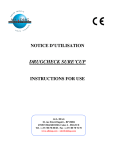
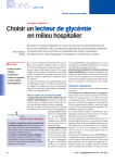
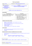
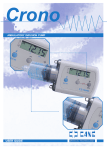
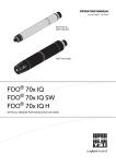
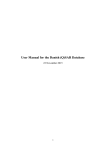
Lead-Finder
v.1.1.10
Software for Drug Discovery
User
Manual
Modeling the HIV-1 protease complex with U100313
is a particularly tough case for docking software.
Lead-Finder successfully predicts correct ligand
position (rendered in licorice) with 1.2Ǻ RMSD as
evidenced from X-ray structure (electron density rendered in wireframe). Lead-Finder v.1.1.10 User Manual
2
Table of contents
Data flow in Lead-Finder ........................................................................ 3
Protein Structure And Its Preparation For Docking ..................................... 4
Ligand Structure And Its Preparation For Docking ...................................... 5
Installation ........................................................................................... 6
Configuration Options for Docking ........................................................... 8
Command Line Options ........................................................................ 11
Example 1: Calculating energy grid maps. ........................................... 12
Example 1: Calculating energy grid maps. ........................................... 12
Example 2: Calculating free energy of binding. ..................................... 13
Example 3: Ligand docking with pre-calculated grid maps. ..................... 13
Example 4: Virtual screening. ............................................................ 14
Example 5: Preparation of trypsin structure for docking. ........................ 15
Example 6: Preparation of progesterone structure for docking. ............... 16
Example 7: Preparation of methanol dehydrogenase structure for docking.
..................................................................................................... 17
Example 8: Docking to cytochrome p450. ............................................ 18
Glossary ......................................................................................... 19
Lead-Finder v.1.1.10 User Manual
3
Data flow in Lead-Finder
Lead-Finder software is a command line application that is available for Windows and Linux
platforms. Lead-Finder takes 3D-coordinates of protein and ligand as input, along with a short
text file containing calculation parameters and global settings. Lead-Finder can be used to
perform:
- protein structure preparation;
- high-precision protein-ligand docking;
- estimation of the free energy of protein-ligand binding;
- virtual ligand screening to search for potent binders for a given protein target.
Protein structure
3D-coordinates of a protein
File formats: pdb, mol, gro, mol2
Source: PDB, in house structures,
molecular modeling…
Ligand structure
3D-coordinates of a ligand
File formats: mol, sdf, mol2, pdb, gro
Source: PDB, CCDC, in house
databases, molecular modeling…
Lead-Finder
1.
2.
2.
3.
Parameter file
Protein structure preparation
Calculation of energy grid maps
Ligand docking
Binding energy calculation
Parameters to specify
calculations
File format: text file
Energy estimations
Docked structure
Free energy of ligand binding, virtual
screening score, detailed output of
energy components for all docked
ligand poses
3D-coordinates of rank-ordered ligand
binding pose(s)
File format: pdb, mol, sdf
File format: text file
Lead-Finder v.1.1.10 User Manual
4
Protein Structure And Its Preparation For Docking
•
Common file formats 1 such as pdb, gro, mol2, containing 3D-coordinates of heavy
atoms and at least functional hydrogen atoms (such as ones attached to N, O, S) are
accepted as input for Lead-Finder.
•
3D-structure of a protein can be taken from publicly available or in-house databases,
or obtained through molecular modeling techniques. Accuracy of 3D protein structure
is crucially important for molecular docking applications. As a rule, high-resolution
(<2.5 Å) X-ray crystal structures perform better when alternatives exist.
•
Frequently, a protein structure, especially if comes from PDB, will contain no hydrogen
atoms. Lead-Finder requires appropriate placement of hydrogen atoms in the protein
structure. At least the functional hydrogens must be placed and the following points
must be considered: (i) protonation state of a protein depends on pH; (ii) protonation
of His should be reviewed as a special case; (iii) protons should be placed on the
fittest atom when alternatives exist, such as for chemically equivalent atoms in His,
Glu, Asp; (iv) proton orientation may have to be further optimized.
•
Lead-Finder can automatically prepare protein structure file by adding hydrogen
atoms, selecting ionization states of amino acids, optimizing positions of hydrogens,
etc. For this purpose special program called Model_build is included in Lead-Finder
distribution. Model_build accounts for electrostatic, Van der Waals and hydrogen
bonding energy when adding hydrogen atoms to a protein and optimizing their
positions, thereby performing high quality automatic structure preparation. Description
of electrostatic calculations and optimization algorithms implemented in Model_build
can be found in Technology and Bencmarking sections of Lead-Finder internet site.
•
Special care should be taken with respect to the defects in experimentally resolved
protein structures, such as missed atoms or residues, incorrect bond lengths, angles
etc., especially in the proximity to the ligand’s binding site 2 . Model_build automatically
repairs some of the widespread defects like incorrect aminoacid labels, missing or
unresolved side chains of aminoacids.
•
Protein cofactors important for ligand binding should be retained within the protein
structure. Model_build automatically adds hydrogen atoms to cofactors.
•
Any non-intrinsic parts of a protein such as ligand, water molecules, buffer ions, etc.
should be removed before docking calculations. Cofactors and (structurally or
catalytically important) metal ions bound to protein should be retained; sometimes,
conservative structural water molecule(s) known to play crucial role in ligand binding
may be retained as well.
1
Format converting programs such as Obabel, freely available at Hhttp://openbabel.sourceforge.net H,
may be used to convert your protein structure into one of the file formats listed above.
2
The quality of a protein structure may be assessed by a number of internet services available at the
PDB site Hwww.rcsb.orgH.
Lead-Finder v.1.1.10 User Manual
5
Ligand Structure And Its Preparation For Docking
•
Common file formats such as pdb, sdf, mol, mol2, gro containing 3D-coordinates of
heavy atoms and all hydrogen atoms are accepted as input for Lead-Finder.
•
3D-coordinates of a ligand can be taken from publicly available or in-house
databases, or obtained through molecular modeling techniques. 2D-structures of
ligands must be converted to 3D-coordinates before docking 3 .
•
Ligand structure must have all hydrogen atoms in correct places. Correct
protonation 4 must be performed before docking with Lead-Finder.
•
A single sdf file containing coordinates for multiple compounds may be used with
Lead-Finder. In this case, an additional brief file listing ligand names and
corresponding binding energies (dG and VS-score) will be produced for user
convenience.
-O
O
CH2
O
O
O
N
HC
CH
O
Crude 2D-structure of a ligand
CH
O
N
O-
2D-structure with all H-atoms
and ionization states adjusted to
pH 7. Note that the hydroxyl
group bound to the nitrogen
atom is ionized!
Complete 3D-structure
ACD/ChemSketch software (Hwww.acdlabs.comH) or Corina Hhttp://www.molecularnetworks.com/software/corina/index.htmlH) can be used for 3D-optimization of 2D-structures.
4
ACD/pKa software (Hwww.acdlabs.comH) can be used for predicting pKa and protonation of ionogenic
groups .
3
Lead-Finder v.1.1.10 User Manual
6
Installation
Once the Lead-Finder software license is obtained you get the installation distributive and the HASP
key (USB device) that manages your license. HASP device stores information about your license, so
when it comes off the license HASP device blocks the program exploitation. Updating license is
performed via a simple patch file described below.
The process of software installation under Windows and Linux is provided below.
Installation under Windows
Install the HASP drivers by executing either haspdinst.exe or HASPUserSetup.exe files located in
‘Drivers’ directory of your installation distributive. 5 Both haspdinst.exe and HASPUserSetup.exe
recognize your operating system and install the correct driver to the required location. The
difference between haspdinst.exe and HASPUserSetup.exe is that the former is a command line
application, while the latter has a graphical user interface.
After you have installed the HASP driver, copy files lead_finder.exe, dock_ff.lib and residue.lib
from the ‘Program’ directory of your installation distributive to the directory where you’d like to
install Lead-Finder. Edit your %PATH% environment variable by adding your installation path to
it. Now software is ready for use. Do not forget to keep HASP key inserted when running LeadFinder.
Installation under Linux
Install the HASP driver (aksusbd daemon) either from rpm package or by running installation
script5.
RPM packages corresponding to various Linux distributions are provided in the ‘Drivers’
directory of your installation distributive. 6 You can find there packages specially designed
for SuSe and RedHat distributions, however, to our experience, they will work for other
RedHat- and Debian-compatible Linux distributions. Also the latest versions of HASP drivers
for various Linux distributions are freely available at: http://www.hasp.com/downloads.
Alternatively, you may launch dinst script provided in the directory setup/aksusbd. This
script designed by HASP suppliers will automatically install the optimal driver for your
operating system. This script also sets up the aksusb daemon to start on boot.
After you have installed HASP driver, you need to have HASP device mounted properly. On
newer Linux distributions (SuSe 8.0 and higher, RedHat 9 and higher) it is mounted
automatically upon HASP driver installation. You can ensure if the device was mounted by
typing:
bash –c “if [ -e /proc/bus/usb ]; then echo 1; else echo 0; fi;”
and examining the output.
If the HASP device was not mounted automatically, type the following string to mount it:
mount -t usbdevfs none /proc/bus/usb5
To install Lead-Finder software copy files lead_finder, dock_ff.lib and residue.lib from the
Program directory of your installation distributive to the directory /usr/local/bin. Please make sure
that ownership and access permissions are properly set up.
Importantly that under Linux you must have aksusbd daemon launched before running Lead-Finder
software. Aksusbd daemon is launched with the following command:
<path>/aksusbd
Contact your system administrator for technical questions.
Do not forget to keep HASP key inserted when running Lead-Finder.
Please note that you will need to install HASP LM in order to communicate with your network
HASP key. Please refer to HASP HL manual for the details.
Administrator/root privileges are required for installation.
Additional information on particular drivers for different Linux distributions can be found in
readme.txt file in the ‘Drivers’ directory.
5
6
Lead-Finder v.1.1.10 User Manual
7
Updating software license
To update your license insert HASP key and run program update_key. The program will
prompt you to retrieve HASP key information (i) or to update the key (u).
Choose option (i), this will generate ‘client-to-vendor’ file, which contains information about
the current status of your license.
Send this file to us by e-mail, and you’ll receive file ‘vendor-to-client’ which contains updated
license information.
Run program update_key, choose option u this time and select your updated file. Now your
license is renewed.
Lead-Finder v.1.1.10 User Manual
8
Configuration Options for Docking
You can set up Lead-Finder tasks by specifying parameters in a par file. par file is a humanreadable text file containing references to ligand and protein structure, instructions to energy
grid map calculations and optional preparation of protein structure. The order of parameters
in the parameter file is arbitrary. Single-line comments are allowed after the semi-colon “;”
symbol.
Ligand file
ligand = "filename"
Optional parameter; no default value
Example: ligand = "ligand.mol"
Specifies name of a file in mol, sdf, mol2, pdb or gro format containing ligand 3Dcoordinates. When Lead-Finder is run with –li as a command line option, this line of par
file is ignored, and the file specified after –li is used instead.
ligand_reference = "filename"
Optional parameter; no default value
Example: ligand_reference = "ligand_reference.pdb"
Specifies name of a file containing such reference ligand from PDB or other source that
is positioned in the same binding site of the same protein and in the same coordinate
system. This option is useful when you already have at least one resolved protein-ligand
structure and want to dock new ligands to the same protein’s site. The reference
structure is then used to determine the center and the size of energy grid maps. If the
reference ligand coincides with the docked ligand, which may be the case when checking
program’s performance on experimentally resolved structures, RMSD between the
reference and predicted ligand poses will be calculated and saved in a log file. Note that
reference ligand does not necessarily be a chemically consistent structure; when actual
reference ligand is missing, you can take some points in the ligand binding site and
write them into reference ligand structure (say in pdb format); this may help in
determining overall placement of docking solutions.
additional_reference = "filename1"
additional_reference = "filename2"
…
Optional parameter; no default value
Example: additional_reference = "ligand_reference1.pdb"
Specifies coordinates of additional reference ligands (if needed). Additional reference
ligands can be used for adequate RMSD calculations of docked ligand pose and reference
ligand when a number of symmetric ligand-binding positions are valid. In such case,
when a number of corresponding (symmetry related) reference ligand positions are
provided, RMSD will be calculated with respect to the nearest reference. Anyway,
accuracy of ligand docking does not depend on additional reference ligands.
covalent = A B X
Optional parameter; no default value
Example: covalent = 2 2542 1.542
When ligand is covalently bound to a protein, numbers of bonded atoms must be
provided. Specify A as the ligand’s bonded atom number, and B as the protein’s bonded
atom number. The enumeration for ligand and protein starts from zero. Additional
parameter X specifies covalent bond length linking ligand to the protein. This parameter
is optional (specifying A and B will be enough in principle) but highly recommended,
since when ligand coordinates are arbitrary (which is a common case), specified bond
length will position ligand correctly with respect to protein. Ignoring X is valid only when
ligand coordinates are already compatible with covalent bond constraints.
Lead-Finder v.1.1.10 User Manual
9
Protein file
receptor = "filename"
Optional parameter; no default value
Example: receptor = "protein.pdb"
Specify name of a file in pdb, gro, mol2 formats containing protein coordinates. When
Lead-Finder is run with –mm as a command line option, this line of par file is ignored
and the file specified after –mm is used instead. Atom names (of amino acid residues,
cofactors, metal ions, etc.) listed in protein coordinate file must correspond to standard
atom names (contained in dock_ff.lib library file).
exclude_water = yes | no
Optional parameter; default value is yes
Example: exclude_water = no
Specify this option to delete water molecules from protein structure file or retain them
during docking calculations. This option can be useful when structurally conserved water
molecule(s) is(are) necessary for ligand binding.
metal = A B
Optional parameter; no default value
Example: metal = 30 6
When automatic determination of a metal coordination number is ambiguous, it may be
useful to explicitly define the metal coordination number. A is the metal atomic number
(enumeration starts from zero). B is the metal coordination number that is used in the
calculation of a directional potential of metal-ligand interactions. When there are several
metal ions in the ligand binding site, each ion can be described by its own string.
Energy grid maps
grid_center = reference | X Y Z
Required parameter; default value is reference
Example: grid_center = 13.5 46.3 10.4
Choose one of the two methods for grid center placement: reference – center at the
reference ligand if one is used. Alternatively, specify three Cartesian coordinates in X, Y
and Z (in Å).
grid_spacing = X
Required parameter; default (and recommended) value is 0.375
Example: grid_spacing = 0.375
Distance between neighboring grid points along particular Cartesian direction (in Å).
grid_size = reference X | A B C
Required parameter; no default value
Example: grid_size = 10 10 10
Choose one of the two methods for grid size definition: (1) reference – a grid with X Å
margin in each dimension from the reference ligand, default value of margin is 6 Å; (2)
an explicit definition grid_size = A B C with three dimensions (lengths) of the grid box
edges (in Å). Alternatively, grid size can be defined by setting the number of grid points
with grid_npoints parameter instead.
grid_npoints = A B C
Optional parameter; no default value
Example: grid_npoints = 120 60 65
Specify grid size in a number of grid points in each dimension. grid_npoints and
grid_size parameters are mutually exclusive.
Lead-Finder v.1.1.10 User Manual
10
grid_atom_types = auto | common | <individual atoms>
Optional parameter; default value is auto
Example: grid_atom_types = auto
Specify atoms for which grid maps will be calculated: auto – automatic extraction of
atoms from ligand structure; common – all basic atoms (C, A, N, NX, O, S, H, P, F, Cl,
Br, I) 7 ; also, individual atoms can be specified, for example: grid_atom_types = C A N
NX O H. Specify atom types with care, since during virtual screening of a big library of
compounds different ligand atom types may emerge, so that it is better to calculate
more grids than to lack them during high-throughput calculations.
reorient_grid = yes | no
Optional parameter; default value is yes
Example: reorient_grid = yes
Specify this parameter to reorient grids to maximally overlap with the binding site, as
determined by the built-in cavity detection algorithm. Reorientation allows reduction of
the grid size due to more efficient spatial arrangement.
exclude_volume = V
Optional parameter; default value is 40.5 (the volume of benzene molecule)
Example: exclude_volume = 40.5
3
Do not consider intramural protein cavities with volume less than V (in Å ).
Other docking parameters
n_out = N
Optional parameter; default value is 20
Example: n_out = 20
Specify number of predicted docked positions (poses) to be output.
rot_sulfamide = yes | no
Optional parameter; default value is no
Example: rot_sulfamide = yes
Turn on/off the rotation of sulfamide bond during docking.
rot_amide = yes | no
Optional parameter; default value is no
Example: rot_amide = yes
Turn on/off the rotation of amide bond during docking.
rot_conjugated = yes | no
Optional parameter; default value is no
Example: rot_conjugated = no
Turn on/off the rotation of conjugated double or aromatic bonds during docking.
environment = solution | membrane
Optional parameter; default value is solution
Example: environment = membrane
Choose the type of environment surrounding the ligand binding site. Choose membrane
when ligand binds to the membrane-buried protein site; in this case specially adjusted
settings of electrostatic calculations (and other energy terms) will be activated. Default
environment is solution.
7
A – is aromatic carbon, NX – is a nitrogen that doesn’t form H-bonds (for example, an amide
nitrogen).
Lead-Finder v.1.1.10 User Manual
11
Command Line Options
You can use command line parameters to set-up Lead-Finder tasks and override some
configuration options specified in a par file. The following command line parameters are
accepted:
-f [file.par]
The docking parameter file. Default docking settings will be used if this parameter is not
specified. See” for more details.
-li [ligand.mol]
The ligand structure in a mol, sdf, mol2, pdb or gro file.
-mm [protein.pdb]
The macromolecule (protein) structure in a pdb, gro, mol2 file.
-og [grid.bin]
Calculate and save grid maps after docking. The acceptable grid file formats are .bin
(binary format) and .map (human-readable format). Grid files with .map extension can
be visualized with VMD software, but they are larger in size and are slower to load than
the binary files.
-g [grid.bin]
Load grid maps from file and skip grid map calculations.
-o [solutions.pdb]
Output 3D-coordinates (pdb) of docked poses.
-os [brief_output.log]
Output only ligand’s name and calculated energy (dG and VS score) into a text file. This
option may be useful when docking many ligands from an sdf file.
-omm [protein.pdb]
Output a protein structure file in pdb, gro, mol2 formats. The binary file with protein
structure can be used subsequently as input for docking as it allows faster processing.
-l [solutions.log]
Output free energy of ligand binding for each docked pose in a human-readable log file.
-task [dock, scr]
Specify one of the two available lists of settings for the conformational search algorithm:
docking (dock) or virtual screening (scr). Settings are optimized with respect to docking
speed/robustness with screening being the fastest and docking – the most precise
(details can be found in the Technology section of Lead-Finder internet site).
-mode [norm, grid, check, dG]
Choose one of the following modes of operation: norm - run docking calculations; grid –
calculate energy grid maps without doing docking; check – verbose mode, check the
ligand and protein structure at input and diagnose problems; dG – calculate free
energies of binding.
-h
Print help for using command-line options.
-v
Display information on structure preparation and docking while calculations are in
progress.
-debug
Dump of intermediate results, which can be sent to Lead-Finder developers for
maintenance purposes.
Lead-Finder v.1.1.10 User Manual
12
Example 1: Calculating energy grid maps.
Lead-Finder may be used to calculate energy grid maps only, without doing docking or other
tasks. This function is useful when you want to calculate grid maps and store them for future
use with other docking experiments, thereby reducing the amount of processing and speeding
up docking calculations.
The folder Example 1 contains a 3D-structure of streptavidin (PDB 1SRE) in protein.gro and
model settings for energy grid calculations in 1sre.par. The settings in 1sre.par are described
below:
receptor = "protein.gro“
exclude_water = yes
; the protein structure will be taken from "protein.gro" file
; water molecules will be removed from the protein
structure
grid_center = 33 13 -6
; set the grid center at (33;13;-6) XYZ coordinates, in Å
grid_size = 15 15 15
; the grid will span 15 Å in each direction
grid_atom_types = common
; grid maps will be calculated for all types of atoms
reorient_grid = no
; do not reorient grid to enable visualization of a ligand
with the protein structure
Start grid calculations with the following command line:
Lead_finder -f 1sre.par -mode grid -og 1sre.map
The calculated energy grid maps will be saved in 1sre.map file. Note that map files are loaded
to memory sufficiently slower than bin files; thus for re-using calculated energy grid maps in
future docking experiments it is practical to save them as bin files.
The advantage of map files (map is AutoDock file format) is that they can be visualized with
freely available VMD software. Also, this option may be useful in qualitative analysis of ligand
binding forces.
This is an example of visualization of grid
map files with VMD software. The
structure of streptavidin, 1SRE, is
rendered in ‘new cartoon’, reference
ligand (2-((4'-hydroxyphenyl)azo)benzoic acid) - in licorice, VdW grid in gray wireframe, H-bond donor grid – in
blue wireframe. The reference ligand’s
shape seems to fit well into the protein’s
binding site and the ligand’s carboxylic
group is placed correctly for H-bonding
interactions.
Other grid maps (electrostatic, etc.) may
be loaded to VMD and visualized as well.
Lead-Finder v.1.1.10 User Manual
13
Example 2: Calculating free energy of binding.
Lead-Finder can be used to calculate free energy (ΔG) of ligand binding to protein when
structure of the protein-ligand complex is available (from X-ray, NMR, molecular modeling
studies, etc.). For this purpose the program should be launched in -mode dG and the
structures of protein and ligand from the complex should be provided in separate files.
The folder Example 2 contains a 3D-structure of HIV-1 protease (PDB 1HSG) in protein.gro
and a structure of the ligand in ligand.mol. The coordinates of protein’s and ligand’s heavy
atoms were taken from PDB and necessary hydrogen atoms were added. As a first step, LeadFinder calculates energy grid maps. Then, it performs optimization of ligand’s position and
calculates free energy of ligand binding. The file 1hsg.par contains parameters for the free
energy of binding calculations:
ligand = "ligand.mol"
; the ligand
ligand_reference = "ligand.mol"
; the reference ligand
receptor = "protein.gro"
; the protein structure
exclude_water = yes
; remove water molecules will from the protein structure
grid_center = reference
; place the grid center in the geometric center of
the reference ligand
grid_size = reference
; determine the grid size by the reference ligand
grid_atom_types = auto
; calculate grid maps for all types of atoms present
in “ligand.mol”
Start calculations of free energy of binding with the following command line:
Lead_finder -f 1hsg.par -mode dG
The free energy of binding and its decomposition to different terms will be saved in text log
file. The experimental value of the free energy of binding is –13.2 kcal/mol.
Example 3: Ligand docking with pre-calculated grid maps.
When energy grid maps are available (were calculated earlier), they can be directly used for
docking without recalculating them. For this purpose key –g in the command line should be
used to point to the grid map file.
Folder Example 3 contains streptavidin 3D-structure (protein.gro) ready for docking, grid map
file (1sre.map), biotin structure (ligand.mol) and parameter file (1sre.par) described in
Example1.
Docking with pre-calculated grid can be launched with the string:
Lead_finder -f 1sre.par -task dock -g 1sre.map
Calculated ligand poses will be stored in solution.pdb file, energies – in solution.log.
Lead-Finder v.1.1.10 User Manual
14
Example 4: Virtual screening.
Lead-Finder can be used for docking a big number of ligands into single protein thus acting as
in silico analogue of high-throughput screening. To achieve the fastest docking with LeadFinder key –task scr should be used in the command line.
To perform virtual screening energy grid maps should be calculated first. Then, depending on
the configuration of your computational resource (PC, a cluster of PCs, etc.), successive or
parallel execution of Lead-Finder should be applied to all ligands of interest. This can be done
in many ways, for example using Perl scripting; consult your system administrator how to
organize screening in your case.
Folder Example 4 contains all necessary files to perform virtual screening in a successive
mode (on a single PC): 3D-structure of human serum albumin (protein.gro) prepared from
PDB entry 1B5J; set of decoy ligands (directory decoys); set of true albumin ligands
(directory ligands); parameter file (screen.par); and a Perl script (do_screen.pl for Linux or
do_screen_win.pl for Windows), which launches grid calculations and successive docking of all
ligands.
Results of screening can be processed (by another script) and gathered in a single file
(stats.txt), which can be analyzed with standard table processing software (say MS Excel).
Enrichment curve for virtual screening of human serum albumin ligands: X-axis represents
the fraction of active albumin ligands (known to bind with albumin with good affinity), Y-axis
represents the fraction of decoy ligands. Each point on the curve reveals the ratio of true
positives (active ligands) to false positives (decoys) at the certain fraction of the library
screened. Totally 16 active ligands and 84 decoys (randomly chosen from the set of drug-like
compounds) were used for screening. Results show that in the list of compounds ranged by
their calculated affinity only one decoy compound occupies 14-th place, while active ligands
occupy 1-13 and 15-17 places. ROC 8 - integral parameter describing the accuracy of virtual
screening – equals 0.9987, which is excellent result.
8
ROC – receiver operating characteristic – corresponds to the area under the normalized enrichment
curve in coordinates: fraction of found true active compounds (from 0 to 1) vs fraction of decoys (from 0
to 1). Ideal curve reflecting 100% of true actives found at 0% of decoys gives ROC = 1.00.
Lead-Finder v.1.1.10 User Manual
15
Example 5: Preparation of trypsin structure for docking.
This example illustrates the necessary steps of protein and ligand structure preparation for
docking.
1. Download trypsin structure (1TNL) from PDB at www.rcsb.org
2. Cut the ligand’s (trans-2-phenylcyclopropylamine) coordinates (with any text editor) and
save it as a separate file ligand_reference.pdb.
3. As far as a number of protein residues, for which alternative protonation states may take
place, appear in the ligand’s proximity, special care should be taken while adding
hydrogen atoms to PDB structure. For this purpose you may launch Model_build program
supplied with Lead-Finder distribution
Model_build –f 1tnl.pdb –o protein.gro –ph 7
Next, ligand structure should be prepared for docking. For this purpose all hydrogen
atoms must be added to ligand. ACD software, for example, may be used for adding all
hydrogens. Note that correct ionization of the ligand must be performed; this can be done
with ACD/pKa module. In principle, any third party software may be used that can
correctly add hydrogens to the ligand.
4.Docking may be launched with the string:
Lead_finder -f 1tnl.par -o 1tnl_solution.pdb -l 1tnl_energy.log
Coordinates of found solutions will be written in 1tnl_solution.pdb file and energies – in
1tnl_energy.log.
Lead-Finder v.1.1.10 User Manual
16
Example 6: Preparation of progesterone structure for docking.
This example outlines the necessity of careful handling with structures from PDB, which may
have experimental defects like unresolved amino acid residues in protein or distorted ligand’s
geometry.
1. Download progesterone receptor structure (1A28) from PDB at www.rcsb.org
2. Cut the ligand’s (progesterone) coordinates (with any text editor) from pdb file and save it
as a separate file ligand_reference.pdb. Note that pdb file contains two identical protein
chains, each having bound ligand. Thus, delete one chain (with its ligand) from the
original pdb file.
3. In 1A28 structure some of the residues are not resolved experimentally. You can build
them with freely available Swiss-PDB Viewer program (or other proper software). Save
structure with added residues in a separate file 1a28_corrected.pdb.
4. As far as a number of protein residues, for which alternative protonation states may take
place, appear in the ligand’s proximity, special care should be taken in adding hydrogen
atoms to PDB structure. You may use Model_build program for this purpose (as described
in Example 5).
5. Next, convert ligand’s coordinates to mol file as it was described in Example 5.
6. Pay attention to the fact that ligand’s geometry in 1A28 structure was initially distorted
and due to this obstacle mol file contains wrong double bonds. You must correct bond
orders before adding hydrogens. This can be done in ACD/ChemSketch (or other proper
software) via graphical menu.
7. When bonds in the mol file are corrected, hydrogen atoms must be added to the ligand.
This can be done as described in Example 5.
8. When protein and ligand structures are ready, docking parameter file should be provided
(by analogy with Example 5) and docking launched with the string:
Lead_finder -f 1a28.par -o 1a28_solution.pdb -l 1a28_energy.log
Coordinates of solutions will be written in 1a28_solution.pdb file and energies – in
1a28_energy.log.
Superposition of deficient ligand
(progesterone) structure extracted from PDB
(white) and energy-optimized structure
(cyan). Seemingly small differences of the
overall ligand geometry (almost all bond
lengths in PDB structure are distorted by 0.20.4 Ǻ) influence docking precision.
Lead-Finder v.1.1.10 User Manual
17
Example 7: Preparation of methanol dehydrogenase structure for
docking.
This example outlines the necessity of careful handling with target protein structure, which
may need to retain non-protein residues (metal ions, water, etc) essential for catalytic
competency of a protein.
1. Download methanol dehydrogenase structure (4AAH) from PDB at www.rcsb.org
2. Cut the ligand’s (pyrroloquinoline quinone) coordinates (with any text editor) from pdb file
and save it as a separate file ligand_reference.pdb. Delete unnecessary protein chains (B
and D) from the original pdb file.
3. This enzyme contains catalytically important calcium ion and water molecule (residue
number 785), which should be retained in the structure, while other water molecules may
be deleted from pdb file.
4. Add hydrogen atoms to protein as it was described earlier. Note that orientation of
hydrogen atoms in water molecule bound to calcium should be correct. Correction of
water orientation can be performed for example with freely available VMD software.
5. Add all hydrogen atoms to ligand as it was described earlier.
6. When protein and ligand structure files are ready, remember that we want to retain
catalytically important water molecule for docking, so chose exclude_water = no in par
file.
7. To demonstrate flexibility of grid maps definition in Lead-Finder try setting the grid center
explicitly via three Cartesian coordinates. Par file will look like:
ligand = "ligand.mol"
; ligand will be taken from "ligand.mol“
ligand_reference = "ligand_reference.pdb " ; reference ligand will be taken from
"ligand.pdb“
receptor = "protein.gro"
; protein will be taken from "protein.gro“
exclude_water = no
; water will not be excluded
grid_center = -15 19 15
; XYZ coordinates of grid center
grid_size = reference
; grid size will be determined by reference ligand
grid_atom_types = auto
; grid maps will be calculated for all types of atoms
present in “ligand.mol”
8. Launch docking with the most precise settings of search algorithm with key –task dock
standing for the default docking regime:
Lead_finder -f 4aah.par -o 4aah_solution.pdb -l 4aah_energy.log -task dock
Coordinates of found solutions will be written in 4aah_solution.pdb fileand energies – in
4aah_energy.log.
9. You may want to compare Lead-Finder docking accuracy achieved with different settings
of docking search algorithm. Launch docking using already generated structure, grid and
parameter files but with different values of key –task equal: dock (default docking
settings), scr (fast screening mode).
10. .Compare accuracy of structure prediction (RMSD is calculated in 4aah_energy.log file for
each pose when reference ligand is provided) and speed of calculations (CPU time is also
provided in 4aah_energy.log) 9 .
9
The following results were achieved by us (using AMD Athlon XP 1.8 GHz with 2 GB RAM) : screening
mode - 0.74 Ǻ (RMSD), 2 s (CPU time); docking mode - 0.70 Ǻ, 5 s.
Lead-Finder v.1.1.10 User Manual
18
Example 8: Docking to cytochrome p450.
This example outlines the necessity of careful handling with target protein structure, which
may need to retain cofactor molecules essential for catalytic competency of a protein.
1. Download cytochrome p450cam structure (5CPP) from PDB at www.rcsb.org
2. Cut the ligand’s (adamantanone) coordinates (with any text editor) from pdb file and save
it as a separate file ligand_reference.pdb.
3. This enzyme contains catalytically important cofactor – heme, which is essential for
cytochrome substrate binding, and must be retained for docking.
4. Add hydrogen atoms to protein as it was described earlier.
5. Add all hydrogen atoms to ligand as it was described above.
6. Use standard docking parameter file (as described in previous examples).
7. Launch docking with key –task scr standing for fast screening regime:
Lead_finder -f 5cpp.par -o 5cpp_solution.pdb -l 5cpp_energy.log -task scr
8. You may want to compare Lead-Finder docking accuracy achieved with different settings
of docking search algorithm. Launch docking with different key –task values and compare
accuracy of structure prediction and speed of calculations (as it was described in Example
7) 10 .
10
The following results were achieved by us (using AMD Athlon XP 1.8 GHz with 2 GB RAM): screening
mode - 1.16 Ǻ (RMSD), 0.9 s (CPU time); docking mode - 0.60 Ǻ, 2 s.
Lead-Finder v.1.1.10 User Manual
19
Glossary
Binding affinity – free energy of protein-ligand binding (kcal/mol).
dG score – Lead-Finder estimation of binding affinity.
Docking – positioning of the ligand in the protein binding site with respect to optimal free
energy of binding. For details see Technology section of Lead-Finder internet site.
Energy grid – 3D function placing in accordance a point in Cartesian space and energy of
particular type of interaction, which would atom of particular type possess being
placed at this point. Correspondingly, grids are calculated for each type of interactions
and each atom type.
HASP key – USB device that controls your software license
Virtual screening – rank-ordering of compounds (from particular library) according to
estimated binding potency with respect to particular protein. Lead-Finder docks each
compound from the library and calculates VS-score for docked ligand, which serves a
quantitative measure of ligand activity (binding potency).
VS score - Lead-Finder estimation of ligand activity (binding potency) for rank-ordering
ligands during virtual screening. For details see Technology section of Lead-Finder
internet site.
Lead-Finder v.1.1.10 User Manual