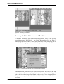
1
Version 6
User Manual
VIDEO
© 2006 BRUKER OPTIK GmbH, Rudolf-Plank-Str. 27, D-76275 Ettlingen, www.brukeroptics.com
All rights reserved. No part of this publication may be reproduced or transmitted in any form or by any
means including printing, photocopying, microfilm, electronic systems etc. without our prior written
permission. Brand names, registered trade marks etc. used in this manual, even if not explicitly marked
as such, are not to be considered unprotected by trademarks law. They are the property of their respective owner.
The following publication has been worked out with utmost care. However, Bruker Optik GmbH does
not accept any liability for the correctness of the information. Bruker Optik GmbH reserves the right to
make changes to the products described in this manual without notice.
This manual is the original documentation for the OPUS spectroscopic software.
Table of Contents
1
Introduction . . . . . . . . . . . . . . . . . . . . . . . . . . . . . . . . . . . . . . . . . . . . . . 1
2
Hardware Requirements . . . . . . . . . . . . . . . . . . . . . . . . . . . . . . . . . . . . 3
2.1
2.2
2.3
2.4
Spectrometer . . . . . . . . . . . . . . . . . . . . . . . . . . . . . . . . . . . . . . . . . . . . . . . . . .3
x/y Sample Stage . . . . . . . . . . . . . . . . . . . . . . . . . . . . . . . . . . . . . . . . . . . . . . .3
Video Camera . . . . . . . . . . . . . . . . . . . . . . . . . . . . . . . . . . . . . . . . . . . . . . . . .3
Microscopes . . . . . . . . . . . . . . . . . . . . . . . . . . . . . . . . . . . . . . . . . . . . . . . . . . .4
3
Software Requirements . . . . . . . . . . . . . . . . . . . . . . . . . . . . . . . . . . . . . 5
4
Installing the Video Hardware . . . . . . . . . . . . . . . . . . . . . . . . . . . . . . . 7
4.1
4.2
4.3
4.4
5
Configuring the Video Hardware . . . . . . . . . . . . . . . . . . . . . . . . . . . . 11
5.1
5.2
5.3
5.4
5.5
5.6
6
Display Settings . . . . . . . . . . . . . . . . . . . . . . . . . . . . . . . . . . . . . . . . . . . . . . . .7
Firmware Check (IRscope II & Hyperion) . . . . . . . . . . . . . . . . . . . . . . . . . . .7
Removing the Optical Camera Adapter (IRscope II and Hyperion only) . . . .8
Orientation of the Video Camera . . . . . . . . . . . . . . . . . . . . . . . . . . . . . . . . . . .8
Setting up the Mapping Device . . . . . . . . . . . . . . . . . . . . . . . . . . . . . . . . . . .11
Setting up the Imaging Device . . . . . . . . . . . . . . . . . . . . . . . . . . . . . . . . . . .12
5.2.1
Imaging Device Options . . . . . . . . . . . . . . . . . . . . . . . . . . . . . . . . .15
5.2.2
Video Options . . . . . . . . . . . . . . . . . . . . . . . . . . . . . . . . . . . . . . . . .20
5.2.3
Define Useful Settings for . . . . . . . . . . . . . . . . . . . . . . . . . . . . . . . .26
Selecting the Motorized Aperture Option . . . . . . . . . . . . . . . . . . . . . . . . . . .28
X/Y Stage . . . . . . . . . . . . . . . . . . . . . . . . . . . . . . . . . . . . . . . . . . . . . . . . . . .29
5.4.1
Checking the operational Reliability of the Stage . . . . . . . . . . . . . .29
5.4.2
Stage Setup . . . . . . . . . . . . . . . . . . . . . . . . . . . . . . . . . . . . . . . . . . .31
Selecting Measurement Parameters . . . . . . . . . . . . . . . . . . . . . . . . . . . . . . . .34
Offset Correction . . . . . . . . . . . . . . . . . . . . . . . . . . . . . . . . . . . . . . . . . . . . . .34
Video-assisted Measurement . . . . . . . . . . . . . . . . . . . . . . . . . . . . . . . . 35
6.1
6.2
6.3
6.4
General Information . . . . . . . . . . . . . . . . . . . . . . . . . . . . . . . . . . . . . . . . . . .35
Window for video-assisted Measurement . . . . . . . . . . . . . . . . . . . . . . . . . . .35
6.2.1
Video Setup Dialog (for FT-IR Microscopes) . . . . . . . . . . . . . . . .38
6.2.2
Video Setup Dialog (for Raman Microscopes) . . . . . . . . . . . . . . . .44
Functions of the Pop-up Menu Item ‘‘Mouse Mode’’ . . . . . . . . . . . . . . . . . 45
Performing a Video-assisted Measurement with 3D Mode . . . . . . . . . . . . . .47
6.4.1
General Procedure . . . . . . . . . . . . . . . . . . . . . . . . . . . . . . . . . . . . . .47
6.4.2
Snapshot and Overview Image . . . . . . . . . . . . . . . . . . . . . . . . . . . .47
6.4.3
Performing a Background Measurement . . . . . . . . . . . . . . . . . . . . .49
6.4.4
Performing a Sample Measurement . . . . . . . . . . . . . . . . . . . . . . . .50
6.4.5
Performing Repeated Video Measurements . . . . . . . . . . . . . . . . . .51
6.4.6
Predefining Measurement Positions . . . . . . . . . . . . . . . . . . . . . . . .53
6.5
7
57
58
58
59
59
60
60
61
62
62
62
63
Special OPUS Windows . . . . . . . . . . . . . . . . . . . . . . . . . . . . . . . . . . . . 65
7.1
7.2
7.3
7.4
7.5
7.6
7.7
8
6.4.7
Editing Measurement Positions . . . . . . . . . . . . . . . . . . . . . . . . . . .
6.4.8
Measuring Marked Positions . . . . . . . . . . . . . . . . . . . . . . . . . . . . .
6.4.9
Displaying the Spectrum of certain Measurement Position . . . . .
6.4.10 Exporting Images and Measurement Positions . . . . . . . . . . . . . . .
6.4.11 Deleting Images or Measurement Positions . . . . . . . . . . . . . . . . .
6.4.12 Image Processing . . . . . . . . . . . . . . . . . . . . . . . . . . . . . . . . . . . . . .
6.4.13 Illumination Correction . . . . . . . . . . . . . . . . . . . . . . . . . . . . . . . . .
6.4.14 Saving Data and Closing the Video Window . . . . . . . . . . . . . . . .
Video-assisted Measurement without 3D . . . . . . . . . . . . . . . . . . . . . . . . . .
6.5.1
General Information . . . . . . . . . . . . . . . . . . . . . . . . . . . . . . . . . . . .
6.5.2
Measurement . . . . . . . . . . . . . . . . . . . . . . . . . . . . . . . . . . . . . . . . .
6.5.3
Saving Video Images . . . . . . . . . . . . . . . . . . . . . . . . . . . . . . . . . . .
Opening a special OPUS Window . . . . . . . . . . . . . . . . . . . . . . . . . . . . . . . .
3D Window . . . . . . . . . . . . . . . . . . . . . . . . . . . . . . . . . . . . . . . . . . . . . . . . .
3D+ Window . . . . . . . . . . . . . . . . . . . . . . . . . . . . . . . . . . . . . . . . . . . . . . . .
Map+Spec-Window . . . . . . . . . . . . . . . . . . . . . . . . . . . . . . . . . . . . . . . . . . .
Map+Video+Spec-Window . . . . . . . . . . . . . . . . . . . . . . . . . . . . . . . . . . . . .
Overview Window . . . . . . . . . . . . . . . . . . . . . . . . . . . . . . . . . . . . . . . . . . . .
VideoPostrun Window . . . . . . . . . . . . . . . . . . . . . . . . . . . . . . . . . . . . . . . . .
65
65
66
67
68
68
69
Motorized Knife Edge Aperture . . . . . . . . . . . . . . . . . . . . . . . . . . . . . 71
8.1
8.2
Changing the Aperture Settings . . . . . . . . . . . . . . . . . . . . . . . . . . . . . . . . . . 71
Measuring at pre-defined Positions . . . . . . . . . . . . . . . . . . . . . . . . . . . . . . . 75
Appendix . . . . . . . . . . . . . . . . . . . . . . . . . . . . . . . . . . . . . . . . . . . . . . . . 77
A.1
A.2
A.3
A.4
A.5
A.6
Rotation of the Camera . . . . . . . . . . . . . . . . . . . . . . . . . . . . . . . . . . . . . . . .
Image as received by the Detector . . . . . . . . . . . . . . . . . . . . . . . . . . . . . . . .
IRscope II and Hyperion without Optical Camera Adapter . . . . . . . . . . . . .
IRscope II and Hyperion with Optical Camera Adapter . . . . . . . . . . . . . . .
IRscope I . . . . . . . . . . . . . . . . . . . . . . . . . . . . . . . . . . . . . . . . . . . . . . . . . . .
Ramanscope . . . . . . . . . . . . . . . . . . . . . . . . . . . . . . . . . . . . . . . . . . . . . . . . .
77
78
79
79
79
80
1
Introduction
OPUS/VIDEO allows to display a live image of a sample as viewed through the
ocular of a microscope on a video screen. In combination with a computer-controlled x/y sample stage and the OPUS/MAP software, a surface can be mapped
and an overview image generated from multiple video frames. Spectral information can be collected from the sample surface in correlation to these images.
In order use to the video functions, a frame grabber board and a video camera
has to be installed.
Bruker Optik GmbH
OPUS/VIDEO
1
Introduction
2
OPUS/VIDEO
Bruker Optik GmbH
Spectrometer
2
Hardware Requirements
To ensure an optimum functioning of the OPUS/VIDEO software certain hardware requirements must be met. The computer requirements depend on the following used components:
• Spectrometer
• x/y-sample stage of the microscope
• Video equipment (video camera and frame grabber)
2.1
Spectrometer
Spectrometers connected to the PC using an Ethernet cable (Tensor, Vertex,
IFS125, Senterra) require an Ethernet interface. In case they are to be integrated
also into a LAN another Ethernet interface (Ethernet card) may be necessary.
Acquisition processor controlled spectrometers (e.g. Vector22, IFS28, IFS55,
IFS66, IFS88, IFS120) need to be equipped with an ISA slot.
2.2
x/y Sample Stage
The x/y sample stage can be controlled either by an internal controller (LSTEP
card of the manufacturer LANG) or an external controller (of the manufacturer
LANG) or a CANBUS controller. The internal controller requires a long PCI
slot and a high-capacity PC power supply unit because the stage stepper motors
are supply with power by the PC. Alternatively, the power supply of the LSTEP
card can be realized by means of an external power supply unit. This variant is
especially recommendable if the PC has to supply also the digital camera with
power via an USB interface. In case of the external LANG controller, the PC
must be equipped with a RS-232 (COM)-interface. The CANBUS controller
does not require any additional PC resources.
2.3
Video Camera
Video images can be acquired by either an analog video camera plus frame
grabber or a digital video camera which is connected to the USB interface. The
frame grabber is a plug-in card (DT3131 or the older version DT3153 manufactured by Data Translation) that occupies a short PCI slot. When using a digital
video camera, an USB 2.0-interface must be available. (USB 1.x is not sufficient!) Moreover, a high image resolution requires a fast CPU (> 1GHz). In
addition, the PC display needs to be DirectShow-compatible and has to be able
to show overlays over the live video image. In case of PCs with the display
implemented on the motherboard, the latter requirement can cause problems
and possibly can make an additional display plug-in board necessary.
Bruker Optik GmbH
OPUS/VIDEO
3
Hardware Requirements
An instrument configuration consisting of a Tensor or Vertex spectrometer, a
Hyperion microscope, a x/y sample stage and the digital video camera Infinity1
require, for example, the following hardware components:
•
•
•
•
Short PCI slot for LAN integration (if desired)
Long PCI slot for LSTEP card (for controlling the x/y stage)
USB 2.0 interface
High-capacity PC power supply unit, or alternatively an external
power supply unit for the LSTEP card
• DirectShow-compatible display that is capable of showing overlays
(possibly a separate display card that requires a AGP or PCI slot)
If an analog video camera is used instead of the digital camera Infinity1, the
USB 2.0 interface and, normally, an external power supply unit are not needed.
Instead, another PCI slot for the frame grabber card is required.
2.4
Microscopes
The following types of microscopes are supported:
•
•
•
•
•
•
•
IRscope I/II
Hyperion 1000/2000/3000
RamanScope I/II/III
Senterra (dispersive Raman microscope)
EOS (FT Raman accessory)
Helios (MIR ATR unit)
IMAC (macrochamber for measurements using the FPA detector)
IRScope II and Hyperion enable a computer-controlled switching between
reflectance mode, transmittance mode, VIS, IR and IR/VIS mode. Moreover,
the software can read theses settings from the microscope and is capable of auto
focussing, provided these options have been purchased. For reading out the
microscope settings, firmware version 1.5 or higher is required.
4
OPUS/VIDEO
Bruker Optik GmbH
Microscopes
3
Software Requirements
The OPUS/VIDEO package is required to display the live video image of the
sample on the PC screen and to save, retrieve and plot the images.
The OPUS/MAP software controls the x/y sample stage (if installed) and the
OPUS/VIDEO software creates an overview image from individual images.
Using the overview image or the live video image, measurement positions can
be defined interactively. Then, the x/y sample stage moves to the measurement
positions selected on the screen.
The advanced mode of the video software writes all data (spectra, images, measurement positions, annotations) into one 3D file. To access these data files, you
need the OPUS/3D package. In particular, the OPUS/3D software is required if
a sample surface has been measured in a grid pattern and you want to display
the results in the form of a 3D plot (or contour plot) showing the intensity versus the measurement position.
It is possible to use the OPUS/VIDEO software without having the optional
OPUS/3D software installed on the PC. In video-assisted measurement mode
without 3D, a single spectrum file is generated for each acquisition point of the
sample surface, similar to a regular measurement.
Bruker Optik GmbH
OPUS/VIDEO
5
Software Requirements
6
OPUS/VIDEO
Bruker Optik GmbH
Display Settings
4
Installing the Video
Hardware
4.1
Display Settings
We recommend a display resolution of 800 x 600 (SVGA) or 1024 x 768
(XGA) pixels. Set the color palette to the True color mode. Other modes will
only display a grey background instead of an image. Problems may arise, if the
PC display controller does not support a color depth of 32 bit per pixel but, for
example, only 24 bit per pixel as it sometimes occurs with controllers that are
integrated into the motherboard. In this case the framegrabbing will not work
unless the PC is equipped with a separate display supporting a color depth of 32
bit per pixel.
Note: It is not recommended to use screen savers during image acquisition, as
they may interfere with the video software.
4.2
Firmware Check (IRscope II & Hyperion)
The OPUS/VIDEO software tries to read the settings of the microscope front
panel buttons and the illumination. But this is only possible if the microscope
operates with firmware version 1.5 or higher. Otherwise, the chip containing the
firmware has to be updated.
Test the installed firmware version using the OPUS Direct Command Entry
function:
1) Select Direct Command Entry from the Measure menu.
2) Enter MSR=4 and press return. The status of the front panel buttons will
be read. If the proper firmware is installed
STA00 ANS MSRhgfedcba (e.g. STA00 ANS MSR01001000 )
will be returned.
Each letter (a to h) represents one bit of the 8 bit response and has either
the value 1 or 0.
Bruker Optik GmbH
Digit
Bit #
Value
a
0
not in use
b
1
not in use
c
2
1=IR
d
3
1=VIS
OPUS/VIDEO
7
Installing the Video Hardware
Digit
Bit #
Value
e
4
1=VIS/IR
f
5
1=MEAS button active
g
6
1=REFLECTANCE
h
7
1=TRANSMITTANCE
3) Enter MSR56=5 and press return. The status of the illumination will be
checked. If the value MOT 120 is returned switch the spectrometer off
and on again. If the return value is still MOT 120 this indicates that the
firmware has to be updated.
Note: Always switch on the IRscope II and the Hyperion before you switch on
the spectrometer. Otherwise, the automatic reading of the microscope settings
will result in errors.
4.3
Removing the Optical Camera Adapter
(IRscope II and Hyperion only)
IRscope II and Hyperion systems are mainly used with an optical camera
adapter between the microscope and the video camera, to provide the same field
of view for the camera and the binocular. We recommend removing of this optical camera adapter if you intend to generate overview images, because the illumination of the video image decreases drastically towards the edges making it
impossible to assemble an overview image with homogeneous illumination.
This effect can partly be corrected by the software, using only the sufficiently
illuminated center part of the image for the assembly of the overview image. In
addition, a flat-field correction option is available to equalize the illumination.
(See section 6.4.13.)
4.4
Orientation of the Video Camera
Although the software is capable of inverting the video images in both directions (x and y direction) we recommend not using this software based image
inversion function, because it increases the CPU time drastically. If the image is
either not inverted or inverted in both axes (as in the case of the IRscope II and
the Raman Scope) it is recommended to turn the camera by 180 degrees instead
of using the software to invert the image. If the IRscope II is used without the
optical camera adapter (as recommended above), the camera must be turned
until the BNC-plug faces the binocular and the power connector points in the
opposite direction. The camera orientation must be checked carefully to ensure
that the edge of the video image runs parallel to the edge of the stage. Otherwise, there will be a visual gap when overview images are assembled, as
explained in the appendix.
8
OPUS/VIDEO
Bruker Optik GmbH
Orientation of the Video Camera
If the camera is mounted in combination with the optical camera adapter, the
camera must be rotated by 180 degrees compared to the camera mounted without the optical camera adapter. If only one image direction is inverted, as in case
of the IRscope I, software image inversion cannot be avoided to assemble overview images. To invert the x- or y-axis of the IRscope I, select the option Flip
image in x or Flip image in y during the setup of the imaging device. (See
figure 24 in section 5.2.) Whether the x- or the y-axis needs to be flipped
depends on the orientation of the camera.
Bruker Optik GmbH
OPUS/VIDEO
9
Installing the Video Hardware
10
OPUS/VIDEO
Bruker Optik GmbH
Setting up the Mapping Device
5
Configuring the Video
Hardware
5.1
Setting up the Mapping Device
If you use a computer-controlled x/y stage you have to register the stage controller. Select the Optic Setup and Service function in the Measure menu. The
Optic Setup and Service window opens. Click on the Devices/Options tab. Activate the Mapping Device check box (figure 1) and click on the Setup button.
Figure 1: Optic Setup and Service Dialog Box – Selecting the Mapping Device
The Devices/Options dialog box opens. Enter the type of x/y sample stage
installed on your microscope. As shown in figure 2 two types of x/y stage controllers (LANG and CANBUS) are available for video mapping. When using an
external LANG controller, the first line must contain the entry “MCL”, see
figure 2. Otherwise, the software automatically assumes the use of a CANBUS
controller.
For the internal LANG controller there are two entries: Lstep and Lstep atr.
These entries should not be modified as they belong to two pre-defined ini-files
having the same name (Lstep.ini and Lstep atr.ini).
The option Lstep enables the manual movement of the stage in x- and y-direction and the manual focus setting (i.e. moving the stage manually in z-direction). The option Lstep atr is only intended for mapping measurements using
Bruker Optik GmbH
OPUS/VIDEO
11
Configuring the Video Hardware
the ATR objective. In this case, the stage movement in z-direction is automatically controlled by the computer. The intention of the exclusively computercontrolled stage movement is z-direction is the prevention of an inadvertent
damage of the ATR crystal as a manual stage movement in z-direction holds a
potential risks for damaging the ATR crystal.
Figure 2: Devices/Options Dialog Box – Registering the Mapping Device
If your microscope is not equipped with a motorized x/y-stage make sure the
Mapping Device check box is not activated.
5.2
Setting up the Imaging Device
Select the Video assisted measurement function in the OPUS Measure menu. If
this function is disabled (i.e. grayed and not black) you may not have the appropriate access rights to run the software. Make sure the option Show all functions
is checked in the Customize menus dialog box (OPUS Setup menu). If the problem persists, a new registration key is required which enables the Video software. A registration key can only be generated by Bruker. (For more detailed
information see the OPUS Reference Manual.)
12
OPUS/VIDEO
Bruker Optik GmbH
Setting up the Imaging Device
Figure 3: Measure Menu
The Video assisted measurement dialog box opens:
Figure 4: Video assisted Measurement Dialog Box
Before starting a video-assisted measurement or a measurement using the FPA
detector you have to select the installed imaging device from the corresponding
drop-down list. The drop-down list, in turn, contains only those imaging devices
that have been set up before. To do this, select in the OPUS Measure menu the
function Optic Setup and Service and click on the Devices/Options tab.
Bruker Optik GmbH
OPUS/VIDEO
13
Configuring the Video Hardware
Figure 5: Optic Setup and Service Dialog Box - Setting up the Imaging Device
Activate the Imaging Device check box as shown in figure 5 and click on the
Setup button. The Devices/Options dialog box (figure 6) opens. This dialog box
contains only those imaging devices that have already been set up. If you want
to set up a new imaging device click on the Add New Item button and enter a
string with the following syntax: number<equal sign<description of the imaging device (e.g. 1=Hyp3000MitFPA).
Note: You can enter any number, however, take into consideration that each
imaging device is assigned to an unambiguous number. Choose a description of
the imaging device that can be used as file name.
Figure 6: Device/Options Dialog Box - Selecting the Mapping Device
14
OPUS/VIDEO
Bruker Optik GmbH
Setting up the Imaging Device
To set up an imaging device, first click on the entry in question and then on the
Setup button next to entry field. The Setup for Imaging Device dialog box
appears:
Figure 7: Setup for Imaging Device Dialog Box
All options and settings that have not yet been set up are indicated by a warning
sign
next to the corresponding button. A warning sign disappears after having set up the corresponding option.
Before starting a measurement, you have to set up all options and settings with a
warning sign.
5.2.1
Imaging Device Options
Microscope/Device
If you click on the Microscope/Device button (figure 7) the following dialog
box opens:
Figure 8: Selecting the Microscope/Device
Bruker Optik GmbH
OPUS/VIDEO
15
Configuring the Video Hardware
This dialog box contains the available imaging devices that allow you to take
pictures of the sample. Note that not all listed imaging devices are
IR microscopes but also special optical systems, e.g. IMAC (external sample
chamber for measurements using a FPA detector), EOS (FT-Raman microscope) and HELIOS (infrared microscope for ATR measurements).
Select the imaging device you want to work with and click on the OK button.
Available Objectives
If you click on the Available Objectives button (figure 7) the following dialog
box opens:
Figure 9: Selecting the Objectives
You have to select the objectives that can be used in conjunction with the
microscope you have selected on the previous dialog box (figure 8).
Note: The selected objectives are offered as options in the Objective drop-down
list of the Video assisted measurement dialog box (figure 4).
Electronic Options
If you click on the Electronic Options button (figure 7) the following dialog box
opens:
16
OPUS/VIDEO
Bruker Optik GmbH
Setting up the Imaging Device
Figure 10: Selecting electronic Options for the Imaging Device
Some microscopes (e.g. IRscope II, Hyperion 1000/2000/3000) are equipped
optional electronic accessories:
• Motorized Aperture (For information about the motorized knife-edge
aperture refer to section 5.3.)
• Autofocus
• Can read frontpanel (i.e. the option to read and set up the settings of
the microscope control panel using the computer)
• Move to saved stage z-position, if available (Select this option only if
you work with a motorized x/y stage. This option causes the stage to
move automatically to a predefined background measurement position as soon as you start a background measurement.)
To check whether the stepper motors really exist for these options, click on the
Check which Devices exist button (figure 10). The options for which the stepper
motors have been found, are shown in blue color.
Note: Do not select the options for which no stepper motors have been found.
Otherwise, instructions will be send permanently to the stepper motors during the
measurement. As no stepper motor exists, an error message appears.
If your microscope is not equipped with at least one of these electronic options,
the message Existing options are shown in blue color is not displayed.
Bruker Optik GmbH
OPUS/VIDEO
17
Configuring the Video Hardware
Figure 11: Microscope without optional electronic Accessories
Optical Options
If you click on the Optical Options button (figure 7) the following dialog box
opens:
Figure 12: Selecting optical Options for the Imaging Device
Some microscopes are equipped with the following optical options:
•
•
•
•
18
Reflection
IR/VIS
Optical camera adapter
Fluorescence
OPUS/VIDEO
Bruker Optik GmbH
Setting up the Imaging Device
The availability of these options can not be checked electronically. If you have
installed or removed the optical camera adapter, the image on the CCD chip
changes. Therefore, always click on the Set Scaling Factors to Default button
(figure 7) after the installation or removal of the optical camera adapter in order
to enter new scaling factors in the imaging device file.
Motorized X/Y Stage
If you click on the Motorized X/Y Stage button (figure 7) the following dialog
box opens:
Figure 13: Selecting the motorized x/y stage
This dialog box contains all motorized x/y stages that are supported by OPUS.
The option MCL stands for the large x/y stage controlled by an external LANG
controller.
For a motorized stage controlled by a LSTEP card, the following options are
available: LStepStage and LStepATRStage. The option LStepStage allows a
manual stage movement in x-, y- and z-direction. In case you want to perform a
mapping measurement using the ATR objective you have to select the option
LStepATRStage. This option allows only a computer-controlled stage movement in z-direction in order to prevent an ATR crystal damage.
Note: The stage movement in z-direction requires an optional third stepper
motor.
The CANBUS controllers are used normally in conjunction with the
RamanScope.
Bruker Optik GmbH
OPUS/VIDEO
19
Configuring the Video Hardware
FPA Device
If you have selected a microscope (e.g. IRscope II and Hyperion 3000) that can
be equipped with a FPA detector (focal plane array detector) you can select a
FPA device by clicking on the corresponding button in the Setup for Imaging
Device dialog box (figure 7). The following dialog box opens:
Figure 14: Selecting a FPA Device
To select a FPA device, you have to set up a transient recorder beforehand
(Measure menu
Optic Setup and Service
Devices/Options tab
Transient Recorder check box). Enter, for example, the following string:
7=FPA (pcdig.dll). Do not forget to enter the supplement (pcdig.dll) as it
invokes the function library (Dll), which is necessary for the FPA detector operation.
5.2.2
Video Options
The Video Options group field on the right side of the Setup for Imaging Device
dialog box (figure 7) contains a number of buttons. Use these buttons to set up
the options that are concerned with capturing video images.
Capture Device
If you click on the Capture Device button the following dialog box opens:
20
OPUS/VIDEO
Bruker Optik GmbH
Setting up the Imaging Device
Figure 15: Selecting a Video Capture Device
This dialog box contains all available video capture devices. The options
DT3153 and DT3131 are frame grabber cards for analog cameras that OPUS
supports. The options for DirectShow-compatible digital cameras are Infinity1
and US 2800 Video. The options Video to 1394 Converter and DFK 31F03 can
not yet used.
Click on the Check which Devices Exist button to check whether which of these
cards is installed in your computer and works. Existing cards are displayed in
blue.
Video Input Channel
If you click on the Video Input Channel button (figure 7) the following dialog
box opens:
Figure 16: Selecting the Video Input Channel
Bruker Optik GmbH
OPUS/VIDEO
21
Configuring the Video Hardware
The frame grabber cards have three analog inputs: one input for the S-video signal and two inputs for the composite signal.
To check whether there is a video signal at the selected video signal input channel click on the Check button. Thereupon, the video image of the selected input
channel is displayed under the list box. See the following figure:
Figure 17: Input Channel with Video Signal
If there is no video signal at the selected input channel either the following error
message (figure 18) or a noisy video image with blue flight strips (figure 19) is
displayed.
Figure 18: Error Message in case of an analog Video Camera
22
OPUS/VIDEO
Bruker Optik GmbH
Setting up the Imaging Device
Figure 19: Video Image with no Video Signal detected
If there is no video signal detected check whether the camera is switched on and
whether the video cable is connected to the right inputs. A completely black or
white video image indicates that there is a video signal detected at the input
channel but the illumination setting is too dark or too light.
In case of a digital video camera the following error message (figure 20)
appears if either the camera is not connected to the USB interface or one of
DirectShow filters is not installed on the PC or the filters can not be connected
with each other for whatever reason.
Figure 20: Error Message in case of a digital Video Camera
Video Camera
If you click on the Video Camera button (figure 7) the following dialog box
opens:
Bruker Optik GmbH
OPUS/VIDEO
23
Configuring the Video Hardware
Figure 21: Selecting a Video Camera
Select the installed video camera. The selection of the correct video camera is of
crucial importance as each video camera has a slightly different CCD chip size
and a different number of pixels and, consequently, individual scaling factors.
Furthermore, the selection of the correct video standard (PAL and NTSC) is
important as you have to set the video standard in the frame grabber driver correspondingly. Most of the video cameras have the PAL standard. A wrong
video standard manifests itself in a test image that is instable in vertical direction (i.e. the image is ‘running’ permanently). Click on the Check button to
change the video standard in the driver. To do this, however, you need to have
the access rights as administrator.
Image Format
If you click on the Image Format button (figure 7) the following dialog box
opens:
Figure 22: Selecting the Image Format
24
OPUS/VIDEO
Bruker Optik GmbH
Setting up the Imaging Device
You can choose between two image formats: Bitmap and JPEG (75). A bitmap
picture is a decompressed image. The bitmap format allows you to store images
without quality loss. The disadvantage of this format is, however, the fairly
large size of the resulting file which requires a large storage space. In contrast to
the bitmap format, the JPEG format uses a compression algorithm to minimize
the resulting file size. The default compression ratio of 75% represents an
acceptable compromise between file compression and information loss.
Image Options
If you click on the Image Options button (figure 7) the following dialog box
opens:
Figure 23: Selecting Image Options
If you activate the option Save all frames the overview images are stored
decompressed, i.e. each individual image of the overview image is stored in full
resolution. Only if the overview image size exceeds 4,000 x 4,000 pixel it is
compressed to this maximum size. By default, this option is deactivated.
If you activate the option Play Sound during Image Capture there will be a
clicking sound during the image capture, provided that your PC is equipped
with an audio card and a loudspeaker system.
In case of the IRscope I microscope (figure 24) two further option may occur:
Flip image in x
Flip image in y
These options flip the image in x- or y-direction. As in case of the IRscope I the
camera image is optically inverted in the x-direction, it is necessary to compensate for the optical inversion by the software. Depending on the camera orientation, the image must be flipped either in x- or in y-direction before overview
images can be correctly assembled. However, as flipping the image requires
CPU power, this option should only be activated if overview images are to be
generated.
Bruker Optik GmbH
OPUS/VIDEO
25
Configuring the Video Hardware
Figure 24: Image Options for IRscope I
5.2.3
Define Useful Settings for
The Define Useful Settings for: group field (figure 7) contains a number of buttons labeled Sources, Channels, Detectors and Beamsplitters. Use these buttons
to define useful settings for these optical components. If you click, for example,
on the Detectors button, the following dialog box opens:
Figure 25: Selecting Detectors
The list box contains all options you have set up beforehand by activating the
corresponding check box on the Optic Setup and Service dialog box (figure 5)
and clicking on the Setup button next to the respective check box.
26
OPUS/VIDEO
Bruker Optik GmbH
Setting up the Imaging Device
You can restrict the available detectors in the list box (figure 25) in such a way
that only reasonable detector options are available when you perform measurements with the imaging device. If you select, for example, only the InGaAs-Te
detector in the list box, measurements can be performed using only this detector. If you select another detector in the Detector Settings drop-down list in the
Video assisted measurement or Continuous Scan FPA Measurement dialog box,
the drop-down list will have a red background as shown is figure 26.
Figure 26: Video assisted measurement dialog box - Optic page
Set Scaling Factors to Default
Do not forget to click on the Set Scaling Factors to Default button (figure 7) if
you have not yet specified the scaling factors for a new imaging device or if you
have changed settings that have an influence on the projection of the video
image on the CCD chip. This is the case if you:
•
•
•
•
select a new microscope,
add new objectives,
install/remove the optical camera adapter or
select a new video camera.
Save Setup
Only if you have clicked on the Save Setup button (figure 7) the options you
have set are stored permanently and are available for video-assisted measurements or measurements using an FPA detector.
Bruker Optik GmbH
OPUS/VIDEO
27
Configuring the Video Hardware
5.3
Selecting the Motorized Aperture Option
The optional motorized knife-edge aperture can be used in conjunction with the
following microscopes: IRscope II, Hyperion1000, Hyperion2000 and
Hyperion3000. This kind of aperture is completely controlled by the
OPUS/VIDEO software package.
Before starting a measurement with the motorized knife-edge aperture you need
to activate this option before. To do this, select in the OPUS Measure menu the
Optic Setup and Service function. The dialog window shown in figure 5 opens.
Click on the Devices/Options tab, activate the Imaging Device check box and
click on the Setup button. Select the connected imaging device from the list
field (figure 6) or, if empty, create a new entry by clicking on the Add New Item
button. Select this entry and click on the Setup button next to the entry field.
The Setup for Image Device dialog box (figure 7) appears. Click on the Electronic Options button. The following dialog window opens:
Figure 27: Selecting the Motorized Aperture Option
Activate the Motorized Aperture check box. Working with the motorized knifeedge aperture requires that the stepper motors MOT32, MOT33 and MOT34 are
known to the spectrometer electronics (i.e. are listed in the spectrometer motor
table). To check whether the stepper motors are already known to the system or
whether the motors are electrically connected (the motorized aperture has to be
installed into the microscope) click on the Check which Devices Exist button.
The options for which the stepper motors have been found, are shown in blue
color.
28
OPUS/VIDEO
Bruker Optik GmbH
X/Y Stage
Figure 28: Imaging Device with Motorized Aperture Option
If the motorized aperture is not found the reasons may be the fact that either the
motor table in the spectrometer electronics needs to be updated or that the
motorized knife-edge aperture is not properly installed. In case of doubt,
switching off the spectrometer for a short time with the microscope switched on
may solve the problem.
Once the Motorized Aperture option is activated and found by the spectrometer
electronics, you can use this aperture in conjunction with the OPUS/VIDEO
software.
5.4
5.4.1
X/Y Stage
Checking the operational Reliability of the Stage
This dialog window allows you to control the x/y sample stage. If you set up the
instrument for the first time, it is advisable to check whether the x/y stage operates correctly.
Bruker Optik GmbH
OPUS/VIDEO
29
Configuring the Video Hardware
Figure 29: Video Assisted Measurement Dialog Box – XY Stage Page
If you have an external LANG controller connected to your x/y sample stage,
MCL will appear grayed in the Device drop-down list. In case of a communication error between controller and computer, the message Error in writing to com
port will be displayed. (See figure 30.) A communication error can occur, for
example, if the controller has been connected to the wrong serial port or if the
joystick switch on the front panel of the LANG controller is set to H instead of
A.
To test the communication with the stage activate and then deactivate Activate
joystick check box. If this command works correctly, the message “Wait...” will
pop up. If this message is not displayed and the communication with the stage
fails, an error message will be displayed.
Figure 30: Error Message – Communication Error
Possible failure causes are:
• the stage controller is connected to the wrong COM port of the PC.
• the cable is defective.
• the joystick switch on the front panel of the LANG-controller is set to
H (manual mode) instead of A (automatic mode).
30
OPUS/VIDEO
Bruker Optik GmbH
X/Y Stage
Calibrate Stage. Clicking on this button moves the stage to its zero position and
resets the counters to zero. If the stage works correctly, this command should
move the stage backwards and to the right side of the microscope.
The X Position and Y Position counters indicate the current stage position. You
can manoeuvre the stage by entering a desired x and y position in the Move to
fields and clicking on Move to Position button. As soon as the stage has reached
its new position the X Position and Y Position counters will be updated to show
the new stage position.
In case the x/y sample stage is controlled by a LSTEP card, bear in mind that
after each computer reboot, the software ‘forgets’ the current stage position
because OPUS does not store it. Therefore, after each computer reboot do not
forget to recalibrate the stage. To do this, click either in the Video assisted measurement dialog window
page XY Stage on the Calibrate Stage button
(figure 30) or in the Video Setup dialog box on the Calibrate button (figure 38).
5.4.2
Stage Setup
To set up the x/y sample stage select in the OPUS Measure menu the Optic
Setup and Service function. Click on the Devices/Options tab, activate the Mapping Device check box and click on the Setup button. (See also section 5.1.)
Select the corresponding mapping unit by clicking with the left mouse button on
it. As a result, a Setup button appears next to the entry field. When you click on
this button a dialog box opens. The definable parameters of the appearing dialog
box depend on the selected mapping unit.
MCL Stage (external LANG Controller)
Figure 31: Setup of a LANG Controller
Bruker Optik GmbH
OPUS/VIDEO
31
Configuring the Video Hardware
By default the Serial Port field is set to COM2. In case you have connected the
LANG controller to COM1, you have to change this value.
The Timeout value defines duration before an error message will be displayed in
case of a communication error with the controller. The default value is 30 and
should not be changed.
Specify the unit for the lateral stage movement in the Units field. Valid units are
centimeters (value 2), millimeters (value 1) and microns (value 0). The X and Y
range is set in these units.
Define in the Ramp field the stage acceleration. The default setting is 50. We
recommend a lower value (e.g. 10) if you operate the stage at a higher speed
(70).
Define in the Speed field the stage speed. The default value is 50. This value
means a rather low speed and should be used when the stage is heavily loaded.
Otherwise, you can set the speed to 70.
Adjust the X Spindle and Y Spindle value until the reading of the LANG controller is identical to the actual distance driven by the stage. Options are 10000 and
20000. The correct value depends on the stage model.
The Resolution value can be smaller than one step of the stepper motor, since
the motor can also perform fractional steps. Enter the desired value in the corresponding field. Note that the unit is microns.
The values for the X Range and the Y Range depend on the stage model. Note
that these values must be defined in the unit specified in the Units field. If you
change the unit in the Units field, you also have to change the X Range and Y
Range values accordingly.
Save your settings by clicking on the Save settings button.
After having selected the correct COM port and switched the LANG controller
to the automatic mode (A), communication should be established. Check the
correct cable connection by calibrating the stage. Switch to the XY Stage dialog
window of the Video assisted measurement dialog box (figure 29) and click on
the Calibrate Stage button. Now the stage should move backwards and to the
right side of the microscope. If this is not the case, either the connections of the
X and Y cable have to be changed, or the stage is not mounted correctly.
LStep Stage Controller (internal LANG Controller)
To specify the parameters for an LStep stage select in the Devices/Options dialog window (figure 2) the corresponding LStep option and click on the Setup
button next to the entry field. The following dialog window opens.
32
OPUS/VIDEO
Bruker Optik GmbH
X/Y Stage
Figure 32: Setup of a LStep Stage
The parameters Units, X Range and Y Range are already described in detail in
the above section. The parameters Z Lowering and Z Lowering Delta are only
relevant to mapping measurements with ATR objective. In case of this kind of
measurement, the stage movement in z-direction is exclusively controlled by
the OPUS software in order to prevent an ATR crystal damage. (A manual stage
movement in z-direction is not possible in this case.) It is recommended to use
the displayed default parameter values.
CANBUS Controller
In Parameter setup for CAN stepper motors dialog window (figure 33) you
specify the type of stage (xy or xyz), the numbers of the internal motors, the
microns per step, and the maximum number of steps the motor is to perform.
Figure 33: Setup of a CANBUS Controller Dialog Box
Bruker Optik GmbH
OPUS/VIDEO
33
Configuring the Video Hardware
5.5
Selecting Measurement Parameters
Before starting a measurement you need to specify suitable measurement
parameters in the Video assisted measurement dialog box (figure 4). For more
information on how to specify a parameter set, refer to the OPUS Reference
Manual. To load an experiment parameter set click on the Basic tab. Then, click
on the Load button and browse the directory to locate an appropriate parameter
set.
5.6
Offset Correction
Ideally, the focus of the IR beam should be identical to the center of the live
video image. If this is not the case, you have to correct the offset. To do this, put
a pinhole on the stage (in sample position), switch to the transmittance mode
and align the pinhole with the center of the video image (indicated by the crosshairs). Focus on the pinhole. Now, move the pinhole slightly off center while
monitoring the IR signal. Make sure that the whole area in the vicinity of the
pinhole center is covered. Ideally, the maximum IR signal should be observed
when the pinhole is centered in the video image. If the maximum IR signal is
found at a pinhole position slightly off center, this offset can be corrected automatically. Right-click on the live video image window and select Setup Hardware
Define Detector Offset in the pop-up menu. Click on the Save button
when the stage is positioned to yield the maximum IR signal.
Figure 34: Correcting the Detector Offset
34
OPUS/VIDEO
Bruker Optik GmbH
General Information
6
Video-assisted Measurement
6.1
General Information
The OPUS/VIDEO software package allows both video-assisted measurements
with and without 3D mode.
When you perform a video-assisted measurements in the 3D mode, all spectra
acquired during a mapping measurement are stored automatically in one single
3D file. Afterwards, you can have these spectra displayed in a 3D plot. 3D plots
(i.e. intensity of a trace over the measured area) are only possible if the measurement positions have been specified in a regular grid pattern form. To open a
3D file and to process the data of such a file, the optional OPUS/3D software
package is required. For displaying the measurement results, a number of special OPUS window types is available.
When you perform a video-assisted measurements not in the 3D mode, each
measured spectrum is stored in a single spectrum file, i.e. the number of generated files is identical to the number of acquired spectra during the video-assisted
measurement. The spectra acquired in this way can not be displayed in a 3D plot
afterwards. So this kind of video-assisted measurement does not require the
OPUS/3D software package. If your microscope is not equipped with a motorized x/y sample stage, all spectra are acquired at the same measurement position. In this case, select the video-assisted measurement mode without 3D.
6.2
Window for video-assisted
Measurement
To perform a video-assisted measurement select the Video assisted measurement function in Measure menu. The Video assisted measurement dialog box
(figure 4) opens. If you click on the Start Video Assisted Measurement button
the OPUS software opens automatically the window for video-assisted measurements in 3D mode, shown in figure 35. This window consists of four subwindows: two video image windows and two spectrum windows.
If you click in the Video assisted measurement dialog box (figure 4) on the Start
Measurement without 3D button the OPUS software opens automatically the
window for video-assisted measurements without 3D mode, shown in figure 67.
The only difference between these two windows are the two missing spectrum
subwindows in case of the window without 3D mode.
Bruker Optik GmbH
OPUS/VIDEO
35
Video-assisted Measurement
(a)
(j)
(b)
(k)
(c)
(l)
(d)
(m)
(e)
(n)
(f)
(g)
(h)
(i)
(o)
Figure 35: Video Window
a) Video setup dialog (hardware control panel)
b) Live video image window
c) Red frame (indicating the used CCD area)
d) Green frame (indicating area detected by the IR detector)
e) Cross hairs
f) Spectrum window (displaying already acquired spectra)
g) Ruler
h) Overview video image window
i) Button to exit the video mode
j) Online help button
k) Image selection drop-down list
l) Spectrum window (displaying the spectrum in real-time)
m) Current stage position
n) Current cursor position
o) Overview image processing status
36
OPUS/VIDEO
Bruker Optik GmbH
Window for video-assisted Measurement
The live video image window (b) displays the video image in real time as
recorded by the camera. The upper spectrum window (f) displays the spectra
which have previously been acquired and already been stored in the 3D file. The
lower spectrum window (l) displays the “live spectrum” in real-time, i.e. the
spectrum that is used to monitor the intensity before the actual measurement is
started, but not stored in the 3D file. The video image window on the right (h) is
intended for the overview video image. Initially, the overview video image window is black. All subwindows can be resized by dragging the splitter bars
between the subwindows to another position.
In the live video image window there is a red frame (c) indicating that part of
the live video image which will be used to assemble the overview video image.
This frame can be resized interactively. The green frame (d) indicates that part
of the sample which is detected by the IR detector. The cross hairs (e) in the
center of the live video image help you to size and position your sample. The
green frame as well as the cross hairs can be deactivated by right-clicking on the
live video image, selecting Video Image... and deselecting Overlay Detector
Size or Overlay Crosshair. (See figure 36.)
Figure 36: Video Image Menu
In area at the bottom of the main window there is a help button (j) providing
information about the OPUS/VIDEO software. The Image drop-down list (k)
contains all video images acquired so far and shows which of the images is currently displayed. Next to the drop-down list the current stage position (m) and
the current cursor position (n) is displayed. Both positions are given in microns
in relation to the zero position of the stage. In addition, these positions are also
displayed in the live video image window. During data acquisition, the amplitude and position of the interferogram are also displayed in the area at the bottom of the main window. The overview image processing status shows current
rendering activities (e.g. resizing or zooming).
Depending on whether you right-click on the live video image or the overview
video image, the appearing pop-up menus are slightly different. See the following figure.
Bruker Optik GmbH
OPUS/VIDEO
37
Video-assisted Measurement
Figure 37: Pop-up Menu of a) Live Video Image and
b) Overview Video Image
These functions are described in detail in the following sections.
6.2.1
Video Setup Dialog (for FT-IR Microscopes)
When the video window opens (figure 35) the Video Setup dialog appears automatically in the upper corner on the left. Use this dialog to define measurement
mode, illumination, sample position etc. To close this dialog click on the
button in the upper right corner of the dialog box. If you want to open the Video
Setup dialog later once again, right-click on the live video image and select
Hardware Control...
Hardware Parameters.
K
A
L
M
B
N
C
O
D
P
Q
R
E
S
F
G
T
H
I
U
J
V
Figure 38: Video Setup Dialog Box (in case of an IR Microscope)
38
OPUS/VIDEO
Bruker Optik GmbH
Window for video-assisted Measurement
A
Objective Drop-Down List
B
Illumination Control
C
Fluorescence ON Button
D
IR/VIS/IR+VIS Mode Buttons
E
Brightness/Contrast/Color Sliders
F
Contrast Button
G
Histogram Button
H
Setting the parameters of the driver for
the camera in question
I
Video Image Snapshot
J
Set+Scan Overview Image Area
K
Scaling Function for Objective and Stage
L
Current Scaling Factor
M
Reflectance Mode Button
N
Transmittance Mode Button
O
Manual Focus Control
P
Automatic Focus Control
Q
Focus Zero Point Selection
R
Stage Position Control
S
Calibrating the Stage
T
Mark Measurement Positions
U
Measure Marked Positions
V
Collect Background at Current Position
Objective Drop-Down List
This list contains all objectives you have selected in the Select Available Objectives dialog box (figure 9). Always ensure that the objective selected in the
drop-down list corresponds to the actually used objective. Otherwise, the scaling factor of the microscope will be incorrect, and neither the interactive nor the
automatic motion mode of the x/y sample stage will work correctly.
Scaling Function
Each objective must be assigned to a proper scaling factor in order to position
the x/y stage exactly with regard to the cursor position on the video image.
Although you can use the default scaling factors provided for each objective, it
may be desirable to fine-tune these values (depending on the requirements of
your experiment).
Bruker Optik GmbH
OPUS/VIDEO
39
Video-assisted Measurement
The scaling factor for the current objective (in microns per image pixel) is displayed below the objective drop-down list. A video image generated by a PAL
standard video camera consists of 768 pixels in the x-direction and 576 pixels in
the y-direction. Consequently, the default value of 1.104 microns/pixel covers a
sample area of 768 x 1.104 = 848 microns in the x-direction and 576 x 1.104 =
636 microns in the y-direction. The scaling factors differ by a factor of 768/640
when using NTSC-type cameras, because this video standard produces images
of 640 x 480 pixels.
Clicking on the Scale button opens a window which prompts the user to select a
prominent image feature by clicking on it in the live video image window.
Based on the current scaling factors, the software determines the distance
between this feature and the center axis of the camera. Then, the software tries
to align the selected sample feature with the center of the camera with the result
that this sample feature is now in the center of the image displayed in the live
video image window. If this is not the case, the current scaling factors need to
be improved. This is done automatically by clicking again on the same image
feature. The software calculates the offset in pixel and adjusts the scaling factor
accordingly.
In order to improve the scaling accuracy maximize the live video image window. Select an image point which is far off-center, preferably close to a corner
of the image or move the stage using the joystick to position the image point
close to a corner. This method, however, is not suitable for microscopes with
image inversion (e.g. IRscope I), as it will result in negative scaling factors.
Negative scaling factors can also occur if the stage cables are not connected correctly. Furthermore, inappropriate spindle factors of the stage and insufficient
stepper motor accuracy may lead to unsatisfactory results. Therefore, the objectives can also be scaled without using an x/y stage, provided a micron ruler is
available.
Reflectance/Transmittance Mode
In order to switch between reflectance and transmittance mode, you can either
use the microscope control panel buttons or click on the respective buttons in
the Video Setup dialog box (figure 38).
Note that you have to activate the Can read Frontpanel check box in the Select
possible Options dialog box (figure 10) in order to read the status of the frontpanel. The (left) reflectance mode button (figure 38) is only visible if you have
activated the Reflection check box in the Select Available Optical Options dialog box (figure 12).
Illumination Control
This feature is only available for the microscopes IRscope II and Hyperion. The
setting controls the brightness of the microscope light source, and will regularly
be updated according to the settings of the potentiometer wheel located on the
microscope control panel.
40
OPUS/VIDEO
Bruker Optik GmbH
Window for video-assisted Measurement
Fluorescence ON Button
This button is only available if you have selected this optical option when setting up the imaging device. (See figure 12.) Clicking on it moves the dichroic
beamsplitter into the fluorescence position and switches the microscope into the
VIS and the reflectance mode.
IR+VIS Mode
This feature is only available for the microscopes IRscope II and Hyperion, provided that you have activated the IR/VIS check box in the Select Available Optical Options dialog box (figure 12). Switch between the IR, VIS or IR+VIS
mode using these buttons.
Manual Focus Control
Providing the microscope is equipped with a motor to control the focus and the
Autofocus check box has been activated (see figure 10), you can use the Manual
Focus Control to change the z-position of the sample in order to focus the optics
on the region of interest. The specified value indicates the stage position in relation to an arbitrarily defined z-position. Note that there is no feedback between
the microscope focus adjustment knob and the Manual Focus Control of the
Video Setup dialog.
Automatic Focus Control
Clicking on the Auto button focuses the microscope on the sample. The effect of
this button is identical to the Autofocus... function of Hardware control... menu
(figure 37). The autofocus function is not available if you work with the ATRobjective.
Focus Zero Point Selection
Clicking on the Zero button assigns the value ‘zero’ to the current z-position of
the stage. See also the above section Manual Focus Control.
Stage Position Control
Using this control panel, you can move the stage in x- and/or y-direction. The
speed of the stage movement depends on the cursor position when clicking on
this control panel. Clicking close to the center results in a slow stage movement,
while clicking in the peripheral region leads to a faster stage movement.
Alternatively, you can move the stage position by clicking on the live or overview video image and selecting the corresponding Mouse Mode function. (See
chapter 6.3.)
Calibrate Button
After a computer reboot, clicking on this button calibrates the motorized stage,
i.e. the stage moves to its predefined home position.
Bruker Optik GmbH
OPUS/VIDEO
41
Video-assisted Measurement
Brightness/Contrast/Color Control
In case of an analog video camera, these sliders allow you to manually control
these parameter settings of the frame grabber card.
Note: In case of a digital video camera, these parameters can only be set using
these sliders if the camera has a DirectDraw interface. Otherwise, these sliders
are deactivated.
Histogram Button
This button is only available if you have selected a DirectShow-compatible
video camera (e.g. Infinity1, USB 2800 Video) when setting up the video capture device. (See figure 15.)
Clicking on this button opens the Histogram window. (See figure 39.) The histogram allows you to assess the image contrast. The contrast is set optimally
when the histogram extends over the complete x-axis range. In the example
shown in figure 39, there are only medium brightness values; high and low
brightness values are missing. In this case, an extension of the histogram over
the complete x-axis range is achieved by increasing the brightness and reducing
the contrast.
Figure 39: Histogram Window
Contrast Button
This button is only available if you have selected a DirectShow-compatible
video camera (e.g. Infinity1, USB 2800 Video) when setting up the video capture device. (See figure 15.)
Clicking on this button opens the window shown in figure 40. Using the contrast level slider you can adjust the contrast setting as desired.
Note: In case of a digital video camera, the contrast can be adjusted in different
ways using the Video Setup dialog box:
• using the corresponding slider (E in figure 38), provided the camera has a
DirectDraw interface,
• using the Contrast button (F in figure 38) and
• using the dialog box of the corresponding camera driver which is invoked
by clicking on the button H in figure 38. (Note that this contrast setting is
not manage by OPUS.)
42
OPUS/VIDEO
Bruker Optik GmbH
Window for video-assisted Measurement
Figure 40: Contrast Setting Window
Setting the Parameters of the Camera Driver
Clicking on this button opens a dialog box which allows the setting of the
parameters of the camera driver (provided the camera has a DirectDraw interface). The appearing dialog box depends on the installed digital camera. The
definable parameters vary from camera to camera, except the following parameters: brightness, contrast and color. They can be defined for all cameras in this
dialog box.
Note: As the driver software for the installed camera is delivered by the manufacture of the camera in question, OPUS is not able to identify and to store the settings you have made in this dialog box.
Start Activity
These buttons are shortcuts for existing pop-up menu functions.
‘Snapshot’ Button
Clicking on this button takes a snapshot of the live video image. The effect of
this button is equivalent to Video Image
Snapshot function of the live
video image pop-up menu.
‘Set+Scan Overview Image Area’ Button
Clicking on this button allows the assembly of an overview image consisting of
several snapshots of adjacent regions. The effect of this button is equivalent to
Video Image
Set+Scan Overview Image Area function of the pop-up menu.
‘Measure Marked Position’ Button
Clicking on this button starts the measurement at all measurement positions
defined so far. The effect of this button is equivalent to Starting Measurement
Measure Marked Positions function of the pop-up menu.
Bruker Optik GmbH
OPUS/VIDEO
43
Video-assisted Measurement
‘Collect Background at Current Position’ Button
Clicking on this button acquires a background spectrum at the current measurement position. The effect of this button is equivalent to Starting Measurement
Collect Background at current Position function of the pop-up menu.
‘Mark Measurement Positions’ Button
Clicking on this button changes the mouse mode in such a way that clicking on
a certain position in either the live video image or the overview image marks
this position as ‘to be measured’. The effect of this button is equivalent to Measurement Spots/Grid
Mark Measurement Positions function of the live
video image pop-up menu.
6.2.2
Video Setup Dialog (for Raman Microscopes)
In case of a Raman microscope (e.g. Senterra), the Video Setup dialog has in
part different buttons. (See figure 41.) In the following, only those buttons are
described that are exclusively relevant to a Raman microscope.
Y
Z
W
X
Figure 41: Video Setup Dialog Box (in case of a Raman Microscope)
44
W
Activates the Raman Mode (Measurement Mode)
X
Activates the VIS Mode
Y
Switches off the Raman Laser
Z
Switches on the Raman Laser
OPUS/VIDEO
Bruker Optik GmbH
Functions of the Pop-up Menu Item ‘‘Mouse Mode’’
6.3
Functions of the Pop-up Menu Item
‘‘Mouse Mode’’
Click with the left mouse button on either the live or the overview video image
and select the Mouse Mode... menu. This menu contains the following functions:
Figure 42: Mouse Mode Control
No action
If you select this function, no special mouse function is activated.
Move to “Click” Mode
If you select this function, the stage moves to any position that has been selected
by positioning the cursor on the video image and pressing the left mouse button.
The stage moves in such a way that afterwards the selected point of the video
image is in the center of the live video image. This applies to both the live video
image window and the overview video image window.
Pan Mode
If you select the Pan Mode function, you can drag an image point in the video
image window to a new position using the mouse. Select an object you want to
move, press the left mouse button, and move the mouse to the desired position.
Release the mouse button. Now the stage moves to the position that corresponds
to the position selected using the software (in other words: the stage moves to
that position at which you have released the mouse button). This applies to both
the live video image window and the overview video image window. Both
modes can be deactivated by selecting No action or any other mouse mode.
If the stage does not move to the desired position, the scaling factors for the
selected objective may be inappropriate. In this case you have to fine-tune the
scaling factors. (See section 6.2.1.) If this measure does not solve the problem
make sure that the optical configuration (microscope type, optical adapter,
objective, default scaling factors etc.) is set correctly. If these settings are OK,
the spindle factors of the x/y sample stage may be set incorrect. (See
section 5.4.) In this case exit the video mode, change the spindle factors by a
factor of two and try it again, until the stage tracks the mouse correctly.
Bruker Optik GmbH
OPUS/VIDEO
45
Video-assisted Measurement
Add Text Annotation
This function allows you to add an annotation in the video image by moving the
cursor to the desired position in the video image, pressing the left mouse button
and entering the desired text.
Edit Measurement Positions & Annotations
If the Edit Measurement Positions & Annotations function in the mouse mode
menu (figure 42) is activated left-clicking on a measurement point invokes a
rectangle (figure 43) which can be moved by dragging and dropping, deleted
using the Del-key on the keyboard or resized. (Resizing only enlarges the
marker for a better visualization but does not change the measurement position.)
Note that a defined measurement position can only be edited (i.e. deleted or
moved to another position) in the run-up to a measurement. After the measurement, editing a measurement position is no longer possible.
Figure 43: Editing a Measurement Position
Measure Distances
This function allows you to measure distances. Place the cursor at a starting
point, press the left mouse button and move the cursor to the desired end point
while keeping the mouse button pressed. As a result, a distance vector is shown
in the live image window and in the overview image window as a white and a
red line, respectively. At the line end point, the distance value is displayed using
the same unit as for the stage control.
Note: Check whether you have selected the correct objective before starting the
distance measurement.
Figure 44: Measuring a Distance in the Overview Image
46
OPUS/VIDEO
Bruker Optik GmbH
Performing a Video-assisted Measurement with 3D Mode
6.4
Performing a Video-assisted
Measurement with 3D Mode
6.4.1
General Procedure
To perform a video-assisted measurement proceed as follows:
1) Adjust focus and illumination and set the measurement mode (reflectance, transmittance) using the Video Setup dialog (See section 6.2.1,
figure 35).
2) Locate a sample region of interest by moving the stage using either the
mouse mode functions (Move to “Click” Mode or Pan Mode), the Video
Setup controls (Stage Position Control) or the joystick.
3) Generate an overview image or take a snapshot of the sample video
image by right-clicking on the live video image and selecting Video
Image...
Snapshot. The snapshot or the overview image will be
displayed in the overview window.
4) Move the object to be measured to the center of the image.
5) Set the aperture in such a way that only the relevant part of the sample is
shown.
6) You can predefine several sample measurement positions in the run-up
to the measurement by right-clicking on the live or overview video
image and selecting Measurement Spots/Grid.... (For detailed information refer to section 6.4.6.)
7) Acquire a background spectrum either at the current position or at a predefined background position using the same aperture settings as later for
the sample measurement. To predefine a background position right-click
on the live or overview video image and select Defining Positions...
Background Position.
8) Move the stage back to the sample measurement position.
9) Start the measurement by right-clicking on the live or overview video
image and selecting Starting Measurement...
Measure Current
Position or Measure Marked Positions.
Note: Before starting an interactive video-assisted measurement, first ensure that
the cursor position correctly tracks the movement of the sample stage.
6.4.2
Snapshot and Overview Image
To be able to identify the measured sample later you need to take an image of
the sample which is stored together with the spectrum. You can either take a
snapshot, showing the current area enclosed by the red frame in the live video
image, or generate an overview image of the sample surface. To take a snapshot
right-click on the live video image and select Video Image...
Snapshot
(figure 36). To generate an overview image right-click on the live video image
and select Video Image...
Set + Scan Overview Image Area. The following window opens:
Bruker Optik GmbH
OPUS/VIDEO
47
Video-assisted Measurement
Figure 45: Scan and Assemble Overview Image Area Dialog Box
Define the sample regions that are be included in the overview image by moving the stage to the desired positions and clicking on the Add Region button.
After you have defined all sample regions of interest, click on Overview Area
now defined to start the overview image assembling. The overview image contains all previously defined sample regions and is displayed in a separate video
image window. (See figure 35.)
Sample
Regions defined by clicking Add Region
Resulting Overview Image
Figure 46: Example for Defining an Overview Image
After you have generated an overview image you can navigate the stage by
clicking in the overview image window on any spot you like.
Text annotations can be added to the overview image by selecting the Add Text
Annotation function from the Mouse Mode menu (figure 42). Left-click on the
image position where the text should appear. A dialog opens in which you can
enter the annotation text. Note that the space bar will act as return (or enter) key
and generate a new text line. The color of the annotations can be changed by
right-clicking on the annotation. This will bring up the pop-up menu. Select the
Annotation function and click on Change Color. Assign another color to the text
by clicking on the desired color. To delete annotations click on the Remove
function.
48
OPUS/VIDEO
Bruker Optik GmbH
Performing a Video-assisted Measurement with 3D Mode
6.4.3
Performing a Background Measurement
Before starting a sample measurement you have to acquire a background spectrum. The background spectrum has to be acquired using the same aperture setting as for the sample measurement(s) later on. Therefore, optimize the aperture
setting for the sample before starting the background measurement. To do this,
proceed as follows:
• Move the stage to the desired sample measurement position by clicking on the respective spot in the overview image. Ensure that the
function Move to „Click“ Mode in the Mouse Mode menu (figure 42)
is activated. Position the stage in such a way that the sample area of
interest is in the center of the overview image.
• Close the aperture manually until only that part of the sample is visible that is to be measured.
Before starting the background measurement check the signal intensity. To do
this, proceed as follows:
• Right-click on the live or overview video image and select Starting
Measurement...
Live Spectrum (figure 47). Now the instrument starts to acquire single-channel spectra. The signal intensity is
displayed in the status bar.
Figure 47: Activating the Live Spectrum Mode
Bruker Optik GmbH
OPUS/VIDEO
49
Video-assisted Measurement
Note: To obtain accurate data the A/D converter should not be oversaturated.
Oversaturation occurs when the signal intensity reaches 32767 counts. In general,
the signal intensity should not be higher than 25000 counts because, in case of a
background measurement, there is no sample to absorb light and therefore, the
signal intensity will be higher compared with a sample measurement. If the signal
intensity is too high, close the microscope apertures to reduce it.
• To stop the data acquisition, right-click on the green status bar and
select "Stop task". Alternatively, you can right-click on the live or
overview image and select Starting Measurement...
Stop Scanning (figure 47). Clicking again on Live Spectrum continues the
acquisition.
To acquire a background spectrum, proceed as follows:
• Move the stage to a sample-free area and start the background measurement by right-clicking on the live or overview video image and
selecting Starting Measurement...
Collect Background at Current Position, i.e. a background spectrum will be acquired at the current stage position. You can save that stage position for further
background measurements by right-clicking on the live or overview
video image and selecting Defining Positions...
Background
Position.
Note: Make sure that the same aperture setting is used for both the sample measurement and the background measurement.
• If you have saved a stage position for the background measurement
and you intend to acquire sample spectra at several measurement
positions, you can specify that a background spectrum is acquired at
regular intervals. To do this, right-click on the live or overview video
image, select Starting Measurement...
Change Measurement
Parameters and define the number of sample measurements after
which a background measurement is to be performed.
6.4.4
Performing a Sample Measurement
To acquire a sample spectrum, proceed as follows:
• Move the stage back to the selected sample measurement position
and start the data acquisition by right-clicking on the live or overview
video image and selecting Starting Measurement...
Measure
Current Position or Measure Marked Positions. (See also
section 6.4.6.) The measurement spot is marked in both the live video
image and the overview image. The resulting sample spectrum
appears in the upper spectrum window (f in figure 35).
• To interrupt a sample measurement click on the green status bar and
select Stop task.
• You can save a sample measurement stage position to find it later
again. To do this, right-click on the live or overview video image and
select Defining Positions...
Load Position1/2 or Special
Position 1/2. After you have changed the stage position you can
move the stage back to the stored position(s) using the Moving To
Defined Positions... command of the pop-up menu.
50
OPUS/VIDEO
Bruker Optik GmbH
Performing a Video-assisted Measurement with 3D Mode
Figure 48: Storing the x/y Stage Position
• If you want to repeat a measurement with a different number of scans
or with a different scan time for the sample and background measurement right-click on the live or overview image and select
Starting Measurement...
Change Measurement Parameters.
The following dialog box opens:
Figure 49: Changing Measurement Parameters
You can specify the Sample/Background Scan Time either as number
of scans or in seconds. For automated measurements you can also
define that a background spectrum is acquired every Nth sample position.
6.4.5
Performing Repeated Video Measurements
The OPUS/VIDEO software allows the repetition of a video measurement in
defined time intervals at a fixed defined measurement position. With this kind
of video measurement, you can document sample changes both in form of video
images and in form of IR spectra. This kind of measurement, however, is only
possible with a microscope that allows of a simultaneous sample viewing and
IR data acquisition (e.g. HELIOS or HYPERION with VIS/IR option).
To perform repeated video measurements, proceed as follows:
• Right-click on the live video image and select Starting Measurement...
Start Repeated Video Measurement. (See figure 47.)
Bruker Optik GmbH
OPUS/VIDEO
51
Video-assisted Measurement
Thereupon, the dialog window shown in figure 50 opens.
Figure 50: Repeated Video Measurements Dialog Window
• Define the time interval (in seconds) between the measurements by
entering the desired value in the field Repeat measurement every x
seconds. (The value 0 means that the next measurement starts immediately after the previous one.)
• Decide whether either one video image is to be taken before each
measurement or a video stream is to be started before the first measurement by activating the corresponding option button.
Note: In the first case (Take a single video image before each measurement), the
type of file in which the spectra and the video images are stored depends on the
video-assisted measurement mode, i.e. with or without 3D:
• Video-assisted measurement mode with 3D: All acquired spectra and all
recorded video images are stored together in one single 3D file., i.e. the
resulting 3D file contains n spectra and n video images. To view the measurement result afterwards, open a VideoPostrun window. This OPUS
window type allows you to switch between the individual images. (See
figure 51.) The spectrum which belongs to the currently selected image is
displayed in the right subwindow of the VideoPostrun window. In this
way, a logical assignment of the spectra to the video images (or vice
versa) is possible.
• Video-assisted measurement mode without 3D: Each single video image
plus the corresponding spectrum is stored in a separate file, i.e. this mode
yields several files. The number of files is identical to the number of the
measurement repetitions. For viewing the measurement results of these
files afterwards, use a VideoPostrun window as well.
Figure 51: Lower Part of a VideoPostrun Window (Detail View)
52
OPUS/VIDEO
Bruker Optik GmbH
Performing a Video-assisted Measurement with 3D Mode
Note: In latter case (Start video stream before first acquisition), a continuous
recording of video images (video film) is started asynchronously and the video
film is stored in an *.avi file. At the moment, the use of this option is still
restricted for the following reasons:
• The resulting *.avi file is not compressed so that the file will increase to
an enormous size within a very short time. (The file can increase to several gigabytes per minute!)
• OPUS does not yet allow of replaying the video film. To do that, another
appropriate commercial program (e.g. Windows Media Player) is
required.
• The video file is not stored in a data block of the corresponding OPUS file
but as an independent *.avi file. So, a logical assignment of the spectra to
the video images of the film (or vice versa) is not possible.
• Specify the criterion (either measurement time or number of repetitions) for the automatic termination of the measurement by clicking
on the corresponding option button and entering either the desired
measuring time (in seconds) or the desired number of repetitions.
• Click on the Start Repeated Video Measurements button to start the
measurement cycle.
After you have started the measurement cycle, a Stop button appears in the
lower part of the video window. See figure 52. Clicking on this button aborts
the measurement cycle, if required.
Figure 52: Lower Part of the Video Window
6.4.6
Predefining Measurement Positions
Apart from performing a measurement at the current stage position, you can
also predefine a number of measurement positions at which the sample is automatically measured in one run. You can specify either arbitrary measurement
positions or use a default pattern of measurement positions (e.g. a rectangular
grid). The measurement positions can be specified either in the live video window or the overview window.
However, if an overview image has been assembled from a large number of
regions, its scale may be too coarse to accurately select the measurement positions. Therefore, the overview image can be zoomed to full image resolution.
To do this, right-click on the overview image, select Mouse Mode
Zoom
Mode and draw a rectangle around that part of the overview image that is to be
enlarged. Alternatively, use the “+” and “-” keys of your keyboard to scale up or
down the defined detail of the image. You can move the overview image using
the arrow keys (up, down, left, right) of the keyboard. Pressing the „Pos 1“ key
resets the image to the original scaling.
Bruker Optik GmbH
OPUS/VIDEO
53
Video-assisted Measurement
Figure 53: Mouse Mode – Zoom Function
Note: If your microscope can operate in the VIS/IR mode, it can be helpful to
acquire single-channel spectra while scanning the sample surface for potentially
interesting measurement positions. To do this, select in the pop-up menu Starting
Measurement...
Live Spectrum. Using this function, single-channel spectra
will be continuously measured using the same parameter set as for the actual
measurement later. The resulting ‘‘live’’ spectrum is displayed in the lower spectrum window (l in figure 35). At the same time, the intensity of the corresponding
interferogram as well as its peak position is shown below the spectrum window.
Figure 54: Live Spectrum Mode
Arbitrary Measurement Positions
To define arbitrarily measurement positions, select in the pop-up menu Measurement Spots/Grid...
Mark Measurement Positions (figure 55) and
click either in the live video image window or in the overview image window
on the positions you want to mark for the sample measurement. These measurement positions will be saved and labeled with the letter M in both the live video
image window and the overview image window.
54
OPUS/VIDEO
Bruker Optik GmbH
Performing a Video-assisted Measurement with 3D Mode
Figure 55: Defining Measurement Positions
Linear Grid of Measurement Positions
If all measurement positions are to be located on a straight line, use the Define
Linear Measurement Grid function in the Measurement Spots/Grid... menu.
Then, place the cursor on the desired first measurement position, press the left
mouse button and drag the cursor to the desired last measurement position. As
soon as you release the left mouse button, a straight line appears, the actual distance between starting and end point (in micron) is displayed in the video image
and a dialog box opens prompting you to enter the total number of measurement
positions, including the coordinates (x and y) of the first measurement position.
Figure 56: Defining a Linear Grid - Setting the Number of Measurement Positions
After you have clicked on the OK button the line will be divided equally by the
number of measurement positions you have entered in the corresponding field.
The measurement positions are marked both in the live video image window
and in the overview video image window as shown in figure 57.
Bruker Optik GmbH
OPUS/VIDEO
55
Video-assisted Measurement
Figure 57: Resulting Linear Grid
Rectangular Grid of Measurement Positions
To define a rectangular grid of measurement positions, select in the pop-up
menu Measurement Spots/Grid...
Define Rectangular Grid and draw a
frame using the cursor. Then, a dialog box (figure 58) opens prompting you to
enter the number of grid positions and the other parameters.
Figure 58: Defining a Rectangular Grid of Measurement Positions
Enter the desired parameters in the corresponding fields and click on the OK
button. As a result, a rectangular grid of measurement positions is displayed
both in the live video image window and in the overview video image window.
The measurement positions are labeled with the letter M as shown in the following figure.
56
OPUS/VIDEO
Bruker Optik GmbH
Performing a Video-assisted Measurement with 3D Mode
Figure 59: Resulting Rectangular Grid of Measurement Positions
Elliptical Grid of Measurement Positions
In case of circular or elliptical objects you can arrange the measurement positions in form of an elliptical grid. To do this, select in the pop-up menu Measurement Spots/Grid...
Define Elliptical Grid.
Figure 60: Elliptical Grid of Measurement Positions
6.4.7
Editing Measurement Positions
You can relocate or delete measurement positions using the Edit Measurement
Positions function in the Measurement Spots/Grid... menu. Select a measurement position marker by left-clicking on it. Then, drag it to another position or
delete it using the Del key on the keyboard. You can also select all measurement
Bruker Optik GmbH
OPUS/VIDEO
57
Video-assisted Measurement
positions belonging to a grid by pressing the Shift key while left-clicking on any
measurement position marker. In this way, the whole grid can be relocated or
deleted. You can edit the measurement positions either in the live video image
window or the overview video image window. Both windows will be updated
accordingly.
Figure 61: Editing Measurement Positions
6.4.8
Measuring Marked Positions
When all measurement positions have been defined, you can start the data
acquisition by selecting Starting Measurement...
Measure Marked Positions (figure 47). As a result, the stage automatically moves to the first measurement position. Then, a spectrum is acquired, stored in the 3D file and displayed
in the upper spectrum window. This procedure is repeated for all defined measurement positions. As soon as a measurement position has been processed, its
label changes from “M” to the corresponding number of the spectrum in the 3D
file. In addition, the color of the label corresponds now to the color of the spectrum in the display window.
6.4.9
Displaying the Spectrum of certain
Measurement Position
During the measurement, only the last acquired spectrum is displayed in the
upper spectrum window. After the measurement, you can display every spectrum by clicking on the corresponding measurement position marker, provided
the Mouse Mode is set to No Action (figure 53). You can also display several
spectra at the same time by pressing the CTRL key while clicking on the corresponding measurement position markers. Apart from the spectra which have
already been displayed you can see the ones you have just selected.
58
OPUS/VIDEO
Bruker Optik GmbH
Performing a Video-assisted Measurement with 3D Mode
6.4.10
Exporting Images and Measurement Positions
You can export the overview image to a file or to the clipboard. To do this,
right-click on the overview image, select Export from the pop-up menu
(figure 62) and specify where the overview image is to be exported to. If you
export the overview image to a file, it will be saved in either JPEG format or
BMP format, depending on which check box you have activated in the Select an
Image Format dialog box (figure 22). You can also export the current measurement positions. They are saved in a XY file and can later be imported to reproduce a measurement.
Figure 62: Export Menu
The list of measurement positions in the XY file is compatible with the
OPUS/MAP software, i.e. you can import a list of measurement positions created with OPUS/MAP into OPUS/VIDEO and vice versa. Another way of reusing a list of measurement positions is to simply drag the annotations (ANNO
block) of a file which has previously been created using the OPUS/VIDEO software into the live video image window.
6.4.11
Deleting Images or Measurement Positions
The Delete menu (figure 63) allows you to delete the current overview image
and all measurement positions which have not yet been measured. Measurement
positions, which have already been processed, cannot be deleted. To open the
Delete menu right-click on the overview image.
Bruker Optik GmbH
OPUS/VIDEO
59
Video-assisted Measurement
Figure 63: Delete Menu
6.4.12
Image Processing
The Image Processing functions are only available (and visible) if the
OPUS/OBJECT software package has been purchased and registered. These
functions are used to automatically detect areas of interest in the video image
and to measure them automatically by moving the x/y sample stage to the
appropriate measurement positions. These functions are described in detail in
the OPUS/OBJECT manual.
6.4.13
Illumination Correction
If the video image is not evenly illuminated (e.g. it gets darker towards the
edges), there are two ways of correcting the illumination:
• Using only the evenly illuminated part in the center of the image.
Select Video Image...
Illumination Correction
Set useful
CCD-area in the pop-up menu. See figure 64.
• Performing a Flatfield Correction. First, place a white, unstructured
sample (e.g. a piece of paper) on the stage and select Measure Flatfield Image in the Illumination Correction menu. Each sample image
is divided by the flatfield image. The result should be an evenly illuminated video image.
Figure 64: Illumination Correction
60
OPUS/VIDEO
Bruker Optik GmbH
Performing a Video-assisted Measurement with 3D Mode
After the flatfield image has been measured, it is smoothed to enhance the quality and the evenness of the image. If the smoothing has been carried out successfully, the following message appears:
Figure 65: Flatfield Correction - Message
The flatfield image is stored as a Bitmap file (FLATFIELD.BMP) in the OPUS
directory. If you want to perform the flatfield correction later, you have to load
the flatfield image using the Load Flatfield Image function before starting the
flatfield correction by selecting the Automatic Flatfield Correction function
(figure 64).
Note: If you look at the flatfield image using a graphics program, you will see
that the y axis is inverted. This has no effect on the flatfield correction.
6.4.14
Saving Data and Closing the Video Window
When you close the video window all image data, measurement position markers and annotations are stored automatically in the same 3D file (i.e. the file
used for this experiment) which will be closed and is no longer accessible for
saving additional data. The 3D file automatically appears in the OPUS browser
window and consists of up to three data blocks: a 3D block containing all
acquired spectra, an image block (GRAPH) containing several images and an
annotation block (ANNO). Both the image block and the annotation block are
OPUS reports which can be displayed in a report window. However, it is more
useful to view the images and the spectra in special OPUS windows. See also
section 7.
After you have finished the video-assisted measurement close the video window by clicking on the
-button in the upper right corner of the window.
Bruker Optik GmbH
OPUS/VIDEO
61
Video-assisted Measurement
6.5
Video-assisted Measurement without 3D
6.5.1
General Information
The video-assisted measurement mode without 3D does not require the optional
OPUS/3D software package to view the video images and spectra after the measurement as all spectra are stored in separate files like during a normal measurement.
Images which have been taken before the measurement and which are displayed
in the overview video image window are stored automatically together with the
single spectra. All images which have been taken during a video session (i.e. as
long as the live video image window is open) are stored in a separate file having
the extension .OIF (OPUS Image File).
To start a video-assisted measurement in the non-3D mode, select in the OPUS
Measure menu the Video assisted measurement function and click on Start
Measurement without 3D button (figure 66).
Figure 66: Starting a video-assisted Measurement without 3D
Apart from the above mentioned difference between the two modes, nearly all
the other functions (including x/y stage control, taking overview images and
microscope front panel control) are available in the video mode without 3D as
well. Therefore, only the differences between video mode with 3D and without
3D are described in the following sections.
6.5.2
Measurement
Another difference between the two video modes are the missing spectrum windows in the video window of the mode without 3D. (Compare figure 67 with
figure 35.) Moreover, in the video mode without 3D, you can select the
Advanced Measurement function (of the OPUS Measure menu) to define different parameter values and to check the signal during a video session. In the video
mode with 3D, it is impossible to start a measurement using the standard Measurement dialog box.
In case you perform a video-assisted measurement without 3D, the acquired
spectra are combined with the video images you have already taken during the
video session. The resulting files contain one spectrum each and possibly several video images.
62
OPUS/VIDEO
Bruker Optik GmbH
Video-assisted Measurement without 3D
Figure 67: Video Window of the Video Mode without 3D
6.5.3
Saving Video Images
During a video session without 3D, the images and the measurement position
markers are collected for the time being in the Graph and Annotation data
blocks of a file having the extension .OIF (OPUS Image File). Name and path
of this file is automatically identical to the file name and the path you have
entered in the Video assisted measurement dialog window on the Advanced
page. When you start a video-assisted measurement without 3D mode, all
images and measurement position marks, which have been collected in the .OIF
file till then, are attached to the resulting spectrum file.
If you have forgotten to take a video image before the measurement the resulting spectrum file does not contain an image. But you can take an image later
and add it to the spectrum file using the Copy Data Block function in the File
menu, if required. If you want the resulting spectrum file to contain a video
image it is recommended to take it before the measurement.
Bruker Optik GmbH
OPUS/VIDEO
63
Video-assisted Measurement
Figure 68: Select the Measurement Parameters
64
OPUS/VIDEO
Bruker Optik GmbH
Opening a special OPUS Window
7
Special OPUS Windows
7.1
Opening a special OPUS Window
To ensure an adequate display of the acquired data resulting from a videoassisted measurement in 3D mode, the OPUS software provides a number of
special window types. To open a window of a special type select the New Registered Window function in the Window menu. The dialog box (figure 69) opens.
The drop-down list contains all available OPUS window types.
Figure 69: Drop-Down List containing the available OPUS Window Types
To 3D files resulting from a video-assisted measurement the following window
types are relevant:
•
•
•
•
•
•
3D
3D+
Map+Spec
Map+Video+Spec
Overview
VideoPostrun
Note: When you load a 3D file the data are displayed by default in a 3D+ window.
7.2
3D Window
This window type displays only one of the possible plots (xyz, xy, zy or xz) at a
time, by default the xyz-plot. (See figure 70.) You can switch between these
plots by selecting the desired plot type in the pop-up menu.(For detailed information about the functionality and the pop-up menu of the 3D window, refer to
the OPUS/3D Manual.)
In the following figure, the 3D properties have been selected in such a way that
the video image is displayed below the 3D plot, for example. Other presentation
variants are also possible. See OPUS/3D Manual.
Bruker Optik GmbH
OPUS/VIDEO
65
Special OPUS Windows
Figure 70: 3D Window
The 3D plot visualizes the spectral changes against space. (In this case one is
talking about spatial-resolved measurements or mapping measurements.) The
three-dimensional data presentation is achieved by assembling the spectra that
have been acquired at each measurement position and stored in the 3D file to a
3D plot. In case the 3D file results from a spatial-resolved measurement the
physical units of the of the x-axis and the z-axis are length units (e.g. micron or
millimeter). And the y-axis shows the absorption intensity versus the space.
7.3
3D+ Window
The 3D+ window consists of four subwindows. Each subwindow displays one
plot type: the xyz plot in the left upper subwindow, the xz plot in the right upper
window, xy plot in the left lower subwindow and the zy plot in the right lower
subwindow. (See figure 71.) The size of the subwindow can be changed by
moving the border lines with the pressed left mouse button. Moreover, you can
define the 3D properties for each subwindow separately. (For detailed information refer to the OPUS/3D Manual.)
66
OPUS/VIDEO
Bruker Optik GmbH
Map+Spec-Window
Figure 71: 3D+ Window
7.4
Map+Spec-Window
The Map+Spec-window consists of two subwindows: a 3D window and a normal spectrum window. (See figure 72.) The upper subwindow (3D window)
displays only one of the possible plots (xyz, xy, zy or xz) at a time; by default
the xyz plot. The lower subwindow displays the spectrum at the current position
of the z-axis marker line. (For detailed information about this window type refer
to the OPUS/3D Manual.)
Figure 72: Map+Spec-Window
Bruker Optik GmbH
OPUS/VIDEO
67
Special OPUS Windows
7.5
Map+Video+Spec-Window
The Map+Vid+Spec window consists of three subwindows: two 3D windows
and a normal spectrum window. (See figure 73.) In the two upper 3D subwindows displays one of the possible plots (xyz, xy, zy or xz). (For the display of
the video image select in the pop-up menu the xz plot. Then, select in the popup menu the Properties function and activate on the 3D properties page the
Video image option button.) The lower subwindow displays the spectrum at the
given intersection point of the x- and z-axis marker lines. (For more information
about the functionality of this special window refer to the OPUS/3D Manual.)
Figure 73: Map+Video+Spec Window
7.6
Overview Window
The Overview window displays snapshots and overview video images without
spectral information. (See figure 74.) It is mainly used to create plot scripts
which contain an video image.
Open an Overview window (as described in section 7.1) and load the 3D file in
that window by dragging and dropping the GRAPH data block from the OPUS
file manager window to the Overview window.
Depending on the number of video images you have taken, you can have the
desired image been displayed by selecting the corresponding image number in
the drop-down list in the lower part of the overview window. To print an image
use the Quickprint function in the OPUS Print menu.
68
OPUS/VIDEO
Bruker Optik GmbH
VideoPostrun Window
Figure 74: Overview Window
7.7
VideoPostrun Window
Open an VideoPostrun window (as described in section 7.1) and load the 3D
file in that window by dragging and dropping the GRAPH data block from the
OPUS file manager window to the VideoPostrun window. Alternatively, you
can open a VideoPostrun window by double-clicking on the GRAPH data block
of the 3D file in question. Depending on the image size, the loading process
may take several seconds.
The VideoPostrun window consists of two subwindows: a video window and a
spectrum window. (See figure 75.) The left subwindow displays both video
snapshots and assembled overview images. Depending on the number of video
images you have taken, you can have the desired image been displayed by
selecting the corresponding image number in the drop-down list in the lower
part of the overview window. To print an image use the Quickprint function in
the OPUS Print menu.
As shown in figure 75, all labels and measurement position markers are displayed together with the video image. When you click on one of these measurement position markers the corresponding spectrum will be displayed in the right
spectrum window. To display several spectra at the same time, press the Ctrlkey while clicking on the desired measurement position markers. The spectrum
window is similar to the standard OPUS spectrum window. To modify the display properties right-click on the spectrum window and use the pop-up menu
functions. (For detailed information refer to the OPUS Reference Manual.)
Bruker Optik GmbH
OPUS/VIDEO
69
Special OPUS Windows
Figure 75: VideoPostrun Window
70
OPUS/VIDEO
Bruker Optik GmbH
Changing the Aperture Settings
8
Motorized Knife Edge Aperture
When you start a video session, the motorized knife-edge aperture is always
open.
8.1
Changing the Aperture Settings
To change the aperture settings you have to define a measurement spot by rightclicking on the live video image or the overview video image (provided an
overview video image has already been created) and selecting Measurement
Spots/Grid...
Mark Measurement Positions from the pop-up menu.
Figure 76: Measurement Spots/Grid Menu
In addition to the red cross (labeled with M) marking the center of the measurement position, a red rectangle appears indicating the aperture size used for this
measurement position. In case of the 15 x objective, the default aperture size is
50 x 50 microns in the object plane.
Red
Rectangle
Red Cross
Figure 77: Knife Edge Aperture indicated in the Video Image
Bruker Optik GmbH
OPUS/VIDEO
71
Motorized Knife Edge Aperture
To change the size, position or rotation angle of the aperture, you first have to
select the aperture either by clicking with the left mouse button on it or by clicking with the right mouse button on it and selecting Aperture
Change
Aperture from the pop-up menu (figure 78).
Figure 78: Aperture Menu
After selecting the aperture, there are small white squares on the red rectangle
indicating that the aperture size, position and rotation angle can now be changed
interactively. Positioning the mouse cursor on the corners of the aperture rectangle, the cursor shape switches to a curved arrow (see figure 79) indicating that
the rotation angle can be changed by dragging the corner to a new position. If
you position the mouse cursor on a white square that is located on median of the
red rectangle the cursor changes to a double-arrow indicating that the aperture
size can be changed.
Figure 79: Changing the Knife Edge Aperture Parameters
72
OPUS/VIDEO
Bruker Optik GmbH
Changing the Aperture Settings
As soon as you have selected one aperture a small dialog box (labeled Knife
Edge Aperture) appears at the button of the video image window.
Note: Do always select only one aperture.
This dialog box allows you to change the size and the rotation angle of the aperture manually by entering the corresponding values. Furthermore, the dialog
box has three buttons: Position, Open and Calibrate. Clicking on the Position
button moves the x/y stage to the measurement position and sets the aperture to
the desired size and angle. If you click on the Open button the aperture opens to
such an extent that it disappears from the field of view, whereas, if you click on
the Calibrate button the aperture closes again by moving all motors to their
front end switches.
Note: Opening and calibrating the aperture may take quite a long time. If the following error message appears:
the timeout value in the registry should be set to a higher value. To do this, go to
the entry HKEY_LOCAL_MACHINE\SYSTEM\ControlSet001\Service\BrukerIR\Parameters and set parameter AQP.Timeout from its default value 6000 milliseconds to 2000 milliseconds.
If the aperture is used for the first time in conjunction with a new computer, the
aperture zero position equals the position after calibrating the knife edge aperture. (See the Knife Edge Aperture dialog window in figure 79.) As shown in
the following figure, the aperture zero position can be a slightly tilted aperture.
Figure 80: Performing an Offset
Bruker Optik GmbH
OPUS/VIDEO
73
Motorized Knife Edge Aperture
In order to obtain a zero tilt at an aperture angle of zero degrees, the aperture
angle has to be offset. To do this, click in the Knife Edge Aperture dialog box on
the Offsets option button and change the Angle value using the arrow buttons
until the aperture and the red rectangle (representing the aperture) are parallel to
each other. Then, click on the Aperture option button. Thereafter, any position
command using an angle of 0, 90,180, 270 or 360 degrees should lead to a
proper vertical/horizontal alignment of the aperture. The X and Y value for the
offset should remain unchanged (i.e. X and Y = 0).
If you have to define several measurement positions proceed as described
above. By default, every new measurement position gets assigned the default or
last-used aperture size. If the objects to be analyzed are of different size, the
aperture size can be adjusted individually to the object size. However, if all
positions are to be measured using the same aperture setting there is no need to
specify the aperture setting for each measurement position individually. In this
case, select one of the measurement positions and assign the specified aperture
setting to it. Then, right-click on this or another measurement position and
select Aperture
Set All Apertures to Selected Aperture from the pop-up
menu. See the following figure:
Figure 81: Aperture Menu
If only a subset of measurement positions is to be measured using the same
aperture setting set the aperture for one of these measurement positions and
leave it selected. Then, right-click on another measurement position of this subset and select Aperture
Set This Aperture to Last Selected Aperture from
the pop-up menu (figure 81).
If you want to analyze a linear or rectangular grid of measurement positions, the
software computes automatically an aperture size that covers the available
space exactly. See the figures 82 and 83.
Figure 82: Linear Grid of Measurement Positions
74
OPUS/VIDEO
Bruker Optik GmbH
Measuring at pre-defined Positions
Figure 83: Rectangular Grid of Measurement Positions
Although the individual measurement positions of these grids can be assigned
to different aperture settings, in practice, this is rarely the case.
8.2
Measuring at pre-defined Positions
In order to obtain usable spectra you have to measure background and sample
with the same aperture setting. Thus, the software attempts to measure also a
new background spectrum if a sample measurement position with a new aperture setting is encountered. Therefore, it is absolutely necessary to define a
stage position at which the background spectrum is to be acquired.
To define a background measurement position, move the sample stage to a
suited position, right-click on the live video image and select Defining Positions
Background Position in the pop-up menu.
Figure 84: Defining Positions Menu
Once the background measurement position is defined you can start the measurement by selecting Starting Measurement...
Measure Marked Positions.
Bruker Optik GmbH
OPUS/VIDEO
75
Motorized Knife Edge Aperture
Figure 85: Starting Measurement Menu
As soon as you have actuated this command the following message box
appears:
Figure 86: OPUS Message
The OPUS/VIDEO software asks you whether you have defined all apertures
and a background measurement position. At this stage, you have a last chance to
specify the aperture settings as desired and to define a background measurement
position before the measurement actually starts.
During the measurement, the x/y stage may move forward and backward quite a
lot because each time a measurement position is encountered of which the aperture setting does not correspond to the one of the current background spectrum,
the x/y stage moves to the background measurement position and a background
spectrum is acquired using the new aperture setting. Thereafter, the x/y stage
moves to the sample measurement position and a sample spectrum is acquired.
76
OPUS/VIDEO
Bruker Optik GmbH
Rotation of the Camera
Appendix
The appendix covers details about the optics of the various instruments available.
A.1
Rotation of the Camera
As already explained in section 4.4, setting the correct scaling factors is not sufficient to generate overview images. In addition, the CCD chip edges of the
video camera must be aligned parallel to the x and y direction of the stage
movement. Otherwise, the resulting overview image, assembled from snapshots, will show discontinuities at the borders of adjacent image frames.
Assuming the x/y sample stage is slightly tilted in relation to the camera the
resulting overview image would look like the overview image in figure 78.
Snapshot 3
Snapshot 2
Snapshot 1
Object of Interest
Direct
tage M
ion of S
nt
oveme
Resulting Overview Image
Figure 78: Overview Image resulting from a tilted Camera Orientation
In this example the camera is tilted clockwise in relation to the stage. Correct
this misalignment by turning the camera counterclockwise.
Bruker Optik GmbH
OPUS VIDEO
77
A.2
Image as received by the Detector
The software allows to display a green rectangle in the live video image to visualize that part of the image that is received by the detector (Show detector size
command from the pop-up menu). This size is determined by the geometry of
the beampath.
CCD camera chip
Detector Image
Object Image
Magnifying element in the
upper part of the scope,
maps intermediate image
onto CCD chip
magnification = MU
Intermediate images
of object and detector
IR-Detector
45° Mirror
Magnifying element, maps
detector into intermediate
image
Objective Magnification = MO
Magnification = MD
Figure 79: Beampath of the Optics
78
OPUS VIDEO
Bruker Optik GmbH
IRscope II and Hyperion without Optical Camera Adapter
A.3
IRscope II and Hyperion without
Optical Camera Adapter
The detector area is a square with a side length of 0.25mm. These side lengths
are magnified by MD x MU times by the optics and projected onto the CCD chip
of the camera. With MD = 10 and MU = 1.5 a square with a side length of
3.75mm is projected on the CCD chip, i.e. the detector receives only that part of
the sample which appears in the center of the 3.75 x 3.75mm area of the CCD
chip. The CCD chip area is 6.3 x 4.72mm of which the detector receives
3.75/6.3 x 100 = 60% of the image in x direction and 3.75/4.72 x 100 = 80% of
the image in y direction measured from the center point. Operating in PAL
video mode, the CCD chip generates an image of 768 pixel in x direction and
576 pixel in y direction. Thus, the detector size corresponds to 3.75/6.3 x 768 =
457 image pixels in x direction and 3.75/4.72 x 576 = 457 pixels in y direction.
Note that the percentage of the image received by the detector does not depend
on MO and thus remains the same when the objective is changed. The size of the
green square in the live video image does not directly correspond to a certain
length in the object plane but to the detector size in the detector plane.
A.4
IRscope II and Hyperion with
Optical Camera Adapter
The above description (section A.3) applies to this configuration as well, with
the only exception that MU is reduced by a factor 0.45. Therefore, the detector
image projected on the CCD chip surface is a square with a side length of about
1.7mm. This is only 27% of the image in x direction and 36% in y direction and
corresponds to 205 pixels in x and y direction.
A.5
IRscope I
The above description (section A.3) applies to the IRscope I as well, with the
only exception that MD is 15. Therefore, the detector image projected on the
CCD chip surface is a square with a side length of 5.6mm. This is 90% of the
image in x direction and 120% in y direction (corresponding to 685 pixels in
both directions), i.e. the detector image in y direction is larger than the CCD
chip.
To avoid the rectangle being larger than the video image the detector area the
live video image is smaller than it actually is.
Bruker Optik GmbH
OPUS VIDEO
79
A.6
Ramanscope
The Raman detector collects scattered radiation from that part of the sample
which is illuminated by the Raman laser. The CCD chip of the video camera is
also sensitive to Raman laser light. Thus, the illuminated circular area of the
laser spot on the sample is visible in the video image at sufficient laser power.
Therefore, no frame is used to mark the sample area in the live video image. In
case of Raman measurements the option Show Detector Size should be deactivated.
80
OPUS VIDEO
Bruker Optik GmbH
Index
Numerics
3D file 5, 37, 58, 61, 65, 66, 68, 69
3D mode 35, 47, 62, 63
3D plot 5, 35, 66
3D window 65
3D+ window 65, 66
A
Add text annotation 46
Analog camera 3, 21, 22, 42
Annotation 5, 46, 48, 59, 61
Aperture angle 74
Aperture position 72
Aperture rotation angle 72, 73
Aperture setting 47, 49, 50, 71, 74, 75, 76
Aperture size 72, 73, 74
Arbitrary measurement positions 54
ATR objective 12, 19, 33
Autofocus 17
Automatic focus control 39, 41
B
Background measurement 17, 49, 50
Background measurement position 17, 75,
76
Background spectrum 44, 47, 49, 50, 51, 75,
76
Beamsplitter 26
Bitmap 25
BMP 59
BNC-plug 8
Brightness 39, 42, 43
C
Calibrate stage 31, 41
Camera driver 39, 42
CANBUS controller 3, 11, 19, 33
Capture device 20, 42
CCD area 60
CCD chip 19, 24, 27, 77, 79, 80
Channel 26
Color 39, 42, 43
COM port 32
Composite signal 22
Computer-controlled x/y stage 11
Contrast 39, 42
Cross hairs 36
Cursor position 36, 37, 39, 47
D
Deleting images 59
Deleting measurement positions 59
Detector 26, 27, 78, 79
Detector offset 34
Dichroic beamsplitter 41
Digital camera 3, 23, 42
DirectDraw interface 42
DirectShow filter 23
DirectShow-compatible digital camera 21,
42
DirectShow-compatible display 3, 4
Display resolution 7
Display settings 7
Driver software 43
DT3131 3
DT3153 3
E
Editing annotations 46
Editing measurement positions 46, 57
Electronic options 16
Elliptical grid of measurement positions 57
Ethernet cable 3
Ethernet interface 3
Exporting images 59
Exporting measurement positions 59
F
Firmware version 4, 7
Flatfield correction 8, 60, 61
Flatfield image 60, 61
Flip image in x 25
Flip image in y 25
Fluorescence 18, 39, 41
Focus zero point 39, 41
FPA detector 13, 16, 20
Frame grabber 3
Frame grabber board 1
Frame grabber card 4, 21, 22, 42
Frame grabber driver 24
Frontpanel 17, 40
G
Green frame 36, 37
H
Histogram 39, 42
I
Illumination 8, 38, 39, 40, 47, 60
Illumination correction 60
Image format 24
Image inversion 8, 9
Image option 25
Image processing 60
Imaging device 9, 12, 13, 14, 15, 16, 27, 28,
41
Input channel 22
Interferogram 54
IR mode 4, 41
IR/VIS mode 4, 18, 41
ISA slot 3
Measuring marked positions 39, 43, 58
Motorized knife-edge aperture 17, 28, 29, 71
Motorized x/y stage 17, 19
Mouse mode 45, 46, 47, 48, 49, 53
Move to “Click” mode 45, 47, 49
N
NTSC standard 40
O
Objective 16, 27, 39, 40, 71
Offset 40, 73, 74
Offset correction 34
Optical camera adapter 8, 9, 18, 19, 27, 79
Optical options 18
Overview video image 1, 5, 25, 37, 38, 41,
43, 44, 45, 46, 47, 48, 50, 51, 53, 59, 68, 71
Overview video image window 36, 37, 45,
54, 55, 56, 58, 62
Overview window 68
J
P
Joystick 30, 40
JPEG 25, 59
PAL standard 40, 79
Pan mode 45, 47
PCI slot 3, 4
Pinhole 34
Predefining measurement positions 53
L
LANG controller 3, 11, 19, 30, 31, 32
Linear grid of measurement positions 55, 74
Live image 1
Live spectrum 37, 49, 50, 54
Live video image 3, 37, 38, 41, 43, 44, 45,
47, 50, 51, 71, 78
Live video image window 36, 37, 40, 45, 54,
55, 56, 58, 59
LSTEP card 3, 4, 19, 31
M
Manual focus control 39, 41
Map+Spec-window 67
Map+Vid+Spec window 68
Mapping device 11
Marking measurement positions 39, 44
MCL 11, 19, 30, 31
Measurement position 5, 46, 50, 54, 55, 56,
57, 58, 59, 71, 73, 74, 75, 76
Measurement position marker 58, 61, 63, 69
Measuring distances 46
R
Raman laser 44, 80
Raman mode 44
Ramp 32
Rectangular grid of measurement positions
56, 74
Red frame 36, 37
Reflectance mode 4, 39, 40, 47
Reflection 18
Registration key 12
Repeated video measurements 51
Resolution 32
RS-232 interface 3
S
Sample measurement 50
Sample measurement position 47, 49, 50, 76
Scaling factor 19, 24, 27, 39, 40, 45, 77
Serial port 32
Signal intensity 49, 50
Snapshot 39, 43, 47, 68
Source 26
Spatial-resolved measurement 66
Spectrum file 5, 35, 63
Spectrum window 35, 36, 37, 50, 54, 58, 62,
69
Speed 32
Spindle factor 40, 45
Stage acceleration 32
Stage controller 11
Stage position 31, 36, 37, 41, 47, 50, 53, 75
Stage position control 41
Stage setup 31
Stepper motor 3, 17, 28, 32, 33
S-video signal 22
T
Timeout 32
Transmittance mode 4, 34, 39, 40, 47
U
USB interface 3, 4, 23
V
Video film 53
Video image 52, 62, 63
Video image window 35
Video input channel 21
Video option 20
Video setup 36, 40
Video signal 22, 23
video standard 24
Video stream 52, 53
VideoPostrun window 52, 69
VIS mode 4, 41, 44
X
X spindle 32
x/y sample stage 1, 3, 4, 5, 11, 29, 30, 31, 35,
39, 45, 77
Y
Y spindle 32
Z
Z lowering 33
Z lowering delta 33