1
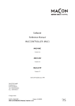
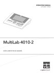
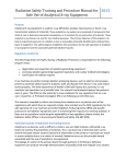
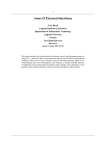
THE UNIVERSITY OF ARIZONA ®
Radiation Generating Machines
Reference Guide
October 2008
1717 E. Speedway Blvd. (Bldg. 151)
Suite 1201
Tucson, AZ 85724
P.O. Box 245101
www.radcon.arizona.edu
Phone (520) 626-6850 Fax (520) 626-2583
Table of Contents
I. INTRODUCTION ................................................................................. 1 II. PHYSICS .......................................................................................... 1 A. ATOMIC STRUCTURE ............................................................................................................ 1 1. Protons ............................................................................................................................ 1 2. Neutrons .......................................................................................................................... 1 3. Electrons ......................................................................................................................... 2 B. IONIZING RADIATION ........................................................................................................... 2 III. PRODUCTION OF X-RAYS ............................................................... 2 A. B. C. D. X-RAY TUBES ...................................................................................................................... 3 BREMSSTRAHLUNG .............................................................................................................. 4 CHARACTERISTIC X-RAYS .................................................................................................... 5 POWER ................................................................................................................................. 6 IV. UNITS OF RADIATION MEASUREMENT ......................................... 6 A. EXPOSURE ............................................................................................................................ 6 B. ABSORBED DOSE ................................................................................................................. 6 C. DOSE EQUIVALENT .............................................................................................................. 6 V. A. B. C. D. BIOLOGICAL EFFECTS ................................................................... 7 DIRECT AND INDIRECT EFFECTS .......................................................................................... 7 ACUTE AND DELAYED EFFECTS ........................................................................................... 7 RECOGNIZING ACUTE EXPOSURE ......................................................................................... 8 DETERMINISTIC AND STOCHASTIC EFFECTS ......................................................................... 8 VI. BACKGROUND RADIATION ............................................................ 9 VII. REGULATIONS ............................................................................. 9 VIII. OCCUPATIONAL RADIATION DOSE LIMITS ............................. 10 A. DOSE LIMIT FOR THE GENERAL PUBLIC ............................................................................. 10 B. DECLARATION OF PREGNANCY .......................................................................................... 11 IX. ALARA ........................................................................................ 11 A. LINEAR, NON-THRESHOLD (LNT) HYPOTHESIS ................................................................. 11 X. RADIATION PROTECTION METHODS .......................................... 12 A. TIME .................................................................................................................................. 12 B. DISTANCE .......................................................................................................................... 12 C. SHIELDING ......................................................................................................................... 13 XI. SIGNS AND LABELS ....................................................................... 13 XII. A. B. C. D. E. F. SAFETY DEVICES AND OTHER PROTECTIVE MEASURES ........ 14 WARNING LIGHTS .............................................................................................................. 14 INTERLOCKS....................................................................................................................... 15 SAFETY DEVICE TESTS ...................................................................................................... 15 OPERATING PROCEDURES .................................................................................................. 15 RECORDKEEPING................................................................................................................ 15 DEDICATED ROOM ............................................................................................................. 15 XIII. SECURITY................................................................................... 15 XIV. RADIATION DETECTION AND MEASUREMENT ........................ 16 A. RADIATION SURVEYS ......................................................................................................... 16 B. AREA RADIATION MONITORS ............................................................................................ 16 XV. RADIATION DOSIMETERS ......................................................... 16 A. INVESTIGATION LEVELS ..................................................................................................... 17 B. RADIATION DOSE RECORDS ............................................................................................... 17 XVI. RADIATION SAFETY COMMITTEES .......................................... 17 XVII. RADIATION MACHINE APPROVALS AND MACHINE
REGISTRATION ...................................................................................... 18 A. B. C. D. E. F. EXEMPTIONS ...................................................................................................................... 18 MACHINE REGISTRATION................................................................................................... 18 DISPOSAL, TRANSFER, OR CHANGE OF LOCATION OF RADIATION MACHINES ................... 18 LEAVES OF ABSENCE ......................................................................................................... 18 APPROVAL AUDITS ............................................................................................................ 19 NON-COMPLIANCE ............................................................................................................. 19 XVIII. TRAINING ................................................................................ 19 XIX. EMERGENCY RESPONSE ........................................................... 20 XX. RESPONSIBILITIES BY ROLE ..................................................... 21 A. B. C. D. APPROVAL HOLDER RESPONSIBILITIES .............................................................................. 21 RADIATION SAFETY COORDINATOR RESPONSIBILITIES...................................................... 21 RADIATION WORKER RESPONSIBILITIES ............................................................................ 22 RADIATION CONTROL OFFICE RESPONSIBILITIES............................................................... 22 I.
Introduction
The University of Arizona (UA) Radiation Control Office (RCO) Radiation Machines Program
exists to promote the safe use of ionizing radiation producing machines and to ensure compliance
with regulations regarding the use of these machines.
The Radiation Machines Program Manual serves to describe the machines program at the UA,
and includes responsibilities of Approval Holders and individual users, as well as information
regarding various types of ionizing radiation producing machines and their safe use.
This manual applies to those individuals who are utilizing ionizing radiation producing
machines, or “radiation machines”, for research purposes.
In this manual and within the Radiation Machines Program, the term “radiation machine”
includes, but is not limited to x-ray fluorescence, x-ray diffraction, cabinet radiography,
accelerators, x-ray spectrometers, and medical radiography units used in non-clinical studies.
Other machines may qualify depending on the intended application of the radiation produced.
II.
Physics
A. Atomic Structure
The basic unit of matter is the atom. The basic atomic model, as described by Ernest
Rutherford and Neils Bohr in 1911, consists of a positively charged core surrounded by
negatively-charged shells. The central core, called the nucleus, contains protons and
neutrons. Nuclear forces hold the nucleus together. The shells are formed by electrons
which exist in structured orbits around the nucleus.
Figure 1 Atomic Model
1. Protons
Protons (p+) are positively charged and located in the nucleus of the atom. The number of
protons determines the element.
2. Neutrons
Neutrons (n) are uncharged and located in the nucleus of the atom. Atoms of the same
element have the same number of protons, but can have a different number of neutrons.
1|Page
3. Electrons
Electrons (e-) are negatively charged and travel in specific orbits or energy levels about
the nucleus. An atom is electrically neutral if the total electron charge equals the total
proton charge. The term ion is used to define atoms or groups of atoms that have a net
positive or negative electrical charge.
B. Ionizing Radiation
Radiation is defined as the transfer of energy in the form of particles or waves through open
space.
Radiation with sufficient energy to physically remove electrons from neutral atoms to create
ions (cause ionization) is referred to as ionizing radiation. Ionizing radiation includes alpha
particles, beta particles, gamma or x-rays, and neutron particles. Figure 2 illustrates the
phenomenon of ionization. From the standpoint of human health and safety, ionizing
radiation is of concern since it can create many energetic ionized atoms which engage in
chemical reactions that interfere with the normal processes of cells.
Figure 2 Ionization
Radiation that lacks the energy to cause ionization is referred to as non-ionizing radiation.
Examples of non-ionizing radiation include radiowaves, microwaves, and visible light.
The energy of ionizing radiation is usually given in electron volts (eV). The electron volt is
defined as the energy of an electron that has been accelerated through an electron potential of
one volt. The eV is a very small amount of energy and therefore keV (thousand electron
volts) and MeV (million electron volts) are used as the units of measurement for the energies
associated with the emissions from radiation machines. The energy of visible light is about
two or three eV.
III.
Production of X-rays
Radiation-producing devices produce x-rays by accelerating electrons through an electric voltage
potential and stopping them with a target. Most x-ray devices emit electrons from a cathode,
accelerate them with a voltage, and allow them to hit an anode, which emits x-ray photons.
X-rays were discovered in 1895 when Wilhelm Conrad Roentgen observed that a screen coated
with a barium salt exhibited fluorescence when placed near a cathode ray tube. Roentgen
concluded that a form of penetrating radiation was being emitted by the cathode ray tube and
called the unknown rays “x-rays”.
Like radio waves, light, and gamma rays, x-rays are a form of electromagnetic radiation. All
electromagnetic radiation is characterized by the movement of massless waves of energy called
photons. Photons travel at the speed of light and move with a characteristic wavelength and
frequency that defines the specific type of electromagnetic radiation.
The amount of energy carried by a photon is directly proportional to the frequency of the
radiation and inversely proportional to the wavelength. Thus, x-rays which have a relatively
short wavelength and high frequency possess a great deal of energy. X-rays are termed ionizing
radiation because they contain sufficient energy to remove orbital electrons from atoms, creating
ions.
X-rays are produced when charged particles, usually electrons, are accelerated by an electrical
voltage (potential difference). Whenever a high voltage potential, a vacuum, and a source of
electrons are present in any scientific device, x-rays can be produced.
A. X-ray Tubes
An x-ray tube has an evacuated envelope, an anode, and a cathode. Figure 3 shows the
construction of a typical x-ray tube. The configuration of an x-ray tube may differ, but the
method of x-ray production is consistent across various configurations.
Figure 3 – X-ray tube
The cathode (filament) is the negative terminal of the x-ray tube. Electrons are produced at
the cathode by heating the filament to incandescence when a high current flows through it.
Adjusting the current through the filament controls the number of electrons produced.
Electrons are accelerated toward the positive anode by a high voltage potential. This potential
difference is expressed in kilovolts (kV). Since the voltage across the tube may fluctuate it is
usually expressed as peak voltage (kVp) An electron drawn across a voltage of one volt has
energy of one electron volt (eV). The peak voltage, therefore, defines the upper limit of the
most energetic photon produced by the energetic electrons interacting in the anode.
The anode is the positive terminal of the x-ray tube. The electrons are accelerated from the
cathode to the anode. The anode material used depends on the x-ray energy that is required.
For clinical diagnostic units the target is made of a thin layer of tungsten embedded in
copper. Other common targets for analytical units are copper, chromium and cobalt.
The term tube current represents the number of electrons traveling from the cathode to the
anode per second. The tube current is measured in amperes. Tube currents in research
radiation machines are usually measured in milliamps (mA) or microamps (µA). The
product of the tube current and the number of seconds that the current flow is expressed in
milliampere-seconds (mAs). The mAs value determines the total amount of x-rays that are
generated during an exposure.
The anode and cathode are sealed into an envelope. This allows for gases and other
impurities to be pumped out of the tube, creating the vacuum necessary for proper
performance. The x-ray creation process must occur in a vacuum so as not to disrupt the
electron beam, and also to allow for proper filament performance and durability.
Generally, more than 99% of the kinetic energy of the accelerated electrons is converted to
thermal energy (heat), leaving less than 1% available for the production of x-rays, making the
production of x-rays a relatively inefficient process. x-rays are produced within the anode
through two processes, Bremmstrahlung and the production of characteristic x-rays.
B. Bremsstrahlung
When high-speed electrons from the cathode hit the anode, some of the negatively charged
electrons are able to get through the target atom’s electron cloud and interact with the
positively charged nucleus. The electrons that are able to penetrate near the target material
nuclei are “braked,” or decelerated, to varying degrees depending on how closely they
approach the target nuclei. When accelerated electrons hit the anode and decelerate, they
emit bremsstrahlung (meaning braking radiation in German).
Figure 4
Bremsstrahlung photons are produced by over a wide energy spectrum depending upon the
degree of braking or deflection that the accelerated electrons experience as they interact with
the target nuclei.
C. Characteristic x-rays
If an accelerated electron interacts with an inner shell electron rather than an outer shell
electron, characteristic x-rays can be produced. The interaction must impart enough energy
to a K-shell electron to overcome its binding energy, removing the orbital electron from the
atom, ionizing the atom. This leaves a temporary electron hole in the K-shell. The atom is in
an unstable state which is corrected by an outer shell electron falling to the K-shell to fill the
hole. The transition results in an x-ray with a characteristic energy equal to the difference
between the binding energy of the K-shell electron and the binding energy of the electron
that falls to the K-shell to fill the hole.
Figure 5
A typical x-rays spectrum consisting of the Bremmstrahlung x-ray spectrum and
characteristic x-rays is shown in Figure 6. The characteristic x-rays of the tungsten target are
superimposed on the Brehmsstrahlung continuum.
Figure 6 – composite x-ray spectrum
D. Power
For some analytical instruments it is important to know the power emitted by the instrument.
This can also be important to for radiation protection purposes. Power, which is measured in
watts (W), equals voltage times current (P = V x I). For example, a 10 kVp device with a
current of 1 mA emits 10 W of power.
IV.
Units of Radiation Measurement
A. Exposure
X-rays, being ionizing radiation, can produce ionization of air. The unit of ionization of air
by x-rays is the Roentgen, where:
-4
1 R = 2.58 x 10 Coulombs/kg of air
B. Absorbed Dose
When predicting biological effects, it is important to determine the energy deposited in
human tissue rather than air. The total x-ray energy deposited per unit mass of material is the
absorbed dose. This definition is not restricted to air, but can be any material. The rad
(sometimes thought of as radiation absorbed dose) is the traditional unit of radiation absorbed
dose.
1 rad = 100 ergs/gram
The System International (SI) unit of radiation absorbed dose is the Gray (Gy).
1 Gray = 1 J/kg
The relationship between the Gray and the rad is:
1 Gray = 100 rad
C. Dose Equivalent
The dose equivalent is used to measure the biological effects of ionizing radiation on the
human body. It is a function of the absorbed dose and the type of radiation absorbed. The
weighting factor used according to the type of radiation absorbed is called the quality factor
(QF). The quality factor for x-rays is 1, but can range up to 20 for other types of ionizing
radiation. This weighting factor is used to address the fact that different types of ionizing
radiation have differing biological effects in the scope of exposure to low levels of radiation.
The rem is the tradition unit of dose equivalent.
1 rem = 1 rad x QF
(QF=1 for x-rays)
The SI unit for dose equivalent is the Sievert (Sv).
As it was between the rad and Gray, the relationship between rem and Sievert is:
1 Sievert = 100 rem
Because the quality factor for x-rays is one, for x-ray radiation safety purposes, it may be
assumed that 1 R = 1 rad = 1 rem.
V.
Biological Effects
X-rays can penetrate deeply into the human body, stripping electrons from atomic orbit, and
breaking or modifying chemical bonds within critical biological molecules that make
up the cells. This process can cause cell injury and even cell death, depending on the
dose and dose rate of the exposure.
Several factors contribute to the biological effects of x-ray exposure. These include:
•
•
•
•
•
Total dose
Dose rate
Energy of radiation
Portion of body exposed
Individual sensitivity
Biological effects of x-rays can be described as direct or indirect, acute or delayed, and
stochastic or deterministic.
A. Direct and Indirect Effects
Biological effects can be categorized by the damage mechanism that occurs. Radiation that
deposits energy directly into intracellular structures, with DNA being the most important
target, results in a direct effect. If radiation interacts with DNA, the damage, if not repaired,
can result in cell death or a genetic mutation. Genetic mutation may lead to cancer.
An indirect effect occurs when energy from ionizing radiation is absorbed by water
molecules in the cell, resulting in the production of free radicals. Free radicals produced by
the ionization of water are extremely chemically reactive and can result in damage to DNA
and other intracellular structures.
B. Acute and Delayed Effects
Biological effects can also be categorized by the time period between exposure and effect.
Acute effects are effects that occur due to a high radiation dose in a short period of time
(seconds to days). Acute exposures and effects are generally associated with accidental
exposure. Many types of research machines are incapable of being operated under conditions
that would cause an acute exposure effect. These effects are more likely to occur with an xray diffraction unit, which has a highly collimated beam and a very high exposure rate (the
primary beam of an x-ray diffraction unit may have an exposure rate of up to 10,000
Roentgen per second). The acute effect such as erythema (skin reddening) may manifest
itself within a day. More severe effects, such as skin ulceration, may take weeks to appear.
Delayed exposure effects can be due to an acute exposure or a chronic exposure with the
effect occurring years after exposure. Among the delayed consequences of overexposure to
x-rays that are of concern are cancer and other genetic effects.
Radiation safety rules and regulations are designed to minimize the probability of receiving
an acute dose by preventing accidental exposures and to minimize chronic exposure during
routine use of a radiation-producing machine.
C. Recognizing Acute Exposure
X-ray burns do not harm the outer, mature, non-dividing skin layers. Rather, the x-rays
penetrate to the deeper, basal skin layer, damaging or killing the rapidly dividing germinal
cells that were destined to replace the outer layers that slough off.
Following this damage, the outer cells that are naturally sloughed off, but are not replaced.
Lack of a fully viable basal layer of cells means that x-ray burns are slow to heal, and in
some cases, may never heal. Frequently, such burns require skin grafts. In some cases,
severe x-ray burns have resulted in gangrene and amputation of one or more digits.
An acute dose greater than 300 rad (3 Gy) to a part of the body causes a radiation burn equivalent
to a first-degree thermal burn or mild sunburn. Typically, there is no immediate pain, but a
sensation of warmth or itching occurs within about a day after exposure. A reddening or
inflammation of the affected area usually appears within a day and fades after a few more days.
The reddening may reappear as late as two to three weeks after the exposure. A dry scaling or
peeling of the irradiated portion of the skin is likely to follow. A typical x-ray unit can produce
this level of radiation dose in just a few second if the exposure occurs very close to the x-ray tube
or the beam is very collimated.
If you have been working with or around an x-ray device and you notice an unexplained
reddening of your skin, notify your supervisor and the Radiation Control Office.
An acute dose greater than 1,000 rad (10 Gy) to a part of the body causes serious tissue damage
similar to a second-degree thermal burn. First reddening and inflammation occurs, followed by
swelling and tenderness. Blisters will form within one to three weeks and will break open
leaving raw, painful wounds that can become infected.
If you develop symptoms such as these, seek immediate medical attention to avoid infection and
help relieve pain. Contact the Radiation Control Office as soon as possible. An even larger acute
dose causes severe tissue damage similar to a scald or chemical burn. Intense pain and swelling
occurs, sometimes within hours.
D. Deterministic and Stochastic Effects
Deterministic effects are effects in which a clear connection between the person that suffered
the overexposure to radiation and the effect can be made. In these cases there is a causal
relationship between the amount of dose and severity of the effects on an individual. A dose
threshold for an effect must be exceeded for that deterministic effect, and the severity of the
effect increases with the dose above that threshold. Deterministic effects can be acute or
delayed. Examples of deterministic effects associated with x-ray exposure include skin
erythema (reddening), epilation (hair loss), and skin ulceration.
Stochastic effects are effects with no threshold dose, and their probability of occurrence
increases with an increase in dose received by an overexposed exposed population. The
stochastic effects are delayed. The stochastic effect most associated with ionizing radiation
exposure is cancer.
VI.
Background Radiation
The average background dose to the general population in the U.S. from both natural and
manmade sources is 360 millirem per year (mrem/yr), but background radiation dose can vary
widely depending on your geographic location and other factors. Figure 7 illustrates the various
sources of background radiation dose.
Figure 7 – Background Radiation
Naturally occurring sources include an average of about 200 mrem per year from radon and its
decay products, about 40 mrem per year from internal emitters such as potassium-40, about 28
mrem per year from cosmic rays, and about 28 mrem per year from terrestrial sources such as
naturally occurring uranium and thorium. Manmade sources of ionizing radiation exposure
include an average of about 10 mrem per year from consumer products such as building
materials and about 53 mrem per year from medical procedures such as diagnostic x-ray and
nuclear machine procedures.
VII.
Regulations
The Arizona Radiation Regulatory Agency (ARRA) regulates the registration, possession, and
use of radiation machines in Arizona. Radiation machines are registered to the University of
Arizona. Their possession and use under this registration is administered by the RCO. Principal
investigators are granted “Approvals”, or sub-registrations, to possess and use under the UA
registrations.
The registration and regulation of radiation machines is codified in Articles of the Arizona
Administrative Code, Title 12, Chapter 1:
Article 2: Registration, Installation, and Service of Ionizing Radiation-Producing Machines; and
Certification of Mammography Facilities
Article 4: Standard for Protection Against Ionizing Radiation
Article 8: Radiation Safety Requirements for Analytical X-Ray Operations
Article 9: Particle Accelerators
Article 10: Notices, Instructions, and Reports to Ionizing Radiation Workers; Inspections
VIII.
Occupational Radiation Dose Limits
A radiation worker is defined by the RCO as an individual who has received both general and
specific radiation safety training regarding the use of radioactive materials and/or radiationproducing devices for which they are authorized. The radiation dose received by radiation
workers in the course of their work is defined as occupational dose.
Limits on occupational doses are based on data regarding the biological effects of ionizing
radiation. The International Commission on Radiological Protection and the National Council
on Radiation Protection and Measurements publish guidance for setting radiation protection
standards. The ARRA sets regulatory requirements related to radiation protection in the state of
Arizona. These limits are set to ensure that the probability of detrimental biological effects due
to the use of ionizing radiation emitting machines and materials is equivalent to what has been
observed in other safe industries. The annual occupational radiation dose limits are shown in
Table 1.
Table 1 – Occupational Radiation Dose Limits
Occupational Dose Limits
(rem/yr)
Whole Body
Lens of the Eye
Extremities
Skin
5
15
50
50
Occupational dose limits for minors are 10% of the adult limits. The University of
Arizona only allows a minor to become a radiation worker and use a radiation machine if
it is part of an educational experience. It cannot be solely associated with employment.
A. Dose Limit for the General Public
The radiation dose limit for the non-radiation workers is 100 millirem per year. Nonradiation workers are defined as all other individuals who frequent or enter the radiation
machine use area, whether faculty, staff, students, visitors and members of the general public.
These include those who work in the lab but have not completed the machine training
requirements nor been added to the machine Approval. It also includes individuals who are
present in areas adjacent to the machine use area.
B. Declaration of Pregnancy
The dose limit to an embryo/fetus is 500 mrem over the entire pregnancy of a declared
pregnant woman. It is also required that every effort be made to avoid substantial variation
above a uniform monthly dose (50 mrem/month). A declared pregnant woman refers to a
woman who has voluntarily informed her employer, in writing, of her pregnancy and the
estimated date of conception.
Should you become pregnant and declare your pregnancy, it is a regulatory requirement that
you be provided with additional information about the potential risks of radiation exposure to
a fetus. Please contact the Radiation Control Office for review of your work situation,
previous exposure history, and to receive the information pamphlet.
It is a woman's right to "undeclare" her pregnancy at any time even though she is pregnant
and has previously declared the pregnancy.
IX.
ALARA
Because the effects of chronic doses of low levels of ionizing radiation are not precisely known,
we assume there is some risk at any dose. The ALARA principle is to maintain radiation dose
As Low As Reasonably Achievable, considering economic and social constraints. The goal of
ALARA is to keep your radiation dose as far below the occupational limits as is reasonably
achievable. It is not only a good radiation safety practice; it is also required by the ARRA
regulations. The RCO is required to demonstrate that we are working to maintain radiation dose
to radiation workers and non-workers ALARA. Training (such as the Radiation Machines
Protection Course), radiation surveys, audits, and distribution of radiation dosimeters are all part
of the ALARA program.
A. Linear, Non-threshold (LNT) Hypothesis
The ALARA policy is based on the linear, non-threshold (LNT) dose-effect hypothesis,
which predicts the risk of developing cancer associated with long-term radiation exposure.
The LNT hypothesis assumes that the high doses of ionizing radiation associated with
observed injurious effects in humans may be used to predict the effects of low doses.
According to the LNT hypothesis, any dose of ionizing radiation, no matter how small, has
some detrimental effect and each incremental increase in the dose increases the probability of
detrimental effects associated with that exposure.
However, there is some controversy associated with the LNT hypothesis. Many safety
professionals maintain that no injurious effects of low-dose and low-dose rate ionizing
radiation have ever been documented to occur in human populations, which is counter to the
hypothesis. Furthermore, they maintain that scientific evidence confirms that there are
probably no injurious effects from low-dose and low-dose rate ionizing radiation at even
many multiples of the dose and dose rates of natural background radiation. They suggest that
the doses and dose rates from natural background radiation are comparable to the doses and
dose rates experienced by most radiological workers in the workplace and by members of the
public. On the other side of the spectrum are those that believe that the LNT hypothesis
underestimates the detriment associated with low dose, low-dose rate ionizing radiation
exposure.
Regardless of the controversy, the ARRA regulations require that the University of Arizona
demonstrate that radiation doses are being maintained ALARA.
X.
Radiation Protection Methods
There are three basic methods for keeping your radiation dose and the dose of others around you
ALARA:
•
•
•
Minimize time
Maximize distance
Use appropriate shielding
A. Time
The dose of radiation a worker receives is directly proportional to the amount of time spent in
a radiation field. Thus, reducing the time by one-half will reduce the radiation dose received
by one-half. Operators should always work quickly and spend as little time as possible
around the equipment while it is operating. Methods of minimizing time of exposure include
planning work carefully and ensuring all equipment is in good working condition before
work begins. Repeating experiments unnecessarily is not good ALARA practice.
B. Distance
Radiation exposure decreases rapidly as the distance between the worker and the x-ray
source increases. The decrease in exposure from a point source, such as an x-ray tube, can be
calculated by using the inverse square law. This law states that the amount of radiation at a
given distance from a point source varies inversely with the square of the distance. This is
illustrated by the equations in Figure 6.
or
Where:
I1 = Radiation Intensity at the First Distance (D1)
I2 = Radiation Intensity at the Second Distance (D2)
Figure 6 – Inverse square law equation
For example, doubling the distance from an x-ray tube will reduce the dose to one-fourth of
its original value, and increasing the separation distance by a factor of three will reduce the
dose to one-ninth of its original value.
Although the inverse square law does not accurately describe scattered radiation, distance
will still dramatically reduce the intensity from this source of radiation. Maintaining a safe
distance, therefore, represents one the simplest and most effective methods for reducing
radiation exposure to workers and non-workers.
Realize that the inverse square law does not apply to exposure to a collimated x-ray beam
such as the primary beam of an x-ray diffraction unit.
C. Shielding
Placing attenuating material between the x-ray device and workers, and non-workers as well,
can also reduce radiation dose. Materials that are dense and have a high atomic number, such
as lead, are effective x-ray shielding. Other materials, such as stainless steel or lead- or tinloaded acrylic viewing windows can also be effective barriers.
Shielding must be of an adequate thickness to reduce the radiation level outside an x-ray
enclosure or outside local components to below regulatory limits. The RCO must survey all
new x-ray equipment prior to use to ensure that the radiation levels on the exterior of the unit
are at appropriate levels. Subsequent surveys are required under the circumstances listed in
section 13.1.
XI.
Signs and Labels
Signs and labels are used to communicate the presence of an ionizing radiation hazard to
both workers and non-workers.
Figure 7 – Entrance posting examples
Each area or room containing analytical x-ray equipment must be conspicuously posted with
a sign or signs bearing the radiation symbol and the words "CAUTION - X-RAY
EQUIPMENT". Figure 7 shows two examples of the signs that must be posted at all
entrances to areas or rooms containing radiation producing machines.
Figure 8 – Control panel label
A label bearing the words CAUTION THIS EQUIPMENT PRODUCES RADIATION
WHEN ENERGIZED (or similar wording) must be affixed to the control device of each
machine. Figure 8 shows one version of this label.
Figure 9 – Emergency response label
An Emergency Response label must be affixed on or near the control panel. Notification
information for the Approval Holder must be included on this label.
A copy of the Form ARRA-6, “Notice to Employees”, must be available in all rooms where
radiation machines are located. The Form ARRA-6 reminds workers of their rights and
responsibilities as radiation workers.
XII.
Safety Devices and Other Protective Measures
Most modern enclosed radiation-producing machines are equipped with safety devices to
prevent accidental exposure to ionizing radiation. These safety devices include warning
lights and interlocks. No bypassing or modifications of radiation machine safety devices
may be made unless these have received prior approval of the RCO.
A. Warning lights
Warning lights must be available as appropriate:
• X-ray tube status (On-Off) indicator in systems where the primary beam is controlled
in this fashion;
• Shutter status (Open-Closed) indicators near each port on the radiation housing for
systems which control the primary beam; and
• A clearly visible warning light labeled with the words “X-RAY ON,” or a similar
warning located near any switch that energizes an x-ray tube, illuminated only when
the tube is energized
The warning lights must be labeled so that their purpose is easily identified.
B. Interlocks
Interlocks are defined as devices arranged or connected such that an occurrence of an event
or condition is required before a second event or condition can occur or continue to occur. In
the context of radiation machine use, this means that an enclosure switch must be closed or a
warning light operating before the machine can produce x-rays or continue to produce x-ray
once production has begun.
Shutter mechanisms and interlocks must not be tampered with or defeated under any
circumstances. Do not use the interlock to turn the unit off. Use the main switch.
Interlocks may be overridden to perform some types of maintenance, such as alignment of a
diffraction x-ray beam. Personnel who intend to override interlocks must receive prior
authorization from the Approval Holder and the Radiation Control Office, be listed on an
interlock override memo maintained for review by the RCO and ARRA, and must be
assigned and wear both a whole body dosimeter and at least one ring badge.
C. Safety Device Tests
Documented safety device tests must be performed monthly. Test documentation must be
available for RCO and ARRA inspection. These safety device tests include, as applicable:
• “X-RAY ON” light on control panel
• X-RAY ON” light on or near source housing
• “SHUTTER OPEN” light
• Door interlocks
D. Operating Procedures
Operating procedures must be available near the machine. The user manual will suffice as an
operating procedure if it is current. If a user manual is not available, the Approval Holder
must ensure that written operating procedures are developed.
E. Recordkeeping
A use log for each machine must be maintained. At a minimum, the log must contain the
name of the user and the date of use. The use log can also be used to document safety device
checks.
F. Dedicated Room
The machine should be placed in a separate room from other work areas or in low traffic
areas, if practical. This helps keep radiation dose to members of the public ALARA.
XIII.
Security
When not in use, radiation-producing machines must be secured in such a manner that it cannot
be used by untrained, unauthorized personnel or members of the public. Key control or
equivalent room or device access control must be maintained for each machine. For older
machines that do not have a keyed control device, this requirement may be fulfilled by
controlling access to the laboratory or an alternate method approved by the RCO.
XIV.
Radiation Detection and Measurement
A. Radiation Surveys
Radiation surveys with portable radiation survey instruments are used to verify the efficacy
of radiation shielding used to minimize radiation exposure to radiation workers and nonworkers. Surveys also ensure that radiation levels are below regulatory limits during
machine operation. Machine users are not required to perform radiation surveys unless it is a
specific condition of the machine Approval. Radiation surveys of local components and xray enclosures are performed by the RCO. Radiation surveys must be performed under the
following circumstances:
•
•
•
•
•
•
Upon installation and prior to initial use of the machine
Every twelve months (this is normally performed during the Approval audit),
Following any change in the arrangement of local components,
Following maintenance or service that requires the removal or disassembly of a local
component,
Any time a visual inspection by a user reveals an abnormal condition, and
When personnel monitoring devices show a significant increase over the previous
monitoring period.
Radiation survey can always be performed by the RCO at the Approval Holder’s, Radiation
Safety Coordinator’s or radiation worker’s request.
B. Area Radiation Monitors
The RCO may, depending on the condition of use of an x-ray system, mount area radiation
monitors to measure radiation dose to members of the public in unrestricted areas. The
Approval Holder should be aware of these dosimeters and report missing or damaged
dosimeters to the RCO.
XV.
Radiation Dosimeters
The RCO determines radiation dosimeter assignment based on the type of machine and the use
of that machine. Users of enclosed x-ray units are not issued radiation dosimeters. Users of
open beam radiation machines that are not equipped with a safety device are required to wear a
dosimeter whenever they work with the machine. This also applies to individuals who are
authorized to perform maintenance (e.g. alignment) that involves access to open beams are
required to wear a personal dosimeter during all use of the machine. Radiation dosimeters
generally consist of a “whole body” badge and one or more extremity dosimeters or ring badges
(Figure 10) depending on the configuration of the unit.
Figure 10 Radiation Dosimeters
Ring badges are assigned if exposure to the extremities could be significantly greater than
exposure to the user’s torso. Dosimeters are required for operators of open beam analytical x-ray
units that are not contained in an enclosure equipped with safety devices. Dosimeters are also
required for individuals who are authorized to remove local components or override safety
devices to perform maintenance or alignment.
If you are assigned radiation dosimeters, follow the directions outlined in the RCO radiation
dosimetry guidelines for proper use and storage of your dosimeter. This guidance includes
wearing the whole body badge on your torso (at or above your waist) on the portion of your body
likely to be exposed to the highest level of radiation. The finder badge is oriented with the white
“chip side” on such that it faces the radiation beam as your hand approaches the beam.
Immediately notify the RCO of a lost or damaged dosimeter. The RCO will replace the damaged
dosimeter free of charge. A lost dosimeter or a dosimeter that is turned in late will result in a
$25.00 charge to the wearer’s department for badge replacement and administrative costs. This
charge may be passed on to the user by the department.
A. Investigation Levels
The RCO sets investigation levels for radiation dose recorded during each wear period. If a
worker exceeds the investigation level in a wear period for a radiation dosimeter, the RCO
will review the worker’s work practices to ensure that radiation doses are ALARA.
The exposure of a radiation dosimeter to deceptively indicate a dose delivered to an
individual is prohibited by state regulations.
B. Radiation Dose Records
All radiation dose records are on file with the RCO. The RCO will supply you with your
radiation dose history upon request. Those individuals who are required to wear a radiation
dosimeter receive an annual report of their radiation dose.
XVI.
Radiation Safety Committees
The administration of UA’s radiation machines registrations and development of radiation safety
policies is directed by two radiation safety committees, the University Radiation Safety
Committee (URSC) and Medical Radiation Safety Committee (MRSC). Details of the functions
and responsibilities of the committees are described in their respective charters. Jurisdiction of
these committees is based on the location of the machine, although the rules are consistent across
the committees. The committees meet bi-monthly and have the responsibility to act in the best
interest of safety. To this end, the duties of the Committees include:
•
•
•
Setting machine safety policy, which is reflected in the Radiation Machine Program
Manual,
Reviewing the operations and procedures of the Radiation Control Office with respect to
radiation machine safety,
Establishing enforcement actions against Approval Holders who are in non-compliance
with the radiation machine program and have not made an attempt to remedy the noncompliance issue in a timely manner, and
•
XVII.
Considering appeals of enforcement actions.
Radiation Machine Approvals and Machine Registration
Radiation machines are possessed and used under a machine Approval system. These Approvals
act as “sub-registrations” under the UA registration with the ARRA. The Approval system
tracks machine inventory, personnel, training, and requirements for machine use. The Approval
process begins with the completion of RCO Form RC-030, “Application for Machine Approval”.
This form can be found on the RCO website. An Approval Holder is generally the faculty
member who has primary responsibility for radiation machine safety within his/her research
group, but may be other personnel under certain circumstances. The RC-030 requires the
applicant to describe the intended use of their radiation machine(s) and is complemented by the
RCO Form RC-005, “RCO Research Machines Registration” Form.
Granting an Approval may include special conditions regarding possession and use of a radiation
machine. The applicant may be required to attend the Radiation Generating Research Machines
Protection Course as a condition of being granted an Approval if their training and experience
with ionizing radiation-producing machines is not sufficient.
New Approval Holders are required to meet with a member of the RCO staff for an Approval
Holder Orientation (AHO) prior to beginning work with their machines. This orientation is
provided to Approval Holders and Radiation Safety Coordinators to ensure that all
authorizations, restrictions, and administrative requirements are clearly understood. Users and
others present in the lab are encouraged to attend.
A. Exemptions
Users of electronic products which produce x-rays only incidental to operation may be
exempted from the requirements contained in this manual depending on the product and/or
the results of radiation survey measurements. Such exemptions may include electron
microscopes (scanning electron microscopes), ion-implanters, and cathode ray tubes. Please
contact the RCO to request an ionizing radiation survey of electronic equipment.
B. Machine Registration
All operable x-ray generating machines must be registered with the RCO prior to operation.
Registration begins with the completion of a RCO Form RC-005, “RCO Research Machines
Registration” Form. The RC-005 requires the registrant to list the specifications of the unit
and describe its use. An evaluation of the unit and its use will be made to determine if any
operation restrictions or additional safety devices are necessary. An initial radiation survey
and safety device check will also be performed.
C. Disposal, Transfer, or Change of Location of Radiation Machines
The RCO must be notified prior to a change in a machine’s location, transfer of ownership,
or disposal. These changes of status must be reported to the ARRA.
D. Leaves of Absence
If an Approval Holder will be absent from the University for a period greater than 45 days,
responsibility for possession and use of the absent Approval Holder’s radiation machines
must be transferred to an appropriate substitute. The RCO must be notified at least 60 days
prior to departure of the Approval Holder’s departure.
E. Approval Audits
Radiation Machine Approvals are audited annually to ensure safe use of the radiation
machines and to ensure regulatory compliance. Areas reviewed during the audit include, but
are not limited to a review of:
• Inventory verification,
• Radiation dosimeter records,
• Availability of radiation dosimeters, if required
• Machine use log and safety device check records,
• certificates for all users,
• Signs and labels,
• Operating and emergency procedures,
• Room and/or machine security,
• Operation of the unit since the previous audit, and
• Operators’ awareness of ARRA regulations.
A radiation survey of the unit is performed to ensure that applicable dose limits are not
exceeded and all radiation exposure is maintained ALARA.
Items of non-compliance identified in the Approval audit will be communicated to the
Approval Holder in an audit memo and, if warranted, in a post-audit meeting. The Approval
Holder is responsible for correcting deficiencies in a timely manner.
F. Non-compliance
Non-compliance with RCO requirements, whether identified during audits or at other times,
will be investigated by the RCO and reported to the appropriate committee. Follow-ups may
be necessary to ensure compliance. Continued non-compliance with RCO requirements may
necessitate attendance of the Approval Holder at a radiation safety committee meeting to
discuss termination of the Approval or machine use restrictions.
XVIII.
Training
Only qualified and authorized personnel are permitted to operate a regulated radiation machine.
The Approval Holder must determine the operator’s qualifications. Each person who operates a
radiation machine must receive two categories of training, laboratory or system-specific training
and RCO radiation machine safety training (Radiation Machines Protection Course). The user
must successfully complete all required training and demonstrate safe use of the radiation
machine to the Approval Holder’s satisfaction prior to commencement of work with the radiation
machine. Registration for the Radiation Generating Research Machine Protection Course is
accomplished by completing and submitting a RCO Form RC-088, “Radiation Worker Data
Sheet and Training Record”.
The Radiation Generating Research Machines Protection Course is completed in two parts. Part
one consists of a review of the Radiation Machines Program Manual and completion of a
problem set. Part two of the training is a lecture followed by an exam.
Upon successful completion of the Radiation Generating Research Machines Protection Course a
certificate will be emailed to the participant, Approval Holder, and Radiation Safety Coordinator.
The Approval Holder must maintain a copy of the Radiation Generating Research Machines
Protection Course certificate for each user in his/her files for RCO and ARRA inspection.
The Radiation Generating Research Machines Protection Course and this manual do not address
operating procedures for specific installations.
Radiation Generating Research Machines Protection Course training must be augmented with
machine-specific training. Machine-specific training should include:
•
•
•
•
•
•
•
XIX.
Description of the system,
Safety device identification and safety device checks
Review of written operating and safety procedures
Selection and use of personal protective equipment, if required
Completion of operating log book
Review of emergency procedures
Identification of ancillary hazards
Emergency Response
Accidents are usually caused by one or more of the following:
•
•
•
•
•
Poor equipment design or configuration (e.g. unused ports not covered or shielding is
inadequate)
Equipment failure (e.g. shutter failure, interlock failure)
Manipulation of equipment when the x-ray tube is energized (moving samples or
aligning cameras while unit is in operation)
Inadequate training and awareness
Improvising and ignoring established procedure (e.g. overriding interlocks)
In the event that an overexposure occurs or is suspected, follow these steps, as applicable:
•
Protect people by keeping them out of the immediate accident area until the situation
has been evaluated and deemed safe. If you are unable to terminate the beam turn the
machine off. Most units have a master emergency “STOP” button.
•
If oil is leaking from the tube area, do not touch it. It can be very hot and can easily
burn the skin. After the oil has cooled it should be treated like any other oil spill,
including contacting the UA Risk Management.
•
If an individual has been exposed to the primary beam contact the RCO immediately.
•
Do not change the configuration of the machine, but you can turn it off. The RCO will
need to evaluate the exposure to the individual.
XX.
•
If a radiation-producing machine is lost or stolen notify the police (621-UAPD) and the
RCO immediately.
•
In the event of a fire, evacuate, and call 911 to notify the fire department. Then call
the RCO immediately.
Responsibilities by Role
The responsibility for maintaining radiation doses to workers and the public ALARA is shared
among the Radiation Control Office, the Approval Holder, the Radiation Safety Coordinator, and
each radiation machine user.
A. Approval Holder Responsibilities
The Approval Holder is the person ultimately responsible for safe use of the radiation
machines under his/her control and those machines listed on his/her Approval.
The Approval Holder responsibilities include:
•
•
•
•
•
•
•
•
•
•
•
•
•
•
•
Complying with requirements specified in the Radiation Machines Program Manual
and conditions listed in their Approval
Ensuring safe use of all radiation machines under his/her Approval.
Providing adequate supervision of authorized users,
Securing their machines from unauthorized use,
Maintaining required training records for each current authorized user,
Responding to information requests by the RCO in a timely manner,
Notifying the RCO of all ionizing radiation producing machines before or upon
their arrival and prior to commencing their use,
Notifying the RCO before a radiation machine is sold or transferred within or
outside of the University,
Notifying the RCO immediately if an exposure has occurred or is suspected,
Ensuring that radiation workers receive appropriate training
Meeting requirements for postings and warning lights (contact the RCO for
postings),
Acquiring and ensuring proper use of personal protective equipment, if required
Notifying the RCO prior to a machines being moved to another room
Ensuring that appropriate engineering and administrative controls are in place as
required by the Radiation Machines Program Manual, and as listed on his or her
Approval.
Notifying the RCO when he or she will be absent from the University for an
extended time that will reduces the effective oversight of laboratory operations. If
an extended absence is foreseen, an agreement must be reached with an appropriate
substitute to oversee the use of all radiation machines while the Approval Holder is
away.
Radiation Safety Coordinator Responsibilities
It is understood that some Approval Holders may not have the time or resources to personally
monitor the day-to-day operation of the lab. Some Approval Holders also need assistance in
B.
responding to RCO requests for information or corrective actions. The Approval Holder may
appoint a Radiation Safety Coordinator to operate under his/her Approval. The role of the
Radiation Safety Coordinator is to act as a point of contact in the lab for RCO personnel.
The Radiation Safety Coordinator must attend the RCO Radiation Generating Research
Machines Protection Course and will receive an Approval Holder Orientation. Radiation
Safety Coordinator duties may include:
•
•
•
•
Identifying new machine users and ensuring that they receive system/lab-specific
training prior to completing and submitting a form RC-088 to the RCO
Ensuring that new users attend the Radiation Generating Research Machines
Protection Course before beginning work, and Providing adequate supervision of
authorized users,
Maintaining required training records for each current authorized user, and
Instituting corrective actions, including shutting the system down, due to unsafe
conditions.
Although many administrative and safety responsibilities may be delegated to the laboratory
RSC, the Approval Holder has the ultimate responsibility for use of the radiation machines
under his/her control.
C. Radiation Worker Responsibilities
Responsibilities of the radiation worker include:
•
•
•
•
•
•
•
Completing RCO Form RC-088,
Completing all required training before operating a registered radiation machine,
Working under the direct supervision of another knowledgeable radiation worker
until competence with the machine is demonstrated,
Completing a new RC-088 if he/she transfers to a new Approval Holder’s group or
laboratory,
Reading and complying with the requirements covered in the RCO Radiation
Generating Research Machine Protection Course,
Complying with all required Standard Operating Procedures (SOP’s) established by
the Approval Holder for operation of the radiation machine, and
Reporting all accidents and exposures (real or suspected) to the Approval Holder and
to the RCO.
D. Radiation Control Office Responsibilities
The Radiation Control Office (RCO) responsibilities include:
•
•
•
•
Provide training (via the Radiation Generating Research Machines Protection Course)
for all users of research machines,
Maintain a current inventory of radiation machines,
Register units with the Arizona Radiation Regulatory Agency,
Evaluate new installations of research radiation machines,
•
•
•
•
Review and approve modifications to x-ray equipment that may affect radiation
protection, such as tube housings, cameras, shielding, enclosures, and safety devices,
Perform annual audits of research machine Approvals,
Investigate exposures to personnel and take remedial actions, and
Provide personnel dosimeters and area monitors, if necessary.