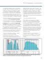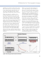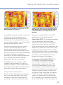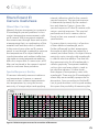
1
The Ultimate Infrared Handbook for R&D Professionals A Resource Guide for Using Infrared in the Research and Development Industry Contents IR Thermography – How It Works 1 IR Detectors For Thermographic Imaging 7 Getting The Most From Your IR Camera 14 Filters Extend IR Camera Usefulness 25 Ultra High-Speed Thermography 35 Published by FLIR AB This booklet may not be reproduced in any form without the permission in writing from FLIR Systems, AB. © Copyright. All rights reserved. Chapter 1 IR Thermography – How It Works IR Thermography Cameras Although infrared radiation (IR) is not detectable by the human eye, an IR camera can convert it to a visual image that depicts thermal variations across an object or scene. IR covers a portion of the electromagnetic spectrum from approximately 900 to 14,000 nanometers (0.9–14 µm). IR is emitted by all objects at temperatures above absolute zero, and the amount of radiation increases with temperature. Thermography is a type of imaging that is accomplished with an IR camera calibrated to display temperature values across an object or scene. Therefore, thermography allows one to make non-contact measurements of an object’s temperature. IR camera construction is similar to a digital video camera. The main components are a lens that focuses IR onto a detector, plus electronics and software for processing and displaying the signals and images. Instead of a charge coupled device that video and digital still cameras use, the IR camera detector is a focal plane array (FPA) of micrometer size pixels made of various materials sensitive to IR wavelengths. FPA IR In NIR MWIR LWIR Detector Cooling Digitization Optics resolution can range from about 160 × 120 pixels up to 1024 × 1024 pixels. Certain IR cameras have built-in software that allows the user to focus on specific areas of the FPA and calculate the temperature. Other systems utilized a computer or data system with specialized software that provides temperature analysis. Both methods can supply temperature analysis with better than ±1°C precision. FPA detector technologies are broken down into two categories: thermal detectors and quantum detectors. A common type of thermal detector is an uncooled microbolometer made of a metal or semiconductor material. These typically have lower cost and a broader IR spectral response than quantum detectors. Still, microbolometers react to incident radiant energy and are much slower and less sensitive than quantum detectors. Quantum detectors are made from materials such as InSb, InGaAs, PtSi, HgCdTe (MCT), and layered GaAs/AlGaAs for QWIP (Quantum Well Infrared Photon) detectors. The operation of a quantum detector is based on the change of state of electrons in a crystal structure reacting to incident photons. These detectors are generally faster and more sensitive than thermal detectors. However, they require cooling, sometimes down to cryogenic Video Processing Electronics User Interface User Control Video Output Digital Output Synchronization In/Out System Status Figure 1. Simplified block diagram of an IR camera 1 Chapter 1 temperatures using liquid nitrogen or a small Stirling cycle refrigerator unit. IR Spectrum Considerations Typically, IR cameras are designed and calibrated for a specific range of the IR spectrum. This means that the optics and detector materials must be selected for the desired range. Figure 2 illustrates the spectral response regions for various detector materials. Because IR has the same properties as visible light regarding reflection, refraction, and transmission, the optics for thermal cameras are designed in a fashion similar to those of a visual wavelength camera. However, the types of glass used in optics for visible light cameras cannot be used for optics in an infrared camera, as they do not transmit IR wavelengths well enough. Conversely, materials that are transparent to IR are often opaque to visible light. IR camera lenses typically use silicon (Si) and germanium (Ge) materials. Normally Si is used for MWIR (medium wavelength IR) camera systems, whereas Ge is used in LW (long wavelength) cameras. Si and Ge have good mechanical properties, i.e., they do not break easily, they are non- MCT PtSi QWIP InSb Microbolometer MWIR 3.0µm LWIR 5.0µm 8.0µm 14.0µm Figure 2. Examples of detector materials and their spectral responses relative to IR midwave (MW) and longwave (LW) bands 2 hygroscopic, and they can be formed into lenses with modern turning methods. As in visible light cameras, IR camera lenses have antireflective coatings. With proper design, IR camera lenses can transmit close to 100% of incident radiation. Thermal Radiation Principles The intensity of the emitted energy from an object varies with temperature and radiation wavelength. If the object is colder than about 500°C, emitted radiation lies completely within IR wavelengths. In addition to emitting radiation, an object reacts to incident radiation from its surroundings by absorbing and reflecting a portion of it, or allowing some of it to pass through (as through a lens). From this physical principle, the Total Radiation Law is derived, which can be stated with the following formula: W = aW + rW + tW, which can be simplified to: 1 = a + r + t. The coefficients a, r, and t describe the object’s incident energy absorbtion (a), reflection (r), and transmission (t). Each coefficient can have a value from zero to one, depending on how well an object absorbs, reflects, or transmits incident radiation. For example, if r = 0, t = 0, and a = 1, then there is no reflected or transmitted radiation, and 100% of incident radiation is absorbed. This is called a perfect blackbody. In the real world there are no objects that are perfect absorbers, reflectors, or transmitters, although some may come very close to one of these properties. Nonetheless, the concept of a perfect blackbody is very important in the IR Thermography – How It Works 1 – emissivity). Since a perfect blackbody is a perfect absorber, r = 0 and e = 1. science of thermography, because it is the foundation for relating IR radiation to an object’s temperature. The radiative properties of a perfect blackbody can also be described mathematically by Planck’s Law. Since this has a complex mathematical formula, and is a function of temperature and radiation wavelength, a blackbody’s radiative properties are usually shown as a series of curves (Figure 3). Fundamentally, a perfect blackbody is a perfect absorber and emitter of radiant energy. This concept is stated mathematical as Kirchhoff’s Law. The radiative properties of a body are denoted by the symbol e, the emittance or emissivity of the body. Kirchhoff’s law states that a = e, and since both values vary with the radiation wavelength, the formula can take the form a(l) = e(l), where l denotes the wavelength. These curves show the radiation per wavelength unit and area unit, called the spectral radiant emittance of the blackbody. The higher the temperature, the more intense the emitted radiation. However, each emittance curve has a distinct maximum value at a certain The total radiation law can thus take the mathematical form 1 = e + r + t, which for an opaque body (t = 0) can be simplified to 1 = e + r or r = 1 – e (i.e., reflection = 4.50 T-1000˚C Blackbody spectral radiant emittance 4.00 3.50 3.00 T-900˚C 2.50 2.00 T-700˚C T-800˚C T-600˚C 1.50 T-500˚C T-400˚C 1.00 T-300˚C 0.50 T-200˚C 0.00 Visible light 2 3 4 5 6 7 8 9 10 11 12 13 14 15 Figure 3. Illustration of Planck’s Law 3 Chapter 1 wavelength. This maximum can be calculated from Wien’s displacement law, lmax = 2898/T, where T is the absolute temperature of the blackbody, measured in Kelvin (K), and lmax is the wavelength at the maximum intensity. Using blackbody emittance curves, one can find that an object at 30°C has a maximum near 10µm, whereas an object at 1000°C has a radiant intensity with a maximum of near 2.3µm. The latter has a maximum spectral radiant emittance about 1,400 times higher than a blackbody at 30°C, with a considerable portion of the radiation in the visible spectrum. From Planck’s law, the total radiated energy from a blackbody can be calculated. This is expressed by a formula known as the Stefan-Bolzmann law, W = σT4 (W/m2), where σ is the Stefan-Bolzmann’s constant (5.67 × 10⁻⁸ W/m2K4). As an example, a human being with a normal temperature (about 300 K) will radiate about 500W/ m2 of effective body surface. As a rule of thumb, the effective body surface is 1m2, and radiates about 0.5kW—a substantial heat loss. The equations described in this section provide important relationships between emitted radiation and temperature of a perfect blackbody. Since most objects of interest to thermographers are not perfect blackbodies, there needs to be some way for an IR camera to graph the temperature of a “normal” object. 4 Emissivity The radiative properties of objects are usually described in relation to a perfect blackbody (the perfect emitter). If the emitted energy from a blackbody is denoted as Wbb, and that of a normal object at the same temperature as Wobj, then the ratio between these two values describes the emissivity (e) of the object, e = Wobj / Wbb. Thus, emissivity is a number between 0 and 1. The better the radiative properties of the object, the higher its emissivity. An object that has the same emissivity e for all wavelengths is called a greybody. Consequently, for a greybody, StefanBolzmann’s law takes the form W = eσT4 (W/m2), which states that the total emissive power of a greybody is the same as that of a blackbody of the same temperature reduced in proportion to the value of e for the object. Still, most bodies are neither blackbodies nor greybodies. The emissivity varies with wavelength. As thermography operates only inside limited spectral ranges, in practice it is often possible to treat objects as greybodies. In any case, an object having emittance that varies strongly with wavelength is called a selective radiator. For example, glass is a very selective radiator, behaving almost like a blackbody for certain wavelengths, whereas it is rather the opposite for other wavelengths. Atmospheric Influence Between the object and the thermal camera is the atmosphere, which tends IR Thermography – How It Works to attenuate radiation due to absorption by gases and scattering by particles. The amount of attenuation depends heavily on radiation wavelength. Although the atmosphere usually transmits visible light very well, fog, clouds, rain, and snow can prevent us from seeing distant objects. The same principle applies to infrared radiation. high-pass filter above 7.5μm (Figure 4). The MW band of 3–5µm tends to be employed with highly sensitive detectors for highend R&D and military applications. When acquiring a signal through the atmosphere with MW cameras, selected transmission bands must be used where less attenuation takes place. For thermographic measurement we must use the so-called atmospheric windows. As can be seen from Figure 4, they can be found between 2 and 5µm, the mid-wave windows, and 7.5–13.5µm, the long-wave window. Atmospheric attenuation prevents an object’s total radiation from reaching the camera. If no correction for attenuation is applied, the measured apparent temperature will be lower and lower with increased distance. IR camera software corrects for atmospheric attenuation. Temperature Measurements Typically, LW cameras in the 7.5–13.5μm range work well anywhere that atmospheric attenuation is involved, because the atmosphere tends to act as a Given this situation, we can derive a formula for the calculation of the object’s temperature from a calibrated camera’s output. The radiation that impinges on the IR camera lens comes from three different sources. The camera receives radiation from the target object, plus radiation from its surroundings that has been reflected onto the object’s surface. Both of these radiation components become attenuated when they pass through the atmosphere. Since the atmosphere absorbs part of the radiation, it will also radiate some itself (Kirchhoff’s law). Figure 4. Atmospheric attenuation (white areas) with a chart of the gases and water vapor causing most of it. The areas under the curve represent the highest IR transmission. 5 Chapter 1 1. Emission from the object = e · t · Wobj, where e is the emissivity of the object and t is the transmittance of the atmosphere. 2. Reflected emission from ambient sources = (1 – e) · t · Wamb, where (1 – e) is the reflectance of the object. (It is assumed that the temperature Tamb is the same for all emitting surfaces within the half sphere seen from a point on the object’s surface.) 3. Emission from the atmosphere = (1 – t) · Watm, where (1 – t) is the emissivity of the atmosphere. The total radiation power received by the camera can now be written: 6 Wtot = (1 – t) · Wobj + (1 – e) · t · Wamb + (1 – t) · Watm, where e is the object emissivity, t is the transmission through the atmosphere, Tamb is the (effective) temperature of the object’s surroundings, or the reflected ambient (background) temperature, and Tatm is the temperature of the atmosphere. To arrive at the correct target object temperature, IR camera software requires inputs for the emissivity of the object, atmospheric attenuation and temperature, and temperature of the ambient surroundings. Depending on circumstances, these factors may be measured, assumed, or found from look-up tables. Chapter 2 IR Detectors For Thermographic Imaging IR Cameras Thermographic imaging is accomplished with a camera that converts infrared radiation (IR) into a visual image that depicts temperature variations across an object or scene. The main IR camera components are a lens, a detector in the form of a focal plane array (FPA), possibly a cooler for the detector, and the electronics and software for processing and displaying images (Figure 1). Most detectors have a response curve that is narrower than the full IR range (900–14,000 nanometers or 0.9–14µm). Therefore, a detector (or camera) must be selected that has the appropriate response for the IR range of a user’s application. In addition to wavelength response, other important detector characteristics include sensitivity, the ease of creating it as a focal plane array with micrometer size pixels, and the degree of cooling required, if any. In most applications, the IR camera must view a radiating object through the atmosphere. Therefore, an overriding concern is matching the detector’s response curve to what is called an IR In NIR MWIR LWIR Detector Cooling Digitization Optics atmospheric window. This is the range of IR wavelengths that readily pass through the atmosphere with little attenuation. Essentially, there are two of these windows, one in the 2–5.6µm range, the short/ medium wavelength (SW/MW) IR band, and one in the 8–14µm range, the longwavelength (LW) IR band. There are many detector materials and cameras with response curves that meet these criteria. Quantum vs. Non-Quantum Detectors The majority of IR cameras have a microbolometer type detector, mainly because of cost considerations. Microbolometer FPAs can be created from metal or semiconductor materials, and operate according to non-quantum principles. This means that they respond to radiant energy in a way that causes a change of state in the bulk material (i.e., the bolometer effect). Generally, microbolometers do not require cooling, which allows compact camera designs that are relatively low in cost. Other characteristics of microbolometers are: • Relatively low sensitivity (detectivity) • Broad (flat) response curve • Slow response time (time constant ~12ms) Video Processing Electronics User Interface User Control Video Output Digital Output Synchronization In/Out System Status Figure 1. Simplified block diagram of an IR camera 7 Chapter 2 1012 K 193 Se Pb 7K e7 S Pb K 95 2 Se Pb 26K :Hg Ge 10 9 :G Si 7K )7 K .2 a4 :A Si PH OT ID OVO EA L L P TAIC HO TOCONDUCTOR 1011 D · (cm√Hz/W) D · (cm√Hz/W) LP H OT OT OC OVO LTAIC ON DU CTO R K 95 s2 Pb 2π STERADIANS FIELD OF VIEW 295K BACKGROUND TEMPERATURE L EA ID 1010 7K s7 Pb EA ID PH L EA ID 1011 1012 2π STERADIANS FIELD OF VIEW 295K BACKGROUND TEMPERATURE K 93 s1 Pb K .2 s4 K 4.2 Sb Si: 7K (PV )7 Te 3K PV Cd ) 19 b( Hg (PC InS e T Cd g H 1010 K 77 V) (P Te 7K d C )7 Hg PC e( dT C Hg 10 9 Bolometer (90Hz) QWIP Pyroelectric Det. (90Hz) 10 8 1.0 1.5 2.0 2.5 3 4 5 6 7 8 9 10 15 20 25 30 40 10 8 1.0 1.5 2.0 2.5 3 Wavelength (µm) 4 5 6 7 8 9 10 15 20 25 30 40 Wavelength (µm) Figure 2. Detectivity (D*) curves for different detector materials For more demanding applications, quantum detectors are used, which operate on the basis of an intrinsic photoelectric effect. These materials respond to IR by absorbing photons that elevate the material’s electrons to a higher energy state, causing a change in conductivity, voltage, or current. By cooling these detectors to cryogenic temperatures, they can be very sensitive to the IR that is focused on them. They also react very quickly to changes in IR levels (i.e., temperatures), having a constant response time on the order of 1µs. Therefore, a camera with this type of detector is very useful in recording transient thermal events. Still, quantum detectors have response curves with detectivity that varies strongly with wavelength (Figure 2). Table 1 lists some of the most commonly used detectors in today’s IR cameras. 8 Table 1. Detector types and materials commonly used in IR cameras. Detector Type/ Material Microbolometer HgCdTe HgCdTe InSb PtSi QWIP Operation Broadband detector SW photon detector LW photon detector MW photon detector MW photon detector LW photon detector Operating Temp. Uncooled (~30°C) 200 K 77 K 77 K 77 K 70 K Operating Principles for Quantum Detectors In materials used for quantum detectors, at room temperature there are electrons at different energy levels. Some electrons have sufficient thermal energy that they are in the conduction band, meaning the electrons there are free to move and the IR Detectors For Thermographic Imaging material can conduct an electrical current. Most of the electrons, however, are found in the valence band, where they do not carry any current because they cannot move freely. (See left-most views of Fig 3.) When the material is cooled to a low enough temperature, which varies with the chosen material, the thermal energy of the electrons may be so low that there are none in the conduction band (upper center view of Figure 3). Hence the material cannot carry any current. When these materials are exposed to incident photons, and the photons have sufficient energy, this energy can stimulate an electron in the valence band, causing it to move up into the conduction band (upper right view of Figure 3). Thus the material (the detector) can carry a photocurrent, which is proportional to the intensity of the incident radiation. There is a very exact lowest energy of the incident photons that will allow an electron to jump from the valence band into the conduction band. This energy is related to a certain wavelength, the cutoff wavelength. Since photon energy is inversely proportional to its wavelength, the energies are higher in the SW/MW band than in the LW band. Therefore, as a rule, the operating temperatures for LW detectors are lower than for SW/MW detectors. For an InSb MW detector, the necessary temperature must be less than 173 K (–100°C), although it may be operated at a much lower temperature. An HgCdTe (MCT) LW detector must be cooled to 77 K (–196°C) or lower. A QWIP detector typically needs to operate at about 70 K (–203°C) or lower. The lower center and right views of Figure 3 depict quantum detector wavelength dependence. The incident Figure 3. Operating principle of quantum detectors 9 Chapter 2 photon wavelength and energy must be sufficient to overcome the band gap energy, ΔE. Cold side Copper Thermoelectrical material Cooling Methods The first detectors used in infrared radiometric instruments were cooled with liquid nitrogen. The detector was attached to the Dewar flask that held the liquid nitrogen, thus keeping the detector at a very stable and low temperature (–196°C). Later, other cooling methods were developed. The first solid-state solution to the cooling problem was presented by AGEMA in 1986, when it introduced a Peltier effect cooler for a commercial IR camera. In a Peltier cooler, DC current is forced through a thermoelectric material, removing heat from one junction and creating a cold side and a hot side. The hot side is connected to a heat sink, whereas the cold side cools the component attached to it. See Figures 4 and 5. For very demanding applications, where the highest possible sensitivity was needed, an electrical solution to cryogenic cooling was developed. This resulted in the Stirling cooler. Only in the last 15 to 20 years were manufacturers able to extend the life of Stirling coolers to 8,000 hours or more, which is sufficient for use in thermal cameras. The Stirling process removes heat from the cold finger (Figure 6) and dissipates it at the warm side. The efficiency of this type of cooler is relatively low, but good enough for cooling an IR camera detector. Regardless of the cooling method, the detector focal plane is attached to the cold side of the cooler in a way that allows 10 Warm side + – DC Figure 4. Single stage Peltier cooler Mounting plate IR detector Figure 5. Three-stage Peltier cooler efficient conductive heat exchange. Because focal plane arrays are small, the attachment area and the cooler itself can be relatively small. Focal Plane Array Assemblies Depending on the size/resolution of an FPA assembly, it has from (approximately) 60,000 to more than 1,000,000 individual detectors. For the sake of simplicity, this can be described as a two-dimensional pixel matrix with each pixel (detector) having micrometer size dimensions. FPA resolutions can range from about 160 × 120 pixels up to 1024 × 1024 pixels. IR Detectors For Thermographic Imaging Figure 6. Integrated Stirling cooler, working with helium gas, cooling down to –196ºC or sometimes even lower temperatures Figure 7. Examples of cooled focal plane array assemblies used in IR cameras In reality, assemblies are a bit more complex. Depending on the detector material and its operating principle, an optical grating may be part of the FPA assembly. This is the case for QWIP detectors, in which the optical grating disperses incident radiation to take advantage of directional sensitivity in the detector material’s crystal lattice. This has the effect of increasing overall sensitivity of a QWIP detector. Furthermore, the FPA must be bonded to the IR camera readout Figure 8. QWIP FPA mounted on a ceramics substrate and bonded to external electronics electronics. A finished QWIP detector and IC electronics assembly is shown in Figure 8. This would be incorporated with a Dewar or Stirling cooler in an assembly similar to those shown in Figure 7. Another complexity is the fact that each individual detector in the FPA has a slightly different gain and zero offset. To create a useful thermographic image, the different gains and offsets must be corrected to a normalized value. This multi-step calibration process is performed by the camera software. See Figures 9–11. The ultimate result is a thermographic image that accurately portrays relative temperatures across the target object or scene (Figure 12). Moreover, actual temperatures can be calculated to within approximately ±1°C accuracy. 11 Chapter 2 Signal Without any correction Signal First correction step Radiation –20°C Radiation +120°C –20°C +20°C +120°C +20°C Figure 9. To normalize different FPA detector gains and offsets, the first correction step is offset compensation. This brings each detector response within the dynamic range of the camera’s A/D converter electronics. Signal First correction step Signal Second correction A/D Dynamics Radiation –20°C Radiation +120°C –20°C +20°C +120°C +20°C Figure 10. After offset compensation, slope correction is applied. Signal Third correction, Non-Uniformity Correction (NUC) Signal After NUC Radiation –20°C +120°C +20°C Radiation –20°C +120°C +20°C Figure 11. After gain factors are brought to the same value, non-uniformity correction (NUC) is applied so that all detectors have essentially the same electronic characteristics. 12 IR Detectors For Thermographic Imaging Figure 12. IR image from a 1024 × 1024 InSb detector camera Application Criteria As indicated earlier, different types of detectors have different thermal and spectral sensitivities. In addition, they have different cost structures due to various degrees of manufacturability. Where they otherwise fit the application, photon detectors such as InSb and QWIP types offer a number of advantages: • High thermal sensitivity • High uniformity of the detectors, i.e., very low fixed pattern noise • There is a degree of selectability in their spectral sensitivity • High yield in the production process • Relatively low cost • They are resistant to high temperatures and high radiation • They produce very good image quality Camera electronics can handle wide variations in absolute detector sensitivities. For example, high sensitivity that might saturate a detector at high thermal intensities can be handled by aperture control and neutral density filters. Both of these solutions can reduce the radiant energy impinging on the FPA. Price aside, spectral sensitivity is often an overriding concern in selecting a detector and camera for a specific application. Once a detector is selected, lens material and filters can be selected to somewhat alter the overall response characteristics of an IR camera system. Fig 13 shows the system response for a number of different detectors. Relative sensitivity 100% InGaAs InSb 90% MCT-SW Microbolometer 80% MCT-LW QWIP VisGaAs 70% 60% 50% 40% 30% 20% 10% 0% 1 2 3 4 5 6 7 8 9 10 Wavelength λ [µm] 11 12 13 14 15 16 17 18 Figure 13. Relative response curves for a number of IR cameras 13 Chapter 3 Getting The Most From Your IR Camera Understanding IR camera calibration and corrections help ensure accurate temperature measurements and thermographic mapping. Quantitative Measurements with IR Cameras For best results, IR camera users need to think carefully about the type of measurements they need to make, and then be proactive in the camera’s calibration process. Of course, the first step is selecting a camera with the appropriate features and software for the application. An understanding of the differences between thermographic and radiometric measurements is very helpful in this regard. Thermography is a type of infrared imaging in which IR cameras detect radiation in the electromagnetic spectrum with wavelengths from roughly 900 to 14,000 nanometers (0.9–14 µm) and produce images of that radiation. Typically, this imaging is used to measure temperature variations across an object or scene, which can be expressed in degrees Celsius, Fahrenheit, or Kelvin. Radiometry is the measurement of radiant electromagnetic energy, especially that associated with the IR spectrum. It can be more simply defined as an absolute measurement of radiant flux. The typical unit of measure for imaging radiometry is radiance, which is expressed in units of Watts/(sr-cm2). (The abbreviation “sr” stands for steradian; a non-dimensional geometric ratio expressing the solid (conical) angle that encloses a portion of the surface of 14 a sphere equivalent to the square of the radius.) In simple terms, one can think of thermography as “how hot” an object is, whereas radiometry is “how much energy” the object is giving off. Although these two concepts are related, they are not the same thing. IR cameras inherently measure irradiance not temperature, but thermography does stem from radiance. When you thermographically calibrate an IR system you are calibrating /measuring based on effective blackbody radiance and temperature. Therefore, the emissivity of the target object you are measuring is vital to achieving accurate temperatures. (Emissivity or emittance is the radiative property of an object relative to a perfect blackbody.) Entry level IR cameras with microbolo meter detectors operate according to non-quantum principles. The detectors respond to radiant energy in a way that causes a change of state in the bulk material (e.g., resistance or capacitance). Calibration software in these cameras is oriented toward thermographic imaging and temperature measurements. Highend IR cameras with photon detectors operate according to quantum physics principles. Although they also provide high quality images, their software is typically more sophisticated, allowing accurate measurements of both radiance and temperature. Some reasons why radiance measurements are important include: • Given a linear sensor, measured radiance is linear with incident energy. Temperature is non-linear with raw Getting The Most From Your IR Camera • • • • digital image counts, even with a linear sensor. Given the radiance and area of an object, radiant intensity can be calculated. Knowing total radiant intensity of a target gives a radiometric analyst the ability to model the irradiance generated by the target over various geometric and atmospheric conditions. The relationship between spectral bands of interest can be much easier to determine if you are working within radiometric units. The comparison between different objects in radiometric terms tends to have less uncertainty because emissivity is not a concern. (One still needs to consider atmospheric and spectral bandpass effects.) One can typically convert a radiometric signature from radiance to effective blackbody temperature given a few assumptions or ancillary measurement data. It tends to be more difficult to go from temperature to radiance. Key Physical Relationships in Camera Operation There are five basic steps in producing radiometric and thermographic measurements with an IR camera system: 1. The target object has a certain energy signature that is collected by the IR camera through its lens. 2. This involves the collection of photons in the case of a photon detector, or collection of heat energy with a thermal detector, such as a microbolometer. 3. The collected energy causes the detector to produce a signal voltage that results in a digital count through the system’s A/D converter. (For example, a FLIR ThermoVision® SC6000 IR camera has a 14-bit dynamic range in its A/D converter, which creates count values ranging from 0–16,383. The more IR energy incident on the camera’s detector (within its spectral band), the higher the digital count.) 4. When the camera is properly calibrated, digital counts are transformed into radiance values. 5. Finally, the calibrated camera‘s electronics convert radiance values to temperature using the known or measured emissivity of the target object. Expanding on Steps 4 and 5, an effective blackbody temperature measurement can be derived from a radiance measurement by applying a radiometric calibration, temperature vs. radiance model, and emissivity of the target object or scene. Every IR camera designed for serious measurements is calibrated at the factory. In the calibration lab, the camera takes a number of blackbody measurements at known temperatures, radiance levels, emissivities, and distances. This creates a table of values based on the A/D counts from the temperature/radiance measurements. Once the counts for each blackbody temperature measurement are entered into the calibration software, the data are then passed through an in-band radiance curve fit algorithm to produce the appropriate in-band radiance vs. count values given the camera system’s normalized spectral response function. This produces a radiometric calibration of in-band radiance [W/(sr-cm2)] versus the digital counts 15 Chapter 3 Radiance vs. Measurement Black Body Source Temperature (°C) 2.9516e–04 0 10 20 30 40 2.8149e–04 2.6781e–04 2.5414e–04 2.4047e–04 Radiance (W/(sr-cm2)) 2.2680e–04 2.1313e–04 1.9946e–04 Curve Fit 1.8579e–04 Measurements 1.7212e–04 1.5845e–04 1.4478e–04 1.3111e–04 1.1744e–04 1.0377e–04 9.0096e–05 1.4938e+04 1.4716e+04 1.4503e+04 1.4280e+04 1.4068e+04 1.3855e+04 1.3632e+04 1.3420e+04 1.3207e+04 1.2985e+04 1.2772e+04 1.2559e+04 1.2337e+04 1.2124e+04 1.1912e+04 1.1689e+04 1.1476e+04 1.1264e+04 1.1041e+04 1.0829e+04 1.0606e+04 1.0393e+04 1.0181e+04 9.9585e+03 9.7460e+03 9.5334e+03 9.3107e+03 9.0981e+03 8.8856e+03 8.6629e+03 8.4503e+03 8.2738e+03 8.0151e+03 7.5899e+03 6.2755e–05 7.8025e+03 7.6425e–05 Measurement (Counts) Figure 1. Example of camera measurements and corresponding in-band radiance values for given black body temperatures with resulting radiance vs. measurement curve. obtained while viewing a blackbody over a range of temperatures. The result is a series of calibration curves. An example of how calibration points are captured is shown in Figure 1. The calibration curves are stored in the camera system’s memory as a series of numeric curve-fit tables that relate radiance values to blackbody temperatures. When the system makes a measurement, it takes the digital value of the signal at a given moment, goes into the appropriate calibration table, and calculates temperature. Due consideration is given to other factors like atmospheric attenuation, reflected ambient temperature, and the camera’s ambient temperature drift before the final result is presented. 16 Ambient Drift Compensation (ADC). Another important consideration in the calibration process is the radiation caused by the heating and cooling of the camera itself. Any swings in camera internal temperature caused by changes in environment or the heating and cooling of camera electronics will affect the radiation intensity at the detector. The radiation that results directly from the camera is called parasitic radiation and can cause inaccuracies in camera measurement output, especially with thermographically calibrated cameras. Certain IR cameras (like the FLIR ThermoVision® product line), have internal sensors that monitor changes in camera temperature. As part of the calibration process, these cameras are placed in an environmental chamber Getting The Most From Your IR Camera and focused at a black body reference. The temperature of the chamber and black body are then varied and data is collected from the internal sensors. Correction factors are then created and stored in the camera. In real-time operation, the camera sensors continually monitor internal temperature and send feedback to the camera processor. The camera output is then corrected for any parasitic radiation influences. This functionality is commonly referred to as ambient drift compensation. Ultimately, the camera must calculate at an object’s temperature based on its emission, reflected emission from ambient sources, and emission from the atmosphere using the Total Radiation Law. The total radiation power received by the camera can be expressed as: Wtot = e · t · Wobj + (1 – e) · t · Wamb + (1 – t) · Watm, where e is the object emissivity, t is the transmission through the atmosphere, Tamb is the (effective) temperature of the object surroundings, or the reflected ambient (background) temperature, and Tatm is the temperature of the atmosphere. The best results are obtained when a user is diligent in entering known values for all the pertinent variables into the camera software. Emissivity tables are available for a wide variety of common substances. However, when in doubt, measurements should be made to obtain the correct values. Calibration and analysis software tools available to users are not always contained onboard the camera. While high-end cameras have many built-in software functions, others rely on external software that runs on a PC. Even high-end cameras are connected to PCs to expand their internal calibration, correction, and analysis capabilities. For example, FLIR’s ThermaCAM® RTools™ software can serve a wide variety of functions from real-time image acquisition to post-acquisition analysis. Whether the software is on the camera or an external PC, the most useful packages allow a user to easily modify calibration variables. For instance, FLIR’s ThermaCAM RTools provides the ability to enter and modify emissivity, atmospheric conditions, distances, and other ancillary data needed to calculate and represent the exact temperature of the object, both live and through saved data. This software provides a post-measurement capability to further modify atmospheric conditions, spectral responsivity, atmospheric transmission changes, internal and external filters, and other important criteria as needed. The discussions that follow below are intended to represent both onboard and external camera firmware and software functions. Where these functions reside depends on the camera. Typical Camera Measurement Functions IR cameras have various operating modes to assure correct temperature measurements under different application conditions. Typical measurement functions include: • Spotmeter • Area • Profile • Isotherm 17 Chapter 3 • Temperature range • Color or gray scale settings Cursor functions allow easy selection of an area of interest, such as the crosshairs of the spot readings in Figure 2. In addition, the cursor may be able to select circle, square, and irregularly shaped polygon areas, or create a line for a temperature profile. Once an area is selected, it can be “frozen” so that the camera can take a snapshot of that area. Alternatively, the camera image can remain live for observation of changes in temperature. Figure 2. IR image of a printed circuit board indicating three spot temperature readings. Image colors correspond to the temperature scale on the right. The spotmeter finds the temperature at a particular point. Depending on the camera, this function may allow ten or more movable spots, one or more of which may automatically find the hottest point in the image. The area function isolates a selected area of an object or scene and finds the maximum, minimum, and average temperatures inside that area. The isotherm function makes it possible to portray the temperature distribution of a hot area. Multiple isotherms may be allowed. The line profile is a way to visualize the temperature along some part of the object, 18 which may also be shown as a graph (Figure 3). Figure 3. Graph of temperature along a selected area of a target object using a camera’s profile function The temperature measurement range typically is selectable by the user. This is a valuable feature when a scene has a temperature range narrower than a camera’s full-scale range. Setting a narrower range allows better resolution of the images and higher accuracy in the measured temperatures. Therefore, images will better illustrate smaller temperature differences. On the other hand, a broader scale and/or higher maximum temperature range may be needed to prevent saturation of the portion of the image at the highest temperature. As an adjunct to the temperature range selection, most cameras allow a user to set up a color scale or gray scale to optimize the camera image. Figure 4 illustrates two gray scale possibilities. In Figure 2 a so-called “iron scale” was used for a color rendering. In a manner similar to the gray scale used in Figure 4, the hottest temperatures can be rendered as either lighter colors or darker colors. Another possibility is rendering images with what is known as a rainbow scale (Figure 5). In some color images, gray is used to indicate areas where the camera detector has become saturated (i.e., temperatures well above the top of the scale). Getting The Most From Your IR Camera Figure 4. Gray scale images of car engine; left view has white as the hottest temperature; right view shows black as the hottest While choice of color scale is often a matter of personal preference, there may be times when one type of scale is better than another for illustrating the range of temperatures in a scene. In the case of isotherm measurements, areas with the same thermal radiance are highlighted. If we use a color scale with ten colors, we will in fact get ten isotherms in the image. Such a scale sometimes makes it easier to see the temperature distribution over an object. In Figure 6, the temperature Figure 5. Rainbow scale showing lower temperatures towards the blue end of the spectrum scale is selected so that each color is an isotherm with a width of 2°C. Still, it is important to realize that an isothermal temperature scale rendering will not be accurate unless all of the highlighted area has the same emissivity, and the ambient temperatures are the same for all objects within the area. This points out common problems for IR camera users. Often, emissivity varies across an object or scene, along with variations in ambient temperatures, accompanied by atmospheric conditions that don’t match Figure 6. Isotherm color scale with each color having an isotherm width of 2°C 19 Chapter 3 a camera’s default values. This is why IR cameras include measurement correction and calibration functions. Emissivity Corrections In most applications, the emissivity of an object is based on values found in a table. Although camera software may include an emissivity table, users usually have the capability of inputting emissivity values for an object ranging from 0.1 to 1.0. Many cameras also provide automatic corrections based on user input for reflected ambient temperature, viewing distance, relative humidity, atmospheric transmission, and external optics. As described earlier, the IR camera calculates a temperature based on radiance measurements and the object’s emissivity. However, when the emissivity value is unknown or uncertain, the reverse process can be applied. Knowing the object temperature, emissivity can be calculated. This is usually done when exact emissivity values are needed. There are two common methods of doing this. The first method establishes a known temperature by using an equalization box. This is essentially a tightly controlled temperature chamber with circulating hot air. The length of time in the box must be sufficient to allow the whole object to be at a uniform temperature. In addition, it is absolutely necessary that the object stabilize at a temperature different from the surroundings where the actual measurements will take place. Usually, the object is heated to a temperature at least 10°C above the surroundings to ensure that the thermodynamics of the measurements are valid. 20 Once the object has reached the set temperature, the lid is drawn off and a thermogram is captured of the object. The camera and/or software for processing thermograms can be used to get the emissivity value. Another (“adjacent spot”) method is much simpler, but still gives reasonably exact values of the emissivity. It uses an area of known emissivity. The idea is to determine the temperature of the object with the camera in the usual way. The object is adjusted so that the area with unknown emissivity is very close to an area of known emissivity. The distance separating these areas must be so small that it can be safely assumed they have the same temperature. From this temperature measurement the unknown emissivity can be calculated. The problem is illustrated in Figure 7, which is an image of a printed circuit board (PCB) heated to a uniform temperature of 68.7°C. However, areas of different emissivities may actually have different temperatures, as indicated in the caption of Figure 7a. Using the technique just described, emissivity correction proceeds by finding a reference spot where a temperature of 68.7°C is indicated and calculating the emissivity at that location. By knowing the emissivity of the reference spot, the emissivity of the target spots can be calculated. The corrected temperatures are shown in Figure 7b. As illustrated in these figures, this technique can be used with a camera’s area selection function (“AR” in the figures) and using the average temperature for that area. The reason for using the average temperature in the reference area is that there is usually a spread of temperatures within the area, Getting The Most From Your IR Camera Figure 7a. PCB heated to a uniform 68.7°C, but digital readouts are incorrect. Figure 7b. PCB with emissivity correction using the “adjacent spot” technique. Digital readouts now indicate the correct temperatures at all locations. especially for materials with low emissivity. In that case, using a spotmeter or an area maximum value would give a less stable result. The isotherm function is not recommended either, as it is not possible to get the averaging effect with it. Knowing a camera’s field of view (FOV) specifications helps determine this. It may also be possible to use a contact sensor to find the temperature of an area of unknown emissivity, but such measurements pose other problems that may not be easy to overcome. Furthermore, it is never possible to measure the emissivity of an object whose temperature is the same as the reflected ambient temperature from its surroundings. Generally, a user can also input other variables that are needed to correct for ambient conditions. These include factors for ambient temperatures and atmospheric attenuation around the target object. Using Camera Specifications When considering IR camera performance, most users are interested in how small an object or area can be detected and accurately measured at a given distance. Field of View (FOV). This parameter depends on the camera lens and focal plane dimensions, and is expressed in degrees, such as 35.5° × 28.7° or 18.2 × 14.6°. For a given viewing distance, this determines the dimensions of the total surface area “seen” by the instrument (Figure 8). For example, a FLIR ThermoVision SC6000 camera with a 25mm lens has an FOV of 0.64 × 0.51 meters at a distance of one meter, and 6.4 × 5.1 meters at a distance of ten meters. Instantaneous Field of View (IFOV). This is a measure of the spatial resolution of a camera’s focal plane array (FPA) detector. The configuration of the FPA in the FLIR ThermoVision SC6000 is 640 × 512 detectors, which makes a total of 327,680 individual picture elements (pixels). Suppose you are looking at an object at a distance of one meter with this camera. In determining the smallest detectable object, it is important to know the area’s IFOV covered by an individual pixel in the array. The total FOV is 0.64 × 0.51 meters at 21 Chapter 3 Figure 10. IFOV (red squares) relative to object size. Figure 8. A camera’s field of view (FOV) varies with viewing distance. a distance of one meter. If we divide these FOV dimensions by the number of pixels in a line and row, respectively, we find that a pixel’s IFOV is an area approximately 1.0 × 1.0mm at that distance. Figure 9 illustrates this concept. Figure 9. A camera’s geometric (spatial) resolution (IFOV) is determined by its lens and FPA configuration. To use this information consider, the pixel IFOV relative to the target object size (Figure 10). In the left view of this figure, the area of the object to be measured covers the IFOV completely. Therefore, the pixel will receive radiation only from the object, and its temperature can be measured correctly. 22 In the right view of Figure 10, the pixel covers more than the target object area and will pick up radiation from extraneous objects. If the object is hotter than the objects beside or behind it, the temperature reading will be too low, and vice versa. Therefore it is important to estimate the size of the target object compared to the IFOV in each measurement situation. Spot Size Ratio (SSR). At the start of a measurement session, the distance between the camera and the target object should be considered explicitly. For cameras that do not have a calibrated spot size, the spot size ratio method can be used to optimize measurement results. SSR is a number that tells how far the camera can be from a target object of a given size in order to get a good temperature measurement. A typical figure might be 1,000:1 (also written 1,000/1, or simply abbreviated as 1,000). This can be interpreted as follows: at 1000 mm distance from a target, the camera will measure a temperature averaged over a 1mm square. Note that SSR is not just for targets far away. It can be just as important for close-up work. However, the camera’s minimum focal distance must also be considered. For shorter target distances, some manufacturers offer close-up lenses. Getting The Most From Your IR Camera For any application and camera/lens combination, the following equation applies: DSSR __–____ , where S 1 • Use of different transmission and/ or emissivity curves or constants for calibration data points D is the distance from the camera to the target, In addition, IR camera software and firmware provide other user inputs that refine the accuracy of temperature measurements. One of the most important functions is non-uniformity correction (NUC) of the detector FPA. This type of correction is needed due to the fact that each individual detector in the camera’s FPA has a slightly different gain and zero offset. To create a useful thermographic image, the different gains and offsets must be corrected to a normalized value. S is smallest target dimension of interest, and SSR is the spot size ratio. The units of D and S must be the same. When selecting a camera, keep in mind that IFOV is a good figure of merit to use. The smaller the IFOV, the better the camera for a given total field of view. Other Tools for Camera Users As mentioned earlier, IR cameras are calibrated at the factory, and field calibration in not practical. However, some cameras have a built-in blackbody to allow a quick calibration check. These checks should be done periodically to assure valid measurements. Bundled and optional data acquisition software available for IR cameras allows easy data capture, viewing, analysis, and storage. Software functions may include real-time radiometric output of radiance, radiant intensity, temperature, target length/area, etc. Optional software modules are also available for spatial and spectral radiometric calibration. Functions provided by these modules might include: • Instrument calibration in terms of radiance, irradiance, and temperature • Radiometric data needed to set instrument sensitivity and spectral range • Adjustments for atmospheric effects This multi-step NUC process is performed by camera software. However, some software allows the user to specify the manner in which NUC is performed by selecting from a list of menu options. For example, a user may be able to specify either a one-point or a twopoint correction. A one-point correction only deals with pixel offset. Two-point corrections perform both gain and offset normalization of pixel-to-pixel nonuniformity. With regard to NUC, another important consideration is how this function deals with the imperfections that most FPAs have as a result of semiconductor wafer processing. Some of these imperfections are manifested as bad pixels that produce no output signals or as outputs far outside of a correctable range. Ideally, the NUC process identifies bad pixels and replaces them using a nearest neighbor replacement algorithm. Bad pixels are 23 Chapter 3 identified based on a response and/or offset level outside user-defined points from the mean response and absolute offset level. Other NUC functions may be included with this type of software, which are too numerous to mention. The same is true of many other off-the-shelf software modules that can be purchased to facilitate thermographic image display, analysis, data file storage, manipulation, and editing. Availability of compatible software is an important consideration when selecting an IR camera for a user’s application or work environment. 24 Conclusions Recent advances in IR cameras have made them much easier to use. Camera firmware has made setup and operation as simple as using a conventional video camera. Onboard and PC-based software provides powerful measurement and analysis tools. Nevertheless, for accurate results, the user should have an understanding of IR camera optical principals and calibration methods. At the very least, the emissivity of a target object should be entered into the camera’s database, if not already available as a table entry. Chapter 4 Filters Extend IR Camera Usefulness Where Filters Can Help Materials that are transparent or opaque to IR wavelengths present problems in noncontact temperature measurements with an IR camera. With transparent materials, the camera sees through them and records a temperature that is a combination of the material itself and that which is behind it. In the second case, when an IR camera needs to see through a material to measure the temperature of an object behind it, signal attenuation and ambient reflections can make accurate temperature readings difficult or impossible. In some cases, an IR filter can be placed in the camera’s optical path to overcome these problems. Spectral Response is the Key IR cameras inherently measure irradiance not temperature. However, a camera’s software coverts radiance measurements into temperatures by using the known emissivity of a target object and applying internal calibration data for the camera’s spectral response. The spectral response is determined primarily by the camera’s lens and detector. Figure 1 shows the spectral response of a few IR cameras with various spectral responses. The spectral performance of most cameras can be found in their user manual or technical specifications. For many objects, emissivity is a function of their radiance wavelength, and is further influenced by their temperature, the angle at which they are viewed by a camera, and other factors. An object whose emissivity varies strongly with wavelength is called a selective radiator. One that has the same emissivity for all wavelengths is called a greybody. Transparent materials, such as glass and many plastics, tend to be selective radiators. In other words, their degree of transparency varies with wavelength. There may be IR wavelengths where they are essentially opaque due to absorption. Since, according to Kirchhoff’s Law, a good absorber is also a good emitter, this opens the possibility of measuring the Relative sensitivity 100% InSb 90% MCT-SW Mikrobolometer MCT-LW FLIR QWIP 80% 70% 60% 50% 40% 30% 20% 10% 0% 1 2 3 4 5 6 7 8 9 10 Wavelength λ [µm] 11 12 13 14 15 16 17 18 Figure 1. Relative response curves for a number of IR cameras 25 Chapter 4 radiance and temperature of a selective radiator at some wavelength. Spectral Adaptation Inserting a spectral filter into the camera’s optical path is called spectral adaptation. The first step of this process is to analyze the spectral properties of the semitransparent material you are trying to measure. For common materials the data may be available in published data. Otherwise, this requires analysis with a spectrophotometer. (The camera manufacturer or a consulting firm may supply this service.) In either case, the objective is to find the spectral location of a band of complete absorption that falls within the IR camera’s response curve. Microbolometer detectors have rather broad response curves so they are not likely to present a problem in this respect. However, adding a filter decreases overall sensitivity due to narrowing of the camera’s spectral range. Sensitivity is reduced approximately by the ratio of the area under the filter’s spectral curve to the area under the camera’s spectral curve. This could be a problem for microbolometer systems, since they have relatively low sensitivity to start with and a broad spectral curve. Using a camera with, for example, a QWIP detector will provide greater sensitivity with a narrower spectral curve. Still, this narrow range may limit the application of such cameras for spectral adaptation. Ultimately, an optical (IR) filter must be selected that blocks all wavelengths except the band where the object absorbs. This ensures that the object has high emissivity within that band. 26 Besides semitransparent solids, selective adaptation can also be applied to gases. However, a very narrow filter might be required for selecting an absorption “spike” in a gas. Even with proper filtering, temperature measurement of gases is difficult, mainly due to unknown gas density. Selective adaptation for a gas has a better chance of success if the objective is merely gas detection, since there are less stringent requirements for quantitative accuracy. In that case sensitivity would be more important, and some gases with very high absorption might still be measurable. Spectral adaptation could also be applied the opposite way, i.e., selection of a spectral band where the transmission through a medium is as high as possible. The purpose would be to enable measurement on an object by seeing through the medium without any interference. The medium could be ordinary atmosphere, the atmosphere of combustion gases inside a furnace, or simply a window (or other solid) through which one wants to measure. Filter Types The simplest filters are broadband neutral density types that are used merely to reduce optical transmission and prevent detector saturation at high temperatures. While necessary sometimes, this is not spectral adaptation. In spectral adaptation, filters are used in order to suppress or transmit certain wavelengths. For discussion purposes, filters can be described as short-pass (SP), long-pass (LP), band-pass (BP), and narrow band-pass (NBP). See Figure 2. SP and LP filters are specified with a cut-on and a cut-off wavelength. BP and NBP filters are Filters Extend IR Camera Usefulness Different types of filter characteristics 100 90 80 70 System response curve 60 Long-pass filter 50 Band-pass filter 40 Narrow band-pass filter 30 Short-pass filter 20 10 0 1.5 2 2.5 3 3.5 4 4.5 5 5.5 Wavelength, µm Figure 2. Response curves for different types of filters specified with a center wavelength and a half-width (half-power) wavelength, the latter being the width where spectral response has decreased to 50% of its maximum. For temperature measurements on transparent materials, the filter selected must provide a band of essentially complete absorption. Incomplete absorption can be used, at least theoretically, provided that both absorptance and reflectance are known and stable at the absorption band. Unfortunately, absorption often varies with both temperature and thickness of the material. An example of applying a NBP filter to the measurement of polyethylene film temperature is shown in Figure 3. The blue curve in the figure shows the absorption band of polyethylene film. The red curve shows the transmittance of a 3.45µm NBP filter, which is designed to match polyethylene film. The green curve shows the resulting transmission through film plus the filter. This curve, running just above the zero line, indicates an excellent filter adaptation, i.e. the film appears to be opaque to the camera, and no background radiation would disturb the measurement of film temperature. Filters can also be classified according to their application temperature. Traditionally, cold filters, filters that are stabilized at or near the same temperature as the detector, are the most accurate and desired filters for thermal signatures. Warm filters, filters screwed onto the back of the optical lens outside of the detector/cooler assembly, are also commonly used but tend to provide more radiometric calibration uncertainty due to varying IR emission with ambient temperature changes. 27 Chapter 4 Filter adaptation 1 0.9 Transmission % 0.8 3.45µm NBP filter 0.7 0.6 Polyethylene transmission 0.5 0.4 Resulting transmission 0.3 0.2 0.1 0 3 3.1 3.2 3.3 3.4 3.5 3.6 3.7 3.8 3.9 4 Wavelength, µm Figure 3. Application of an NBP filter to achieve nearly complete absorption and high emittance from polyethylene film, allowing its temperature measurement Once a filter is selected for use with a particular camera, the camera/filter combination needs to be calibrated by the camera manufacturer. Then the performance of the system should be characterized since accuracy and sensitivity will be affected due to a reduction in energy going to the detector. Transparent Material Measurement Techniques Production of sheet glass and thin plastic film requires fairly tight temperature control to maximize production quality and yield. Traditionally, temperature sensors have been embedded at the orifice of the extruder, which provides rather coarse information about sheet/film temperature. An IR machine vision system can make noncontact temperature measurements and supply more usable data about the material as it is extruded. However, as described above, an appropriate filter is needed for 28 the IR camera to make the material appear opaque. To ensure that the proper filter was selected, spectral response curves for the camera/filter system can be created by the camera manufacturer. (See the green curve in Figure 3.) In fact, this is generally required for permanent cold filter installations to validate filter response. Otherwise, (with supportive spectral data) the user can proceed by checking emissivity. This is a verification of emissivity efficiency for the overall system response, including the target material and camera with installed filter. Recalling Kirchhoff’s law, rl + el + tl = 1, or el = 1 – tl – rl , it is clear that in order to get an emissivity value, transmittance and reflectance at the pass band of the filter must be known. The transmittance, t l , can be taken directly from a transmission diagram like the one Filters Extend IR Camera Usefulness Spectral transmission of polyethylene 100 90 80 70 60 50 40 25µm 125µm 250µm 30 20 10 0 2 3 4 5 6 7 8 9 10 11 12 13 14 Wavelength, µm Figure 4. Transmission bands for polyethylene films of three different thicknesses in Figure 3 (a value of about 0.02 in that example). Reflectance is less easy to characterize and usually is a function of material thickness. However, a transmission diagram like the one in Figure 4 provides some indication of this parameter’s value. Using the blue curve for the thinnest polyethylene material in Figure 4, which has the lowest absorption, the transmission between absorption bands is seen to be approximately 90%. If there were no absorption bands at all, we could conclude that the reflection would be 10%. Since there are some narrow absorption bands under the curve, we can estimate the reflection to be 8% in the spectral regions where absorption is very low. However, we are interested in the reflectance where the absorption is high (i.e., where the material appears to be opaque). To estimate the reflectance of this polyethylene film, we must first make the reasonable assumption that its surface reflectance stays constant over the absorption bands. Now recognize that the 8% value is the result of reflections from both sides of the film, i.e., approximately 4% per surface. At the absorption band, however, since the absorption in the material is almost complete, we get reflection only on one side. Thus rl = 0.04. From this rl , and the t l value obtained from the transmission graph (Figure 3 in this example), emissivity can be calculated: el = 1 – 0.02 – 0.04 = 0.94. This value is entered into the camera’s measurement database before having it calculate the temperatures from radiance observations. Sheet and plate glass production have similar temperature measurement requirements. The most common industrial varieties are variations of sodalime-silica glass. Although they may vary 29 Chapter 4 Spectral transmittance of Soda-Lime-Silica glass. Glass thickness in mm. 100 0 .23 Transmittance % 80 0.7 60 1 .6 40 3. 2 20 5. 9 0 2 .5 3 3 .5 4 4 .5 5 5 .5 6 6 .5 7 7 .5 8 Wavelength, µm Figure 5. Transmission curves for a common industrial glass in five thicknesses from 0.23 to 5.9mm in composition and color, their spectral characteristics do not change much. Looking at the spectral transmittance of such a glass with different thicknesses (Figure 5), one can conclude that IR temperature measurement must be restricted to wavelengths above 4.3µm. Depending on glass thickness, this may require either a midwavelength (MW) or long wavelength (LW) camera/detector. MW cameras cover some portion of the spectrum from 2–5μm, and LW cameras cover some portion within 8–12μm. In selecting a filter, the temptation might be to go for an LP type with a cut-on wavelength near the point where transmittance drops to zero. However, there are other factors to consider. For example, LP filter characteristics can interfere with the negative slope of the spectral response curve of thermo-electrically cooled HgCdTe (MCT) detectors, which are used in both 30 MW and LW cameras. A better choice may be a NBP filter. In Figure 6, transmission characteristics of a glass, an SW camera, and two filters are superimposed. The green curve represents the LP filter response curve, whereas the NBP filter response is shown in blue. The latter was selected for the spectral location where glass becomes “black,” and has a center wavelength of 5.0µm. The reflectance of this glass is shown in Figure 7. Note the peak between 8 and 12µm, which must be avoided when using an LW camera to measure the glass. Another consideration is the camera’s viewing angle, because glass reflectance can change with angle of incidence. Fortunately reflectance does not change much up to an angle of about 45° relative to normal incidence (Figure 8). From Figure 8, a value 0.025 for the glass reflectance is valid when using either the Filters Extend IR Camera Usefulness Spectral adaptation to glass 100 90 Glass transmission curve Transmission % 80 70 SW/TE MCT spectral response 60 50 4.7µm LP filter curve 40 30 5.0µm NBP filter curve 20 10 0 1. 5 2 2. 5 3 3. 5 4 4. 5 5 5. 5 6 Wavelength, µm Figure 6. Two alternative filters for glass measurement with a SW camera Spectral reflectance of Soda-Lime-Silica glass at normal incidence 50 40 30 20 10 0 2 4 6 8 10 12 14 16 Wavelength, µm Figure 7. Reflectance of a common glass at normal (perpendicular) incidence 4.7µm LP or the 5.0µm NBP filter (Figure 6), because they both operate in the 5µm region. Consequently a proper value for the glass emissivity in those cases would be 1 – 0.025 = 0.975. Transmission Band Applications For many applications, the user will need to find a spectral band where the medium through which the camera is looking has minimum influence on the measurement. The object of interest is at the end of a measurement path on the other side of the medium. The medium is in most cases 31 Chapter 4 Reflectance of Soda-Lime-Silica glass for a varying angle of incidence at 5µm 0 .1 4 0 .1 2 0 .1 0 0 .0 8 0 .0 6 0 .0 4 0 .0 2 0 .0 0 0 10 20 30 40 50 60 70 Angle of incidence, degrees Figure 8. Glass reflectance as a function of camera viewing angle relative to normal incidence ordinary atmosphere, but it could also be a gas or a mixture of gases (e.g., combustion gases or flames), a window, or a solid semitransparent material. As is the case in absorption band applications, a spectral transmission measurement of the actual medium would be the ideal starting point. The objective is to find a band within the camera’s response curve where the medium has minimum influence on IR transmission from the target object. However, it is often impractical to perform such a measurement, particularly for gases at high temperatures. In such cases it may be possible to find the spectral properties of gas constituents (or other media) in IR literature, revealing a suitable spectrum for the measurement. In most cases, IR camera manufacturers have anticipated the atmospheric attenuation problem. Camera manufacturers typically add a filter that reduces measurement errors due to inaccurate and/or varying atmospheric 32 parameters by avoiding absorption bands of the constituent gases and water vapors. This is especially needed at long measurement distances and shorter wavelengths. For MW cameras, an appropriate filter utilizes the atmospheric window between the absorption bands of H2O+CO2 around 3µm or CO2 at 4.2µm. Atmospheric effects on an LW camera are much less, since the atmosphere has an excellent window from 8 to 12µm. However, cameras with a broad response curve reaching into the MW spectrum may require an LP filter. This is particularly true for high temperature measurements where the radiation is shifted towards shorter wavelengths and atmospheric influence increases. An LP filter with a cut-on at 7.4µm blocks the lower part of the camera’s response curve. An interesting transmission band application is temperature measurements on a gas-fired furnace, oven, or similar heating equipment. Objectives could be Filters Extend IR Camera Usefulness Relative Intensity 3.9µm flame filter 4.3µm CO2 filter 1.0 0.9 0.8 0.7 0.6 0.5 0.4 0.3 0.2 0.1 0.0 1 1.5 2 2.5 3 3.5 4 4.5 5 Wavelength λ [µm] Figure 9. Flame absorption spectrum of a gas-fired furnace with two types for filters for different measurement applications the measurement of flame temperature or the measurement of internal components through the flames. In the latter case, an unfiltered IR camera will be overwhelmed by the intense radiation from the flames, making measurement of the much weaker radiation from internal objects impossible. On the other hand, any transmission through the flames from cooler internal objects will make flame temperature measurements inaccurate. The flame absorption spectrum in Figure 9 reveals the spectral regions where these two types of measurement could be made. There is very little radiation from the flames in the 3.9µm area, whereas there is a lot of radiation between the 4.2 and 4.4µm range. The idea is to employ filters that utilize these spectral windows for the desired measurements. For measurement of internal components, you need to avoid strong absorption bands because they attenuate the radiation from the target object and they emit intensely due to the high gas temperature, thus blinding the camera. Although gas-fired combustion gases consist mostly of CO2 and water vapor, an atmospheric filter is unsuitable because gas concentrations and temperatures are much higher. This makes the absorption bands deeper and broader. A flame filter is needed for this application. See Figure 9. This is a BP filter transmitting between 3.75 and 4.02µm. With this filter installed, the camera will produce an image where the flames are almost invisible and the internal structure of the furnace is presented clearly (Figure 10). To get the maximum temperature of the flames, a CO2 filter will show they are as high as 1400°C. By comparison, the furnace walls as seen with the flame filter are a relatively cool 700°C. 33 Chapter 4 Conclusions Filters can extend the application of IR cameras into areas that might otherwise restrict their use. Still, some preliminary spectrophotometer measurements may be needed on the objects and media of interest if spectral information cannot be found in IR literature. Once a filter is selected and installed, the camera/filter system should be calibrated by the camera manufacturer. Even with a well-calibrated system, it is a good idea to avoid errors by not using spectral regions of uncertain or varying absorption relative to the camera/ filter system response spectrum. 34 Figure 10. FLIR ThermaCAM® image of furnace tubes with flame filter to allow accurate temperature measurement Chapter 5 Ultra High-Speed Thermography Recent Advances in Thermal Imaging We have all seen high-speed imagery at some point in our lives, be it a video of a missile in flight or a humming bird flapping its wings in slow motion. Both scenarios are made possible by high-speed visible cameras with ultra short exposure times and triggered strobe lighting to avoid image blur, and usually require high frame rates to ensure the captured video plays back smoothly. Until recently, the ability to capture high-speed dynamic imagery has not been possible with traditional commercial IR cameras. Now, recent advances in IR camera technologies, such as fast camera detector readouts and high performance electronics, allow high-speed imagery. Challenges prohibiting high-speed IR cameras were based primarily on readout electronic designs, camera pixel clocks, and backend data acquisition systems being too slow. Older readout designs only allowed minimum integration times down to about 10µs, which in some cases were insufficient to stop motion on a fast moving target without image blur. Similarly, targets with very fast temperature changes could not be sampled at an adequate frame rate to accurately characterize the object of interest. Even with the advent of faster IR cameras, there still remains the hurdle of how to collect high resolution, high-speed data without overwhelming your data collection system and losing frames of data. Not all challenges for high-speed IR cameras were due to technology limitations. Some were driven by additional requirements that restricted the maximum frame rates allowed. For example, cameras that required analog video output naturally restricted the maximum frame rate due to the NTSC and PAL format requirements of 30Hz or 25Hz, respectively. This is true regardless of the detector focal plane array’s (FPA) pixel rate capabilities, because the video monitor’s pixel rates are set by the NTSC or PAL timing parameters (vertical and horizontal blanking periods). However, with new improvements in high-end commercial R&D camera technologies, all these challenges have been overcome and we can begin exploring the many benefits of high-speed IR camera technology. The core benefits are the ability to capture fast moving targets without image blur, acquire enough data to properly characterize dynamic energy targets, and increase the dynamic range without compromising the number of frames per second. Reducing Image Blur with Short Integration Times With advanced FPA Readout Integrated Circuits (ROIC), IR cameras can have integration times (analogous to exposure time or shutter speed in visible cameras) as short as 500ns. In addition, new ROIC designs maintain linearity all the way to the bottom of their integration time limits; this was not true for ROICs developed only a few years ago. The key benefit again is to avoid motion blur as the target moves or vibrates through the field of view of the camera. With sub-microsecond integration times, these new cameras are more than sufficient 35 Chapter 5 for fast moving targets such as missiles or in the following example, a bullet in flight. Faster Than a Speeding Bullet In the following experiment, a high speed IR Camera was used to capture and measure the temperature of a 0.30 caliber rifle bullet in flight. At the point of image capture the bullet was traveling at supersonic speeds (800–900 meters per second) and was heated by friction within the rifle barrel, the propellant charge, and aerodynamic forces on the bullet. Due to this heat load, the IR camera could easily see the bullet even at the very short integration time of 1µs; so unlike a visible camera, no strobe source is needed. A trigger was needed to start the camera integration time to ensure the bullet was in the Field of View (FOV) of the camera at the time of frame capture. This was done by using an acoustic trigger from the rifle shot, which locates the bullet along the axis of fire to within a distance of several centimeters. Figure 1a. Infrared image of a 0.30 caliber bullet in flight with apparent temperatures 36 Figure 1a shows a close-up IR image of the bullet traveling at 840m/s (~1900 mph); yet using the 1µs integration time, effectively reduced the image blur to about 5 pixels. Figure 1b shows a reference image of an identical bullet imaged with a visible light camera set to operate with a 2-microsecond integration time. The orientation of the bullets in the two images is identical – they both travel from left to right. The bright glow seen on the waist of the image is a reflection of bright studio lights that were required to properly illuminate the bullet during the exposure. Unlike the thermal image, the visible image required active illumination, since the bullet was not hot enough to glow brightly in the visible region of the spectrum. High-Speed Imaging for Fast Transients Short integration times and high-speed frame rates are not always paired together in IR cameras. Many cameras have fast frame rates but not fast integration times or vice versa. Still, fast frame rates are critical Figure 1b. Visible-light image of an identical 0.30 caliber bullet in flight Ultrahigh-Speed Thermography for properly characterizing targets whose temperatures change very quickly. An application where both short integration time and fast frame rate are required is overload testing of integrated circuits (ICs). See Figure 2. The objective of this test is to monitor the maximum heat load the IC experiences when biased and reverse biased with current levels outside the design limits. Without high-speed IR technology, sufficient data might not be captured to characterize the true heat transients on the IC due to under sampling. This would not only give minimal data to analyze, but could also give incorrect readings of the true maximum temperature. Figure 2. Integrated circuit with 800ms overcurrent pulse When the IC was sampled at a frame rate of 1000Hz, a maximum temperature of 95°C was reported. However, when sampled at only 500Hz, the true maximum temperature was missed and a false maximum of 80°C was reported (Figure 3). This is just one example of why high-speed IR cameras can be so valuable for even simple applications that don’t necessarily Integrated Circuit Example 100 80 60 40 20 0 1 2 3 4 5 6 7 8 9 10 11 Time (ms) Actual Data Under Sampled Data Figure 3. Maximum IC temperature data – actual vs. undersampled appear to benefit from high speed at first consideration. Pixel Clock vs. Analog to Digital Taps High-speed IR cameras require as a prerequisite a combination of a fast pixel clock and a higher number of analog to digital (A/D) converters, commonly called channels or taps. As a frame of reference, most low performance cameras have two channels or A/D converters and run at lower than 40 megapixels/second clock rates. This may sound fast, but when you consider the amount of data, that translates into around 60Hz in most cases. High-speed IR cameras on the other hand typically have a minimum of four channels and have clock speeds of at least 50 megapixels/second. In turn they offer 14-bit digital data at frame rates of over 120Hz at 640 × 512 window sizes. In order to increase frame rates further, IR cameras usually allow the user to reduce the window size or number of pixels read out from FPA. Since there is less data per frame to digitize and transfer, the overall frame rate increases. Figure 4 illustrates the increase in frame rates relative to user defined window sizes. 37 Chapter 5 range from 14 bits to around 18–22 bits per frame. Figure 4. Example of FPA window sizes relative to frame rates Newer camera designs offer 16 channels and pixel clocks upwards of 205 megapixels/second. This allows for very fast frame rates without sacrificing the window size and overall resolution. Preset Sequencing Increases Dynamic Range High-speed IR cameras have an additional benefit that does not relate to highspeed targets, but rather to increasing the dynamic range of the camera. By coupling a high-speed IR camera with a data capture method known as superframing, you can effectively increase the camera’s dynamic Superframing involves cycling the IR camera through up to four multiple integration times (presets), capturing one frame at each preset. This results in multiple unique data movie files, one for each preset. This data is then combined by using off-the-shelf ABATER software. The software selects the best resolved pixel from each unique frame to build a resultant frame composed of data from all the collected data movie files at varying integration times. This method is especially beneficial for those imaging scenes with both hot and cold objects in the same field of view. Typically a 14-bit camera cannot image simultaneously both hot and cold objects with a single integration time. This would result in either over exposure on the hot object or under exposure on the cold object. The results of superframing are illustrated in the Beechcraft King Air aircraft images in Figure 5, captured at two different Figure 5. Active aircraft engine imaged at integration rates of 2ms (left) and 30µs (right) 38 Ultrahigh-Speed Thermography integration times. While the aircraft can be clearly seen in the left image (Preset 0 = 2ms integration time), there are portions of the engine that are clearly over exposed. Conversely, the right image in Figure 5 (Preset 1 = 30µs integration time), shows engine intake and exhaust detail with the remainder of the aircraft underexposed. When the two images in Figure 5 are processed in ABATER software, the best resolved pixels are selected and used to build a single resultant superframed image with no over or under exposed pixels (Figure 6). Figure 6. Superframed image created with ABATER software from Preset 0 and Preset 1 data. As you may have figured out, the down side to this method of data collection and analysis is the reduction in the frame rate by the number of Presets cycled. By applying some simple calculations a 100Hz camera with two Presets will provide an overall frame rate of 50Hz, well under the limits of our discussion of high speed IR imagery. This only reinforces the need for a high speed camera. If a 305Hz camera is superframed as in the example above, a rate of over 150Hz per preset frame rate is achieved. This rate is well within the bounds of high speed IR imaging. Conclusions Sophisticated IR cameras are now available with advanced readout electronics and high speed pixel clocks, which open the door for high speed IR imagery. This allows us to expand the boundaries of which applications can be solved using IR camera solutions. Furthermore, it allows us to begin capturing more data and increase our accuracy for demanding applications with fast moving targets, quick temperature transients, and wide dynamic range scenes. With the release of this new technology in the commercial IR marketplace, we can now begin to realize the benefits of high speed data capture, once only available to the visible camera realm. 39 A wide range of thermal imaging cameras for R&D and Science applications FLIR markets a full product range of thermal imaging cameras for R&D applications. Whether you are just discovering the benefits that thermal imaging cameras have to offer or if you are an expert thermographer, FLIR offers you the correct tool for the job. Discover our full product range and find out why FLIR is the world leader for thermal imaging cameras. FLIR SC325 FLIR SC655 FLIR SC660 Made in Europe FLIR SC2500 FLIR SC7000 FLIR SC5000 FLIR SC7900-VL FLIR Orion SC7000 Software for demanding thermal imaging professionals At FLIR, we recognise that our job is to go beyond just producing the best possible infrared camera systems. We are committed to enabling all users of our thermal imaging camera systems to work more efficiently and productively by providing them with the most professional camera-software combination. Our team of committed specialists are constantly developing new, better and more user-friendly software packages to satisfy the most demanding thermal imaging professionals. All software is Windows-based, allows fast, detailed and accurate analysis and evaluation of thermal inspections. For more information on FLIR products or software please contact your nearest FLIR representative of visit www.flir.com What’s your application? What kind of infrared camera is best for your needs? To speak to an infrared camera expert, please contact: 5150 AG Drunen T: +31 (0)416 369473 F: +31 (0)416 369483 w w w . s e n s o r b v . n l You are always welcome to visit us at: www.sensorbv.nl M: +31 (0)6 45522929 E: [email protected] T559243{en-SV}_A James Wattlaan 7 Postbus 270