1
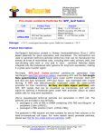
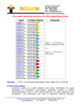
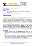
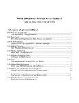
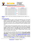
7930 Arjons Drive, Suite B
San Diego, CA 92126
Phone: (858) 6788683
Email: [email protected]
Fax:
(800) 3804198
Pre-made Expression Lentivirus for
Monitoring CRE Recombination Reactions
Catalog
Number
LVP460Puro
&
LVP460Puro-PBS
LVP460Neo
&
LVP460Neo-PBS
LVP460Bsd
&
LVP460Bsd-PBS
Product Name / Description
LoxP GFP/RFP ColorSwitch
lentivirus (Puro): Pre-made lentiviral
particles expressing "LoxP-GFP-StopLoxP-RFP-Stop" cassette under suCMV
promoter. It also contains a puromycin
antibiotic selection marker under Rsv
promoter.
LoxP GFP/RFP ColorSwitch
lentivirus (Neo): Pre-made lentiviral
particles expressing "LoxP-GFP-StopLoxP-RFP-Stop" cassette under suCMV
promoter. It also contains a Neomycin
antibiotic selection marker under Rsv
promoter.
LoxP GFP/RFP ColorSwitch
lentivirus (Bsd): Pre-made lentiviral
particles expressing "LoxP-GFP-StopLoxP-RFP-Stop" cassette under suCMV
promoter. It also contains a Blasticidin
antibiotic selection marker under Rsv
promoter.
Amount
200ul /per vial x
(1x10⁷ IFU/ml) in
DMEM medium with
10% FBS and 10x
polybrene
Or
200ul /per vial x
(5x10⁷ IFU/ml) in
PBS solution
Storage: < -70 °C, avoid repeat freeze/thaw cycles. Stable for > 6 months.
Product Description
1. Instruduction:
GenTarget’s lentivector system is Human Immunodeficiency Virus-1 (HIV) based
plasmids for gene expression and knockdown. The lentivectors are used to
generate lentiviral particles (lentivirus) that can be transduced into almost all
kinds of mammalian cells, including stem cells, primary cells, and non-dividing
cells both in vivo and in vitro. Lentiviral Particles stably integrate into the
transduced cells’ genome for long term expression, making it a great gene transfer
agent.
CRE recombinase, from bacteriophage P1, catalyzes recombination between 34
base-pair target sequences called lox sites and can join individual plasmids
User manual: Pre-made lentivirus of "LoxP-GFP-Stop-LoxP-RFP" / CRE reporting lentivirus, Page 1 of 7
www.gentarget.com; GenTarget Inc Copyrights, 2015
7930 Arjons Drive, Suite B
San Diego, CA 92126
Phone: (858) 6788683
Email: [email protected]
Fax:
(800) 3804198
containing lox sites. CRE recombination provides an excellent tool for conditional
gene targeting in transgenic animal models by linking genotypic alterations to the
biological outcomes (phenotypes).
By inserting a "LoxP-flanked expression target" into a host's genome, target
expression can be controlled via CRE recombinase. Expression of LoxP-flanked
target genes may occur prior to the addition of CRE enzyme; when CRE is applied,
it deletes the LoxP flanked target segment and stops the target expression.
Simultaneously, CRE-mediated recombination can activate expression of a second
target downstream from the deleted segment.
GenTarget provides pre-made CRE reporting lentivirus for easy, fast and
convenient testing and monitoring of CRE recombination efficiency in vivo and in
vitro. This lentivirus has been engineered to constitutively express the "LoxPGFP-stop-LoxP-RFP-Stop" cassette under a super CMV promoter. (See the
expression cassette scheme below). It reports the occurrence of CRE-mediated
recombination events via a “color switch” mechanism, thereby providing an esay,
fast and continual monitoring for the presence of CRE or CRE recombination event.
2. Multiple Selection Markers
GenTarget offers the CRE reporting lentivirus with a broad variety of antibiotic
selection markers including puromycin, neomycin, or blasticidin. The selection
markers are expressed under an RSV promoter (not shown in the schematic above).
User manual: Pre-made lentivirus of "LoxP-GFP-Stop-LoxP-RFP" / CRE reporting lentivirus, Page 2 of 7
www.gentarget.com; GenTarget Inc Copyrights, 2015
7930 Arjons Drive, Suite B
San Diego, CA 92126
Phone: (858) 6788683
Email: [email protected]
Fax:
(800) 3804198
These markers allow easy selection for transduction positive cells (those expressing
the CRE detection cassette) by antibiotic resistance.
3. Two Formats
The LoxP ColorSwtich particles are provided as at 200 µl/vial in two formats:
DMEM medium with 10 % FBS and 60 µg/ml polybrene (10x)
PBS solution, which is best for in vivo applications, cell cultures requiring
serum-free conditions, or hard-to-infect cells.
4. How ColorSwtich Reporting Works
The CRE reporting lentivirus is used to monitor or confirm the efficiency of CRE
recombination in vivo. It is an easy and very effective tool for verifying the
performance of CRE-loxP systems (CRE expression plasmids, pre-made CRE lentiviral
particles, and purified CRE enzyme) in vivo, and can serve as a control indicator for
the same systems.
The CRE reporting lentivirus demonstrates a strong GFP fluorescence after infection
into mammalian cells but does not show an RFP fluorescence signal. Once the CRE
protein is present in the nucleus, CRE excises/deletes the DNA fragment between two
loxP sites. As a result, the GFP is removed and RFP is expressed, with a resulting
switch to RFP fluorescence. The ratio of RFP-expressing to GFP-expressing cells can
be easily monitored via fluorescence cell sorting, visualized by microscopy, or the
fluorescence intensity may be measured by a fluorometer with GFP and RFP filter sets.
See the sample results below.
Notes:
*Like any mammalian pol II promoter, the CMV promoter could seek any possible
ORFs, and in some cell types, it may slightly express the 2nd ORF (the RFP in this
case) which is considered the basal or leaking RFP signal.
** It is not possible for CRE to be delivered into all cells; therefore, some GFP
positive cells may still be detected even after the application of CRE.
***Also, because some cells may integrate multiple copies of the LoxP-GFP-LoxP-RFP
cassette, and since 100% CRE recombination is not possible for all sites in every cell,
there will be many cells demonstrating both GFP and RFP signals after the addition of
CRE recombinase. The important observation is the dramatic increase in RFP positive
cells following addition of CRE. And the RFP/GFP intensity ratio reflects the CRE
recombination rate.
User manual: Pre-made lentivirus of "LoxP-GFP-Stop-LoxP-RFP" / CRE reporting lentivirus, Page 3 of 7
www.gentarget.com; GenTarget Inc Copyrights, 2015
7930 Arjons Drive, Suite B
San Diego, CA 92126
Phone: (858) 6788683
Email: [email protected]
Fax:
(800) 3804198
Application protocol (for reference only):
1. Adhesive cells Transduction Protocols:
Note: A quick transduction protocol is: add 50ul virus into one well in 24-wellplate where cell density is at 50% ~ 75%. At 72 hours after virus added (no need to
change medium), visualize the positive rate under fluorescent microscope. For
stable cell line generation, pass cell into antibiotic containing medium, or sort the
cells via fluorescent signal. Or simply select the cells by antibiotics.
Day 0: Seed the desired cells in complete medium at appropriate density incubate overnight.
(Note: at the time of transduction, it grows to 50% ~75% confluent.)
For example, seed Hela cells at 0.5 x 10 5/ml x 0.5ml in a well of a 24-well plate;
Day 1: Remove the culture medium. Add fresh, warmed, complete medium (0.5ml). Thaw the
Pre-made lentiviral stock at room temperature. Add appropriate amount of virus stock to obtain
the desired MOI. Return cells to 37°C/CO2 incubator. (Try to avoid thaw and freeze cycles for
pre-made lentivirus. But if you cannot use all virus in one time, you still can re-freeze the virus
at -80oC for future use. But virus titer will decrease by ~10% for each re-thaw.)
Day 3: At ~72hr after transduction, check the transduction rate via fluorescence image with a
suitable filter under fluorescent microscope, or calculate the exact transduction rate via Flow
Cytometry System (FACS) or any flow cytometry (such as Guava machine). Note: You should
only see GFP signal at this stage before you apply CRE enzyme to the cells.
Day 3 + : Transduced cells can be sorted out via FACS, selected by its specific antibiotics. A
pilot experiment should be done to determine the antibiotic’s kill curve for your specific cell line.
(Refer to any literatures about How to generate stable cell lines.).
CRE enzyme delivery: The selected cell should demonstrate strong GFP signal and should
have no RFP signal. After the cell selection, the cells are ready used as an indicator cell line
for CRE recombination activity.
Apply the CRE enzyme into the cells (which can be achieved by infected cell with
CRE expression lentivirus, or by regular lipid-transfection of a CRE expression plasmid,
or even simply by adding purified penetrating CRE protein enzyme. (Note: Gentarget
provides ready-to-use CRE expression lentivirus with different antibiotic selection
marker for CRE delivery into cells).
Put cells in normal culture conditions for 48-72 hours.
Detect CRE recombination reaction: The RFP signal will gradually showed up and
peaked at 48 hours or longer times (dependent upon CRE delivery methods) after the
CRE delivery. The RFP/GFP cell population ratio or the RFP signal intensity reflects
the CRE-LoxP recombination efficiency (rate). You can sort the cell by FACS machine,
other meters, or visualize the RFP positive cell under fluorescent signal.
User manual: Pre-made lentivirus of "LoxP-GFP-Stop-LoxP-RFP" / CRE reporting lentivirus, Page 4 of 7
www.gentarget.com; GenTarget Inc Copyrights, 2015
7930 Arjons Drive, Suite B
San Diego, CA 92126
Phone: (858) 6788683
Email: [email protected]
Fax:
(800) 3804198
2. Suspension cells transduction Protocols:
1. Grow your cell in your completed suspension culture medium, shaking in flask in CO 2
incubator if necessary;
2. Measure cell density. When cell grow to ~3 x 10 6 cell/ml, measure cell viability (should
be > 90%), then diluted cells into 1 x 10⁶ cell/ml in completed medium;
3. Transduction: thaw lentiviral particles at room temperature. Simply add premade
lentiviral particle into the diluted cells at ratio of: 50 to 100ul virus per 0.5 ml of cells
(Note: depending on the cell types; you may need to use more or less viruses). Grow
cells in flask, shaking in CO2 incubator.
4. At 24 hours after transduction, add equal amount of fresh medium containing related
antibiotics (Note: each particles contain an antibiotic marker and the antibiotic amounts
to use depends upon cell types). Grow cell in CO2 incubator.
5. At 72 hours after transduction, check fluorescence under microscope or calculate the
transduction efficiency using cell sorting machine (like FACS or Guava machine).
6. You can sort the fluorescent positive cells, and maintain the antibiotic selection to
generate stable cell lines.
(Note: GFP filter wavelength: Ex450-490 ~Em525; RFP filter: ~Ex545/~Em620 ).
Sample images of CRE-loxP recombination detection:
Left panel / without CRE: CRE reporter cell line (Cat#: SC018-Bsd) was
created by LoxP460-Neo particles, cultured in a 24-well-plate. Images were taken
with a GFP filter set (Ex 490nm/Em 525nm) and an RFP filter set (Ex 545nm/Em
620nm).
Right panel / with CRE: CRE reporter cell line (Cat#: SC018-Bsd) was created
by LoxP460-Neo particles, cultured in completed in 24-well plate. 50 µl of CRE
expression lentiviral particle (Cat#: LVP339) was added into the cells in one well.
Images were taken at ~ 72 hours after the addition of CRE expression lentivirus.
User manual: Pre-made lentivirus of "LoxP-GFP-Stop-LoxP-RFP" / CRE reporting lentivirus, Page 5 of 7
www.gentarget.com; GenTarget Inc Copyrights, 2015
7930 Arjons Drive, Suite B
San Diego, CA 92126
Phone: (858) 6788683
Email: [email protected]
Fax:
(800) 3804198
Safety Precaution:
Gentarget lentiviral particles adapts must advanced lentiviral safety features (using the third generation
vectors with self-inactivation SIN-3UTR), and the premade lentivirus is replication incompetent.
However, please use extra caution when using lentiviral particles. Use the lentiviral particles in Biosafety II cabinet. Wear glove all the time when handling Lentiviral particles! Please refer CDC and
NIH’s guidelines for more details regarding to safety issues.
References:
1. Sauer, B. (1987) "Functional expression of the Cre-Lox site-specific recombination system in the
yeast Saccharomyces cerevisiae", Mol Cell Biol 7: 2087-2096
2. Stanislaw J. Kaczmarczyk and Jeffrey E. Green. Nucleic Acids Res. 2001 June 15; 29(12): e56.
Warranty:
This product is for research use only. It is warranted to meet its quality as described
when used in accordance with its instructions. GenTarget disclaims any implied warranty
of this product for particular application. In no event shall GenTarget be liable for any
incidental or consequential damages in connection with the products. GenTarget’s sole
remedy for breach of this warranty should be, at GenTarget’s option, to replace the
products.
Related Products:
Product
Category
GenTarget's Pre-made lentivirus Products
Product Description
(please click category name to see product's pages)
Human,
mouse or rat
ORFs
Fluorescent
markers
Premade lentivirus expressin a human, mouse or rat gene with
RFP-Blastididin fusion dual markers.
Luciferase
expression
Premade lentivirus for all kinds of luciferase protein expression:
firefly and Renilla with different antibiotic selection markers.
0TU
U0T
0TU
U0T
Preamde lentivirus express human codon optimized fluorescent
protein, GFP / RFP/ CFP/ BFP / YFP.
CRE
recombinase
Premade lentivirus for expressing nuclear permeant CRE
recombinase with different flurescent and antibiotic markers.
LoxP
ColorSwitch
Premade lentivirus expressing "LoxP-GFP-Stop-LoxP-RFP"
cassette, used to monitor the CRE recombination event in vivo.
0TU
U0T
0TU
U0T
CRISPR /hu
CAS9
Preamde lentivirus express humanzied wild-type Cas9
endonuclease for genomic editing with CRISPR
TetR
inducible
expression
repressor
Premade lentivirus expressin TetR (tetracycline regulator)
protein, the repressor protein for the inducible expression
system.
0TU
U0T
User manual: Pre-made lentivirus of "LoxP-GFP-Stop-LoxP-RFP" / CRE reporting lentivirus, Page 6 of 7
www.gentarget.com; GenTarget Inc Copyrights, 2015
7930 Arjons Drive, Suite B
San Diego, CA 92126
Phone: (858) 6788683
Email: [email protected]
Fax:
(800) 3804198
0TU
iPS factors
U0T
T-antigen
Expression
Premde lentivirus for human and mouse iPS (Myc, NANOG,
OCT4, SOX2, FLF4) factors with different fluorescent and
antibitoic markers
Express SV40 large T antigen with different selection markers
Cell
Organelle
imaging
Premade lentivirus for cell organelle imaging. The fluorescent
marker GFP/RFP/CFP was sub-cellular localized in different
cell organelle for living cell imaging.
LacZ
expression
Anti-miNA
lentivirus
FluorescentORF fusion
Pre-made
shRNA
lentivirus
microRNA
and antimicroRNA
lentivirus
Negative
control
lentiviruses
Express different full length β- galactosidase (lacZ) with different
selection markers
Pre-made lentivirus expression a specific anti-miRNA cassette.
0TU
U0T
0TU
U0T
0TU
U0T
0TU
Pre-made lentivirus expression a "GFP/RFP/CFP-ORF" fusion
target.
Premade shRNA lentivirus for knockdown a specific genes (P53,
LacZ, Luciferase and more).
U0T
0TU
Premade lentivirus expression human or mouse precursor
miRNA. And anti-miRNA lentivector and virus for human and
mouse miRNA.
U0T
0TU
U0T
Other
Enzyme
expression
Premade negative control lentivirus with different markers:
serves as the negative control of lentivurs treatment, for
validation of the specificity of any lentivirus target expression
effects.
Ready-to-use lentivirus, expressing specific enzymes with
different selection markers.
User manual: Pre-made lentivirus of "LoxP-GFP-Stop-LoxP-RFP" / CRE reporting lentivirus, Page 7 of 7
www.gentarget.com; GenTarget Inc Copyrights, 2015