1
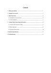
Miltenyi Biotec GmbH Friedrich-Ebert-Str. 68 51429 Bergisch Gladbach, Germany Phone: +49 2204 83060 Fax: +49 2204 85197 [email protected] Miltenyi Biotec Inc. 12740 Earhart Avenue Auburn CA 95602, USA Phone: 800 FOR MACS, +1 530 888 8871 Fax: +1 530 888 8925 [email protected] Miltenyi Biotec Pty. Ltd. (Australia) Phone: +61 02 8877 7400 Fax: +61 02 9889 5044 [email protected] Miltenyi Biotec B. V. (Benelux) Customer services Netherlands phone: 0800 4020120 Belgium phone: 0800 94016 Luxembourg phone: 800 24971 [email protected] Miltenyi Biotec Shanghai Office Phone: +86 21 62351005 Fax: +86 21 62350953 [email protected] μMACS™ Protein A/G MicroBeads MultiMACS™ Protein A/G Kit Miltenyi Biotec (France) Phone: +33 1 56 98 16 16 Fax: +33 1 56 98 16 17 [email protected] Miltenyi Biotec S.r.l. (Italy) Phone: +39 051 646 0411 Fax: + 39 051 646 0499 [email protected] User manual Miltenyi Biotec K.K. (Japan) Phone: +81 3 56 46 8910 Fax: +81 3 56 46 89 11 [email protected] Miltenyi Biotec Asia Pacific Pte. Ltd. (Singapore) Phone: +65 6238 8183 Fax: +65 6238 0302 [email protected] Miltenyi Biotec S.L. (Spain) Phone: +34 91 512 12 90 Fax: +34 91 512 12 91 [email protected] Protein A MicroBeads For 20–40 μMACS™ Isolations For 192 MultiMACS™ Isolations For 384 MultiMACS Isolations Order no. 130-071-001 130-092-944 130-092-945 Protein G MicroBeads For 20–40 μMACS Isolations For 192 MultiMACS Isolations For 384 MultiMACS Isolations 130-071-101 130-092-946 130-092-947 Miltenyi Biotec Ltd. (UK) Phone: +44 1483 799 800 Fax: +44 1483 799 811 [email protected] For further information refer to our website www.miltenyibiotec.com For technical questions, please contact your local subsidiary or distributor. 140-002-003.01 Technical Support Team, Germany: E-mail [email protected] Phone +49 2204 8306-830 Unless otherwise specifically indicated, all Miltenyi Biotec products and services are for research use only and not for therapeutic or diagnostic use. 1. Description Index Index 1. Description 1. Description 1.1 Components and size 1.2 Principle of immunopurification with MACS® Technology 1.3 Product applications 1.4 Reagent and instrument requirements 1.5 Related products 2. Protocols for immunopurification 2.1 Sample preparation and magnetic labeling 2.2 Immunopurification using μ Columns and μMACS™ Separator 2.3 Immunopurification using Multi-8/96 Columns and MultiMACS™ Separator 3. Tips & hints 3.1 Lysis buffer and buffer components 3.2 Enzymatic reactions on the column μMACS™ Protein A/G MicroBeads Components Protein A MicroBeads, 2 mL Protein G MicroBeads, 2 mL Size 20–40 immunoprecipitations * Order no. 130-071-001 130-071-101 MultiMACS™ Protein A/G Kits (24×8) Order no. Components Protein A MicroBeads, 5×2 mL 130-092-944 Protein G MicroBeads, 5×2 mL 130-092-946 Multi-8 Column Box (two boxes), each containing: 12 Multi-8 Columns, separately packaged 1 MultiColumn Frame 1 Deep Well Block, sealed, 96×2.5 mL 1 Microtiter Plate, U-bottom, with adhesive sealing foil Size 192 immunoprecipitations * 4. Troubleshooting 5. Appendix 5.1 Protease inhibitors 6. References ▲ This manual either refers to μMACS Protein A MicroBeads, to μMACS Protein G MicroBeads, or to μMACS Protein A/G MicroBeads. ProteinA/G is used when the information is valid for both μMACS types. In all other cases the specific μMACS MicroBead type is mentioned. 2 1.1 Components and size 140-002-003.01 MultiMACS Protein A/G Kits (4×96) Order no. Components Protein A MicroBeads, 10×2 mL 130-092-945 Protein G MicroBeads, 10×2 mL 130-092-947 Multi-96 Column Box, containing: 4 Multi-96 Columns, packaged sterile in a Deep Well Block, 96×2.5 mL 4 Microtiter Plates, U-bottom, with adhesive sealing foil 140-002-003.01 3 1. Description 1. Description Size 394 immunoprecipitations * *Amounts used per reaction 50 μL of Protein A/G MicroBeads are used for each immunoprecipitation with 1–2 μg of monoclonal antibody, 10–100 μL of hybridoma supernatant (20–50 μg/mL), or 0.1–1 μL of ascites (1–10 mg/mL). 100 μL of Protein A/G MicroBeads are used for each immunoprecipitation with 0.5–5 μL of serum (containing 1–10% antigen-specific antibody) or 2–4 μg of affinity-purified polyclonal antibody. Labeling of target proteins in cell lysate. Apply lysate to μ Column or Multi-8/96 Column. Product format Protein A/G MicroBeads are supplied in a solution containing 0.05% sodium azide. Storage Store MicroBeads protected from light at 2–8 °C. Do not freeze. The expiration dates are indicated on the vial labels. Store Columns, Deep Well Blocks, and Microtiter Plates at room temperature, dry, and protected from light. Remove unbound material by stringent washing. Elute target protein while the Protein A/G MicroBeads remain in the column. Figure 1: Isolation of target proteins using Protein A/G MicroBeads. 140-002-003.01 4 140-002-003.01 1. Description 1. Description 1.2 Principle of immunopurifcation with MACS® Technology Immunoprecipitation (IP) is the selective purification of a particular protein from complex mixtures using a specific antibody and an immunoglobulinbinding protein attached to a particle. Coimmunoprecipitiation (Co-IP) is applied to investigate protein-protein interactions. Analysis of the immunoprecipitated samples by SDS-PAGE or Western blotting provides information about the presence, size, post-translational modification as well as the quantity of protein antigens. However, traditional IP using agarose matrices is a time-consuming and tedious method, especially when numerous samples need to be processed. MACS® Technology provides a convenient and easy way to enable researchers to isolate high-purity proteins: Immunoprecipitation with μMACS Protein A/G MicroBeads does not involve a centrifugation step. Instead, after a short incubation of cleared lysate with the MicroBeads — coated with the specific antibody — the magnetizable immune complex is passed over a separation column placed in the magnetic field of a MACS Separator. The labeled complex is retained within the column while other proteins are efficiently washed away. For SDS-PAGE analysis, the immunoprecipitated protein is eluted from the column with SDS gel loading buffer. Alternatively, enzymatic reactions with the precipitated immune complex can be performed on the separation columns2, see section 3.2, Enzymatic reactions on the column. 6 5 140-002-003.01 μMACS™ Protein A and μMACS Protein G MicroBeads are colloidal, super-paramagnetic MicroBeads, which are conjugated to Protein A or Protein G, respectively. Protein A is a 42 kDa protein component of the cell wall of Staphylococcus aureus while Protein G is a 33 kDa cell surface protein of the group G streptococci. Both of these proteins bind to the Fc region of IgG with high affinity and avidity, leaving the Fab region of the antibody free for interaction with its antigen; thus, resulting in the formation of multimeric complexes between antigen, antibody, and MicroBeads. The binding specificities of Protein A and G for immunoglobins differ depending on the subclass and origin of the antibody (table 1). The extremely small MicroBeads, 50 nm in diameter, allow fast reaction kinetics while the column technology provides effective washing steps to minimize contaminations. Use a single column or up to 96 columns in parallel Immunoprecipitation using Protein A/G MicroBeads can be performed in either low or high throughput formats. Individual IP reactions can be carried out on single μ Columns placed in one of the four positions of the matching μMACS Separator. For larger numbers of samples, up to 96 IPs can be processed in parallel using the microtiter plate-format Multi96 Columns — consisting of twelve strips of Multi-8 Columns — which are placed in the corresponding MultiMACS™ 96 Magnet, operated in the MultiMACS Separation Unit. The instrument can be used either manually on the bench or fully automated after integration into a liquid-handling robot. 140-002-003.01 7 1. Description 1. Description Species Affinity for Protein A* Affinity for Protein G* Chicken - + Cow ++ ++++ Goat - ++ Guinea pig ++++ ++ Hamster + ++ Horse ++ ++++ Human IgG1 ++++ ++++ Human IgG2 ++++ ++++ Human IgG3 - ++++ Human IgG4 ++++ ++++ Mouse ++ ++ Mouse IgG1 + ++++ Mouse IgG2a ++++ ++++ Mouse IgG2b +++ +++ Mouse IgG3 ++ +++ Pig +++ +++ Rabbit ++++ +++ Rat +/- ++ Rat IgG1 - + Rat IgG2a - ++++ Rat IgG2b - ++ Rat IgG2c + ++ Sheep +/- ++ • • μMACS™ Protein A/G MicroBeads were developed for the analytical immunoprecipitation of proteins from cell lysates. The immunoprecipitated proteins can be analyzed by SDS polyacrylamide gel electrophoresis (SDS-PAGE) and, optionally, by subsequent Western Blot or consecutive enzymatic reactions performed on the column. μMACS Protein A/G MicroBeads are an ideal solution for analytical coimmunoprecipitations of proteins and their binding partners from cell lysates. The (co-)immunoprecipitated proteins can be analyzed by SDS polyacrylamide gel electrophoresis (SDS-PAGE) and, optionally, by subsequent Western Blot or by consecutive enzymatic reactions performed on the column. 1.4 Reagent and instrument requirements Requirements for low throughput Table 1: Protein A/G affinities for antibodies from various species. ▲ Note: The affinity of Protein A or Protein G for an antibody is always dependent on the salt concentration in the buffer. 140-002-003.01 8 1.3 Product applications • • • • μMACS Separation Unit (# 130-042-602) MACS® MultiStand (# 130-042-303) μ Columns (#130-042-701) Heating block for heating elution buffer to 95 °C 140-002-003.01 1. Description 1. Description MultiMACS™ 96 Separation Unit (# 130-091-937) • • Heating block plus reagent reservoir compatible with 8-well or 96well (8/96-well) format for heating Elution Buffer to 95 °C, e.g., thermocycler, heating block, or thermoshaker compatible with 8/96well format • • Requirements for medium to high throughput • • • • For large-scale complete lysis of cells (including nucleus): Bead mill, e.g., Mini-Bead Beater-96, BioSpec Products. This is compatible with 8-well strips plus cap strips Stainless steel beads for parallel lysis/homogenization (see supplier of bead mill) For manual use • • 8-channel pipette with tips, e.g., 8-Channel Impact® Pipettor from Matrix Technologies, Corporation: i) Volume range 15–1250 μL with 1250 μL TallTip (102 mm) Filter Tips for transferring lysate and dispensing wash buffers quickly without foaming ii) Volume range 5–250 μL for dispensing hot elution buffer in a single pipetting mode Disposable reagent reservoirs for multichannel pipettes as reservoirs for lysis buffer and wash buffers For automated use • 10 9 Liquid handling platform with four to eight pipetting channels, range 20 μL to 1 mL, and a gripper tool to grip plates sideways 140-002-003.01 MultiMACS™ Adapter (please contact technical support) Reservoir holders and reagent reservoirs for lysis buffer, wash buffers, and MicroBeads Reagent requirements 1×PBS buffer (pH 7.5) Antibody or serum specific for target protein Recommended Buffers Cell lysis buffers Triton® X-100 lysis buffer: 150 mM NaCl, 1% Triton X-100, 50 mM Tris HCl (pH 8.0). High-salt lysis buffer: 500 mM NaCl, 1% Igepal® CA630 (NP-40), 50 mM Tris HCl (pH 8.0). Low-salt lysis buffer: 1% Igepal CA630 (NP-40), 50 mM Tris HCl (pH 8.0). ▲ Note: The suitability of a lysis buffer for the experiment has to be determined experimentally. If no previous experience exists, we recommend using Triton X-100 lysis buffer. ▲ Note: To prevent protein degradation, we recommend adding proteinase inhibitor to the lysis buffer, e.g., from Roche Appplied Science, Germany; please see chapter 5, Appendix. ▲ Note: For further recommendations on lysis buffer, see also chapter 2, Protocols for immunopurification. Wash buffers RIPA wash buffer: 150 mM NaCl, 1% Igepal CA630 (NP-40), 0.5% sodium deoxycholate, 0.1% SDS, 50 mM Tris HCl (pH 8.0). High-salt wash buffer: 500 mM NaCl, 1% Igepal CA630 (NP-40), 50 mM Tris HCl (pH 8.0). 140-002-003.01 11 2. Protocols for immunopurification 1. Description Low-salt wash buffer: 20 mM Tris HCl (pH 7.5). ▲ Note: In initial experiments the same buffer that was used for the lysis of the cells should be used for column washes. In order to reduce the background of unspecifically bound proteins, a more stringent wash buffer can be chosen for subsequent experiments, e.g., RIPA buffer or high-salt buffer. ▲ Note: For coimmunoprecipitations, we recommend using only lysis buffer for all column washes. Too stringent wash conditions may remove the coimmunoprecipitated proteins. Buffer for elution and subsequent SDS-PAGE analysis SDS gel loading buffer (1×): 50 mM Tris HCl (pH 6.8), 50 mM DTT, 1% SDS, 0.005% bromphenol blue, 10% glyercol. Buffer for subsequent enzymatic reaction on the column Suitable buffers and solutions for the enzymatic reaction. See section 3.2 for further details. 1.5 Related products • • • • • Multi-8 Columns (# 130-092-444) Multi-96 Columns (# 130-092-445) Deep Well Block (DWB, 2.5 mL, with adhesive sealing foil, # 130-092-549) MACS® products for cell separation: www.miltenyibiotec.com MACSmolecular products and services for molecular analyses: www.miltenyibiotec.com 2. Protocols for immunopurification Before starting ▲ The lysis of cells is the most crucial step during an immunoprecipitation. The lysis buffer must not impair the antigen-antibody binding. Therefore, the lysis buffer conditions must be carefully chosen. Factors such as ionic strength, the pH, the concentration and type of detergent, the presence of divalent cations, cofactors and stabilizing ligands all influence the effectiveness of a lysis buffer. Generally, the lysis buffer should not contain SDS as it may disrupt the cell nuclei. See also section 3.1, Tips and hints. ▲ To prevent protein degradation, it is best to perform the lysis on ice. Protease inhibitors should be added to the lysis buffer, see section 5.1, Protease inhibitors. ▲ For lysis of yeast cells or bacteria we refer to any of the common protocols found in the literature.1 ▲ An immunoprecipitation is dependent on the affinity of the antibody to the antigen. It is further dependent on the affinity of Protein A/G to a particular antibody which varies with different species and subclasses, see also table 1. ▲ Pre-clearing of the lysate is usually not necessary as very stringent washes can be performed in the μ Column and Multi-8/96 Columns. If a pre-clearing step is required, it can be performed as follows: 1. Before immunolabeling, add 50 μL of Protein A/G MicroBeads to the lysate and incubate for 30 minutes on ice. 140-002-003.01 12 140-002-003.01 2.2 Protocols | μ Columns 2.1 Protocols | Sample preparation and magnetic labeling 2. Pass the lysate over a μ Column or a Multi-8/96 Column and collect the flow-through fraction as the pre-cleared lysate. 3. Use this pre-cleared lysate for immunolabeling. For analytical purposes, the bound material can be eluted from the μ Column or Multi-8/96 Column using SDS gel loading buffer. 2.1 Sample preparation and magnetic labeling ▲ Pre-cool PBS, lysis buffer, and centrifuge to 4 °C. ▲ Cells must be thoroughly washed with pre-cooled PBS before lysis. Residual antibody from the serum, with which the cell culture medium was supplemented, may bind to Protein A/G. 2.1.1 Lysis of adherent cells 1. Remove medium from culture dish. 2. Place culture dish on ice and wash cells up to three times with precooled PBS. 3. Add 1 mL of pre-cooled lysis buffer to a 9 cm culture dish containing 1−10×106 cells. Scrape the lysate from the culture dish using a cell scraper and transfer to a 1.5 mL tube. Mix vigorously. 4. Incubate for 10−30 minutes on ice with occasional mixing. 5. Centrifuge for 10 minutes at 10,000×g at 4 °C to sediment the cell debris. (Optional) The lysate can be stored at this step at –20 °C. 6. Transfer the supernatant to a fresh 1.5 mL tube and proceed to magnetic labeling, section 2.2 when using single μ Columns or to section 2.3 when using MultiMACS™ Columns. 14 13 140-002-003.01 2.1.2 Lysis of suspension cells 1. Transfer the cells of one culture dish (9 cm diameter) containing 1−10×106 cells to a centrifugation tube and centrifuge for 5 minutes at 300×g at 4 °C. 2. Resuspend the cell pellet in 1 mL of pre-cooled PBS and transfer the cells to a fresh 1.5 mL tube. Centrifuge the cells for 5 minutes at 300×g at 4 °C and discard the supernatant. 3. (Optional) Repeat wash step using pre-cooled PBS one or two more times. 4. Place the tube containing the cell pellet on ice and add 1 mL of lysis buffer. Mix vigorously. 5. Incubate on ice for 10−30 minutes with occasional mixing. 6. Centrifuge for 10 minutes at 10,000×g at 4 °C to sediment the cell debris. (Optional) The lysate can be stored at this step at –20 °C. 7. Transfer the supernatant to a fresh 1.5 mL tube and proceed to magnetic labeling, section 2.2 when using single μ Columns or to section 2.3 when using MultiMACS™ Columns. 2.2 Immunopurifi cation using μ Columns and μMACS™ using MultiMACS Columns. Separator Before starting ▲ Pre-heat water bath or heating block to 95 °C. ▲ For efficient immunoprecipitation, we recommend titrating the specific antibody in an initial experiment. 140-002-003.01 15 2.2 Protocols | μ Columns 2.2 Protocols | μ Columns ▲ Note: In initial experiments the same buffer that was used for the lysis of the cells should also be used for the column washes. In order to reduce the background of unspecifically bound proteins, a more stringent wash buffer can be chosen for subsequent experiments, e.g., RIPA buffer or high-salt buffer (see section 1.4, Recommended buffers). 2.2.1 Magnetic labeling 1. Add 1−2 μg of monoclonal antibody, 10−100 μL of hybridoma supernatant (20−50 μg/mL), 0.1−1 μL of ascites (1−10 mg/mL), 0.5−5 μL of serum (containing 1−10% specific antibody), or 2−4 μg of affinity-purified polyclonal antibody to the cell lysate. Mix well. 2. Rinse column with 1×100 μL of low-salt wash buffer and let the buffer flow through. ▲ Note: It is important that high concentrations of residual salt and detergent are removed from the immune complex prior to elution as both may interfere with a subsequent SDSPAGE analysis. ▲ Note: Pre-clearing the lysate with Protein A/G MicroBeads is usually not necessary. 2. • If monoclonal antibody, hybridoma supernatant, or ascites was used for immunolabeling, add 50 μL of Protein G MicroBeads to magnetically label the immune complex. Mix well. If serum or polyclonal antibody was used for immunolabeling, add 100 μL of Protein A/G MicroBeads to magnetically label the immune complex. Mix well. 3. Incubate for 30 minutes on ice. • 2.2.2 μ Column preparation and loading 1. Place μ Column in the magnetic field of the μMACS™ Separator and a suitable waste container under the μ Column. Prepare the μ Column by rinsing with 200 μL of lysis buffer. 2. Place elution buffer in the pre-heated block. Pre-heat 90 μL of buffer for each separation column. 3. Apply the cell lysate onto the column and let the lysate run through. The non-bound fraction can be collected in a fresh tube for analysis. Columns are “flow stop” and do not run dry. Magnetically labeled protein is retained in the μ Columns. 2.2.3 Wash ▲ Note: To perform enzymatic reactions on the column, proceed to section 3.2, Enzymatic reactions on the columns. 2.2.4 Elution 1. Apply 20 μL of pre-heated (95 °C) 1× SDS gel loading buffer onto the column matrix using a fresh pipette tip for each column and incubate for 5 minutes at room temperature. 2. If a drop is present on the column tip, this should be removed by contacting the column tip with the waste tube or by using a fresh pipette tip. 3. Place a fresh collection tube under the μ Column. 4. Apply 50 μL of pre-heated (95 °C) 1× SDS gel loading buffer onto the column matrix using a fresh pipette tip for each column. If a drop is present on the column tip, this should also be collected by contacting the column tip with the tube or by using a pipette. Thereby, a reproducible elution volume will be ensured. The eluted immunoprecipitate can now be analyzed by SDS-PAGE. 1. Rinse column with 4×200 μL of a suitable buffer. 16 140-002-003.01 140-002-003.01 2.3 Protocols | Multi-8/96 Columns 2.3 Protocols | Multi-8/96 Columns recommend heating the buffer in tubes or a plate that are compatible with an 8- or 12-channel multipipette. 2.3 Immunopurification using Multi-8/96 Columns and MultiMACS™ Separator ▲ Caution: Read the MultiMACS™ Separation Unit user manual carefully before running a process. Read the section Warnings and precautions before switching on the instrument. Always be sure that the MultiMACS 96 Magnet, the MultiMACS Column Holder, and the plates are in the same orientation, see MultiMACS Separator user manual for details. ▲ The MultiMACS Separator comes with a list of pre-defined separation programs to choose from, please visit www.miltenyibiotec.com for updates. MULTI-8/96 POS is the standard process used for Multi-8 or Multi-96 Columns with the MultiMACS Protein A or G kits. At the end of this process the target is eluted from the beads during the elution step. If the flow-through should also be collected, then the predefined process MULTI-8/96 NEG/POS should be used. ▲ To run a process with different process parameters, a new program can be created or the parameters of an existing one can be edited. Please see details in the corresponding sections of the MultiMACS Separator user manual. 2.3.1 Before starting ▲ Pre-heat a heating block, e.g., PCR cycler, to 95 °C to heat the elution buffer. For rapid and convenient pipetting of the elution buffer, we 18 17 140-002-003.01 ▲ Elution at temperatures below 95 °C is also possible. However, to ensure a complete reduction of the target protein we recommend heating the eluate to 95 °C for 5 minutes following the elution step. ▲ For efficient immunoprecipitation, we recommend titrating the specific antibody in an initial experiment. ▲ If using plates with other than standard height dimensions, use plates that comply to the ANSI/SBS standards and adjust the process parameter Plate Height, see MultiMACS™ Separator user manual. 2.3.2 Magnetic labeling 1. Add 1−2 μg of monoclonal antibody, 10−100 μL of hybridoma supernatant (20−50 μg/mL), 0.1−1 μL of ascites (1−10 mg/mL), 0.5−5 μL of serum (containing 1−10% specific antibody), or 2−4 μg of affinity-purified polyclonal antibody to the cell lysate. Mix well. ▲ Note: Pre-clearing the lysate with Protein A/G MicroBeads is usually not necessary. 2. • If monoclonal antibody, hybridoma supernatant, or ascites was used for immunolabeling, add 50 μL Protein G MicroBeads to magnetically label the immune complex. Mix well. If serum or polyclonal antibody was used for immunolabeling, add 100 μL Protein A/G MicroBeads to magnetically label the immune complex. Mix well. • 3. Incubate for 30 minutes on ice. 140-002-003.01 19 2.3 Protocols | Multi-8/96 Columns 2.3 Protocols | Multi-8/96 Columns 2.3.3 Multi8/96 Column preparation and loading BJAI>"-$.+ EDH 1. Switch on the MultiMACS™ 96 Separation Unit and touch the Welcome Screen or wait for a few seconds until the Process Selection Screen appears. >CH:GI BJAI>"-$.+ 8DAJBCH C:L H:I JE ▲ Note: The scroll function is only visible if there are more programs than displayed. 2. The last process performed on the MultiMACS is displayed on the upper left segment (default: MULTI-8/96 POS). The second last process is listed below. If necessary, scroll through the list of available process names by touching the symbol or until MULTI-8/96 POS is displayed. Touch MULTI-8/96 POS to go to the Process Management Screen. 140-002-003.01 20 :H8 3. If necessary, check the process parameters. See MultiMACS™ Separator user manual for details. Touch to start the process and to move the magnet to the start position. Follow the instructions given on the Touch Display. L:A8DB:IDI=: BJAI>B68HH:E6G6IDG 7NB>AI:CN>7>DI:8 BJAI>"-$.+ EDH BJAI>"-$.+ C:<$EDH K>:L :9>I 4. • • BDK: 768@ D@ :H8 For purification of less than 96 samples Unpack the necessary number of individual, sterile-packed Multi-8 Columns and put them in the MultiColumn Frame. Avoid touching the column tips. For purification of 96 samples Unpack Multi-96 Columns. Avoid touching the column tips. 5. Insert a MultiColumn Frame with up to twelve Multi-8 Columns or a pre-packed Multi-96 Column into the MultiMACS Column Holder. Touch OK to move the magnet upwards and for the next screen. 140-002-003.01 21 2.3 Protocols | Multi-8/96 Columns 2.3 Protocols | Multi-8/96 Columns 2.3.4 Wash BDK: >CH:GI L6HI:EA6I:/ 768@ D@ :H8 9L7))BB 1. Rinse the columns with 4×200 μL of a suitable buffer. Let the buffer pass through the columns. 6. Place the waste plate, e.g., Deep Well Block, 2.5 mL, onto the TipTouch Plate. If using a plate with a different height, adjust the process parameter Plate Height of the waste plate, for details see MultiMACS™ Separator user manual. Touch OK to move the MultiMACS 96 Magnet downwards. Column tips now slightly immerse in the waste plate. The following screen appears. 2. Rinse the columns with 1×100 μL of low-salt wash buffer and let the buffer pass through. ▲ Note: It is important that high concentrations of residual salt and detergent are removed from the immune complex prior to elution as both may interfere with a subsequent SDSPAGE analysis. ▲ Note: To perform enzymatic reactions on the column, proceed to section 3.2, Enzymatic reactions on the columns. G>CH:!6EEAN BDK: H6BEA:!L6H=# 768@ D@ >;G:FJ>G:9/ :H8 EG:":AJI: 2.3.5 Elution 7. Prepare the Multi-8 columns by adding 200 μL of lysis buffer and let it pass through. Columns are “flow stop” and do not run dry. 8. Place elution buffer in the pre-heated block. Pre-heat 90 μL buffer for each separation column. 9. Apply the cell lysate to the columns and let the lysate pass through. Magnetically labeled protein is retained in the Multi-8/96 Columns. 22 ▲ Note: In initial experiments the same buffer that was used for the lysis of the cells should also be used for the column washes. In order to reduce the background of unspecifically bound proteins, a more stringent wash buffer can be chosen for subsequent experiments, e.g., RIPA buffer or high-salt buffer. For details see section 1.4, Recommended buffers. 140-002-003.01 1. Apply 20 μL of pre-heated (95 °C) 1× SDS gel loading buffer directly onto the Multi-8 Column matrix using a fresh pipette tip for each column. Avoid contact with the Multi-8 Column reservoir. Incubate for 5 minutes at room temperature. 2. Touch OK for the next screen. I>E"IDJ8= 8DAJBCH>C EA6I: 140-002-003.01 D@ :H8 23 2.3 Protocols | Multi-8/96 Columns 2.3 Protocols | Multi-8/96 Columns 3. Move the Tip-Touch Plate firmly back and forth once so that the inner walls of the wells of the Deep Well Block touch the tips of the Multi-8 Columns. This process removes any drops on the column tips that did not fall off by gravity. 4. Touch OK to move the MultiMACS™ 96 Magnet upwards. BDK: >CH:GI :AJI>DCEA6I:/ 768@ BIE&)BB :H8 6. Touch OK and the MultiMACS 96 Magnet will move downwards until column tips slightly immerse (1 mm) in the elution plate. BDK: 768@ 8. Wait approximately 1 minute until the 1× SDS gel loading buffer has passed through. Touch OK for the next screen. I>E"IDJ8= 8DAJBCH>C EA6I: D@ 5. Remove Deep Well Block. If some wells were unused, pour off waste liquid and store plate. Insert the elution plate (Microtiter Plate). If using a plate with a different height, adjust the process parameter Plate Height of the elution plate, see MultiMACS Separator user manual. 6EEAN :AJI>DC 7J;;:G 7. Apply 50 μL of pre-heated (95 °C) 1× SDS gel loading buffer directly onto the Multi-8 Column matrix using a fresh pipette tip for each column. Avoid contact with the Multi-8 Column reservoir. D@ D@ :H8 9. Move the Tip-Touch Plate firmly back and forth once so that the inner walls of the wells of the elution plate touch the tips of the Multi-8 Columns. This process removes any drops on the column tips that did not fall off by gravity. Touch OK to move the MultiMACS 96™ Magnet upwards. BDK: 768@ G:BDK: D@ :AJI>DCEA6I: :H8 :H8 10. Remove elution plate and use samples for downstream application. Alternatively, seal the plate with adhesive foil and store it at –20 °C or –70 °C. 140-002-003.01 24 140-002-003.01 25 3. Tips & hints 2.3 Protocols | Multi-8/96 Columns 11. Touch OK to move the MultiMACS 96 Magnet away from the Column Holder. 3. Tips & hints 3.1 Lysis buffers and buffer components G:BDK: 8DAJBCH! IDJ8=D@ID :C9EGD8:HH BDK: 768@ D@ Lysis buffers :H8 ▲ In case of coimmunoprecipitations with very weak protein-protein interactions we recommend using a low-salt lysis buffer: 1% Igepal® CA630 (NP-40), 50 mM Tris HCl (pH 8.0). 12. Remove Column Frame with Multi-8/96 Columns. If less than twelve Multi-8 Columns were used, remove the used Multi-8 Columns and store the Column Frame. Touch OK to finish the process. ▲ If strong background ionic interactions are expected, we recommend using a high-salt lysis buffer: 500 mM NaCl, 1% Igepal CA-630 (NP-40), 50 mM Tris HCl (pH 8.0). Buffer components Detergents — these are partially hydrophobic and partially hydrophilic and can solubilize membranes and membrane proteins. They work to increase protein solubility and decrease aggregation. Non-ionic detergents tend to be more gentle in their actions than ionic detergents and are more suitable for protein-protein interaction studies. Salts — increasing the salt concentration in the buffer will decrease ionic interactions between proteins in a cell lysate. DTT — this reducing agent is often used to prevent loss of enzyme function via oxidation during protein isolation. EDTA — this chelation agent binds divalent cations and can be used to prevent the action of certain enzymes that require ions such as Mg2+ or Ca2+. 26 140-002-003.01 140-002-003.01 27 3. Tips & hints 3. Tips & hints It can also prevent protein-protein interactions that are dependent on the presence of cations. The Protein A/G MicroBead storage buffer contains 5 mM EDTA. 3.2.1 Enzymatic reactions on μ Columns Phosphatase inhibitors — when active kinases or phosphorylated proteins are to be isolated, we recommend the addition of 1 mM activated sodium orthovanadate (not compatible with DTT) and 1–10 mM NaF to inhibit phosphatase activity. ▲ Note: Neither lysis nor wash buffer should contain SDS since it may impair the biological activity of the immunoprecipitated complex. Choose type of wash buffer and number of wash steps to remove nonspecific protein. pH — increasing or decreasing the pH of the buffer will change the net charge of the proteins depending on their pI and influence the extent of non-specific ionic interactions. Before starting 3.2 Enzymatic reactions on the column Enzymatic reactions with the immunoprecipitated protein complex can be carried out while the protein remains bound to the μ Column or Multi-8 Column. The immobilized protein can be either the enzyme or the substrate of the reaction. Performing the reaction on the column offers the advantage of very convenient handling, especially when working with radioactively labeled proteins or substrates2. It also allows a serial enzymatic reaction on the same μ Column or Multi-8 Column. A few guidelines are listed below on how to perform the enzymatic reaction on the column; however, individual enzymatic reactions may have to be optimized. 140-002-003.01 28 Cell lysis and magnetic labeling should be performed as described in section 2. Proceed after completing wash steps (section 2.2.3, step 2). ▲ For incubation at 37 °C or 42 °C, the heatable thermoMACS™ Separation Unit should be used. For other incubation temperatures, the μ Column and μMACS Separator should be placed in an incubator set at the appropriate temperature. ▲ For incubations in the thermoMACS™, we recommend pipetting 1 μL of μMACS Sealing Solution (# 130-091-060) onto the column matrix to eliminate evaporation of buffer. 1. Rinse column with 1×100 μL of low-salt wash buffer and let the buffer pass through. 2. Rinse the μ Column with 1×100 μL of enzymatic reaction buffer. 3. If the product to be analyzed will be present in the non-bound fraction, collect the flow-through in a fresh tube. Apply substrate or enzyme solution to the μ Column in 25 μL aliquots. ▲ Note: The void volume of the μ Column is 25 μL. Thus, buffers and solutions used for incubation with the immobilized immunoprecipitate should always be applied in 25 μL 140-002-003.01 3. Tips & hints 3. Tips & hints aliquots. If it is necessary to incubate the immobilized immunoprecipitate with a volume >25 μL, sequentially incubate in steps of 25 μL aliquots until the total volume has been applied. After the enzymatic reaction has been performed either the non-bound fraction (pooled flow-through) can be analyzed or the immunoprecipitated protein can be eluted for SDS-PAGE analysis. 3.2.2 Enzymatic reactions on Multi-8/96 Columns Cell lysis and magnetic labeling should be performed as described in section 2. ▲ Note: Neither lysis nor wash buffer should contain SDS since it may impair the biological activity of the immunoprecipitated complex. Choose type of wash buffer and number of wash steps to remove nonspecific protein. Wash Proceed after completing wash steps (section 2.3.4, step 2). 1. Rinse the μ Column with 1×100 μL of reaction buffer to remove remaining enzyme or substrate for analysis. Place a fresh waste tube under the μ Column to collect the wash fraction. Before starting 2. Remove residual salt by rinsing the μ Column with 1×100 μL of lowsalt wash buffer. Elution 1. Apply 20 μL of hot (95 °C) elution buffer to the column and incubate for 5 minutes at room temperature. ▲ Note: If a drop is present on the column tip, this should be removed by contacting the column tip with the waste tube or by using a fresh pipette tip. 2. Place a fresh tube under the μ Column. Apply 50 μL of pre-heated (95 °C) 1× SDS gel loading buffer onto the column matrix using a fresh pipette tip for each column. To ensure a reproducible elution volume, any drop present on the column tip should also be collected by contacting the column tip with the elution tube or by using a pipette. The eluted target protein can now be analyzed by SDS-PAGE. 30 29 140-002-003.01 ▲ To collect both the flow-through and column-bound fractions, the predefined process MULTI-8/96 NEG/POS should be used. 1. Rinse the columns with 1×100 μL low-salt wash buffer and let the buffer pass through. 2. Rinse the Multi-8 Column with 1×100 μL of enzymatic reaction buffer. 3. • If the target to be analyzed is to be found in the flow-through, carry out a tip-touch and remove the Deep Well Block. Insert the elution plate (Microtiter Plate) and press OK. • If the target to be analyzed is bound to the column, continue with step 4. 4. Apply enzyme solution to the Multi-8/96 Column in 30 μL aliquots. ▲ Note: The void volume of the Multi-8/96 Column is 30 μL. Thus, buffers and solutions used for incubation with the immobilized immunoprecipitate should always be applied in 30 μL aliquots. If it is necessary to incubate the immobilized immunoprecipitate with a volume >30 μL, sequentially incubate in steps of 30 μL aliquots until the total volume has been applied. 140-002-003.01 31 4. Troubleshooting 3. Tips & hints After the enzymatic reaction has been performed, either the non-bound fraction (pooled flow-through) can be analyzed, or the immunoprecipitated protein can be eluted for SDS-PAGE analysis. Wash 1. Rinse the Multi-8/96 Column with 1×100 μL of reaction buffer to remove remaining enzyme or substrate for analysis. 2. Proceed with section 2.3, Wash, step 14: Rinse the columns with 1×100 μL low-salt wash buffer and let the buffer pass through. Elution 4. Troubleshooting Slow column Cell debris remaining in the lysate can occlude the column. Thus, cell debris should be removed by high-speed centrifugation (> 10,000×g) before addition of the antibody and Protein A/G MicroBeads. Air bubble formation within the column may also clog the column. To prevent air bubble formation, use room-temperature buffers for the wash steps or, where possible, degas the buffers before use. No protein recovery after the immunoprecipitation 1. Proceed with section 2.3.5, Elution Insufficient cell lysis may cause low protein yield. The lysis buffer should be altered to optimize the recovery of the target protein, see section 2.1, Before starting and 3.1, Lysis buffers and buffer components. Protein A or Protein G do not bind to the antibody subclass used in the experiment. Check antibody specificities using table 1. Too stringent lysis or wash buffers also can lead to poor protein recovery. Binding of antibody to antigen may have been inhibited or the antigen may have been denatured. A lower salt concentration and alternative detergents may help. Elution has failed. Please check elution buffer recipe. Protein background Ineffective wash steps may cause artifacts. If many background protein bands are present following SDS-PAGE analysis of the eluates, more 32 140-002-003.01 140-002-003.01 33 5. Appendix 4. Troubleshooting stringent lysis and/or wash buffer should be used, e.g., with higher salt concentrations, see section 3.1, Lysis buffers and buffer components. 5. Appendix 5.1 Protease inhibitors Background smear due to protease activity The presence of a smear following SDS-PAGE analysis of the eluates may indicate protease activity. Suitable protease inhibitors should be added to the lysis and wash buffers, see section 5.1, Protease inhibitors. Variable elution volumes Reuse of pipette tips to pipette hot 1× SDS gel loading buffer often leads to the transfer of inexact volumes. Always use fresh tips to pipette hot gel loading buffer. Drops remaining on the column tips following the pre-elution or elution steps can result in raised or lowered elution volumes. Carrying out a tiptouch step (MultiMACS™ Separation) or removing drops from the column tips using a pipette (μMACS™ Separation) will ensure reproducible elution volumes. Inhibitor (specificity) Final concentration Stock solution α1-Antitrypsin (S) 10 μM 6 mg/mL (1000×) in ddH2O pH 7 Aprotinin (S) 0.3 μM 1 mg/mL (500×) in ddH2O pH 7 Benzamidin (S) 2 mM 3 mg/mL (10×) in ddH2O pH 7 EDTA-Na2 (M) 1 mM 0.5 M (500×) in ddH2O pH 8 E-64 (C) 10 μM 0.36 mg/mL (100×) in 1 : 1 mixture ddH2O pH 7 : EtOH Leupeptin (S, C) 10 μM 5 mg/mL (1000×) in ddH2O pH 7 PMSF (S) 1 mM 17 mg/mL (100×) in Ethanol, Isopropanol or Methanol. Inactivated by DTT Cocktails recommended, e.g., PMSF, Leupeptin, Aprotinin: S Serineproteases M Metalloproteases C Cysteineproteases Important Note μ Columns and Multi-8/96 Columns cannot be used for cell separation. 34 140-002-003.01 140-002-003.01 35 Notes 6. References 6. References 1. Harlow, E. and Lane, D. (1988) Immunoprecipitation; in Antibodies: A Laboratory Manual, Cold Spring Harbor Laboratory, New York, USA. 2. Fischer, A. et al. (2002) Cyclin-Dependent Kinase 5 Is Required for Associative Learning. J. Neurosci. 22: 3700-3707. Warning Reagents contain sodium azide. Sodium azide yields hydrazoic acid under acid conditions, which is extremely toxic. Azide compounds should be diluted with running water before discarded. These precautions are recommended to avoid deposits in plumbing where explosive conditions may develop. Warranty The products sold hereunder are warranted only to be free from defects in workmanship and material at the time of delivery to the customer. MILTENYI BIOTEC GmbH makes no warranty or representation, either expressed or implied, with respect to the fitness of a product for a particular purpose. There are no warranties, expressed or implied, which extend beyond the Technical Specifications of the products. MILTENYI BIOTEC GmbH’s liability is limited to either replacement of the products or refund of the purchase price. MILTENYI BIOTEC GmbH is not liable for any property damage, personal injury or economic loss caused by the product. Unless otherwise specifically identified, Miltenyi Biotech products and services are for research use only and not for diagnostic or therapeutic use. Triton X-100 is a trademark of various third parties. Igepal is a registered trademark of various third parties. Impact is a registered trademark of Matrix Technologies, Corp. MACS is a registered trademark of Miltenyi Biotec GmbH. thermoMACS, MultiMACS, and μMACS are trademarks of Miltenyi Biotec GmbH. © 2006 Miltenyi Biotec GmbH. Printed in Germany. 36 140-002-003.01 140-002-003.01 Notes Notes 38 37 140-002-003.01 140-002-003.01 39