1
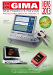
DC-N2
Diagnostic Ultrasound System
Datasheet
1
iStationTM
iVisionTM
Integrated 320G hard drive
2 active transducer connectors
DVD-RW Driver
3 USB ports
Shared Service Application Package
(Abdominal, Obstetrical, Gynecological,
Cardiac, Small Parts, Urological, Vascular,
Pediatric Packages)
Auto Doppler Calculation
iStorage (Direct Network Storage)
Tutorial function: iScanHelper
1.5 Optional features
Height adjustable control panel
3rd transducer connector
iScapeTM View
IMT (Auto Intima-Media Thickness
Evaluation)
Smart 3D
Built-in Battery
Footswitch
- 971-SWNOM (2-pedal)
- 971-SWNOM (3-pedal)
- FS-81-SP-2 (1-pedal)
Nerve Application Package
Emergency Medicine Application
Package
DICOM Basic (including DICOM Task
Management, Print, Storage, Storage
Commitment, Media Exchange)
DICOM Worklist
DICOM MPPS (Modality Performed
Procedure Step)
DICOM OB/GYN structured report
DICOM Vascular structured report
DICOM Cardiac structured report
DICOM Query/Retrieve
1.6 Language support
Software display: English, Chinese,
German, Spanish, French, Italian,
Portuguese, Russian, Czech, Polish,
Turkish
Keyboard input: English, Chinese,
German, Spanish, French, Italian,
Portuguese, Russian, Czech, Polish
System Overview
1.1 Application
Abdomen
Obstetrics
Gynecology
Small parts
Vascular
Urology
Musculoskeletal
Orthopedics
Emergency Medicine
Nerve
Cardiology
Pediatrics
Others
1.2 Transducer types
Curved array
Linear array
1.3 Imaging modes
B-Mode
Tissue Harmonic and PSH (Phase Shift
Harmonic Imaging)
M-Mode
Color Doppler Imaging
Power Doppler Imaging/Directional PDI
Pulsed Wave Doppler
Smart 3D
iScapeTM View (Panoramic Imaging)
1.4 Standard features
B-Mode
THI and PSH
M-Mode
Color Doppler Imaging
Power Doppler Imaging and Directional
PDI
Pulsed Wave Doppler
HPRF (High Pulse Repeat Frequency)
iClearTM (Speckle Suppression Imaging)
iBeamTM (Spatial Compounding
Imaging)
iTouchTM (Auto Optimization)
Zoom/iZoomTM (Full Screen Zoom)
FCI (Frequency Compounding Imaging)
B steer
ExFOV Imaging
2
Icelandic, Norwegian, Swedish, Finnish,
Turkish, Danish
Control panel overlay: Chinese, Italian,
Portuguese, Spanish, German, Russian,
French, Czech, Polish
User manual: English, Chinese, Russian,
Spanish, Portuguese, Turkish, French,
German, Italian
3 User Interface
3.1 Control panel
Power/Battery indicator
Alphanumeric keys
Function keys
Knobs
Backlit keys, ensuring accurate work in
the dark room
8-segment TGC control
Programmable keys, available for
user-defined functions
Trackball, sensitivity adjustment
Key brightness adjustment
Independent up/down of control panel
- Down/Up: 1265~1415mm (150mm
range)
3.2 System boot-up
Boot-up in about 30 sec
Shut down in about 20sec
Boot-up from standby mode in about
15 sec
Shut down from standby mode in about
6 sec
3.3 Comments
Support text input and arrow
Adjustable text size, arrow size and
direction
Support home position
Covers various application
User customizable
3.4 Bodymark
More than 140 bodymarks for versatile
application
User customizable
3.5 Screen information*
Common info:
- Mindray logo
- Hospital name
- Exam date
- Exam time
- Acoustic power
- Mechanical index
- Tissue thermal index
- ID, Last name, First Name, Middle
initial, Gender, Age
2 Physical Specification
2.1 Dimension and weight
Width: 520mm
Height:
- 1265~1415mm (with adjustable
height)
- 1315mm (without adjustable height)
Depth: 670mm
Weight: approx. 55kg (with battery)
2.2 Monitor
15-inch high resolution color LCD
monitor
Resolution: 1024×768
Brightness adjustable
Screen saver
Monitor: tilt of 20°up, 90°down, and
swivel of 90° right, 90° left
2.3 Transducer Connector
Maximum 3 transducer connectors
2.4 Electrical power
AC adapter Input:
- Voltage:100-240V~
- Frequency: 50/60Hz
- Power consumption: 420VA
Built-in Battery: Lithium-ion Battery
14.8V, 6600mAh
2.5 Operating Environment
Ambient temperature: 0-40 °C
Relative humidity: 30%-85% (no
condensation)
Atmospheric pressure: 700hPa-1060hPa
2.6 Storage & Transportation Environment
Ambient temperature: -20~55 °C
Relative humidity: 20%-95% (no
condensation)
Atmospheric pressure: 700hPa-1060hPa
3
Auto merge
Lithotrity
4.3 THI and PSH
Available on all types of transducers
Patent PSH technology, obtains purer
harmonic, better contrast resolution
iClearTM available
4.4 M-mode
Display formats: V1:2, V2:1, V1:1, Full (V:
vertical)
Acoustic output power: 7%-100%
Dynamic range: 30-220, 5/step
Gain: 0-100, 1-2/step
Speed: 1-6, 6 steps
M soften: 0-14, 1/step
Tint map: off; 16 types
Gray Map: 8 types
Edge enhance: 0-14, 1/step
4.5 Color Doppler Imaging
Dual live
iTouchTM: Auto optimization (Gain)
Frequency (depend on probe)
Max. velocity: 241cm/s
Steer: 6° (linear transducer)
Max. frame rate: 233f/s
Acoustic output power: 7%-100%
Gain: 0-100, 2/step
ROI size/position: adjustable
Scale: 30 steps (depend on exam mode)
Baseline: -8~+8, 17 steps
Wall filter: 0-7, 8 steps
PRF: max. 15.7kHz, min. 0.1kHz
Packet size: 0-3, 4 steps
Smooth: 0-4, 5 steps
B/C Align: on/off
Priority: 0%-100%, 10%/step
Map: V0-V10, VV0-VV9, 21 types
Invert: on/off
Persistence: 0-4, 5 steps
Line density: L/M/H/UH, 4 steps
4.6 Power Doppler Imaging
Dual live
Support directional PDI
Frequency (depend on probe)
Acoustic output power: 7%-100%
Dynamic range: 10-70, 5/step
- Probe model
- Operator
- TGC Curve
- Focus position
- Thumbnail
- Imaging parameters
- iScanHelper guidance
*Not all items are listed in this part, detail info
please refer to user manual
4 Imaging Parameters
4.1 Overview
Echo-enriched Beamformer
Up to768 channels
4-beam forming
4.2 B-mode
Display formats: Single(B), Dual(B+B),
Quad(4B)
iClearTM: Off; On, 1-4 steps
iBeamTM: Off/On
iTouchTM: Auto optimization (TGC, Gain)
Frequency (depend on probe)
B steer: available on linear transducers
ExFOV: extended FOV available on
convex, linear transducers
Depth: 0.9-37cm (depend on
transducer )
Frame rate (max): 400f/s
Acoustic output power: 7%-100%
TGC: 8 pods on control panel
Dynamic range: 30-220, 5/step
Gain: 0-100, 1-2/step
Focus number: 1-4, adjustable
Focus position: Max. 16, adjustable
FOV (Field of View): continuously
adjustable
Line density: L/M/H/UH
Persistence: 0-7, 8 steps
Horizontal Scale: on/off
L/R flip: Left/Right
U/D flip: Up/Down
TSI (Tissue Specific Imaging):
general/muscle/fluid/fat
Gray Map: 8 types
Tint map: off; 16 types
Gray Invert
4
Smart 3DTM
- Angle: 10-80, 2/step
- Distance: 10mm-200mm, 10mm/step
- Display formats: Single, Dual, Quad
- Reset: Reset All, Reset curve, Reset
orientation
- Quick Rotation: 0°, 90°, 180°, 270°
- Render type: Gray, Invert
- Accept VOI: on/off
- Render: Surface, Max, Min, X-ray
- Direct: D/U, U/D, L/R, R/L, F/B, B/F (D:
down, U: up, L: left, R: right, F: front, B:
back)
- Threshold: 0%-100%, 1%/step
- Opacity: 0%-100%, 5%/step
- Smooth: 0-20, 21 steps
- Bright: 0%-100%, 2%/step
- Contrast: 0%-100%, 2%/step
- Tint: off; 8 types
- Current window: VR, A, B, C
- iClear: Off; On,1-4 steps
Edit
- Rotation control: X, Y, Z axis
- Tool: inside contour, outside contour;
inside rect, outside rect
- Other operations: undo, undo all
4.9 iScapeTM View (option)
Panoramic imaging
Available on all transducers
Acquisition method: B mode
Imaging length: 120cm
Tint map: off; 16 types
Rotation: 0°~355°, 5°/step
4.10 Zoom
iZoomTM
- Full screen zoom
- Normal image, Zoom standard area,
Zoom image area, 3 steps
Spot zoom (write zoom) 0.8-10x
Pan zoom (read zoom) 0.8-10x
Gain: 0-100, 2/step
ROI size/position: adjustable
Steer: 6° (linear transducers)
Scale: 30 steps (depend on exam mode)
Wall filter: 0-7, 8 steps
PRF: Max. 15.7kHz, min. 0.1kHz
Packet size: 0-3, 4 steps
Smooth: 0-4, 5 steps
B/C Align: on/off
Priority: 0%-100%, 10%/step
Map: P0-P3, dP0-dP3, 8 types
Persistence: 0-4, 5 steps
Line density: L/M/H/UH, 4 steps
4.7 PW Mode
Display formats: V1:2, V2:1, V1:1, Full (V:
vertical)
iTouchTM: Auto optimization (Baseline,
PRF)
Frequency (depend on probe)
Acoustic power
PW velocity: max. 739cm/s
Sample volume size: 0.5-20mm (PW
only), 0.5-5mm/step
Sample gate depth: adjustable
Scale: 30 steps
Baseline: -4~4, 9 steps
PW Steer: 6° (linear transducer)
Audio: 0%-100%, 2%/step
PW PRF: max. 24kHz, min. 0.7kHz
Gain: 0-100, 2/step
Dynamic range: 24-72, 2/step
Speed: 1-6, 6 steps
Wall filter: 0-6, 7 steps
Invert: on/off
Angle: -89°~89°, 1/step
Quick angle: 0°, -60°, 60°
Gray map: 8 types
Tint map: Off; 16 types
Time/frequency resolution: 0-4, 5 steps
Auto calc: on/off
Trace area: above, below, all
HPRF: on/off
Duplex/Triplex: on/ off
Auto calc cycle: 1~5
Auto calc param
4.8 Smart 3D (option)
5 Cine Review and Post Processing
5.1 Cine review
Available in all modes
Frame by frame manual cineloop review
or auto playback with variable speed
5
Independent cine review in 2D Dual and
Quad mode one by one
Maximum cine memory is up to 17265
frames and PW mode up to 169.6s
Cine length: 1-60s
Frame compare: compare different
frames for one cine in dual format
Cine compare: compare two or more
than two cines in dual or quad format
Skip to first and skip to last: one
keystroke review the first or last frame
Start point and end point: selectable
5.2 Post Processing
B-mode:
Zoom
Gray map
Tint map
Flip
iClear
Gray invert
Auto merge
M-mode:
Gray map
Tint map
Color/Power:
Invert
Baseline
Map
Priority
Smooth
Dual Live
PW:
Gain
Baseline
Dynamic Range
Angle correction
Quick angle
Invert
Gray map
Tint map
Auto calc
Distance
Angle
Area: Ellipse, Trace, Spline, Cross
Trace Length
Double Distance
Parallel
Volume: 3-Distance, Ellipse, Ellipse +
Distance)
- Length Ratio
- Area Ratio
- IMT
- B Histogram
- B Profile
- Volume Flow
- Color Velocity
M-mode
- Distance
- Time
- Slope
- Heart Rate
- Velocity
Doppler mode
- D Velocity
- Time
- Heart Rate
- Acceleration
- D Trace
- PS/ED
- Volume Flow
Automatic Doppler Spectrum Analysis
- Heart cycle pre-settable (1, 2, 3, 4, 5)
- Automatic tracing in real-time
- User configurable display of items
- Support PI, RI, TAMAX, TAMEAN,
Volume Flow calculations
- Appropriate factory setting according
to applications
6.2 Clinical option measurement package
Abdominal
- Liver
- Renal Length
- Renal Height
- Renal Width
- Renal Cortical Thickness
- Adrenal Length
- Adrenal Height
-
6 Measurement/Analysis and Report*
6.1 Generic measurements
2D-mode
- Depth
6
-
Adrenal Width
Common bile duct
Portal Vein Diameter
Common hepatic duct
Gallbladder Length
Gallbladder Height
Gallbladder wall thickness
Pancreatic duct
Pancreatic head
Pancreatic body
Pancreatic tail
Spleen
Aorta Diameter
Iliac Diameter
Pre-void Bladder Length
Pre-void Bladder Height
Pre-void Bladder Width
Post-void Bladder Length
Post-void Bladder Height
Post-void Bladder Width
Ureter
Renal Artery Origin
Arcuate Artery
Segmental Artery
Interlobar Artery
Renal Artery
Main Renal Artery
Renal Vein
Aorta
Celiac Axis
Superior Mesenteric Artery
Common Hepatic Artery
Hepatic Artery
Splenic Artery
Inferior Vena Cava
Portal Vein
Main Portal Vein
Hepatic Vein
Left Hepatic Vein
Right Hepatic Vein
Middle Hepatic Vein
Splenic Vein
Superior Mesenteric Vein
Renal Volume
Pre-void Bladder Volume
Post-void Bladder Volume
- Micturated Volume
Gynecology
- Uterine Length
- Uterine Height
- Uterine Width
- Uterine Cervix Length
- Uterine Cervix Height
- Uterine Cervix Width
- Endometrium Thickness
- Ovary Length
- Ovary Height
- Ovary Width
- Follicle 1~16 Length
- Follicle 1~16 Width
- Follicle1~16 Height
- Follicle Volume
- Ovary Volume
- UT Volume
- Uterus Body
- UT-L/ CX-L
Obstetrics
- EGestational Sac Diameter
- Yolk Sac
- Crown Rump Length
- Nuchal Translucency
- Biparietal Diameter
- Occipital Frontal Diameter
- Head Circumference
- Abdominal Circumference
- Femur Length
- Abdominal Transversal Diameter
- Anteroposterior Abdominal Diameter
- Cerebellum Diameter
- Cist Magna
- Lateral Ventricle Width
- Hemisphere Width
- Outer Orbital Diameter
- Inter Orbital Diameter
- Humerus Length
- Ulna Length
- Radius Length
- Tibia Length
- Fibula Length
- Clavicle Length
- Length of Vertebrae
- Middle Phalanx Length
7
-
Foot Length
Ear Length
Anteroposterior trunk diameter
Transverse trunk diameter
Fetal Trunk Cross-sectional Area
Thoracic Diameter
Heart Circumference
Thoracic circumference
Umbilical Vein Diameter
Fetal kidney Length
Matrix Kidney Length
Cervical Length
Amniotic Fluid
Nuchal Fold
Orbit
Placental Thickness
Gestational Sac Diameter 1
Gestational Sac Diameter 2
Gestational Sac Diameter 3
Amniotic Fluid 1
Amniotic Fluid 2
Amniotic Fluid 3
Amniotic Fluid 4
Left Ventricular Internal Diameter at
End-diastole
Left Ventricular Internal Diameter at
End-systole
Left Ventricular Diameter
Left Atrium Diameter
Right Ventricular Internal Diameter at
End-diastole
Right Ventricular Internal Diameter at
End-systole
Right Ventricular Diameter
Right Atrium Diameter
Interventricular Septal Thickness at
End-diastole
Interventricular Septal Thickness at
End-systole
Interventricular Septal Thickness
Aorta Diameter
Main Pulmonary Artery Diameter
Right Ventricular Outflow Tract
Diameter
Right Ventricular Outflow Tract
Diameter
8
Left Ventricular Area
Left Atrium Area
Right Ventricular Area
Right Atrium Area
Heart area
Facial Angle
Mitral Valve diameter
Pulmonary valve Diameter
Ascending Aorta Diameter
Descending Aorta Diameter
Ductus Arteriosus Diameter
Tricuspid valve Diameter
Left pulmonary Artery Diameter
Right pulmonary Artery Diameter
Inferior vena cava Diameter
Aorta Valve Diameter
Mean Gestational Sac Diameter
AFI
Estimated Fetal Weight
Estimated Fetal Weight 2
HC/AC
FL/AC
FL/BPD
AXT
CI
FL/HC
HC(c)
HrtC/TC
TCD/AC
LVW/HW
LVD/RVD
LAD/RAD
AoD/MPAD
LAD/AoD
Fetal Heart Rate
Left ventricular short-axis diameter at
end diastole
Left ventricular short-axis diameter at
end systole
Right ventricular short-axis diameter
at end diastole
Right ventricular short-axis diameter
at end systole
Interventricular septal thickness at en
diastole
Interventricular septal thickness at en
systole
- Umbilical Artery
- Ductus Veno
- Placenta Artery
- Middle Cerebral Artery
- Fetal Aorta
- Descending Aorta
- Uterine Artery
- Ovarian Artery
- Fetal Heart Rate
Cardiology
- Left Atrium Diameter
- Left Atrium major Diameter
- Left Atrium minor Diameter
- Right Atrium major Diameter
- Right Atrium minor Diameter
- Left Ventricular major Diameter
- Left Ventricular minor Diameter
- Right Ventricular major Diameter
- Right Ventricular minor Diameter
- Left Atrium area
- Right Atrium area
- Left Ventricular area at end-diastole
- Left Ventricular area at end-systole
- Right Ventricular area at end-diastole
- Right Ventricular area at end-systole
- Left Ventricular Internal Diameter at
end-diastole
- Left Ventricular Internal Diameter at
end-systole
- Right Ventricular Diameter at
end-diastole
- Right Ventricular Diameter at
end-systole
- Left Ventricular Posterior wall
thickness at end-diastole
- Left Ventricular Posterior wall
thickness at end-systole
- Right Ventricular Anterior wall
thickness at end-diastole
- Right Ventricular Anterior wall
thickness at end-systole
- Interventricular Septal thickness at
end-diastole
- Interventricular Septal thickness at
end-systole
-
9
Aorta Diameter
Aorta arch Diameter
Ascending Aorta Diameter
Descending Aorta Diameter
Aorta Isthmus Diameter
Aorta ST junct Diameter
Aorta Sinus Diameter
Ductus Arteriosus Diameter
Previous ductal Diameter
Posterior ductal Diameter
Aortic Valve Cusp Separation
Left Ventricular Outflow Tract
Diameter
Aorta Valve Diameter
Aortic Valve Area
Pulmonary valve Diameter
Left pulmonary Artery Diameter
Right pulmonary Artery Diameter
Main pulmonary Artery Diameter
Right Ventricular Outflow Tract
Diameter
Mitral Valve diameter
Mitral Valve area
Mitral Valve Cusp Separation
Distance between point E and
Interventricular Septum when mitral
valve is fully open
Tricuspid valve Diameter
Tricuspid Valve Area
Inferior vena cava inspiration
Diameter
Inferior vena cava expiration Diameter
Superior vena cava inspiration
Diameter
Superior vena cava expiration
Diameter
Left Coronary Artery
Right Coronary Artery
Ventricular Septal defect Diameter
Atrial Septal defect Diameter
Patent ductus Arteriosus Diameter
Patent Oval Foramen Diameter
Pericardial Effusion at diastole
Pericardial Effusion at systole
Heart Rate
End-diastolic Left Ventricular
-
Measurement
End-systolic Left Ventricular
Measurement
Left Atrium Diameter/Aorta Diameter
Aorta Diameter/Left Atrium Diameter
Left Atrium Diameter
Left Ventricular Internal Diameter at
end-diastole
Left Ventricular Internal Diameter at
end-systole
Right Ventricular Diameter at
end-diastole
Right Ventricular Diameter at
end-systole
Left Ventricular Posterior wall
thickness at end-diastole
Left Ventricular Posterior wall
thickness at end-systole
Right Ventricular Anterior wall
thickness at end-diastole
Right Ventricular Anterior wall
thickness at end-systole
Interventricular Septal thickness at
end-diastole
Interventricular Septal thickness at
end-systole
Aorta Diameter
Aorta arch Diameter
Ascending Aorta Diameter
Descending Aorta Diameter
Aorta Isthmus Diameter
Aorta ST junct Diameter
Aorta Sinus Diameter
Left Ventricular outflow tract Diameter
Aortic valve Cusp Separation
Left pulmonary Artery Diameter
Right pulmonary Artery Diameter
Main pulmonary Artery Diameter
Right Ventricular outflow tract
Diameter
Amplitude of the Mitral Valve E wave
Amplitude of the Mitral Valve A wave
Mitral Valve E-F slope
Mitral Valve D-E slope
Amplitude of the Mitral Valve DE wave
Mitral Valve Cusp Separation
- Distance between point E and the
interventricular septum
- Pericardial effusion at diastole
- Pericardial effusion at systole
- Left Ventricular pre-ejection period
- Left Ventricular ejection time
- Right Ventricular pre-ejection period
- Right Ventricular ejection time
- Heart Rate
- End-diastolic Left Ventricular
Measurement
- End-systolic Left Ventricular
Measurement
- Left Atrium diameter/Aorta diameter
- Aorta Diameter/Left Atrium Diameter
- Mitral Valve Maximum Velocity
- Mitral Valve E-wave Velocity
- Mitral Valve A-wave Velocity
- Mitral Valve E-wave Velocity-Time
Integral
- Mitral Valve A-wave Velocity-Time
Integral
- Mitral Valve Velocity-Time Integral
- Mitral Valve Acceleration Time
- Mitral Valve Deceleration Time
- Isovelocity Relaxation Time
- Isovelocity Compression Time
- Mitral Valve E-wave Duration
- Mitral Valve A-wave Duration
- Left Ventricular Outflow Tract Velocity
- Left Ventricular Outflow Tract
Velocity-Time Integral
- Left Ventricular Outflow Tract
Acceleration Time
- Ascending Aorta Maximum Velocity
- Descending Aorta Maximum Velocity
- Aorta Valve Maximum Velocity
- Aorta Valve Velocity-Time Integral
- Left Ventricular Pre-ejection Period
- Left Ventricular Ejection Time
- Aorta Valve Acceleration Time
- Aorta Valve Deceleration Time
- Right Ventricular Ejection Time
- Right Ventricular Pre-ejection Period
- Tricuspid Valve Maximum Velocity
- Tricuspid Valve E-wave Flow Velocity
10
-
Velocity-Time Integral
- Aortic Valve Regurgitation
Deceleration Time
- Aortic Valve Regurgitation Pressure
Half Time
- Aortic Valve Regurgitation Velocity at
end-Diastole
- Tricuspid Valve Regurgitation
Maximum Velocity
- Tricuspid Valve Regurgitation
Velocity-Time Integral
- Pulmonary Valve Regurgitation
Maximum Velocity
- Pulmonary Valve Regurgitation
Velocity-Time Integral
- Pulmonary Valve Regurgitation
Pressure Half Time
- Pulmonary Valve Regurgitation
Velocity at end-Diastole
- Ventricular Septal Defect Maximum
Velocity
- Atrial Septal Defect Maximum Velocity
- Patent Ductus Arteriosus Velocity at
End-diastole
- Patent Ductus Arteriosus Velocity at
End-systole
- Coarctation of Pre-Ductus
- Coarctation of Post-Ductus
- Heart Rate
- Right Atrium Pressure
- Mitral Valve E-Vel/A-Vel
- Mitral Valve Orifice Area (PHT)
- Tricuspid Valve E-Vel/A-Vel
- Tricuspid Valve Orifice Area (PHT)
Urology
- Renal Length
- Renal Height
- Renal Width
- Renal Cortical Thickness
- Adrenal Length
- Adrenal Height
- Adrenal Width
- Prostate Length
- Prostate Height
- Prostate Width
- Seminal Vesicle Length
Tricuspid Valve A-wave Flow Velocity
Tricuspid Valve Velocity-Time Integral
Tricuspid Valve Acceleration Time
Tricuspid Valve Deceleration Time
Tricuspid Valve A-wave Duration
Right Ventricular Outflow Tract
Maximum Velocity
Right Ventricular Outflow Tract
Velocity-Time Integral
Pulmonary Valve Maximum Velocity
Pulmonary Valve Velocity-Time
Integral
Pulmonary Valve Acceleration Time
Main Pulmonary Artery Maximum
Velocity
Right Pulmonary Artery Maximum
Velocity
Left Pulmonary Artery Maximum
Velocity
Pulmonary Vein S-wave Flow Velocity
Pulmonary Vein D-wave Flow Velocity
Pulmonary Vein A-wave Flow Velocity
Pulmonary Vein A-wave Duration
Pulmonary Vein S-wave Velocity-time
Integral
Pulmonary Vein D-wave Velocity-time
Integral
Pulmonary Vein Deceleration Time
Inferior Vena Cava Inspiration
Maximum Velocity
Inferior Vena Cava Expiration
Maximum Velocity
Superior Vena Cava Inspiration
Maximum Velocity
Superior Vena Cava Expiration
Maximum Velocity
Mitral Valve Regurgitation Maximum
Velocity
Mitral Valve Regurgitation
Velocity-Time Integral
Mitral Valve Stenosis Maximum
Velocity
Rate of Pressure change
Aortic Valve Regurgitation Maximum
Velocity
Aortic Valve Regurgitation
11
- Seminal Vesicle Height
- Seminal Vesicle Width
- Testicular Length
- Testicular Height
- Testicular Width
- Ureter
- Pre-void Bladder Length
- Pre-void Bladder Height
- Pre-void Bladder Width
- Post-void Bladder Length
- Post-void Bladder Height
- Post-void Bladder Width
- Prostate Mass1 Distance 1~3
- Prostate Mass2 Distance 1~3
- Prostate Mass3 Distance 1~3
- Testis Mass1 Distance 1~3
- Testis Mass2 Distance 1~3
- Testis Mass3 Distance 1~3
- Renal Volume
- Prostate Volume
- Testicular Volume
- Pre-void Bladder Volume
- Post-void Bladder Volume
- Micturated Volume
Vascular
- Common Carotid Artery IMT
- Bulbillate IMT
- Internal Carotid Artery IMT
- External Carotid Artery IMT
- Stenosis Diameter
- Stenosis Area
- Intima-Media Thickness
- Common Carotid Artery
- Bulbillate
- Internal Carotid Artery
- External Carotid Artery
- Vertebral Artery
- Innominate Artery
- Subclavian Artery
- Axillary Artery
- Brachial Artery
- Ulnar Artery
- Radial Artery
- Subclavian Artery
- Axillary Vein
- Cephalic Vein
- Basilic Vein
- Ulnar Vein
- Radial Vein
- Common Iliac Artery
- External Iliac Artery
- Common Femoral Artery
- Superficial Femoral Artery
- Popliteal Artery
- Tibial Peroneal Trunk Artery
- Peroneal Artery
- Posterior Tibial Artery
- Anterior Tibial Artery
- Dorsalis Pedis Artery
- Common Iliac Vein
- External Iliac Vein
- Femoral Vein
- Great Saphenous Vein
- Popliteal Vein
- Tibial Peroneal Trunk Vein
- Sural Vein
- Soleal Vein
- Peroneal Vein
- Posterior Tibial Vein
- Anterior Tibial Vein
- Anterior Cerebral Artery
- Middle Cerebral Artery
- Posterior Cerebral Artery
- Ant.communicating br.
- Post.communicating br.
- Basilar Artery
- Internal Iliac Artery
- Deep Femoral Artery
- Basilar Vein
- Brachial Vein
- Internal Iliac Vein
- Common Femoral Vein
- Superficial Femoral Vein
- Deep Femoral Vein
- Small Saphenous Vein
- Ankle Systolic Pressure
- Brachial Systolic Pressure
Small Parts
- Thyroid Length
- Thyroid Height
- Thyroid Width
- Isthmus height
12
7.2 Exam management
iStationTM workstation dedicated for
patient exam management
Patient exam query/retrieve
Support review of current and past
exam
New exam, Activate exam, Continue
exam functions, End exam are available
Support measurements and
calculations on archived exam and
images
Export image as
BMP/JPG/TIFF/DCM/FRM format (FRM:
system format)
Export cine as DCM/AVI/CIN format (CIN:
system format)
Support backup/send to USB devices,
CD-RW/DVD-RW media
- Testicular Length
- Testicular Height
- Testicular Width
- Breast Mass1 d1-d3
- Breast Mass2 d1-d3
- Breast Mass3 d1-d3
- Thyroid Mass1 d1~d3
- Thyroid Mass2 d1~d3
- Thyroid Mass3 d1~d3
- Thyroid Volume
- Superior Thyroid Artery
- Inferior Thyroid Artery
Orthopedics
- Hip
- d/D
6.3 IMT
Intima-Media Thickness measurement
Automatic detection of IMT when ROI is
set
Support CCA, ICA, ECA, Bulb IMT
Near wall and far wall detection
Angle selectable
6.4 Report
Specific report template to the
application
Editable value in report
Images are selectable
Titles are pre-settable in setup
Export as PDF/RTF format
* Not all measurements are listed in this part;
For more detailed information please refer to
User Manual
8 Connectivity
8.1 Ethernet Network Connection
Wired connection
8.2 DICOM 3.0
DICOM Basic (option)
- Task management
- Print
- Storage
- Storage Commitment
- Media Exchange
DICOM Worklist (option)
DICOM Modality Performed Procedure
Step - MPPS (option)
DICOM OB/GYN structure report
(option)
DICOM Cardiac structure report (option)
DICOM Vascular structure report
(option)
DICOM Query/Retrieve (option)
8.3 iStorage(included in UltraAssist tool)
Direct network storage tool between
ultrasound system and personal
computer
7 Exam Storage and Management
7.1 Exam storage
320GB hard drive. About 270GB internal
hard drive reserved for patient data
storage
Capable of storage up to approximately
232,397 single frames (FRM format)
Storage area
- Pre-settable: image area, standard
area, full-screen
- Image area: 640*480
- Standard area: 800*600
- Full-screen: 1024*768
9 Probes
9.1 Curve array
35C50EA
13
- Application:
Abdomen, Gynecology,
Obstetrics, Urology
- Bandwidth: 2.5-5.2 MHz(-6dB); 1.8-6.0
MHz(-20dB)
- Center Frequency: 3.5MHz
- Number of Elements: 80
- FOV (max): 70°
- ExFOV: 85°
- Convex Radius: 50mm
- Footprint: 62.4mm×16.5mm
- B-mode Frequencies: 2.0, 3.5, 4.5, 5.0
MHz
- Harmonic Frequencies: 5.0, 6.0MHz
- Doppler Frequencies: 2.5, 3.0MHz
- Biopsy Guide: available, multi angle,
reusable
35C20EA
- Application: Gynecology, Obstetrics,
Pediatric, Abdominal, Cardiac
- Bandwidth: 2.3-4.9 MHz(-6dB); 1.7-6.0
MHz(-20dB)
- Center Frequency: 3.5MHz
- Number of Elements: 80
- FOV (max): 90°
- ExFOV: 110°
- Convex Radius: 20mm
- Footprint: 36.8mm×16.2mm
- B-mode Frequencies: 2.5, 3.5, 4.5,
5.0MHz
- Harmonic Frequencies:5.0, 6.0MHz
- Doppler Frequencies: 2.5, 3.0MHz
- Biopsy Guide: available, multi angle,
reusable
65C15EA
- Application: Pediatric Abdomen,
Cardiology
- Bandwidth: 4.2-9.5 MHz(-6dB); 3.0-11.2
MHz(-20dB)
- Center Frequency: 6.5MHz
- Number of Elements: 80
- FOV (max): 96°
- ExFOV: 108°
- Convex Radius: 15mm
- Footprint: 29.3mm×10.2 mm
- B-mode Frequencies: 5.0, 6.5, 7.5,
8.5MHz
- Harmonic
Frequencies:8.0, 9.0MHz
- Doppler Frequencies: 4.4, 5.0MHz
- Biopsy Guide: available, multi angle,
reusable
65EC10EA
- Application: Gynecology, Obstetrics,
Urology
- Bandwidth: 4.9-9.7MHz(-6dB);
3.5-12.2MHz(-20dB)
- Center Frequency: 6.5MHz
- Number of Elements: 80
- FOV (max): 140°
- ExFOV: 150°
- Convex Radius: 10mm
- Footprint: 22.1mm× 9.3 mm
- B-mode Frequencies: 5.0, 6.5, 7.5,
8.5MHz
- Harmonic Frequencies:8.0, 9.0MHz
- Doppler Frequencies: 4.0, 5.0MHz
- Biopsy Guide: available, reusable
65EC10ED
- Application: Gynecology, Obstetrics,
Urology
- Bandwidth: 4.0-9.5 MHz(-6dB);
3.0-11 .0MHz(-20dB)
- Center Frequency: 6.5MHz
- Number of Elements: 80
- FOV (max): 140°
- ExFOV: 150°
- Convex Radius: 10mm
- Footprint: 22.1mm×9.3mm
- B-mode Frequencies: 5.0, 6.5, 7.5, 8.5
MHz
- Harmonic Frequencies: 8.0, 9.0MHz
- Doppler Frequencies: 4.0, 5.0MHz
- Biopsy Guide: available, reusable
65EB10EA_S / 65EB10EA_T
- Application: Urology
- Bandwidth: 4.3-10 MHz(-6dB); 3.0-12.5
MHz(-20dB)
- Center Frequency: 6.5MHz
- Number of Elements: 80
- FOV (max):140°
- ExFOV:150°
- Convex Radius: 9mm
- Footprint: 20.1mm×9.0 mm
14
- B-mode
Frequencies: 5.0, 6.5, 7.5,
8.5MHz
- Harmonic Frequencies: 8.0, 9.0MHz
- Doppler Frequencies: 4.0, 5.0MHz
- Biopsy Guide: available, reusable
9.2 Linear array
75L38EA
- Application: Thyroid, Breast, Small Part,
Vascular, MSK, Nerve, Orthopedic
- Bandwidth: 5.0-11.0 MHz (-6dB);
3.4-12.9 MHz (-20dB)
- Center Frequency: 7.5MHz
- Number of Elements: 80
- Field of View (max): 37mm
- Steered Angle: +/-6°(B, Color, PW)
- Footprint: 43.5mm×10.2mm
- B-mode Frequencies: 5.0, 7.5, 8.5, 10.0
MHz
- Harmonic Frequencies: 8.0, 10.0MHz
- Doppler Frequencies: 5.0, 5.7MHz
- Biopsy Guide: available, multi angle,
reusable
10L24EA
- Application: Thyroid, Breast, Small Part,
Vascular, MSK, Nerve, Orthopedic
- Bandwidth: 5.0-11.9 MHz (-6dB);
3.7-16.2 MHz (-20dB)
- Center Frequency: 10.0MHz
- Number of Elements: 80
- Field of View (max): 24mm
- Steered Angle: +/-6°(B, Color, PW)
- Footprint: 30.0mm×8.0mm
- B-mode Frequencies: 8.0, 10.0, 12.0,
14.0MHz
- Harmonic Frequencies: 10.0, 12.0MHz
- Doppler Frequencies: 5.7, 6.6MHz
- Biopsy Guide: available, multi angle,
reusable
10.3
10.4
10.5
10.6
10.7
SONY UP-20
MITSUBISHI CP910E
Digital Black and White Video Printer
SONY D897
Graph/text printer
HP LaserJet 1020 plus (not applicable
for FDA region)
HP Deskjet 1050
HP Deskjet Ink Advantage 2020hc
HP Deskjet Ink Advantage 2010
HP Deskjet Ink advantage Printer K109g
HP Officejet Pro 8100
HP Deskjet 1000-J110a
EPSON Stylus PHOTO R230
EPSON Stylus PHOTO R270
Footswitch
USB port: FS-81-SP-2 (1-pedal)
USB port: 971-SWNOM (2-pedal)
USB port: 971-SWNOM (3-pedal)
Support User-definable functions
(Freeze, Save, Print)
Built-in Battery
Replaceable and rechargeable lithium
battery
Continuous work time: about 1.5h in B
mode
Full battery lasts about 24h in standby
mode
Empty battery recharged to full in less
than 3h
Built-in DVD R/W
DVD R/W drive
11 System Inputs and Outputs
11.1 Video/Audio output
Video out: 1 port
Audio out: 2 ports
S-Video out: 1 port
VGA out: 1 port
11.2 Other input/output
USB: 3 ports
Ethernet: 1 port
Remote control: 1 port
10 Peripheral Devices and Accessories
(Option)
10.1 Analog Black/white video printer
SONY UP-897MD
MITSUBISHI P93W-Z
10.2 Analog Color video printer
12 Safety and Conformance
12.1 Quality standards
15
ISO 9001:2008
ISO 13485:2003
12.2 Design standards
EN 60601-1 and IEC 60601-1
EN 60601-1-2 and IEC 60601-1-2
EN 60601-2-37 and IEC60601-2-37
EN ISO 14971 and ISO 14971
EN ISO10993-1 and ISO10993-1
EN 62366 and IEC 62366
EN ISO 17664
EN 62304 and IEC 62304
EN 1041
EN ISO 15223-1
12.3 CE declaration
DC-N2 system is fully in conformance with
the
Council
Directive
93/42/EEC
Concerning Medical Devices, as amended
by 2007/47/EC.. The number adjacent to
the CE marking (0123) is the code of the
EU-notified body that certified meeting
the requirements of Annex II of the
Directive.
NOTICE:
Not all features or specifications described in
this document may be available in all probes
and/or modes.
Mindray reserves the right to make changes in
specifications and features shown herein, or
discontinue the product at any time without
notice or obligation. Contact Mindray
Representative for the most current
information
healthcare within reach
16