1
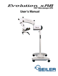
OPERATING MICROSCOPE
OWNERÕ S MANUAL
We are here to serve you!
If you have any questions regarding Seiler’s products or services,
please feel free to contact us.
(:
(:
2:
*:
8:
800.489.2282
314.968.2282
314.968.3601
[email protected]
www.seilerinst.com
• Division Manager
Dane Carlson
ext. 365 • [email protected]
• Corporate Accounts/Marketing
Nicole Rasch
ext. 345 • [email protected]
• Technical Product Information
Tony Leise
ext. 363 • [email protected]
• Accounts/Tracking/Bulbs
Sandy Jeremich
ext. 336 • [email protected]
Contents
1.INTRODUCTION2
Dear customer, thank you for choosing this product.
2
2.COMPONENTS3
Structural3
Illumination:3
Control3
Optical Module3
Control Pedal4
3.ASSEMBLY
8
Unpacking8
Column, Control Box and Pantographic Arm9
Optical Module10
Electrical Connections11
4. USING THE MICROSCOPE12
Moving the Optical Module12
Switching on the unit15
Diopter Adjustment.16
Interpupillary Distance Adjustment16
Adjusting the Focus, Diopters and Interpupillary Distance17
Using the XY system (optional)19
Using the Inclinable Binocular20
Ultra Violet (UV) Filter20
5. MAINTENANCE, HANDLING AND HYGIENE21
When the bulb burns out during use21
Changing the bulb22
Changing the fuses23
General cleaning23
Cleaning the lenses23
Control Items Handling and Hygiene24
Discard25
6.TECHNICAL DETAILS26
General Details26
Electrical Details26
Optical Details26
7.ACCESSORIES27
Beam Splitter27
Camera Adapter27
Video Camera27
Digital Photographic Camera Adapter27
Second Observer Accessory27
Image Inverter27
XY System27
Multifunction Pedal27
Binocular Head27
Zoom System27
Filters27
8.WARRANTY27
Seiler has developed this device in due accordance with global quality requirements, giving priority to safety,
electromagnetic compatibility and failure management, thereby establishing the Seiler Microscopes Evolution Zoom
as a highly safe, reliable and robust device. The main quality standards employed were based upon the standards set out
below:
DESIGN STANDARDS
•
•
•
•
•
•
•
•
•
IEC 60601-1:1988 - Medical Electrical Equipment, Part 1 - General Requirements for Safety
EN IEC 60601-1-2:2001Medical Devices - EMC Requirements
EN 55011:1991- Radiated and Conducted Emission (Class 1, Group A)
IEC 61000-4-2: 1995 (Electrostatic Discharge)
IEC 801-3:1984 (Radiated Electromagnetic Field)
IEC 61000-4-4:1995 (Electrical Fast Transient / Burst)
IEC 61000-4-5:1995 (Electrical Surge)
CISPR 14 (Click)
BS EN 1441: 1998 (Risk Analysis)
SAFETY GUIDELINES
The Seiler Microscopes Evolution Zoom is a class 1 low risk device - in accordance with FDA (Federal Drug Association EE UU) 21CFR, European Directive 93/42 on medical devices and ANVISA-PC1, 26/01/1996. We do recommend,
however, that the Guidelines set out below be followed:
•
•
•
•
•
•
•
•
•
•
Before switching on and using the device, follow the instructions set forth on the labels and carefully read this User’s Manual. Carefully read the recommendations set out below any “Please Note” headings;
This device IS NOT SUITABLE FOR USE IN THE PRESENCE OF INFLAMMABLE MIXTURES
CONTAINING AIR, OXYGEN OR NITROUS OXIDE;
This device is not to be used in the home;
This device must always be used correctly - see the Declaration of Conformity;
Correctly check the output power level (visible light). Use the visible light output with caution - this could
damage the patient’s eyes if there is prolonged, improper exposure to the eyes;
Ensure the appropriate protection is used during each procedure.
Check the availability of a spare bulb for use before embarking upon any procedure. Burned out bulb should only be replaced after checking if the device is switched off and has cooled down;
Connect the device to a no break device using high-quality batteries, for which a minimum power of 1000W (semi-sinusoidal) has been properly calibrated. If this is not done, the device may not support the high voltage;
Take care when handling the Optical Fiber Cable. It is made up of extremely fine, very sensitive fibers.
Improper use can break these fibers affecting the performance of the Cable.
Please contact Seiler should any special filters be required.
–1–
1.Introduction
DEAR CUSTOMER, THANK YOU FOR CHOOSING THIS PRODUCT.
The Seiler Microscopes Evolution Zoom, you have chosen to purchase is a modern, reliable device, which has been
designed using the most advanced optical design tools available on the market. In addition to its optical superiority, the
design was based around refining image quality and providing a product, which is ergonomic and easily handled by
users.
The Seiler Microscopes Evolution Zoom in the range are intended to be used exclusively for magnifying the visual
field, giving the user an excellent level of comfort and improved visual acuity during the activities. This equipment is
not specified for carrying out measurement work.
It is important to emphasize that its use as support equipment does not remove the need for professional skill. The
equipment uses halogen bulbs to produce visible light for illuminating the visual field.
The Seiler Microscopes Evolution Zoom is manufactured according to stringent standards, involving precision
mechanics and optics in order to ensure a high-quality final product.
All optical components have undergone antireflective, multi-layer treatment, which ensures the efficiency of said
components across the entire visible spectrum.
Please contact us immediately should you require any further information or wish to make a suggestion or complaint or
register a doubt concerning the components, devices or accessories. Our team is always ready to provide assistance and
guidance so that the full potential of this device can be realized.
–2–
2. Components
Tomakeiteasiertoidentifythesub-partsofthemicroscope,theSeilerMicroscopesEvolutionZoomisdividedinto
modules,asshowninthefigurebelow:
Modules
STRUCTURAL
The mechanical module, with structural function, consists of Column (I) and Pantographic Arm (IV).
ILLUMINATION:
Joined to the Pantographic Arm (IV) is the Illumination Box (III), which generates light. The light is supplied to the
working area by means of an Optical Fiber Cable to the microscope’s Optical Assembly (VII), which is made up of a
seriesoflensesincludingtheObjectiveLens.
CONTROL
The Control Box (II) joins the electronic circuits for the Microfocusing, Illumination Brightness Control, Zoom and XY
System functions of the microscope.
OPTICAL MODULE
TheOpticalModule(VII)comprises:ObjectiveLens,MagnificationSystem(Zoom),andBinocularHead(Fixedat45º
or Inclinable).
–3–
-9-
Seiler Microscopes Evolution Zoom – Evolution Zoom
CONTROL PEDAL
CONTROL PEDAL
The Control Pedal (VIII) is fundamental to the perfect functioning of the microscope. If it is not connected to the miControlwill
Pedal
croscope,The
the device
not (VIII)
work. is fundamental to the perfect functioning of the microscope. If it is not
connected to the microscope, the device will not work.
Its functions include adjusting the Motorized Microfocus and Zoom.
Its functions include adjusting the Motorized Microfocus and Zoom.
Thefollowingfigurespresentthemainselementsofthemicroscopeinmoredetail.
The following figures present the mains elements of the microscope in more detail.
Mains Elements
–4–
MICROSCOPES-7.5.1-MOP-01 – Review A - Mach 2010
Issue: 2010
Zoom Optical Module with 200º Inclinable Binocular Head (Standard
Binocular Head)
In this configuration the two knobs on the sides of the Optical Module can be assembled in six different positions, in
accordance with user preference. To do this, unscrew the nut that fastens the knob to the body, remove it, change its
position and fasten it in place again.
Optical Module with a stepwise increase in magnification system
–5–
–6–
–7–
3.Assembly
Please note: this manual should be read carefully in order to properly assemble the Seiler Microscopes Evolution
Zoom. The user shall be solely responsible for any other type of procedure not set out in this manual to assemble/install
this product and any misuse of tools.
UNPACKING
The Seiler Microscopes Evolution Zoom is placed in boxes to protect it during transportation.
Check the boxes for damage or signs of violation: contact Seiler immediately in the event of any irregularity. The Seiler
Microscopes Evolution Zoom should ideally be transported in its original packaging. We recommend that said packaging be kept in case it is required in the future.
Please check whether box contains the items set out below:
•
This User’s Manual;
•
Column (I) with Control Box (II) already attached;
•
Illumination Module (III) and Pantographic Arm (IV) (with optical fiber and electrical cables installed) already attached;
•
Optical Module (VII) – Zoom - already connected to the Optical Module Support System;
•
Screw to attach the Base to the Column (I);
•
Accessories, if required.
•
AC Multivoltage Cable 90-240 ~ Volt
•
Protective cover for the Optical Module;
•
2 pairs of sterilizable protectors for the Optical Drum Knobs;
•
1 spare halogen bulb;
•
Two 5A fuses;
•
Optical Module –Zoom – already connected to Optical Module Support system;
•
1 pair of knobs of the Optical Module;
•
1 pair of sterilizable protectors knobs of the Optical Module;
•
Multifunction Pedal (*1) and/or (*2)
–8–
PleaseCONTROL
note: theBOX
greatest
care should be taken
COLUMN,
AND PANTOGRAPHIC
ARM when
handling modules II, III and IV,
since module III are finished with an outer coating consisting of injected layers which
Pleasenote:thegreatestcareshouldbetakenwhenhandlingmodulesII,IIIandIV,sincemoduleIIIarefinishedwith
have no structural function. Any sharp impact on one of these layers may result in
an outer
coating
consisting of
layers which have no structural function. Any sharp impact on one of these laychinks
or scratches
orinjected
other damage.
ers may result in chinks or scratches or other damage.
assemble the
the Microscope,
follow
the instructions
below: below:
To To
assemble
Microscope,
follow
the instructions
- Connect the Connectors according to identification;
–9–
Please note: The Optical Module is the most delicate of the Modules. Please take care
when
assembling, using and transporting the module. This Module is assembled to the
OPTICAL
MODULE
Optical Module Support.
Please note: The Optical Module is the most delicate of the Modules. Please take care when assembling, using and
transporting the module. This Module is assembled to the Optical Module Support.
Fitting the Optical Module Support in the axis of the Extend Arm, being attempted the guide;
• Fitting
Supportstrongly
in the axis
of the
Extension
Put the
theOptical
screwModule
and tighten
with
5mm
AllenArm.
key, next that, tighten the three other
• Putthescrewandtightenstronglywith5mmAllenkey,nextthat,tightenthethreeotherscrewslocatedonthe
screws located on the Optical Module Support with the 3 mm Allen key;
OpticalModuleSupportwith the3mmAllenkey;
Insert the Optical Fiber Cable terminal into the Rapid Coupling Mouth.
• InserttheOpticalFiberCableterminalintotheRapidCouplingMouth.
Please note: Take care when handling the Optical Fiber Cable. It is made up of
Pleasenote:TakecarewhenhandlingtheOpticalFiberCable.Itismadeupofextremelyfine,verysensitivefibers.
extremely fine, very sensitive fibers. Improper use, repeated stretching and bending
Improper use, repeated stretching and bending movements, amongst other things, can cause them to break affecting the
movements, amongst other things, can cause them to break affecting the performance of
performance of the Cable or even rendering it useless.
the Cable or even rendering it useless.
Optical Module Support Assembly
– 10 –
Please note: Please take care when connecting the cables as the connector pins are
ELECTRICAL CONNECTIONS
fragile. When it is necessary to use a transformer, it is recommended to use one that is
insulated.
Please note: Please take care when connecting the cables as the connector pins are fragile. When it is necessary to use a
Connect thetocables
following
the steps below:
transformer, it is recommended
use one
that is insulated.
Connect the cables following the steps below:
•
•
•
•
•
Connect
the Power Cable to the
ConnectthePowerCabletotheMultivoltageACCable
Multivoltage
AC Cable Connector on
ConnectoronelectricalpaneloftheColumn;
electrical panel of the Column;
ConnectthePedalCabletotheControlPedalConnector;
Connect the Pedal Cable to the Control
Connectthecableleadingoutofthemicrofocussystemtothe
Pedal Connector;
Microfocus/ZoomCableConnector;
Connect the cable leading out of the
ConnecttheXYConnectioncabletotheXYSystem;
microfocus system to the Microfocus/Zoom
Connectthevideosystempowercabletothe12VDCPower
Cablevideo
Connector;
Outupt connector. (where
system is included)
Connect the XY Connection cable to the
XY System ;
Connect the video system power cable to
the 12VDC Power Outupt connector.
(where video system is included)
Lower View of Arm, showing where electrical connections are made.
MICROSCOPES-7.5.1-MOP-01 – Review A - Mach 2010
Issue: 2010
– 11 –
4. Using the Microscope
Please note: Users are requested to carefully read this manual and familiarize themselves with the possible movements
and settings so as to be able to use the Seiler Microscopes Evolution Zoom properly. This device should only be used
by qualified personnel.
MOVING THE OPTICAL MODULE
The Microscopes Evolution Zoom has been designed to be easily adjusted and handled while also guaranteeing maximum comfort and stability.
•
•
•
•
•
•
•
To move the Optical Module and put into the position to be used, hold it by the Sterilizable Knobs to move the
Optical Module until the desired position; note it is important to check ALL the knobs: Extension Arm Rotation System Fixing Knob
Control Box Rotation System Fixing knob
Pantographic Arm Rotation System Fixing Knob
Pantographic Arm Vertical Movement Fixing Knob
Loosen all knobs before moving the Optical Module
Place the device on the desired position, tighten again the Knobs to ensure stability to the device. It is
possible to regulate the turning force, by tightening them slightly.
– 12 –
The Optical Module of the Microscopes Evolution Zoom can be turned to left or right. It can be made by loosing the
Optical Module Right/Left Rotation System Fixing Knob, positioning the module as desired and tighten again this
knob. The Optical Module can also be turned to back and front position by loosing the Optical Module Forward/Backward Movement Fixing Knob, adjusting its position and tighten it again.
These knobs can be left slightly tightened in order to allow the Optical Module to rotate, but not freely.
– 13 –
•
Please note: there is a spring inside the Pantographic Arm to ensure that it moves smoothly and accurately; the spring’s tension is not preset at the factory, because its tension needs to be slackened during transport. It must
therefore be adjusted using the Spring Adjustment Knob. Where new accessories are added to the Optical Module,
increasing the weight to be balanced by the Pantographic Arm, the Spring Adjustment Knob should be turned
anti-clockwise. Pay attention to any possible involuntary downward movement of the set when increasing the weight by adding accessories.
Ideal Setting: when moved upwards or downwards, the Pantographic Arm remains in the new position without
having to tighten the Locking Knob of the Pantographic Arm Joint.
– 14 –
SWITCHING ON THE UNIT
•
•
•
•
ConnectthePowerSupplyCabletotheElectricalPanel’sACCableConnectorandtogroundedelectricalsocket;
PresstheMainOn/OffSwitchlocatedontheControlBox;
AdjusttheilluminationbrightnessbyturningtheIlluminationBrightnessAdjustmentButtonlocatedonthe
ControlBox;
ThePedalcanbeusedtocontroltheZoomandMicrofocus.
Please note:thePedalelectricalcircuitisnotcompletelysealed(waterproof);avoidspillingwaterdirectlyontoit
and protect it when cleaning the area where the unit is installed, hanging it by its Transport Handle from the Pedal
Handle attached to the Column.
– 15 –
DIOPTER ADJUSTMENT.
DIOPTER ADJUSTMENT.
DIOPTER ADJUSTMENT.
The diopter adjustment aims at adapting
the microscope
the user’s
eyes, reaching
The diopter
adjustmenttoaims
at adapting
• Thediopteradjustmentaimsatadaptingthemicroscopetothe
the
maximum
efficiency
in
the
image view
the microscope to the user’s eyes, reaching
user’seyes,reachingthemaximumefficiencyinthe
in the microscope.
adjustment
the maximum
efficiency in This
the image
view allows
image viewininthe
themicroscope.
microscope.
This
adjustment
allows
for both
for
both eyesThis
to see
focused-way
object.
adjustment
allows
eyestoseefocused-wayobject.
for both
eyesdiopter
to see focused-way
object.be done in
The
adjustment should
• Thediopteradjustmentshouldbedoneinthering,whichis
the ring,
whichshould
is located
the
The diopter
adjustment
be donebelow
in
located below
the
eyepiece.
There
is
a
reference
eyepiece.
There
is
a
reference
trace
–
fixed
the ring, which is located below the
trace–fixed–forbetterguidingthisadjustment.
– for
better
thistrace
adjustment.
eyepiece.
There
is aguiding
reference
– fixed
• Checkbeforestartinganyadjustment,ifzero(0)ofthering
– forbetter
guiding
thisstarting
adjustment.
Check
before
any adjustment, if
coincideswiththefixedreferencetrace.
zero (0)starting
of the ring
with ifthe fixed
Check before
anycoincides
adjustment,
• Itispossibletoadjustbetween–6and+6diopters.
zero (0)reference
of the ringtrace.
coincides with the fixed
reference
It trace.
is possible to adjust between –6 and +6
diopters.
It is possible
to adjust between –6 and +6
diopters.
INTERPUPILLARY
DISTANCE ADJUSTMENT
INTERPUPILLARY
DISTANCE ADJUSTMENT
INTERPUPILLARY DISTANCE ADJUSTMENT
• Theinterpupillarydistanceadjustmentaimsattheadaptationoftheequipmenttoyourinterpupillarydistanceso
The interpupillary distance adjustment aims at the adaptation of the equipment to your
that to reach
the
ideal
work adjustment.
The
reached
when
user
a single
interpupillary
distance
soideal
that distance
toaims
reach
workthe
adjustment.
The
ideal
distance is
The interpupillary
distance
adjustment
atis the
the ideal
adaptation
of
the views
equipment
toimage
your
(overlappingofeyepiecesimages);
reacheddistance
when thesouser
a single
of eyepieces
images);
interpupillary
thatviews
to reach
theimage
ideal (overlapping
work adjustment.
The ideal
distance is
• FormodelswithanInclinableBinocularHead,adjustmentsarecarriedoutmanuallybyturningthesideknob
reached
when
the user
views
single image
(overlapping
eyepieces images);
For
models
with
an aInclinable
Binocular
Head,ofadjustments
are carried out manually by
below. For models
turning
thean
sideInclinable
knob below.
with
Binocular Head, adjustments are carried out manually by
turning the side knob below.
Inclinable Binocular
Inclinable Binocular
MICROSCOPES-7.5.1-MOP-01 – Review A - Mach 2010
MICROSCOPES-7.5.1-MOP-01 – Review A - Mach 2010
– 16 –
Issue: 2010
Issue: 2010
ADJUSTING THE FOCUS, DIOPTERS AND INTERPUPILLARY DISTANCE
• CentertheMicrofocusSystem,presstheMicrofocusCenteringButton;
• LoosentheLockingKnobofthePantographicArmJoint,thenpositionthemicroscopeOpticalModuleata
distance from the working area which is roughly the same focal distance from the objective lens in use and
retightenitlightlyifrequired;
• ThefinefocusadjustmentismadeusingthemicrofocusadjustmentpedalonthePedal;
• OntheControlPaneltherearetworedindicatorlights,whichcorrespondtothemicrofocusfunction.Theselightup
duringadjustment, indicating thedirectionofthemovement.Whenthemicrofocusingend-of-rangeisreached,a
beepisemittedwhenthepedalispusheddown.Whenthemicrofocusisatitsend-of-range,theindicatorlight
remainslitup;
• Foranextra-finefocusadjustment,youmayselectthemaximumpossiblemagnificationoftheOpticalModuleand
findthebestpossiblefocususingthemicrofocussystem.Thiswillmeanthat,forincreasestoothercontrols,the
focuswillalreadybeatitsoptimum;
– 17 –
– 18 –
USING THE XY SYSTEM (OPTIONAL)
•
•
•
TheXYsystemcanbemovedusingtheFootStick.
Justaswiththemicrofocusfunction,lightindicatorsshowthedirectionofmovementandabeepisemittedwhen
theend-of-rangeisreached;
Tocenterthetwoshaftsatthesametime,presstheCenterbuttonlocatedbetweenthefourXYlightindicators.
– 19 –
USING THE INCLINABLE BINOCULAR
USING
THE INCLINABLE
BINOCULAR
Seiler Microscopes
Evolution Zoom – Evolution
Zoom
The Inclinable Binocular Head can be
moved (inclined) manually;
• TheInclinableBinocularHeadcanbemoved(inclined)
In order to do this, incline the eyepieces
manually;
manually to the position desired.
• UInordertodothis,inclinetheeyepiecesmanuallytothe
SING THE INCLINABLE BINOCULAR
position desired.
The Inclinable Binocular Head can be
moved (inclined) manually;
In order to do this, incline the eyepieces
manually to the position desired.
- 25 -
ULTRA VIOLET (UV) FILTER
Besides the conventional filters, there is also an Ultraviolet (UV) Filter. These are all attached to the
filter drum. The Ultraviolet (UV) Filter is assembled at the factory and its purpose is to prevent the
ULTRA VIOLETpassage
(UV) FILTER
of ultra violet light, which is why the image viewed through the binocular appears slightly
yellow.
ULTRA VIOLET (UV) FILTER
Besidestheconventionalfilters,thereisalsoanUltraviolet(UV)Filter.Theseareallattachedtothefilterdrum.The
To select the filters, just turn the knob on the drum, as shown in below figures.
Ultraviolet (UV) Filter is assembled at the factory and its purpose is to prevent the passage of ultra violet light, which is
Besides
the conventional
filters,
there is also
an Ultraviolet
(UV) Filter. These are all attached to the
why
the image
viewed through
the binocular
appears
slightly yellow.
filter drum. The Ultraviolet (UV) Filter is assembled at the factory and its purpose is to prevent the
passage of ultra violet light, which is why the image viewed through the binocular appears slightly
Toselectthefilters,justturntheknobonthedrum,asshowninbelowfigures.
yellow.
To select the filters, just turn the knob on the drum, as shown in below figures.
SEQUENCE SEEN FOR
THE MAIN OBSERVER
Drum Knob
SEQUENCE SEEN FOR
THE MAIN OBSERVER
Drum Knob
MICROSCOPES-7.5.1-MOP-01 – Review A - Mach 2010
– 20 –
MICROSCOPES-7.5.1-MOP-01 – Review A - Mach 2010
Issue: 2010
Issue: 2010
5. Maintenance, Handling and Hygiene
Please note: this manual must be read in order for it to be possible to clean and maintain the Seiler Microscopes
Evolution Zoom. This shall only be performed by qualified personnel. The use of corrosive chemical or other chemical
products is not advisable.
WHEN THE BULB BURNS OUT DURING USE
• In order to avoid possible interruptions to work caused by a bulb burning out, the microscope is fitted with an extra bulb ready for use. To switch bulbs, simply turn the Bulb Selection Switch.
– 21 –
Check whether the set is hot, if it is hot, wait a few minutes until it has cooled down;
CHANGING
BULB
LoosenTHE
the four
screws holding the Illumination Box Cover in place, and remove it;
Release the two Electrical Sockets which supply the Bulbs;
• ShifttheOn/OffswitchontheControlBoxtotheoffpositionanddisconnectthedevicefromthemains;
Release the two Locking Screws from the Bulb Support;
• Checkwhetherthesetishot,ifitishot,waitafewminutesuntilithascooleddown;
• LoosenthefourscrewsholdingtheIlluminationBoxCoverinplace,andremoveit;
Remove the Bulb Support by pulling it out. It has a slide fitting;
• ReleasethetwoElectricalSocketswhichsupplytheBulbs;
Replace the burnt out bulb
• ReleasethetwoLockingScrewsfromtheBulbSupport;
Insert the Bulb Support
• RemovetheBulbSupportbypullingitout.Ithasaslidefitting;
• Replacetheburntoutbulb
Connect the two Electrical Sockets, taking care not to get them the wrong way round
• InserttheBulbSupport
Assemble it again.
• ConnectthetwoElectricalSockets,takingcarenottogetthemthewrongwayround
• Assembleitagain.
NOTE: Never touch the bulb.
note:Nevertouchthebulb.
Please note: when reassembling the device, do not invert the position of the electrical sockets. If this occurs, a bulb
Please note: when reassembling the device, do not invert the position of the electrical
willalwayslightup,butthepositioningoftheopticalfiberinsidetheIlluminationBoxwillnotbealignedwiththe
sockets. If this occurs, a bulb will always light up, but the positioning of the optical
lamp beingused,withtheresultthatthelightwillnotpassdowntheopticalfiberandthesurgicalfieldwilltherefore
fiber inside
Illumination
Box
will nota light
be aligned
with thethe
lamp
being
used,immediately
with the
not be illuminated.
It isthe
highly
desirable that,
whenever
bulb is changed,
device
be tested
result that the light will not pass down the optical fiber and the surgical field will
afterwards.
therefore not be illuminated. It is highly desirable that, whenever a light bulb is
changed, the device be tested immediately afterwards.
MICROSCOPES-7.5.1-MOP-01 – Review A - Mach 2010
– 22 –
Issue: 2010
CHANGING
FUSES
CHANGING
THETHE
FUSES
Next to the AC cable connector on the column, there is a fuse holder. There are two fuses
• NexttotheACcableconnectoronthecolumn,thereisafuseholder.Therearetwofusesinsidetheholder.Should
inside
holder. Should
the device
functioning
completely,
switch
it off,
it
the device
stopthe
functioning
completely,
switch itstop
off, disconnect
it from
the mains,
open the
fuse disconnect
door and replace
from
the mains,
fuseonly
doorbeand
replace
the fuses
if with
necessary.
5Afuses.
fuses should only be
the fuses
if necessary.
5Aopen
fusesthe
should
used.
The device
comes
two spare
used. The device comes with two spare fuses.
Changing the fuses
GENERAL CLEANING
It is highly recommended that the Seiler Microscopes Evolution Zoom is not exposed to
GENERAL
CLEANING
dust and other contaminating substances. Always cover the Optical Module with the
Protective Cover when the unit is not being used;
• ItishighlyrecommendedthattheSeilerMicroscopesEvolutionZoomisnotexposedtodustandother
contaminating
substances.
Always
cover
Module
the Protective
Cover
when
the unit
is not being
Dust, dirt
and stains
should
onlythe
beOptical
removed
usingwith
a clean,
moist cloth
and
neutral
soap;
used;
Before cleaning the floor of the area where the unit is installed, it is very important to store
• Dust,dirtandstainsshouldonlyberemovedusingaclean,moistclothandneutralsoap;
the Pedal in a safe place, away from possible splashing or spattering. The Multifunction Pedal
• Before cleaning the floor of the area where the unit is installed, it is very important to store the Pedal in a safe place,
has a Handle so that it can be hung from the Column.
away from possible splashing or spattering. The Multifunction Pedal has a Handle so that it can be hung from the
Column.
CLEANING THE LENSES
CLEANING THE LENSES
Please note: Never use commercially available cotton wool buds; the adhesive
Please note:Neverusecommerciallyavailablecottonwoolbuds;theadhesiveattachingthecottonwooltothe
attaching the cotton wool to the cotton wool bud could be dissolved by the alcohol and
cotton wool subsequently
bud could be dissolved
by the
and subsequently
deposited on
the lenses
during cleaning,
deposited
onalcohol
the lenses
during cleaning,
thereby
compromising
thethereby
compromising
the qualities
the aforesaid
lenses.
qualities
of theofaforesaid
lenses.
Do notDo
remove
the lenses
theirfrom
holders,
as holders,
this will lead
to misalignment.
prisms
and lenses prisms
do not
not remove
thefrom
lenses
their
as this
will lead to Microscope
misalignment.
Microscope
requirefrequentcleansing,apartfromtheEyepiecesandtheObjectiveLens.
and lenses do not require frequent cleansing, apart from the Eyepieces and the Objective Lens.
•
•
•
•
Wrap a wad of cotton wool around the end of a toothpick without using any kind of glue or
Wrapawadofcottonwoolaroundtheendofatoothpickwithoutusinganykindofglueoradhesive;
adhesive;
Slightlymoistenthebudinethanol,gentlyrubthisagainstthesurfaceofthelensinacircularmotionstartingatthe
centerofthelensandmovingoutwards;
Slightly moisten the bud in ethanol, gently rub this against the surface of the lens in a circular
Performthecircularmotionagainusingaseconddryandcleanbud;
motion starting at the center of the lens and moving outwards;
Repeatthestepsaboveforeachlensyouwishtoclean.
Perform the circular motion again using a second dry and clean bud;
Repeat the steps above for each lens you wish to clean.
MICROSCOPES-7.5.1-MOP-01 – Review A - Mach 2010
Issue: 2010
– 23 –
CONTROL ITEMS HANDLING AND HYGIENE
CONTROL ITEMS HANDLING AND HYGIENE
The Seiler Microscopes Evolution Zoom has been developed so as to enable the control elements used during the proTheeasily.
Seiler Microscopes Evolution Zoom has been developed so as to enable the control elements
cedure to be cleaned
during the
procedure
to be cleaned easily.
The following canused
be detached
from
the microscope:
The following can be detached from the microscope:
Eyepiece Eyecup these are threaded onto
• EyepieceEyecupthesearethreadedontotheeyepiecesandcan
the eyepieces and can be detached for
be detached for cleansing purposes. In this case, sterilization
cleansing purposes. In this case,
usingautoclaveand/orstoveisnotrecommended.
sterilization using autoclave and/or stove is
not recommended.
The items below resist to the sterilization in autoclave and/or in stoves:
Sterilizable Silicone Covers of Knob of the Optical Module;
Sterilizable Silicone Covers – the microscope comes with silicone protectors;
NOTE: The other control items (buttons, knobs and others) can be cleaned using a
moist,
clothknobs
and and
neutral
soap
or stains.
Covering
these soap
note: The other control
itemsclean
(buttons,
others)
cantoberemove
cleaneddust,
usingdirt
a moist,
clean cloth
and neutral
elements
with
plastic
film
does
not
disrupt
the
operation
of
the
microscope
provided
any
toremovedust,dirtorstains.Coveringtheseelementswithplasticfilmdoesnotdisrupttheoperationofthemicrosuch film is applied correctly.
scopeprovidedanysuchfilmisappliedcorrectly.
MICROSCOPES-7.5.1-MOP-01 – Review A - Mach 2010
– 24 –
Issue: 2010
2002/96/EC, through recycling companies or disposal of solid residues permitted in a performance
DISCARD country.
In case of discarding of the equipment or pieces, the sending to the plant is not necessary.
Theequipment’sdiscardorownpartsitmustbeinaccordancewithambientregulations,directive2002/96/EC,through
Consults or
thedisposal
authorized
deliverer
Seiler
before the
recycling companies
of solid
residues
permitted
in adiscard.
performance country.
In case of discarding of the equipment or pieces, the sending to the plant is not necessary.
Do not discard the product or parts together with the common domestic residues.
Consults the authorized deliverer Seiler before the discard.
Verifies constantly if all device’s components don’t present any risks to the environment, to the
team
the public
and,together
if the hospital
local ambient
protocols can in accordance with
Do not discard
theand
product
or parts
with theprotocols
commonor
domestic
residues.
be discard with safety.
Verifies constantly if all device’s components don’t present any risks to the environment, to the team and the public
The discarding of the Seiler Microscopes Evolution Zoom and the used accessories, after its useful
and, if the hospital protocols or local ambient protocols can in accordance with be discard with safety.
life, it’s a user’s responsibility and they must take care with the local and effective legislation in his
region.
The discarding of the Seiler Microscopes Evolution Zoom and the used accessories, after its useful life, it’s a user’s
responsibility
andoperation
they must
care with
the local and
effectiveZoom
legislation
in his
region.against not qualified
The
oftake
the Seiler
Microscopes
Evolution
must be
protected
inadvertent use.
TheoperationoftheSeilerMicroscopesEvolutionZoommustbeprotectedagainstnotqualifiedinadvertentuse.
MICROSCOPES-7.5.1-MOP-01 – Review A - Mach 2010
Issue: 2010
– 25 –
6.Technical Details
GENERAL DETAILS
Vertical Working Height: minimum 27.5 inches and maximum 56 inches (without XY)
(with XY Positioning System, minimum 26 inches and maximum 55 inches)
Dimensions [Height x Length x Width]: 73 inches x 73 inches m x 31 inches.
Microfocus vertical adjustment limit: 40 mm.
Weight: 200 lbs. (not including accessories).
Gross weight including packaging: 330 lbs.
ELECTRICAL DETAILS
Power supply: 90 - 240~ – automatic selection
Power supply to the Video Camera: 12VDC.
Fuses: 5A.
Average Power Consumption: 200W.
Illumination bulb: PHILIPS EJM 21V 150W 50 Dichroic Reflector or similar.
OPTICAL DETAILS
Standard objective lens: f = 250mm.
Eyepieces: 12.5X.
Field of vision: 10-60mm.
Diopter setting: -6 to +6.
Minimum Interpupillary Distance: 55 to 110 mm for the Binocular Head Fixed at 45º and 48 to 78 mm for the Inclinable Binocular Head.
Magnification available in the Zoom version: 3X to 24X (continuous).
Illumination: provided by optical fibers.
Illumination field: 50mm.
Illumination brightness in the working area at full power: 80,000 lux (min.).
– 26 –
7.Accessories
The accessories available can be seen in below Figures.
BEAM SPLITTER
This accessory splits the image viewed by the Optical Module and redirects part of the image to the Camera Adapter
and the other part to the 2nd Observer. This must be installed between the Optical Module and the Binocular Heads set.
XY SYSTEM
It moves the Optical set along the X and Y axes of the Horizontal Plane, with a movement of 22.5mm to either side
from the central position. Requires the Multifunction Pedal in order to be operated.
MULTIFUNCTION PEDAL
Controls the following functions:
Switches the illumination on and off;
Adjusts the magnification of the Zoom system;
Motorized microfocusing;
Movement of XY system by means of the Footstick;
Illumination brightness adjusting.
8.Warranty
Seiler guarantees the flawless functioning of the Seiler Microscopes Evolution Zoom – except for the Bulbs – in according with its specifications for twelve (12) months, as from the date the customer receives the product.
The guarantee only covers manufacturing and functional defects of the systems and components of this device, which
have been duly identified by an Seiler accredited representative.
In the event of technical assistance and maintenance, the purchaser shall bear all transportation, storage and postage
costs. Any posting shall be performed using the original packaging while the guarantee is in force. Failure to comply
with this requirement shall invalidate the guarantee.
This guarantee shall be automatically cancelled should the device be used in any manner other than that recommended
in this User’s Manual.
Seiler shall not be liable for any personal injury, harm or damages arising out of the misuse of this device.
– 27 –
3433 Tree Court Industrial Blvd.
St. Louis, MIssouri 63122
(800) 489-2282
(314) 968-2282
Fax: (314) 968-3601
Email: [email protected]
Web: www.seilermicro.com