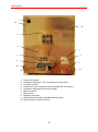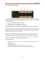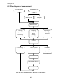
1
Hemolyzer ® 5 Servicemanual Release 1.0.20 Analyticon Biotechnologies AG Am Muehlenberg 10 35104 Lichtenfels - Germany [email protected] www.analyticon-diagnostics.com 1 agile - affordable - accurate Servicemanual This service manual is intended to give detailed information for service engineers of Analyticon Biotechnologies AG Hemolyzer 5 optical hematology analyzer. All information contained herein is the intellectual property of Analyticon Biotechnologies AG and should not be used or reproduced without prior agreement of Analyticon Biotechnologies AG, the manufacturer. This manual was written with the intention to give the most precise and up-to-date, detailed description of operation and use of the analyzer for laboratory purposes. Descriptions contained herein are relevant to Hemolyzer 5, software version 1.1.623 Despite careful revision and multiple grammar and content control, mistakes can still be present in this manual. Analyticon may from time to time issue errata, or a new revision of the manual. Would you find things unclear, please contact [email protected] for assistance. Analyticon Biotechnologies AG Technical Support Team 1 Hemolyzer 5 Table of Contents 1 INTRODUCTION ........................................................................................................................................ 7 1.1 1.2 1.3 2 PRINCIPLES OF OPERATION ................................................................................................................ 8 2.1 2.2 2.3 3 NAME AND SERIAL NUMBER ................................................................................................................... 7 INTENDED USE ........................................................................................................................................ 7 INTEGRATED SOFTWARE ......................................................................................................................... 7 VOLUMETRIC IMPEDANCE METHOD ........................................................................................................ 8 PRINCIPLE OF HGB MEASUREMENT ....................................................................................................... 9 PRINCIPLES OF OPTICAL MEASUREMENT ............................................................................................... 9 FOR YOUR SAFETY ................................................................................................................................ 11 3.1.1 3.1.2 3.1.3 3.1.4 3.1.5 3.1.6 3.1.7 3.1.8 3.1.9 3.1.10 4 Who Should Use This Manual ......................................................................................................... 11 Special Symbols Used In This Manual ............................................................................................ 11 General Precautions ....................................................................................................................... 11 Environmental Factors .................................................................................................................... 12 Electrical Requirements .................................................................................................................. 13 Space Requirements ........................................................................................................................ 13 Weight Requirements....................................................................................................................... 15 Waste Disposal ................................................................................................................................ 16 Known Limitations .......................................................................................................................... 16 Emergency Situations.................................................................................................................. 16 STRUCTURE OF THE ANALYZER ...................................................................................................... 17 4.1.1 Opening the front panel................................................................................................................... 23 4.1.2 Closing the front panel .................................................................................................................... 23 4.1.3 Removing side panels ...................................................................................................................... 23 4.2 COMPONENTS LOCATED ON THE FRONT PANEL ..................................................................................... 25 4.2.1 Display screen and the touch sensitive surface ............................................................................... 25 4.2.2 Start Button and LEDs .................................................................................................................... 26 4.3 COMPONENTS ACCESSIBLE AFTER OPENING THE FRONT PANEL ............................................................ 26 4.3.1 Shear Valve Assembly ..................................................................................................................... 26 4.3.2 Sample rotor .................................................................................................................................... 28 4.3.3 Main Dilutors .................................................................................................................................. 29 4.3.4 Dilutor opto sensor boards.............................................................................................................. 29 4.3.5 Tube organizer ................................................................................................................................ 30 4.3.6 Temperature Control Unit ............................................................................................................... 30 4.3.7 The optical unit ............................................................................................................................... 31 4.3.8 Laser Head Assembly + Sample Injector ........................................................................................ 31 4.3.9 Laserdiode Driver Board ................................................................................................................ 33 4.3.10 Pin Photodiode and Amplifier (OPTSENSOR_2v1) ................................................................... 33 4.4 LEFT SIDE ............................................................................................................................................. 34 4.4.1 Valve boards.................................................................................................................................... 34 4.4.2 WBC/BASO Preheater Assembly..................................................................................................... 35 4.4.3 Counting chamber with electrodes and measuring aperture .......................................................... 35 4.4.4 HGB Measuring Head ..................................................................................................................... 36 4.4.5 Cell counter Amplifier Board .......................................................................................................... 37 4.4.6 Pressure Sensor Board .................................................................................................................... 39 4.4.7 Reagent and Vacuum buffers ........................................................................................................... 40 4.4.8 Reagent Sensor Board ..................................................................................................................... 40 4.4.9 Opening the valve assembly plate ................................................................................................... 40 4.4.10 Vacuum buffer ............................................................................................................................. 40 4.4.11 Pneumatic and Power Boards (PPB1 and PPB2) ...................................................................... 41 4.4.12 Pump assembly ........................................................................................................................... 42 4.5 RIGHT SIDE ........................................................................................................................................... 43 4.5.1 H&V moving unit ............................................................................................................................ 43 4.5.2 XYROpto Board ............................................................................................................................... 43 4.5.3 Sampling needle .............................................................................................................................. 44 2 Servicemanual 4.5.4 Blood detector ................................................................................................................................. 44 4.5.5 Wash head ....................................................................................................................................... 46 4.5.6 Processor unit (LS-DACQ board with DIMM-PC) ......................................................................... 46 4.5.7 Mainboard Rear Panel I/O Ports .................................................................................................... 52 4.6 REMOVING THE COMPUTER MODULE .................................................................................................... 53 4.6.1 PC Mainboard ................................................................................................................................. 53 4.6.2 Mass Storage Device (hard drive) ................................................................................................... 53 4.6.3 Audio Amplifier and Speaker .......................................................................................................... 54 4.6.4 Keyboard, Mouse (optional)............................................................................................................ 54 4.6.5 CD/DVD (optional) ......................................................................................................................... 54 5 ELECTRONIC BLOCK DIAGRAM ....................................................................................................... 55 5.1 5.2 6 OPERATION OF THE FLUIDIC SYSTEM ........................................................................................... 59 6.1 6.2 6.3 6.4 6.5 6.6 6.7 6.8 6.9 6.10 6.11 6.12 6.13 6.14 6.15 6.16 6.17 7 COMPUTER MODULE ............................................................................................................................. 55 DATA ACQUISITION UNIT ..................................................................................................................... 55 THE REAGENT SYSTEM ......................................................................................................................... 59 FLOW DIAGRAM OF MEASUREMENT ...................................................................................................... 60 INITIALIZATION OF THE FLUIDIC SYSTEM ............................................................................................. 62 REAGENTS PRIMING .............................................................................................................................. 62 PIERCING PROCESS ............................................................................................................................... 62 SAMPLING PROCESS .............................................................................................................................. 62 NEEDLE WASHING PROCESSES .............................................................................................................. 63 DILUTING PROCESSES ........................................................................................................................... 63 LYSING PROCESS .................................................................................................................................. 63 RBC COUNTING PROCESS ..................................................................................................................... 64 WBC/BASO COUNTING ....................................................................................................................... 64 WBC 4DIFF COUNTING ........................................................................................................................ 64 CHAMBER DRAINING PROCESSES .......................................................................................................... 65 CLEANING(RINSING) PROCESSES .......................................................................................................... 65 STANDBY PROCESS ............................................................................................................................... 65 WAKE UP PROCESS ............................................................................................................................... 65 SHUTDOWN PROCESS ............................................................................................................................ 66 ADJUSTMENTS ........................................................................................................................................ 67 7.1 MECHANICAL SETTINGS ....................................................................................................................... 67 7.1.1 Opto wheel setting ........................................................................................................................... 67 7.1.2 Sampling needle setting ................................................................................................................... 68 7.1.3 Shear valve opto setting .................................................................................................................. 68 7.2 AMPLIFIER OFFSET SETTING ................................................................................................................. 69 7.3 USER MAINTENANCE ............................................................................................................................ 70 7.3.1 Daily maintenance........................................................................................................................... 70 7.3.2 Rinse function .................................................................................................................................. 70 7.3.3 Cleaning the shear valve ................................................................................................................. 70 7.3.3.1 7.3.3.2 Shear valve with 2 circles of nozzles ...................................................................................................... 72 Shear valve with one circle of nozzles .................................................................................................... 76 7.3.4 Cleaning the wash head .................................................................................................................. 77 7.4 PERIODIC MAINTENANCE BY SERVICE................................................................................................... 79 7.4.1 Check self test and error log ........................................................................................................... 79 7.4.2 Cleaning and Greasing Dilutor Blocks ........................................................................................... 79 7.4.3 Checking and Lubricating Dilutor Piston Tips ............................................................................... 79 7.4.4 Cleaning and Lubricating Needle Moving Mechanics .................................................................... 79 7.4.5 Measuring chambers ....................................................................................................................... 79 7.4.6 Check HGB head ............................................................................................................................. 79 7.4.7 Check and clean sampling needle ................................................................................................... 79 8 VERIFICATION PROCEDURES ............................................................................................................ 81 8.1 8.2 8.3 8.4 SELF TEST ............................................................................................................................................. 81 WARNING FLAGS .................................................................................................................................. 82 ERROR MESSAGES ................................................................................................................................ 83 SERVICE MENU .................................................................................................................................... 87 3 Hemolyzer 5 8.4.1 Service functions ............................................................................................................................. 89 8.4.1.1 8.4.1.2 8.4.1.3 8.4.1.4 8.4.1.5 8.4.1.6 8.4.1.7 8.4.1.8 8.4.1.9 8.4.1.10 8.4.2 8.4.3 8.4.4 8.4.5 8.4.6 8.4.7 8.4.8 8.4.9 Test functions ......................................................................................................................................... 90 Amplifier offset adjustment .................................................................................................................... 90 Low level file management .................................................................................................................... 90 Low level reboot (DIMMPC) ................................................................................................................. 90 Needle and wash head adjustment .......................................................................................................... 91 Pneumatical System – Initialize.............................................................................................................. 91 Network management ............................................................................................................................. 91 RAW data saving mode .......................................................................................................................... 91 Export RAW files ................................................................................................................................... 91 Windows Control Status ......................................................................................................................... 91 Service testing ................................................................................................................................. 92 Service calibration .......................................................................................................................... 92 Stress measure ................................................................................................................................. 93 Auto alignment ................................................................................................................................ 93 AS .................................................................................................................................................... 94 Multiuser Settings ............................................................................................................................ 94 MDA view ........................................................................................................................................ 95 Software upgrade ............................................................................................................................ 95 8.4.9.1 8.4.9.2 8.4.9.3 8.4.9.4 8.4.9.5 High level software upgrade ................................................................................................................... 96 Low level software upgrade.................................................................................................................... 96 Firmware upgrade*................................................................................................................................. 96 Laser upgrade* ....................................................................................................................................... 96 Auto Sampler software upgrade* ........................................................................................................... 96 8.4.10 Reagent Lock............................................................................................................................... 97 8.4.11 Printer installation ...................................................................................................................... 98 8.4.12 Factory Settings .......................................................................................................................... 98 8.4.13 Service mode OFF ...................................................................................................................... 98 8.5 SOFTWARE SYSTEMS ............................................................................................................................ 99 8.5.1 Install operating system .................................................................................................................. 99 8.5.2 Installing a Printer .......................................................................................................................... 99 9 INSTALLATION ..................................................................................................................................... 100 9.1 CHECK THE DELIVERY ....................................................................................................................... 100 9.2 PREPARE FOR INITIAL INSTALLATION ................................................................................................ 100 9.2.1 Select a Suitable Location ............................................................................................................. 100 9.2.2 Make Any Special Arrangements................................................................................................... 101 9.2.3 Gather Your Peripherals Devices ................................................................................................. 101 9.3 PERFORMING THE INSTALLATION ....................................................................................................... 101 9.3.1 Visual Inspection ........................................................................................................................... 101 9.3.2 Move the ‘Hemolyzer 5’ to the Selected Location ......................................................................... 101 9.3.3 Remove the Protective Card from the Shear Valve ....................................................................... 102 9.3.4 Connect the Optional Autosampler ............................................................................................... 102 9.3.5 Connect the Reagents .................................................................................................................... 102 9.3.6 Connect the Power Cord ............................................................................................................... 104 9.3.7 Verify the ‘Hemolyzer 5’ Computer Operation ............................................................................. 104 9.3.8 Connect the Peripherals ................................................................................................................ 104 9.3.9 How to install a printer (driver) .................................................................................................... 105 9.3.10 Set Up Reagents ........................................................................................................................ 105 9.3.11 Initializing the Optional Autosampler ....................................................................................... 107 9.3.12 Using the Settings Menu ........................................................................................................... 107 9.3.13 Adjust the Normal Ranges ........................................................................................................ 108 9.3.14 Set Up a Laboratory Information System (LIS) ........................................................................ 109 9.3.15 Set Up a Serial LIS Connection ................................................................................................ 110 9.3.16 Set Up an Ethernet LIS Connection .......................................................................................... 111 9.3.17 Running Blank Samples and Blood Samples for the First Time ................................................ 111 9.4 CHECKLIST FOR INSTALLING HEMOLYZER 5 HEMATOLOGY ANALYZER.............................................. 113 10 TROUBLESHOOTING ........................................................................................................................... 114 10.1 MEASUREMENT RELATED PROBLEMS ................................................................................................. 114 10.1.1 Small scattergram in the lower left corner ............................................................................... 114 10.1.2 Scattergram shifted/bent left or right ........................................................................................ 114 4 Servicemanual 10.1.3 Scattergram smeared to upper right corner.............................................................................. 114 10.1.4 Triangular populations above / below normal scattergram area ............................................. 115 10.1.5 Thick or contimuous lines on X or Y axis end ........................................................................... 115 10.1.6 Long, smeared population ........................................................................................................ 116 10.1.7 Thick group of cells shited up and left ...................................................................................... 116 10.1.8 No, or very few cells on scattergram ........................................................................................ 116 10.1.9 Concentrated or collapsed scattergram .................................................................................... 117 10.1.10 Smeared scattergram with concentrated center ........................................................................ 117 10.1.11 No cells on the scattergram ...................................................................................................... 117 10.2 MECHANICAL PROBLEMS.................................................................................................................... 118 10.2.1 General guidelines to overcome motor or moving part related problems ................................ 118 10.2.2 Sample Rotor (SR) failures ....................................................................................................... 118 10.2.2.1 10.2.2.2 10.2.2.3 10.2.3 10.2.3.1 10.2.3.2 10.2.4 10.2.4.1 10.2.4.2 10.2.4.3 10.2.4.4 10.2.5 10.2.6 10.2.7 10.2.7.1 SR gives grinding noise and / or SW displays SR error messages ........................................................ 118 SR error appears during initialization process: ..................................................................................... 118 The SR does not turn into the analyzer even with open front panel...................................................... 118 Needle mechanics, Vertical motor (MVert) problems ............................................................... 118 The needle carriage keeps dropping back (down) at initialization ........................................................ 118 MVert cannot reach the optosensor (Home or End) ............................................................................. 119 Shear Valve (SV) related errors ................................................................................................ 119 SV error at the first startup ................................................................................................................... 119 Grinding noise after SV cleaning, (after SV reinstallation) .................................................................. 120 SV leakage............................................................................................................................................ 120 A tube pops off from the SV................................................................................................................. 120 Dilutor errors ............................................................................................................................ 121 A tube comes off of a valve ....................................................................................................... 121 Priming problems ..................................................................................................................... 121 The analyzer would not prime liquids .................................................................................................. 121 10.2.8 Liquid under the analyzer ......................................................................................................... 122 10.3 ELECTRONICS RELATED PROBLEMS .................................................................................................... 123 10.3.1 Touch screen / display errors.................................................................................................... 123 10.3.1.1 10.3.1.2 10.3.1.3 10.3.1.4 10.3.1.5 10.3.1.6 No image on display ............................................................................................................................. 123 No backlight ......................................................................................................................................... 123 Touch sensitive surface not working .................................................................................................... 123 Touch (click) is inaccurate ................................................................................................................... 123 The cursor seems to be moving with good ratios, but in a smaller area ................................................ 123 XY coordinates seem to be interchanged.............................................................................................. 123 10.3.2 Missing DIMMPC info .............................................................................................................. 123 10.3.3 The analyzer does not power on ............................................................................................... 124 10.3.4 I2C errors displayed at startup ................................................................................................. 125 10.4 CLEANING PROCEDURES ..................................................................................................................... 125 10.5 USEFUL INFORMATION ....................................................................................................................... 126 10.5.1 Possible causes of noise ............................................................................................................ 126 10.5.2 Contaminated reagent............................................................................................................... 126 10.5.3 How can a good reagent become bad by time? ........................................................................ 126 10.5.4 Bad earth grounding ................................................................................................................. 126 10.5.5 External electrical noise ........................................................................................................... 127 10.5.6 Internal noise sources ............................................................................................................... 127 10.5.6.1 10.5.6.2 10.5.6.3 10.5.6.4 11 A. Bad chamber insulation: .................................................................................................................. 127 B. Bad insulation of electronic signal paths:......................................................................................... 127 C. Bad components, or connections: .................................................................................................... 128 D. Pneumatic failures, liquid paths that conduct noise into the chamber:............................................. 128 REMOVAL AND REPLACEMENT PROCEDURES ......................................................................... 129 11.1 OPENING THE INSTRUMENT ................................................................................................................ 129 11.2 SHEAR VALVE ASSEMBLY .................................................................................................................. 130 11.2.1 To replace or adjust the opto board of the Shear Valve ........................................................... 130 11.2.2 Visual checking of correct sampling on the A5. ........................................................................ 133 11.3 HORIZONTAL & VERTICAL UNIT ........................................................................................................ 134 11.4 DILUTORS ........................................................................................................................................... 135 11.5 TCS MODULE..................................................................................................................................... 135 11.6 PUMP ASSEMBLY................................................................................................................................ 136 11.7 HARDWARE MODULE ......................................................................................................................... 137 11.8 LASER HEAD ASSEMBLY .................................................................................................................... 138 5 Hemolyzer 5 11.8.1 To calibrate scattergrams to allow correct analysis of 5 part parameters ............................... 142 11.9 MEASUREMENT BLOCK ...................................................................................................................... 142 12 APPENDICES .......................................................................................................................................... 145 12.1 HEMOLYZER5 TECHNICAL SPECIFICATION ......................................................................................... 145 12.2 MENU TREE ........................................................................................................................................ 146 12.3 TUBING SCHEMATICS ......................................................................................................................... 149 12.4 REAGENT CONSUMPTION .................................................................................................................... 150 12.5 LIST OF SPARE PARTS.......................................................................................................................... 151 12.6 SERIAL COMMUNICATION PROTOCOL DESCRIPTION ............................................................................ 154 12.6.1 Characters and basic structure ................................................................................................. 154 12.6.2 Details of the protocol .............................................................................................................. 155 12.6.3 Sample transmission ................................................................................................................. 156 12.7 HL7 INTERFACE DESCRIPTION ............................................................................................................ 157 12.7.1 Description ............................................................................................................................... 157 12.8 RECOMMENDED KIT OF TOOLS ............................................................................................................ 160 12.9 HOW TO SEND .RP FILES TO ASSESSMENT OF ANALYZER PERFORMANCE............................................. 161 12.10 HOW TO USE THE „COLLECT” FUNCTION OF THE HEMOLYZER 5 HEMATOLOGY ANALYZER ........... 161 12.11 GENERAL SW INSTALLATION GUIDE .............................................................................................. 162 12.12 REINSTALLING WINDOWS XP IMAGE ............................................................................................. 164 13 INDEX ....................................................................................................................................................... 166 6 Servicemanual 1 INTRODUCTION Please read this manual carefully to have the knowledge for servicing the instrument perfectly and avoid extra costs and wasting precious time. This Hemolyzer 5 Service Manual contains the functional descriptions of analyzer, operation of the fluidic system, adjustments and settings, and very important information for the Service Personnel about the service operations and possible problems. 1.1 Name and serial number Name: Serial No.: Hemolyzer 5 Hematology Analyzer Every instrument has its own serial number, which is printed on the rear panel label. 1.2 Intended use Complete 24-parameter CBC profile including the optical determination of the 5 part WBC differential count with a throughput of 60 samples per hour from whole human blood sample. Hemolyzer 5 is a fully automated high quality hematology analyzer for in vitro diagnostic use in clinical laboratories. It provides precise and accurate 5-part differential measure with laser based optical measuring technology. It implements the Coulter method for WBC, RBC, PLT and measures the hemoglobin content of red blood cells and with optical measuring head, light scattering five-part differentiation (LYM, MON, NEU, EOS, BAS). 1.3 Integrated software Instrument software allows sending results to an external printer Via USB port (Widows XP printer driver required – the driver CD is provided by the printer‟s manufacturer) Hemolyzer 5 internal memory is capable of storing 100.000 records including flags, scatter- and histograms. QC measurements are also stored in separate database. The software operating the instrument is easy to upgrade using a USB memory device. 7 Hemolyzer 5 2 Principles of operation Hemolyzer 5 uses combined methods to provide hematology reports. Volumetric impedance method is used to determine cell counts regarding WBC RBC and PLT parameters. Photometric measurement of light absorbance is used to determine hemoglobin (HGB) concentration. Optical measurement of light scattering and diffraction is used to determine 5 part WBC parameters by means of a special component. 2.1 Volumetric impedance method The volumetric impedance method (a.k.a. Coulter method) counts and sizes cells by detecting and measuring changes in electrical impedance, when a particle within a conductive liquid passes through a small aperture. Internal electrode + Aperture Blood cell suspension External electrode - Impedance method Each cell passing through the aperture – where a constant DC current flows between the external and internal electrodes – causes some change in the impedance of the conductive blood cell suspension. These changes are recorded as increases in the voltage between the electrodes. The number of pulses is proportional to the number of particles. The intensity of each pulse is proportional to the volume of that particle. The volume distribution diagrams of the particles are WBC, RBC, and PLT histograms. Pulses are counted only in channels (in terms of femtoliter, fl), which are between the lower and upper discriminators. 8 Servicemanual 2.2 Principle of HGB Measurement The lysed sample dilution can be analyzed for HGB based on stable chromogen content. The reagent lyses the red blood cells, which release hemoglobin. Subsequently, the HGB concentration is measured in a photometrical way across the WBC chamber. The actual sample HGB calculated as a different of a blank and a bloody measure with and without illumination to reduce the effect of liquid refraction and disturbing light. 2.3 Principles of Optical Measurement Optical measurement of light scattering and diffraction is used to determine 5 part WBC parameters. The optical measuring head contains focused laser source to illuminate the stream of White blood cells. The intensity changes of the scattered laser light, coming from the cells, determined by the cell volume and structure. The changes recorded as an increase in the voltage of the detector system outputs. The number of pulses is proportional to the number of particles. The intensity of each pulse is proportional to the volume and granularity of the blood cells. The WBC 5 population separation is based on the two dimension volume and granularity distribution diagram. Laser beam Scattered laser light Optodetector Laser Diode Flow Cell Stream of WBC-s The light beam is focused on a linear stream of cells. Light scatters on the internal structure and on the membrane of the cell producing low and high anlge diffraction. Every cell type has 9 Hemolyzer 5 typical low and high angle intensity pairs. Individual data (light intensity of cells) are plotted on scattergrams. Scattergrams are used to identify and calculate cell populations. Hemolyzer5 measurement technology uses different reagents for the different subpopulations of WBC. The reagent system allows measuring WBC populations in two steps. First measurement identifies LYM, MON, EOS and NEU populations, while keeping RBC and BAS populations in the insensitive range of the measurement. The reagent lyses the RBC allowing measurement of WBC only. BAS population is measured separately, where the BAS specific reagent keeps BAS cells more intact than the other 4 populations of WBC. RBC is lysed during the BAS measurement cycle as well. 10 Servicemanual 3 For Your Safety 3.1.1 Who Should Use This Manual This user manual is intended for Analyticon certified Service Engineersprovideing product support for the Analyticon ‘Hemolyzer 5’ automated hematology analyzer. The manual includes information about the operation, maintenance and servicing of the ‘Hemolyzer 5’ analyzer. It also contains the steps necessary to perform the setup procedures to tailor the operation of the analyzer to the requirements of your laboratory. This manual also maintenance requirements to keep the analyzer functioning properly. 3.1.2 Special Symbols Used In This Manual Label Meaning Explanation Biohazard Blood samples and infectious materials. analyzer waste are potentially Corrosive Reagents may cause corrosion or skin irritation. Warning General warning of possible hazard conditions. Sharp needle warning The sampling needle may be a hazard to the operator. 3.1.3 General Precautions The analyzer weighs 35kg (~77lbs).Please do not attempt to move it alone. The analyzer should always be moved by two persons holding the analyzers by its sides in an upright position. Always use safe lifting procedures when lifting the analyzer. Make sure to retain the original packaging material for safe transportation and storage in the future. To prepare the analyzer for shipping, storage or extended periods of inactivity, please drain the reagents and repackage the „Hemolyzer 5‟ in its original packaging. Do not expose the „Hemolyzer 5‟ to direct sunlight, extreme temperature or humidity (>80%). The analyzer operates with chemically and biologically active reagents. Physical contact with these reagents should be avoided. Please read reagent descriptions carefully for possible emergency actions. 11 Hemolyzer 5 To ensure reliable operation and reliable results: Only human blood samples should be analyzed Only genuine Analyticon reagents should be used Required maintenance (user and service level) should be performed as recommended in this manual Only Analyticon certified service personnel should perform service actions Only genuine Analyticon service materials and spare parts should be used Genuine reagents and service materials and spare parts are available from Analyticon. Only Analyticon certified service personnel that have successfully completed the „Analyticon Hemolyzer 5 Service Training„ program are qualified to service the „Hemolyzer 5‟ analyzer. Before operating the „Hemolyzer 5‟ analyzer, all operators should complete a „Analyticon Hemolyzer 5 Operator Training‟ program. This program is offered by Analyticon or by Analyticon certified service personnel. Replacement materials or spare parts (tubes, valves, etc.) which might have been in contact with human blood or reagents should be handled as a potentially biologically hazardous and chemically dangerous material. All applicable laws and regulations must be observed in the handling and disposal of these materials. The „Hemolyzer 5‟ is designed for laboratory operation. Mobile operation is not supported. Operate „Hemolyzer 5‟ within the ambient temperature range described in section 3.1.4. The IVD equipment complies with the emission and immunity requirements described in relevant part of the IEC 61326 series. This equipment has been designed and tested to CISPR 11 Class A. In a domestic environment it may cause radio interference, in which case, you may need to take measures to mitigate the interference. Electromagnetic environment should be evaluated prior to operation of the device. This analyzer contains electronic components. Please handle electronic waste adhering to local or federal regulations. 3.1.4 Environmental Factors Operate the ‘Hemolyzer 5’ analyzer within the ambient temperature range of 15-30°C (59-86 °F) and a relative humidity range of 10% - 80%. The optimum operating temperature is 25°C (~77°F). Avoid exposing the ‘Hemolyzer 5’ analyzer to direct sunlight or to extreme high or low temperatures. If the ‘Hemolyzer 5’ analyzer was subjected to extreme temperatures during shipment or storage, the analyzer must be placed for at least one hour in a room whose temperature is within the operational range before installation or use. Reagents should be stored at a temperature range of 15-30°C (59-86 °F). 12 Servicemanual The analyzer should be placed in a well-ventilated location. Operation at an altitude above 3000 meters (9800 ft) is not recommended. 3.1.5 Electrical Requirements The analyzer should only be operated from a wall outlet meeting these power input requirements: 100-127VAC/200-240VAC; 47Hz to 63Hz Power Consumption: maximum 400 VA Please ensure that the wall outlet is also capable of supplying the power consumption of any additional devices (such as a printer). Only the power cord supplied with the instrument should be used. Avoid using extension cords. The ‘Hemolyzer 5’ analyzer comes with a power cord appropriate for your power system. Proper use of the appropriate power cord assures adequate grounding of the system. If the power network is not reliable, contact your representative for options such as the installation of an external UPS module. Failure to properly ground the „Hemolyzer 5‟ bypasses important safety features and may result in electrical hazard. The instrument should not be placed near potentially interfering devices capable of emitting radio frequencies (e.g. radio or television transmitters/receivers, radars, centrifuges, X-ray devices, fans, etc.). This analyzer is designed to be safe for transient voltages to INSTALLATION CATEGORY II and POLLUTION DEGREE 2. 3.1.6 Space Requirements It is important to install the instrument in a suitable location. A poor location can adversely affect its performance. Select a well-ventilated location near a power source and close to a suitable drain. Place the unit on a clean, level surface. Leave at least 0.5 m (18 inches) space on both sides and above the instrument to access pneumatics. A minimum of 0.2 m (8 inches) must be maintained between the rear panel and the wall to allow for heat dissipation and tube clearance. Ensure there is enough clearance in front of the ‘Hemolyzer 5’ analyzer to open the front panel. Allow enough space if you want to use optional external keyboard, mouse or bar code reader. Your selected location should allow placement of the reagents in an unobtrusive location below the laboratory bench that the instrument is placed on, or on the same surface. Placement below the laboratory bench also allows for storage of a spare set of reagents. Never place the reagents above the ‘Hemolyzer 5’ analyzer. See Figure 1 and Figure 2 for more information about proper analyzer location and clearance. 13 Hemolyzer 5 Figure 1. Hemolyzer 5 with Autosampler Space Requirements 14 Servicemanual Figure 2. Hemolyzer 5 Without Autosampler Space Requirements 3.1.7 Weight Requirements The ‘Hemolyzer 5’ analyzer weighs 35 Kg (77 lb) without the Autosampler. The ‘Hemolyzer 5’ with optional Autosampler weighs 47 Kg (104 lb). Adding an external keyboard, documents etc. can bring the total weight up to 60 Kg (132 lb). If you decide to store the reagents on the same surface, then the combined weight can reach 100 Kg (220 lb). Please select a table, laboratory shelf, or other location which can support the weight of the ‘Hemolyzer 5’ with accessories and is free from vibration. 15 Hemolyzer 5 To allow reliable operation and to provide a safe working environment, make sure that the table supporting the unit is stable enough to carry the weight of the instrument and accessories. 3.1.8 Waste Disposal ‘Hemolyzer 5’ analyzer waste contains human blood and reagents that are chemically and biologically active, and should be considered to be a potential infection and biohazard threat. Safe laboratory practices must be followed including the use of personal protective when operating the ‘Hemolyzer 5’ and handling blood, reagents, and waste. 3.1.9 Known Limitations The ‘Hemolyzer 5’ is not intended for analysis of animal blood samples. Anti-coagulated and homogenized human blood samples must be free from contamination. Blood samples must be analyzed within 12 hours of venipuncture. 3.1.10 Emergency Situations Always follow all applicable laws and regulations with regard to emergency situations. If the ‘Hemolyzer 5’ needs to be powered off due to an emergency situation (like fire, thunderstorm etc.), follow the procedures in chapter Fehler! Verweisquelle konnte nicht gefunden werden.. In case of fire, do not use water to extinguish the fire unless the „Hemolyzer 5‟ is disconnected from the electrical network! 16 Servicemanual 4 Structure of the analyzer Touch Screen Display Front Panel START Button Sampler Rotor The front panel 17 Hemolyzer 5 1 10 10 2 3 7 4 9 5 8 10 10 6 Rear side 1. Power on/off switch 2. Ventillation opening for TCU (Temperature Control Unit) 3. Computer module 4. Connectors of the computer module (standard PC connectors) 5. Ventillation openings for the power supply 6. Mains connector 7. Mains switch 8. Reagent connectors 9. Label with Serial number and Manufacturimg data 10. Thumb screws of the side panels 18 Servicemanual 1 5 2 3 4 1. 2. 3. 4. 5. Left side view Pressure meter board Opening to vacuum chamber Valve block Reagent chambers with level sensors Impedance measurement unit 19 Hemolyzer 5 1 2 3 5 4 6 Left side with pneumatic plate opened 1. 2. 3. 4. 5. 6. PPB boards (2pcs) Vacuum buffer valve blocks, solenoids level sensor connector board for reagent chambers pump module reagent connectors 20 Servicemanual 1 2 4 11 3 3 5 10 6 7 9 8 3 12 3 Front view, with front panel open 1. Display screen assembly 2. Front panel support 3. Thumb screws of the side panels 4. Optical unit 5. Tube organizer 6. Shear valve unit 7. Entry tubes for the TCU (Temperature Control Unit) 8. Dilutor units 9. Sample rotor 10. XY (needle moving) unit 11. Power on / off switch (rear view) 12. Front panel lock levers 21 12 Hemolyzer 5 1 2 3 4 6 5 7 8 Right side 1. 2. 3. 4. 5. 6. 7. 8. Optical unit LSDACQ board XY (needle moving) unit sampling needle wash head Compiuter module (side view) sample rotor (side view) Autoloader electrical connector 22 Servicemanual 4.1.1 Opening the front panel To access the pneumatic system, and to be able to perform some maintenance actions, you have to be able to open the front panel of the instrument. Make sure that nothing is placed on the top, or in front of the analyzer. Grab the lower sides of the front panel, gently pushing the sides. Pull the lower part towards you, and lift it up. Upon opening, a bar becomes visible. Make sure to tilt the front panel upwards so that you can push the lever into the secure position. 4.1.2 Closing the front panel Do not push on the front panel while the safety bar is in the “lock” position. Gently lift the front panel so that the safety support bar can be moved to the free position. Gently lower the front panel. When it reaches its lowest position, gently push on the front side to click the lock-levers in place. 4.1.3 Removing side panels With the front panel open and secured, thumb screws become visible. Both side panels are fixed to the instrument with these screws: 2 in the front, and 2 in the back. Left side covers valves and measuring chambers. It needs to be removed if chamber cleaning is requested by the analyzer. Right side covers sampling unit and wash head and sampling needle. You need to open the right side if wash head needs to be cleaned or replaced. To remove the LEFT panel (facing the analyzer) you have to open and secure the front panel. The left side panel can be removed by simply loosening the thumb screws (all four of them) and pulling the 23 Hemolyzer 5 panel away from the analyzer sideways. Store it in a secure position to avoid injury and damage to the panel. To remove the RIGHT panel (facing the analyzer) you have to open and secure the front panel. The right side panel can be removed by simply loosening the thumb screws (all four of them) and pulling the panel away from the analyzer sideways. Store it in a secure position to avoid injury and damage to the panel. Would the Autloader be installed, the removal procedure is different. You can decide whether to remove the Autoloader, and you can use the above described method OR You keep the Autoloader connected, but then you will have to REMOVE all four screws, and instead of pulling the panel away in the sideways, slide the right side panel upwards so that you can tilt it out below the front panel, and at the same time pulling away from the autoloader. 24 Servicemanual After removing the cover, potentially hazardous parts become accessible, electronic boards, motors, moving parts, sampling needle, chambers, tubes and valves. These components may cause injury, or can get damaged if handled incorrectly. Only certified personnel should open the covers. Running measurements with opened cover is not recommended due to the risk of possible injury. Always wear safety gloves while performing maintenance actions. 4.2 Components located on the front panel The display panel and related components are covered with a metal plate, to avoid electronic interference causing problems, and to protect sensitive electronics. The small metal box covers the interface of the touch sensitive interface. 4.2.1 Display screen and the touch sensitive surface The Hemolyzer 5 uses a colour TFT-LCD touchscreen. The size of the LCD is 10.4”, the resolution is 800x600. The LCD is used in portrait mode, so the real resolution is 600x800. The image is rotated on the display by a software driver. The high voltage, necessary for the backlight is generated by an inverter. The built-in touchscreen is a 4 wire resistive type. It is interfaced to the mainboard by a USB controller. 25 Hemolyzer 5 4.2.2 Start Button and LEDs The START Button board is mounted on the front panel. It comprises a start button and two LEDs. The two LEDs are the same type, and controlled in parallel. Two LEDs are used for more light power and smoother illumination of the START button. The LEDs are bicolour, red and green LEDs, the two colours are independently switchable. When both red and green are switched on, the resultant colour is yellow. 4.3 Components accessible after opening the front panel 4.3.1 Shear Valve Assembly There are 2 versions of the shear valve and of the shear valve assembly. Both sub parts are compatible with one another. old SV disc new SV disc old mechanics OK OK new mechanics OK OK Shear Valve assembly consists of the following parts: - Shear Valve holding plate, - moving mechanism, - stepper motor, - Ceramic Shear Valve, - shear valve optoboard. 26 Servicemanual Ceramic Shear Valve (old type) Ceramic Shear Valve (new type) Shear Valve unit (old) Shear Valve unit(new) This part has has its own optoboard, located on the right side of the shear valve holding plate. Optoboards are identical for old and new mechanics and shear valves. To moving mechanics position indicator 27 Hemolyzer 5 4.3.2 Sample rotor Hemolyzer 5 hematology analyzer has a sample rotor for safety and more precise sample handling. The Sample rotor unit uses a stepper motor, connected to the PPB through the XY opto board. The rotor has micro switches for positioning. The unit blocks itself in the home and end position with mechanical parts and has a special cap that prevents the damage of the electronic and mechanic parts caused by any fluid. Sample rotor is maintenance-free. Micro switches for positioning Tube adapter 28 Servicemanual 4.3.3 Main Dilutors Hemolyzer 5 has two separate main dilutor modules. There are two stepper motors, a common motor opto board, four syringes and piston rods with gear transmission in each module. Maintenance should be provided to the piston tips, by applying A599 to the cogged end of the pistons, between the syringe and the tip itself. This will ensure optimum sealing and longer lifetime of piston tips. Greasing of the cogged transmission parts (cogwheel and cogged bar) should be done regularly using grease - A597. It is recommended to check and repeat greasing of piston tips, and transmission gear every year, or after 10000 measurements. The software identifies and moves four dilutors(Dil1, Dil2, Dil3, Dil4), each dilutor consists of two syringes. Dilutor 1 and 2 is in Main Dilutor module 1, dilutor 3 and 4 is in Main Dilutor module 2, as represented in the picture below. Main Dilutor Module 1 Main Dilutor Module 2 Dil 1 Dil 2 Dil 3 Dil 4 Important notice: for the LYSE and STOP reagent we have BLACK syringe link. This different color indicates that these syringes mustn‟t be greased, because the reagents could easily solve it. 4.3.4 Dilutor opto sensor boards Dilutors have their own separated opto-boards, located directly in the units. On the front side of the optoboards there are 4 optosensors and 4 control LED-s, on the back side are mounted the motor connectors and the flat cable connector. 1 - Dilutor opto sensor board 29 Hemolyzer 5 4.3.5 Tube organizer This is the component that provides arrangement for the tubes going from the valves to the dilutor, shaer valve, optical head and Temperature Control Unit. It is intended to allow easy identification and access to tubes for service related cleaning procedures. Four tubes have metal through tubes to allow easier removal and replacement when necessary in case the TCU needs to be cleaned and rinsed. 4.3.6 Temperature Control Unit The Temperature Control System provides the necessary temperature for reagents. It is able to heat or cool the reagents, depending on the ambient temperature. It contains a massive, molded aluminium block high heat capacity. There are multiple, curved and interconnected stainless steel tubes (fluid paths) inside to ensure proper volumetric capacity and allow movement of liquids through the temperature controlled block. The TCU assembly incorporates: - - - an in-line mixer, designed to force the liquid through sudden cross section changes and thus causing a „natural” way for homogenizing sample, diluting substance and specific reagents. Temperature Controller Board, including a microcontroller to constantly monitor the temperature of the aluminium block, and enable power transistors (heating elements) or the Peltier cooling circuitry a thermal insulation for the necessary temperature stability. 30 Servicemanual 4.3.7 The optical unit The detection unit is an optimized amplifier board with two PIN photodiodes for detecting low- and high-angle scattering.The whole unit is mounted together on a robust aluminium basis. 3 1. LASERDRV BOARD 2. LENS ASSEMBLY 3. OPTSENSE BOARD 1 4. SAFETY SWITCHES 5. SAMPLE INJECTOR 6. OPTICAL FLOW CELL 7. OPTICAL CABLE 2 4 6 7 5 Black anodised aluminium plates are responsible for the laser safety covering. Two different micro-switch protects the customer against direct exposure to beam. When the rear cover holder screw is released the laser activity will be cut immediately by the laser control board. When the rear cover is removed, the laser system will inactive until both switches are closed and the system is restarted. Optical measurement unit has a sheath and sample inlet, and a waste outlet from fluidic side, laser driver cable, analogue output cable and auto-alignment cable from the electronics. 4.3.8 Laser Head Assembly + Sample Injector Laser Head Assembly is responsible for detecting the 4diff and BASO cells from the prepared blood sample. The laser head is responsible for the precise illumination of the sample. The temperature controlled laser diode source is mounted on a huge brass basis which holds it tight and also responsible for the cooling. Just beside the laser diode aspheric and achromatic lenses performs the focusing of the laser beam. 31 Hemolyzer 5 For accurate and stable adjustment, this optical unit is mounted on an aluminium block with a sphere-to-cone contact. Powerful springs hold the unit in place. Using stainless steel levers, the direction of the laser can be rotated around two axes. This means, the laser spot on the flow-cell can be tilted in horizontal and vertical directions. Precise linear motion stepper motors perform the accurate setting of the laser. Coarse adjustment of the laser can be made by setting the rough adjustment screws. The flow-cell unit is responsible for the precise flow control and the pre-detection of the pulses. An optically clear flow-cell (cross section for flow is 0.25x0.25mm)is mounted with 2 side cone-to-cone connection into its holder. Below the flowcell an injector helps to insert the sample in the middle of the main sheath streamline. The sheath puffer opened to free air supplies the sheath and the sample flow. Cross section of the flow cell – sample injector assembly The size and position of the sampler needle, the different tube resistance of the sheath and sampler lines and the applied vacuum result an about 40nm wide sample stream in the middle of the flow-cell. Concentric ring shaped optical cable is also mounted to the flow-cell unit. This collects the scattered light from the cells, and transfers it to the detection unit. Just before the insertion zone of the optical cable, there is a laser dump for filtering direct laser beam. 32 Servicemanual 4.3.9 Laserdiode Driver Board The LASERDRV board is based on a PIC 24FJ64GA004 (Microchip) microcontroller that performs all the control functions of the laser diode and the communication with the LS-DACQ (DIMM-PC). The LASERDVR board incorporates a laser diode with built-in APC (automatic power control). The laser power can be set by a programmable digital potentiometer in the 1.8 – 5.5 mW range. The laser can be swithed on and off from program. The laser diode temperature can be set, and then the microcontroller will keep the diode temperature at the preset value. As the temperature control uses only heating (2 transistors), stable diode temperature can be maintained only above room temperature. The LASERDRV board provides laser safety functions too. When removing the laser cover, the safety switches cut off laser power immediately. In order to turn the laser on the cover must be in replaced and the instrument must be turned off and back on. The controlling interface between the LS-DACQ and the LASERDRV is I2C. 4.3.10 Pin Photodiode and Amplifier (OPTSENSOR_2v1) When a blood cell in the diluted and lysed blood stream crosses the focused laser light, the light scatters and two pin photodiodes sense the scattered light. The current, generated by the two photodiodes has to be handled separately, so two independent analog channels are used on the OPTSENSOR board. The photodiode‟s current is amplified by one transimpedance amplifier per channel. Then the DC level is removed, and more amplification is applied. The DC level of output 0 an 1 (AOUT0, AOUT1) are clamped to +1V. The output span is 2V, so the output range is +1V..+3V. This is appropriate for the A/D converter on the LS-DACQ, that has a 2V input voltage span, only the offset has to be set according to the A/D‟s requirement (+1.5V..+3.5V). The 3rd analog channel (AOUT2) outputs the DC level of channel 0, amplified by 2. It can be used for the auto alignment of the laser. The OPTSENSOR card contains the connectors and LEDs for AutoAlignment motors, as well as position sensing. 33 Hemolyzer 5 4.4 Left side 4.4.1 Valve boards The Hemolyzer 5 system incorporates 44 valves. The valve boards are controlled by two Pneumatic and Power Board (PPB) boards. The used valve boards are the following: 2x Valve_1-5 2x Valve_6-12 2x Valve_13-18 2x Valve_19-22 4 valves are not used, and thus not installed. Valve coils are not installed on valve driver boards behind either. 34 Servicemanual 4.4.2 WBC/BASO Preheater Assembly WBC/BASO preheater assembly The WBC/Baso preheater assembly is located on the right side of the impedance measurement block, near the WBC/BASO counting chamber. It consists of two stainless steel holding plates, thermal insulation, heater block(see illustration below) and electronic board with heating transistors. Thermostated on 36-37 °C and its function is to heat the WBC/BASO dilution for better lysing. Diluted blood and lyse reagent is pushed through the tubing of the heater block, diluent used for rinsing and cleaning the chamber also passes through this assembly thus the chamber itself is warmer than the ambient temperature. 4.4.3 Counting chamber with electrodes and measuring aperture Impedance method is used for determination of volume and number of cells. In this method a known volume of dilution is drawn through a small aperture. Constant current is passed through the aperture from one side to the other. When a cell passes through the aperture, it causes a change in resistance, which generates a voltage pulse. The amplitude of the voltage pulse is proportional to the ratio of cell volume per aperture volume. This is used to determine the volume of cells. The number of cells can be obtained by counting the pulses. In the instruments there are two cell-counter chambers: separate for RBC and WBC. In the RBC chamber the instruments counts red blood cells, and uses no lyse at all in this chamber. It has a smaller draining outlet made of plastic and its measuring tube contains a 70 µm-sized aperture. In the WBC chamber the instrument counts all kind of WBC. It has a measuring tube with an aperture size of 100 µm and a bigger draining outlet made of PTFE (Teflon). 35 Hemolyzer 5 Both chambers have a reference electrode and a draining outlet. The next picture shows the chambers and the measuring tubes. The aperture is made of ruby and it is molded into the measuring tube. RBC chamber Reference electrode WBC chamber Reference electrode RBC measuring tube with the aperture (70m) (no grooving) WBC measuring tube with the aperture (m) (1 grooving) Aperture O-ring 4.4.4 HGB Measuring Head Hemoglobin head is placed around the WBC measuring chamber in the instrument. It contains: a light source (LED) at 540 nm wavelength and Photo Detector (TSL235). The Photo Detector converts the light to frequency. The HGB concentration is a logarithmic function of this frequency measured by the FPGA circuit of the COMB card. TSL235 LED Connection to the amplifier The analyzer performs enhanced Hemoglobin measurement technology for HGB 36 Servicemanual measurement. The output of HGB head is frequency (TSL235 detector is light to frequency converter). A digital counter in the FPGA circuit counts this signal. This counter counts up while the LED is on and counts down while the LED is off, the LED and the counter directions are switched with a 250 Hz signal. This method provides “real time backlight correction”, which makes the HGB measurement more precise in changing backlight environment situation as well. There are two kinds of HGB measurements: Sample measurement Diluent measurement (before RBC counting) (in WBC washing phase) The HGB result is calculated from these measurements by: HGB log (CNTdiluent light / CNTsample light) Due to backlight correction, Hemolyzer 5 is less sensitive to incident light changes. 4.4.5 Cell counter Amplifier Board Amplifier board includes its own voltage regulators, connection interfaces to HGB head, to high voltage board and to LSDACQ board. There is a current generator circuit on this board, which works from 50V measuring voltage (generated by the High Voltage Board) and the probe voltage (DC) is amplified with a voltage follower (output: ELV). Nominal measuring current is 870 µA. Connection to: CSA1 on COMB Connection to: HVB Connection to: COMB (DIGIO) 37 Hemolyzer 5 Connection to the electrodes Offset potentiometer Connection to HGB head Amplifier board includes one input connector for each measuring chamber (measuring electrode). There is one opto switch (OPT1) and a relay (REL1) to connect high voltage to one of the probes with HSW signal and to isolate the input of the amplifier. Test circuit allows generating test pulses (with TEST and PLS signals through Q1, Q2 FETs) for checking proper operation of each amplifier channel. Amplifier board includes a 3-stage main amplifier channel, which gains input signal to the 0...5 V range (this is the input range of the A/D converter (IC10), which is placed on the LSDACQ card). The RSW signal (with Q8 transistor) changes the input electrode through REL2 relay. There is an offset potentiometer, P1 in the third amplifier stage, manufacturer sets the correct offset voltage. Adjust the offset voltage only in case it is out of the +/- 5mV range. To adjust offset, the preheater unit must be removed from the amplifier block. The bottom side of the amplifier board contains special connectors for the electrodes and the HGB head (JP2). DHON signal - from the LSDACQ board - switches on (with Q4) the LED and the PLS signal switches off the Photo Detector in the HGB head, to prevent noise generated by the HGB detector. 38 Servicemanual 4.4.6 Pressure Sensor Board 1 2 3 The Pressure Sensor Board incorporates three differential pressure sensors. The pressure values are read out by the LS-DACQ card through I2C interface. The sesors are responsible for reading pressures of: (1) vacuum for impedance count (“chamber”) (2) vacuum used for draining chambers (“drain”) (3) big puffer vacuum for optical aspiration (“big puffer”) Upon replacing the pressure meter board, it is recommended to adjust pressure meter offset Hemolyzer 5 is equipped with sensitive pressure meters. These pressure meters are used to monitor and control measurement, and chamber draining processes. In some cases, the pressure meters can develop an offset value that can cause “Timeout” or “Chamber draining” errors. The new analyzer software can compensate for this offset value drift. Locate the “Start adjustment” button in the lower part of the screen – and tap it. The SW will perform a process, where the pressure meters will be “vented” to atmospheric pressure – and this value will be noted by the SW. This will minimize the occurrence of the “Timeout” or “draining” related errors. (On the screen you will see the actual value of the pressure meter, corrected values can be seen only if a Self Test is performed after the adjustments). If the above process fails… 1. Perform a Low Level Reboot (service menu, service functions) to get low level PC in a default known state 2. Run Test Function 11 3. Low Level Reboot 2 times 4. Perform Pressure Sensor Offset function (Service Menu, Adjustmens) 5. Perfomr a 3-measurements based Calibration using normal level control blood 39 Hemolyzer 5 4.4.7 Reagent and Vacuum buffers The lower part of the assembly plate holds six plastic cylinders, called cahmbers or buffers. The last one (marked as „1‟ in the image) holds vacuum used in the optical measurement process (for moving the solution). This vacuum is always adjusted according to measured atmospheric pressure. The four next chambers are used as temporary storage volumes for individual reagents for one measurement cycle. The first two (in the front, marked as „5-6‟ are linked in parallel to double the capacity. Chambers 2-5 have internal floating (magnetic) level sensors. The volume of chambers 2-6 allow one measurement cycle to be preformed after the reagent low warning. 4.4.8 Reagent Sensor Board The Reagent Sensor Board monitors the liquid level in the reagent puffers continuously. If the liquid level too high (puffer full), it signals to the PPB2 board and the software can stop the filling process. 4.4.9 Opening the valve assembly plate The plate holding the valves and the measurement block can be folded out of the analyzer. The plate is secured with 2 screws, similar to the ones securing the side panels. The screws are designed to stay on the analyzer to avoid losing them. 4.4.10 Vacuum buffer The glass chamber is located on the internal side of the assembly plate, and is used to store the vacuum for the optical measurements. 40 Servicemanual 4.4.11 Pneumatic and Power Boards (PPB1 and PPB2) PPB card contains the main power regulator circuits, valve and motor driver circuits and other connections for the fluidic and pneumatic system‟s parts. Power system generates +5V (Digital power), +8V (Printer power) and +12V (Motor and valve power) from the single +12V DC input signal. Motor driver part consists of six separated PIC micro-controllers with power drivers. Horizontal, Vertical and Sample rotor motors have one combined ribbon cable connection. Main Dilutor (with two motors) and Micro-dilutor have separated connectors. Valve driver section is based on the valve driver PIC micro-controller and three 8-bit, powered output shift registers (with built in protection diodes) and there are two common ribbon cable connections for the 4 valve boards. The pump assembly has a separated Darlington driver circuit for more reliable operation. All microcontrollers have 2 LEDs: a yellow one and a green one. The yellow one indicates motor moving or holding and active valve or pump moving. (it means current flows into motors, valves or pump) The green one has 3 states: dark: (after initialization phase) error state, blinking: communication in progress - normal state on (just lighting): OK - normal state 41 Hemolyzer 5 4.4.12 Pump assembly Pump assembly generates regulated vacuum and drains the fluidic system. There are two pumps in the system. They are connected to the two PPB boards. For further information see chapter 4.8. 42 Servicemanual 4.5 Right side 4.5.1 H&V moving unit This unit contains slides to move the sample sampling needle in Horizontal and Vertical directions, two stepper motors, XYR opto board, opto wheel, washing head and the sampling needle. It moves the needle to the desired position: from sampling position, to washing head, and by means of the washing head it press the sample tube during the sampling process . Both stepper motors have optical end-switch sensors for detecting these positions. These are required for correct initialization and error detection. All sensors have status LEDs to show actual conditions. The Vertical motor works with a special opto wheel for detecting home & end positions. See the Adjustment section of this manual to place this wheel to the proper position. Greasing of the horizontal/vertical guiding rods should be done regularly using A598 It is recommended to check and repeat greasing of guiding rods every year, or after 10000 measurements. 4.5.2 XYROpto Board Horizontal and Vertical motors and the Sample Rotor unit have a common Opto-board 43 Hemolyzer 5 Connections for Horizontal & Vertical motors LEDs for Sample rotor Opto switches & LEDs for Horizontal motor Opto switches & LEDs for Vertical motor The other (rear) side of the board contains the connection for the Sample rotor and a ribbon cable connection to PPB# 4.5.3 Sampling needle Sampling needle is assembled in the H&V moving unit and it makes the piercing and the sample aspirations. Correct setting of sampling needle is necessary and very important (see Chapter Adjustments). Warning! Be careful, the needle is very sharp and it can cause injury! 4.5.4 Blood detector Blood detector is a component that will allow determining if the sampling process was successful. It is measuring the length of the sample. If the sample is found short, a “sampling error” is going to be displayed. It also has anemergency mode in case the blood detector failed. In this case, the analyzer is not going to consider blood detector data, but it will move the sample to a predefined position. Both parts of the sampling control must be adjusted. Blood sensor adjustments You will need 1 vial of D-Chek3P Normal blood control There are two kinds of analyzers on the market: - A5’s that DO NOT HAVE blood sensors, and - A5’s that HAVE blood sensors installed. The blood sensor can be found near the sampling needle: a black plastic box on the right assembly plate. The below must be performed on both types of analyzers. Start up the pneumatic system. (tap the “Measurement” icon (sample tube)) 44 Servicemanual The blood sample needs to be moved to a specific location through the shear valve. You will need to open the front cover of the analyzer and observe the beginning of the measurement process. The below adjustment is necessary to allow the sample reach this location – to avoid sampling errors and thus avoid optical measurement errors (low optical cell count). The SW has default built-in values for this location. (See images in the end of this document) During sampling, the blood sample must go beyond the last loop of the shear valve, as indicated on the image. This tube is connected to V41/1 The purple area is a schematic representation of the purple area on the shear valve in the Hemolyzer5 (old and new shear valve). Please locate this tube in the analyzer. (the tube is connected to the V41/1 tube in the vertical tube organizer (black bar with nozzles) You will need to watch this distance during sampling. Run a control blood sample. If the distance is between 2 and 8 mm measured from the end of the metal nozzle and the end of the blood sample, then you do not need to change the default position. If it is beyond this value, then please change this value to 0.182. Tap the “Set default position” button. This will save the value. Rerun a control sample. Observe the position. If it is still beyond 2-8mm, please set the value to 0.174. Tap the “Set default position” button. This will save the value. Rerun the sample to verify the position. If you do not have blood sensor installed, please proceed to scatter calibration step If you have a blood sensor installed, please continue with the following process. 45 Hemolyzer 5 Tap the “Calib length” button. The analyzer will start moving liquid (diluent) towards the shear valve. You will need to observe the tube indicated with the purple rectangle. The analyzer will stop moving the liquid, and will ask you whether you can see the bubble in the tube. If you DO NOT see the bubble, tap “Cancel”. Keep tapping the “Cancel” button until you see the bubble. When the bubble is there, tap the “OK” button. The value will be saved. Tap the “Back” button to return to the service menu. Go to Service Functions, and make sure the “Enable Blood Sensor” checkbox is filled. Save the settings. Now your blood sensor is set, and ready to use. 4.5.5 Wash head Wash head is located at the bottom of the H&V moving unit and it is for cleaning the outer surface of the sampling needle. This washing process is made with diluent reagent and the fluid is drained by the pump. The arrows on the picture show the direction of diluent flow during sampling needle washing. Replace washing head yearly, or after 10 000 measurements. 4.5.6 Processor unit (LS-DACQ board with DIMM-PC) The LS-DACQ board is based on a credit card size embedded PC (DIMM-PC), manufactured by Kontron Gmbh. The LS-DACQ implements the following functions: - Receiving commands from the Analytical Unit, Blood sampling and sample handling control, Motor and valve control, Measurements control, Amplifying and A/D converting of 4 input channels simultaneously, Data preprocessing, Transmitting data to the Analytical Unit, Interfacing the Laser Driver and the TCS, Interfacing the Start Button and Status LEDs, 46 Servicemanual The LS-DACQ board incorporates a credit-card size PC, called DimmPC*. The processor on the DimmPC is a 133MHz Pentium-class core, with 32Mbytes on-board RAM, and 32Mbytes on-board IDE compatible Flash Disk. * DimmPC® is the Trade Mark of Kontron Embedded Modules GmbH USB controller 32 Mbytes RAM Flash Disk controller Flash Disk (Hard Disk) Flash BIOS chip Clock generator IC Super I/O Chip On-board SMPS AMD ElanSC520 CPU Edge connector 47 Hemolyzer 5 DIMM-PC peripheral devices The floppy and the parallel port are implemented, but are used only for service purposes. The USB host interface is implemented, a pendrive can be connected for software upgrade. As the USB connector is not available from outside of the equipment, the housing has to be opened for software upgrade. (The DIMM-PC software can be upgraded from the Hemolyzer 5 program too, and this method does not require the opening of the housing.) The PS2 keyboard connector is implemented, but are used only for service purposes. The COM1 port is used for connecting the Auto Sampler. The IDE interface is not implemented on the LS-DACQ board. FPGA interface The FPGA is connected through the ISA bus of the DIMM-PC. The FPGA implements several registers, and the DIMM-PC can reach these registers as memory-mapped. Buffer memory During optical measurement, a vast amount of data has to be transferred to the Analytical Unit through the USB connection. It is necessary to use a buffer memory to store the data temporarily, because the USB transfer speed may not be enough to transfer the data real-time. The LS-DACQ uses a 2 MB SRAM memory as buffer. The SRAM is organized as FIFO (first-in first-out memory). Connection with the Analytical Unit The Analytical Unit is connected to the Data Acqusition System by a full-speed USB interface. The USB interface is implemented by a FT2232L USB chip. The USB chip implements 2 channels, one of them works as a virtual COM port, the other is a parallel data channel. The VCOM channel is used as a command channel, through which the Analytical Unit sends commands to the Data Acqusition System. The measurement data are sent to the Analytical Unit through the parallel data channel. FPGA The FPGA is a Xilinx Spartan II type. The FPGA preprocesses the digitized data of the optical measurement and sends them to the Analytical Unit through the USB. It preprocesses the volumetric impedance measurement data too, produces data packets, and sends them to the Analytical unit. Controls the SRAM, to make a FIFO data buffer of it. Implements an I2C interface to control the PPB (Pneumatic and Power Board) boards. Implements a MDA display for service purposes. FPGA configuration As the FPGA is SRAM based, it is configured after every power on. The program is stored in a Xilinx configuration flash memory. The flash memory can be programmed through a JTAG port by a Xilinx Parallel Cable or in-circuit from the Hemolyzer 5 program. 48 Servicemanual Analog inputs The LS-DACQ has 4 analog inputs (AIN0-AIN3). AIN0 and AIN1 for the 2 channel optical measurement, AIN2 is for the autoalignment, AIN3 is for the volumetric impedance measurement. The input signal range for AIN0 and AIN1 is 1V..+3V, the DC offset is programmable, to adapt it to the A/D converter‟s input level of +1.5V..+3.5V. The input signal range for AIN2 and AIN3 is 0V..+5V, and the gain is programmable. A/D converter The A/D converter is a THS1007 type, four channel A/D, manufactured by Texas. The input voltage level is +1.5V..+3.5V, so AIN0 and AIN1 can be connected after DC level setting, but AIN2 and AIN3 channels require not only level conversion, but attenuation. The input amplifier and level converter perform these functions. The sampling frequency is 1 MHz on all input channels. Temperature and Power Voltage Measurement, I2C Interfaces The LS-DACQ card incorporates a PIC microcontroller that is connected to the DIMM-PC through the FPGA. The PIC with a built-in I2C controller controls the Laser Driver Board, the Opto Sensor Board, the Pressure board and the TCS. The PIC measures the board temperature and there is an input for measuring an external temperature. The PIC measures the board power voltages and the DIMM-PC battery voltage. 49 Hemolyzer 5 Connectors POWER Connector The LS-DACQ board is powered directly by the PC power supply through a standard IDE power connector. It supplies the board with +5V and +12V. Power voltages on the LS-DACQ board: +12V – Provides +12V to the Cell Counter Amplifier board (AJ5-MEAS), Pin Photodiode and Amplifier board (OPTSENSOR), High Voltage Board (AJ-HVB) and the Laser Driver board (LASERDRV). -12V – Generated by a DC-DC converter. Not used on the LS-DACQ, output to the Cell Counter Amplifier board (AJ5-MEAS) and the Pin Photodiode and Amplifier board (OPTSENSOR). +5V – Supply voltage to the DIMM-PC, and to some other logic. +3V3 – It is generated from the +5V by a low dropout voltage regulator. Supply voltage to the 3.3V logic, among them to the FPGA IO pins. +2V5 – It is generated from the +5V by a low dropout voltage regulator. FPGA core voltage. PPB Connector The PPB connector connects the 2 Pneumatic and Power Boards (PPB) to the LS-DACQ board. DIGIT IO Connector The Cell Counter Amplifier board (AJ5-MEAS) is connected to the LS-DACQ board through the DIGIT IO Connector. HVB Connector The HVB connector connects the High Voltage Board (AJ-HVB). ANALOG INPUT Connector The ANALOG INPUT connector connects the Pin Photodiode and Amplifier board (OPTSENSOR) to the LS-DACQ board. AINCH2 Connector The AINCH2 connector is not used in the actual design. AINCH3 Connector The AINCH3 connector connects the analog output of the Cell Counter Amplifier board (AJ5-MEAS) to the LSDACQ board. XILINX JTAG Connector (not used on the field) The FPGA configuration flash memory can be programmed through this connector by a Xilix Parallel Cable. DEBUG DISPLAY Connector (not used on the field) Only for test and debug purposes. LASER DRIVER Connector The LS-DACQ board provides power and an I2C interface to the LASERDRV board through the LASER DRIVER connector. 50 Servicemanual PRESSURE Connector The LS-DACQ board provides power and an I2C interface to the PRESSMEAS board through the PRESSURE connector. TCS Connector The LS-DACQ board provides an I2C interface to the TEMPCTRL board through the TCS connector. As the TCS requires high current power, the TCS is powered directly by an IDE connector of the PC power supply. FRONT PANEL Connector The FRONT PANEL connector connects the Front Panel board (STARTBUT) to the LS-DACQ board. FLOPPY Connector A standard 3.5” floppy drive can be connected. It is used only for test and debug purposes. KEYBOARD Connector A standard PS2 keyboard can be connected to the DIMM-PC‟s keyboard interface by this connector. It is used only for test and debug purposes. COM1 Connector The DIMM-PC‟s COM1 port is output here. The AutoSampler is connected to this port. NTC TEMP Connector An external temperature measuring NTC resistor can be connected, to measure external temperature. USBA Connector The DIMM-PC‟s USB upstream port is output here. It can be used for DIMM-PC software upgrade. USBB2 Connector It is connected to the USB downstream port of the PIC microcontroller. Not used. USBB1 Connector It is the USB interface between the Data Acqusition System and the Analytical System. It is an USB downstream connector. 51 Hemolyzer 5 4.5.7 Mainboard Rear Panel I/O Ports The mainboard I/O ports are accessible on the back of the Hemolyzer 5 housing. It is possible to attach optional mouse, keyboard, serial connection, LAN connection, external VGA display, USB 2.0 devices, audio devices to the Hemolyzer 5. • 1 x PS2 Mouse port to connect optional mouse • 1 x PS2 Keyboard to connect optional keyboard • 1 x Serial port not used • 2 x RJ45 LAN port to connect the hospital data collection system • 1 x VGA port optional VGA monitor connection • 4 x USB 2.0 ports to connect printer • 3 x Audio jacks: Line-out, Line-in and MIC-in (Horizontal, Smart 5.1 supported) not used 52 Servicemanual 4.6 Removing the computer module 4.6.1 PC Mainboard The analytical unit is based on a mini-ITX size (170x170 mm) PC mainboard. The mini-ITX size is an industry standard, and is available from more than one manufacturer. The Hemolyzer 5 uses a VIA EPIA-SN mainboard with a VIA C7 1.8GHz processor. The mainboard is equipped with 1GB DDR2 SDRAM. There is an integrated graphics controller on the board and an add-on card (LVDS-08G) is used to interface the mainboard to the TFT-LCD display. The EPIA-SN board provides extensive I/O capabilities, including 2 PS2 ports, a serial port, 2 RJ45 LAN ports, 6 USB ports and more. 4.6.2 Mass Storage Device (hard drive) The Hemolyzer 5 uses a 160 GB(or bigger) IDE hard disk as a mass storage device. As the hard drive manufacturing technology is improving very fast, it is possible that analyzer will be equipped with higher capacity hard disks. 53 Hemolyzer 5 4.6.3 Audio Amplifier and Speaker The PC main board is equipped with a High Definition Audio Codec and headphones output. The audio output signal level is too low to drive a speaker, so the system uses an audio amplifier and a broadband speaker to make the system sounds audible to the user. 4.6.4 Keyboard, Mouse (optional) PS2 or USB keyboard and mouse can be connected, if necessary for service purposes, or for easier data input. 4.6.5 CD/DVD (optional) The basic configuration does not contain CD or DVD reader. If it is necessary to connect a CD or DVD reader to the system (eg. for software upgrade), an USB CD or DVD reader can be easily connected. 54 Servicemanual 5 Electronic block diagram The analyzer hardware consists of two systems, an Analytical Unit and a Data Acquisition Unit. The two autonomous systems communicate via an USB interface. A 400W ATX PC power supply generates the necessary voltages for the two units. 5.1 Computer module The function of the Analytical Unit is to implement the user interface, to start and control the measurement processes, to receive and process the measurement data, handle the database, display, store, and print the processed measurement data. The components of the Analytical Unit are: - PC mainboard (mini-ITX mainboard). - Mass storage device (160 GB Winchester) - Touchscreen LCD display + Inverter + Touch USB controller - Audio amplifier and speaker - Keyboard (optional) - Mouse (optional) - USB CD/DVD drive (optional) - Mainboard back panel I/O ports 5.2 Data Acquisition Unit This unit executes the commands of the Analytical Unit. It controls all the processes of the sampling, sample actuation, motor and valve control, measurement control and the data acquisition. It preprocesses the raw sampled data and forwards it to the analytical unit. The components of the Data Acquisition Unit are: - Processor unit (LS-DACQ board with DIMM-PC) - Laserdiode driver and diode - PIN photodiode and amplifier - Cell counter amplifier and HGB head - High voltage board - Pressure sensor - TCS (Temperature Control System) - 2 PPB boards (Pneumatic and Power Board) - Stepper motor units with opto boards (dilutors, X-Y module, shear valve) - 44 Valves - 2 Pumps - Reagent sensor board 55 Hemolyzer 5 Analytical Unit 10.4” TFT-LCD 800x600 Touch screen connector Data and Power connector Touch screen USB interface board Back light connector Power supply USB LVDS Power Power supply connector Mini-ITX main board PS2 Keyboard PS2 Mouse IDE USB Keyboard optional Mouse optional 3.5” 160G HDD CD-DVD optional Audio USB Audio amplifier board Speaker Data acquisition system Data acquisition system 56 Servicemanual I2C PPBIF (I2C) PPB1 Analog connector Cell counter amplifier HVB connector Parallel port (debug) Floppy IF Debug DIMM-PC HGB IF COM1 Start Button Connector Reagent Sensor Shear Valve Dilutor 2 chamber + electrode HGB head HVB board I2C Pressure sensor I2C TCS Analog connector Laser driver + laser diode Optical measurement USBB Autosampler (optional) Analytical unit Start Button + LEDs 57 +50V +150V Pin photo diode + amplifier Laser driver connector USBA Auto Alignment Impedance measurement LS-DACQ board JTAG connector (FW upgrade) Pump2 34-37 valves PPB2 I2C DIGITIO connector PS2 keyboard 17-22 valves 38-44 valves 12-16 valves Dilutor1 X-Y module Pump1 23-26 valves 6-11 valves 27-33 valves 1-5 valves Data Acquisition Unit Hemolyzer 5 58 Servicemanual 6 OPERATION OF THE FLUIDIC SYSTEM This section describes the main fluidic steps of Hemolyzer 5 measurement cycle. The following figures show total measurement flow diagram and detailed descriptions of processes for understanding the fluidic system work. The following steps are introduced in this section: In the detailed process description figures, the active tube is filled with colour, while an arrow () shows the direction of the flow. Moving mechanic parts have another arrow indicating direction of movement. In Hemolyzer 5 the cleaning process executed parallel to the measures and the standby process are executed in the background. It means that the database and other functions (except pneumatic) are accessible while the analyzer is performing the measurement cycle and while it is going to standby. The cleaning process does not block the next measurement cycle, so after getting the results the next measurement can be started. Hemolyzer 5 employs a software waste full checking feature. Software integrates volume of the reagents used, and gives a message when this volume reaches the preset tank capacity. 6.1 The Reagent system Name Hemolyzer-Diluent Hemolyzer-5-Lyser Hemolyzer-5-Diff 5P Hemolyzer-Hypocleaner Description Isotonic solution, used to dilute whole blood and quantitative and qualitative determination of RBC, WBC, PLT and HGB concentration Reagent for stromatolysis of RBC and quantitative determination of WBC 5part differentiation (LYM, MON, NEU, EOS, BAS) and HGB concentration measurement of human blood. Quantitative determination of WBC, leukocyte five-part differentiation (LYM, MON, NEU, EOS, BAS) and HGB concentration Capillaries, tubing and chambers, removing blood component precipitates. 59 Function dilution of sample, rinsing and cleaning the tubing system 4-part (LYM, MON, NEU, EOS) lysing, and keeping tubing system clean responsible for BAS reaction timed Cleaning system (external liquid, not part of standard reagent system) Hemolyzer 5 6.2 Flow diagram of measurement START BLANK START ER Needle to ER Sample in Needle piercing Cleaning processes Generating Vacuums Sampling Needle washing (outside) Preparing for making dilutions HGB blank SV to CH position Generating Vacuums dilutions in parallel WBC, MIX, 4diff Needle Prewashing (Inside) SV to NP position Making RBC dilution SV to CH position Note: CH means “CHamber position”; NP means “Needle Position” 60 Servicemanual Cleaning Needle (Inside) Making 4diff dilution + RBC measurement refilling sheath puffer Generating vacuum 4diff measurement Regenerate vacuum SV to NP position HGB measurement + reset vacuum Cleaning WBC measurement MIX,RBC chambers, TCS loops 1st time Taking BASO sample SV to CH position BASO measurement RESULT Cleaning WBC, RBC chambers TCS loops 2nd time SV to NP position START NEXT AVAILABLE 61 Hemolyzer 5 6.3 Initialization of the Fluidic System Fluidic initialization process performs the following steps: Checking the hardware conditions of the unit Checking of pump and pressures sensor by generating measuring vacuum Positioning all mechanical components by scanning moving range (with endswitches) Checking prime conditions of reagent buffers Cleaning of aperture with high-pressure back-flush and high-voltage burning Preparing for compulsory blank measurement 6.4 Reagents priming Sample measurement or most of the fluidic functions is not available if the reagents are not fully primed. This can be done manually or automatically from the Priming and cleaning menu, but the instrument also performs automatic priming in several points of the pneumatic functions. (e.g. starting fill-up process, Preparing for measurement, wake up…etc.) The fluidic system is connected to the reagent containers in a closed fluidic way. This means when a syringe is priming reagent from the internal reagent buffer, the generated vacuum will automatically fill up the buffer with a little delay. Hereby the priming of the reagent during the measurement is automatically. During measurement the system automatically double-check the reagent levels in the buffers, and notice the user at the end of that measure, if any of the reagents are running low and replacement is needed. 6.5 Piercing process After the Start Button was pressed and all of the necessary initializations had done, the measurement cycle starts with the piercing process. The Sample rotor immediately turns in, the needle comes forward and start to pierce the sample. 6.6 Sampling process When the needle reaches the sampling position, a dilutor The blood sample is separated from the diluent by air bubble. The air bubble was taken right after the needle washing process of the previous measurement or during the preparing for measurement or wake-up processes. When the needle is pulled out of the sample tube and washed by the wash-head from outside, the primary blood sample will be transferred into the Shear Valve loops. 62 Servicemanual 6.7 Needle washing processes When the primary sample is taken out, the needle will be washed outside by the wash-head using diluent and continuous drawing. During Diluting process, the needle will be washed through twice. First a preliminary flow of diluent slowly washes out the remnant of the pure blood into the washing head. Finally 2,5ml of diluent cleans out the needle with high speed. When the needle is clean, the system will take air bubble into the end of the needle preparing for the next sampling. 6.8 Diluting processes After the primary blood sample was taken out and transferred to the first 3 SV loops, the SV rotates to Chamber Position(CP). The loops in the SV counting from the needle side are: 1st – WBC (~16μl) 2nd – MIX (~16μl) 3rd – 4diff (~40μl) || 4th – RBC (~16μl) 4diff has a special loop, it is transparent and has higher volume. The system will make mix dilution, wbc dilution and 4diff pre-dilution in parallel. WBC dilution is made by 2.5 ml mixture of diluent and lyse. The concentration of the lysing mixture is approx. 1:4. The WBC dilution is mixed with air bubbles from the bottom of the WBC chamber. During 4diff pre-dilution the primary blood sample is forwarded with diluent into the TCS unit to preset of the required temperature. Mix dilution made by 2.2 ml of diluent and mixed with air bubbling from the bottom and the side connector of the Mix chamber. When the mix dilution is ready, a dilutor syringe moves the mix sample through the SV into the 4th sampling loop. Then the SV rotates back to Needle Position(NP). The RBC dilution is made in the RBC chamber with 2.5ml of diluent and mixed with air bubbling from the bottom. 6.9 Lysing process The dilution for the WBC and optical BASO count is the same. The quick lyse reagent breaks down the WBC-s to its nucleus except the BASOPHILES. After measured the dilution in capillary mode, the remaining sample is transferred to the optical head for basophile count. The main and most precise lysing process is making the 4diff dilution. It requires precise timing, volume control and temperature conditions. The accurate temperature control is made by the TCS module. 63 Hemolyzer 5 In the TCS unit, there are loops for lyse, stopper, pre-diluted blood, blood + lyse mixture. Temperature of these loops are controlled accurately. After the 4diff primer sample is pre-diluted and its temperature has been set, 3 syringes (lyse, stop, diluent) make the precise dilution through the TCS unit, the SV 4diff sample loop, ending in the big vacuum puffer. Previously generated vacuum supports the smooth flow of the mixture. The pre-diluted blood meets with the lysing reagent by a small “T” connector first. Right after this point there is an inline mixer part in the TCS module, where the blood and the lyse will be mixed in its tube by flowing through. After that the precisely mixed lysing dilution runs through one of the TCS loops for better temperature conditions, and reaches the stopper “T” connector. The volume between the lyse and stopper “T” connectors is about 1ml, and this is the incubation zone for the lysing. After adding the stopper reagent the mixture goes through another inline mixer, the SV 4diff sampler loop, and flows into the vacuum puffer. The SV 4diff sampler loop is volumetrically set by the software, so the precisely lysed and stabilized 4diff mixture will be stopped in this loop, ready for measurement. The parameters of the lysing process are the temperature of the dilution, the dilution ratios of the blood-diluent-lyse-stopper mixture and the speed of the flow. These are determining the quality of the 4diff dilution. 6.10 RBC counting process In this case the regulated vacuum aspirates the RBC dilution (RBC) from the RBC chamber through the aperture. The instrument counts the cells for 8 seconds in this case. 6.11 WBC/BASO counting In this case the regulated vacuum aspirates the WBC dilution from the WBC chamber through the aperture. The instrument counts the cells for 6 seconds in this case. After the WBC dilution has measured in capillary mode, the system transfers the remnant of the sample to the BASO loop in the SV. The last measure in measurement cycle is the optical BASO count. The measuring principle is similar to the 4diff measurement. 6.12 WBC 4Diff counting In this case the regulated vacuum aspirates the WBC 4diff dilution and the sheath fluid from Sheath puffer, through parallel tubes (one for sheath flow, one for sample flow), through the flow-cell into the vacuum chamber. The instrument counts the cells for 6 seconds in this case. During measurement the core diameter of the sample stream in the flow-cell is approx. lines. 64 Servicemanual The 4diff measurement ends with a self-cleaning process. The system changes the insertion point of the vacuum, which starts to wash back the sampler needle and the 4diff sample loop in the SV with sheath fluid. This process prepares the optical head for the following BASO count. 6.13 Chamber draining processes Chamber draining is made under pressure control. Pressure controlled draining starts with vacuum generation in the puffer reservoir. After that pump drains chamber while puffer reservoir and thus the pressure sensor is connected to the draining tube. The instrument can detect the empty state of the chamber from drop of vacuum. 6.14 Cleaning(rinsing) processes All cleaning processes during a measurement cycle uses mainly diluent, and for special cases small volume of lyse, and stopper reagent (e.g.: cleaning the adequate loops in the TCS). The instrument performs the cleaning processes parallel to the measurement. When any of the fluidic part has finished its work with the pure or diluted blood, cleaning process will start. The main cleaning processes during measurement are: - Needle washing outside, 2x inside into washing head - Mix chamber washing - RBC, WBC chamber washing, back flush and high voltage burn of apertures - 4diff diluting loop (SV+TCS loops) washing 2x - Optical head and sampling line washing with sheath fluid 6.15 Standby process Because of the high throughput of the Hemolyzer 5, the a measuring cycle ends with empty WBC chamber, and leaves the BASO sampling line is not cleaned. During continuous measurement the beginning of the next measurement will automatically clean this loop. On the other hand, when there is no more measurement started, the instrument will go to stand-by mode after some minutes. Standby process performs the cleaning of the not yet cleaned lines, drains all vacuum chambers, drains all chambers then refill them with diluent just above aperture level. 6.16 Wake up process In this case the instrument prepares itself for the upcoming measurement cycle. 65 Hemolyzer 5 6.17 Shutdown process The fluidic shutdown performs the following steps: Priming chambers with diluent to avoid drying out of aperture and prevent the chamber from dirt. Needle is in up position and washed. All of the syringes are positioned down. Sample rotor moved out 66 Servicemanual 7 ADJUSTMENTS Mechanical and hardware adjustments are described in this section. Software settings are included in Section 5.2. 7.1 Mechanical settings There are two important mechanical settings in the system: Opto wheel setting (Vertical motor) Sampling needle setting The manufacturer adjusts the analyzer during production. However, in case of repairs in the mechanical system, these adjustments should be checked. The omission of these settings can cause malfunction or damages to the instrument. 7.1.1 Opto wheel setting This setting is necessary for the vertical motor movements because this adjustment sets the opto end-switches of the H&V moving unit. The top of this block is called HV head and it is shown in the figure below. Set the distance to 1-2 mm between the moving carriage and the fixed part of the head. Loosen „A” screws to allow free movement of the timing belt. Adjust the opto wheel to home position, i.e. home hole must be in home sensor, and LED corresponding to home opto sensor goes on. Fasten „A” screws A Opto wheel End hole End opto Home opto Home hole 67 B Hemolyzer 5 Check the end position as well: move the needle down. Adjustment is successful if end LED goes on before moving part reaches end of mechanical range. Once this adjustment is necessary, never miss sampling needle setting described in the next section. 7.1.2 Sampling needle setting This adjustment sets the sampling needle to the operational position. In Service menu, in Miscellaneous submenu select Needle setting. The software moves the needle back and up, and turns on horizontal and vertical motors to keep needle in place. Check the washing head position: unfix the sample rotor and turn it in by 90 degrees. If the top of the door cannot reach the bottom of the washing head, then it is correctly adjusted. If it hits the washing head you have to loosen the upper fixing screws of the washing head leading rods. Push them to upper position where the top of the washing head almost reach the bottom of the main moving carriage. Fix the screws. Check the setting of the needle. If end of the needle is at the bottom of the washing head, needle is set correctly. If not, open screws “B” (see above), and adjust the needle to the bottom of the washing head. Fasten “B” screws. Set the end of the tip to the washing head‟s bottom plane, while the carriage is held by motors. (Needle setting menu). Fix the „B” screws. Be careful with the bent upper end of the sampling needle, because if badly aligned, during movement it can hit other mechanical components causing mechanical jam, and therefore damages or error. Warning! Be careful, the needle is very sharp and it can cause injury! 7.1.3 Shear valve opto setting This opto setting is necessary for the correct movement of the Shear Valve. Although the SV is stopped mechanically in its end positions, the optoswitches provide the necessary feedback signal for the electronics that the movement has been done. 68 Servicemanual Shear Valve optoboard Opening for SV opto adjustment Front opto fixing screw 7.2 Amplifier offset setting Amplifier offset should be between ±5mV. Run self test to determine whether offset is within this range. If it is out of range, it should be re-set, by the following way. 1. Locate the opening for offset setting potentiometer on the measuring block (see enclosed picture). 2. In Service menu select Offset adjustment menu. 3. Adjust the potentiometer to reach 0 mV. 69 Hemolyzer 5 7.3 User maintenance 7.3.1 Daily maintenance Hemolyzer 5 requires minimal maintenance during daily routine. After each measurement the chambers and the shear valve are rinsed thoroughly with diluent, the needle is washed and the chambers are drained waiting for next sample . If the analyzer stay unused for 5 minutes, a Standby cycle is initiated automatically, this fills up the chambers with diluent to avoid drying of the apertures. However, at the end of day an additional cleaning of the system is recommended. To perform this cleaning, prepare a vial containing 2-4 ml Diatro-Hypoclean® and place it in the Sample Rotor. From Main Menu go to: Maintenance/Functions and press Clean. This will initiate a cleaning cycle, all chambers, needle, shear valve and the TCS is cleaned with diluted Diatro-Hypoclean®, than the system is rinsed with diluent. 7.3.2 Rinse function The rinse function is an additional feature, used generally to prevent the system from contamination with blood, after e.g. a mechanical jam or malfunction occurred during measurement. It can be accessed from Main/Maintenance/Functions by pressing the Rinse button. The cycle starts with a Pneu Init. process, than the chambers, shear valve, sampling needle and all related tubing is rinsed with diluent to push out the blood remnants that might be present, finally an automatic background cycle is performed to check the cleanliness of the instrument. 7.3.3 Cleaning the shear valve Salt build-up on the inner surface of the Shear Valve may cause malfunction during operation, therefore it is recommended to inspect and clean the SV monthly or whenever it is necessary. Use a soft cloth dampened with water to clean this area. You need the following materials to complete the shear-valve cleaning: A piece of soft cloth dampened with water; A piece of soft, dry cloth; A piece of lint-free soft, dry-cloth (like a non-woven sheet); A pair of gloves; A pair of tweezers; A few tooth-picks; Water. The shear-valve is in contact with the sample blood. Wear gloves while cleaning the shear-valve. Handle the materials use to clean the shear-valve as potentially infectious material. The parts which should be disassembled / re-assembled are fixed with so called thumb-screws. No screwdriver or other similar tool required to tighten these screws. 70 Servicemanual Go to the Maintenance menu, and select Shear Valve Clean function to initiate the procedure. The ‘Hemolyzer 5’ asks for confirmation to start the procedure. After clicking/ tapping the ’OK’ button the ‘Hemolyzer 5’ empties the shear-valve and the connecting tubes. Some liquid can remain inside the tubes and inside the shear-valve. As the preparations completed the ‘Hemolyzer 5’ displays the following message: Don’t press the ‘OK’ until the shear valve is assembled again! Perform the shear-valve cleaning according to the instructions below! Open the front cover, and secure it with the latch. Locate the shear valve. Prepare a soft cloth dampened with water to clean this area. 71 Hemolyzer 5 7.3.3.1 Shear valve with 2 circles of nozzles When instructed, loosen the milled-edge screw then push the latch left. This will release the shear valve. Gently remove the shear valve by pulling it towards yourself and gently lifting it at the same time. Avoid hitting optical sensors located on the right side of the shear valve holding plate. With the Shear Valve in your hands, unscrew the closing screw and pull it off. 72 Servicemanual Slide the two discs apart (do not pull on it, since the closing force of the smooth surfaces will not let the two discs part that way). Gently wipe any possible salt crystals off with a wet, lint-free cloth. To reassemble: Push the closing screw back into the center and push the discs together. Make sure that the joggle of the end of the closing screw is against the joggle of the lower disc inside. Make sure to align the grooves as indicated on the image below. Gently turn the upper disc to a position so that grooves are aligned as indicated on the image. Tighten the screw well. Make sure the discs are laying on one another. There are two metal pins on the shear valve. The longer one must point towards the analyzer. Make sure to drive this longer pin INTO the opening on the receiving part. 73 Hemolyzer 5 The lower disc has a flat section on its side. This must be aligned with the right sidewall of the shear valve rail unit. Gently slide the assembled shear valve back into the rails. Be careful not to hit or break the black optosensors. This a very important step, please pay extra attention to it! Refer to the pictures below to check the alignment. If you are not sure that the lower disc is in correct position, better split the two discs again and install the lower disc first. INCORRECT ALLIGNMENT! INCORRECT ALLIGNMENT! CORRECT ALLIGNMENT 74 Servicemanual Holding the Shear valve push the sliding latch to the right and secure the latch with the thumb screw. Close the front panel. When finished, end the operation by clicking “finished” on the screen. The software will check and adjust shear valve operation automatically. 75 Hemolyzer 5 7.3.3.2 Shear valve with one circle of nozzles Any salt build-up on the inner surface of the shear-valve may cause malfunction during operation. To avoid this problem, it is recommended to clean the shear valve after every 1500 samples. (Below instructions are valid for analyzers manufactured after August 2010) 1.: Open the front cover, and secure it with the latch and locate the shear valve (it is in the center of the analyzer. 2: Unscrew and remove the “Axis screw” Clean the “Axis screw” by using water and wipe it dry. 3: Slide off the upper disk of the shear valve. Due to the extremely smooth surface of the ceramic discs it is not possible to simply lift away the upper part. If the shear-valve was not in use for a few days, then apply a few drops of water to the contact of the upper and lower disks. With time the salt will get dissolved and the upper disk gets released. 4: Clean the connecting surfaces of the disks of the shear-valve, the housing of the valve, the tube connections. Remove any salt-built-ups. Use the tweezers to push-in the dampened and the dry cloths. You may apply a few drops of water to soften-up the contaminations. You may use a toothpick to remove salt-crystals from narrow places. Do 76 Servicemanual not use any sharp/ metal / hard object which can scratch the surface of the shear-valve. Clean the surrounding and the housing/ mounting of the shear-valve as well if necessary. Pay attention to clean the aligning surface. Be sure that no lint/ fibers remain on the connection surfaces of the ceramic disks. 5: After cleaning the shear-valve, the housing and surrounding area, put the disks together. 6: Put the “axis screw’ into the upper disk. There is a spring applied to the axis screw. This will guarantee the necessary closing force for the two disks. Gently press and rotate the “axis screw” clockwise until it clicks into the lower part. Twist the axis screw until it stops. The mechanical design of the screw prevents over-tightening. 8: Mop the surrounding of the shear-valve again. You can let the salt crystals and other small parts fall down. Sweep any particles at the bottom of the ‘Hemolyzer 5’ though the ventilation holes. Remove the gloves. Close the front door. Click/ tap the ‘OK’ button: this informs the ‘Hemolyzer 5’ that you completed the operation. The ‘Hemolyzer 5’ will check the movement and end-positions of the shear-valve. 7.3.4 Cleaning the wash head The wash head cleans the outer surface of the aspirating tip with diluent. Any salt build-up on the lower surface may cause malfunction during operation. The wash head must be removed from the needle assembly for correct cleaning. Open the front panel, and secure it with the support bar. The right side panel of the analyzer must be removed to access the wash head. Loosen the 2+2 screws on the front and rear of the side panel. The panel can be pulled off from the analyzer. Locate the sampling needle. Be careful! The sampling needle is sharp, and can cause injury! The wash head should be “twisted off” from the needle, and pulled off downwards. Inspect the wash head for presence of heavy salt buildup or blood debris. This indicates that the wash head is weared and need to be changed. Small salt buildup and blood remnants can normally be present on the wash head. Use a soft cloth dampened with water to clean the bottom of the wash head. 77 Hemolyzer 5 If the wash head needs to be replaced then gently pull down the tubing from the ports(because the tygon tube can adhere to the metal loosing the tube by means of a screwdriver might be necessary), then push them on to the new wash head. To put the wash head back: Locate the sampling needle. Be careful! The sampling needle is sharp, and can cause injury! Push the wash head onto the needle. Push it up as much as (taking care of the sharp needle) and lock it back by twisting it on the holding rods. Replace the side cover and close the front panel. 78 Servicemanual 7.4 Periodic maintenance by service 7.4.1 Check self test and error log The Self test data can be saved for further reference. It is also recommended to print the report. 7.4.2 Cleaning and Greasing Dilutor Blocks The dilutor block driving wheels and gear bar should be cleaned from dirt and must be greased between the gear bar and the support, and between cogged wheels. Use A597, MACHINE GREASE 'LX2' FOR DILUTOR GEAR (2ml) 7.4.3 Checking and Lubricating Dilutor Piston Tips The cogged end of PTFE dilutor pistons should be cleaned and lubricated by neutral silicon grease. Apply just a thin layer, and move it along the perimeter of the piston, so that some of the material goes into the gaps between the sealing rings. Repeat this step for lyse and dilutor pistons as well. Check the condition of the micro piston sealing, and replace if necessary. Use A599, SILICON GREASE FOR DILUTOR PISTONS (2ml) 7.4.4 Cleaning and Lubricating Needle Moving Mechanics The H&V moving mechanics sliding bars should be cleaned from dust. Use A598, PHOTOLUBE 007 FOR SLIDING BARS (2ml) GREASE OR PURE LUBRICATING OIL IS NOT SUITABLE. 7.4.5 Measuring chambers Observe and clean the chambers if necessary. It is recommended to perform a Hypochlorite cleaning procedure as well, which will 7.4.6 Check HGB head Observe the self test results. If the HGB light and dark values are near the limits () then it is advised to replace the component. 7.4.7 Check and clean sampling needle The sampling needle can collect some residual blood from the rubber caps of sample tubes. This will most likely be present on the upper half of the needle, the part that usually resides above the wash head. This does not interfere with measurement results, however it is recommended to clean the needle from time to time. Use cleaning tissue or paper and alcohol to clean the surface of the sampling needle. 79 Hemolyzer 5 80 Servicemanual 8 Verification Procedures 8.1 Self test The analyzer has built in test functions to check and evaluate operation of internal modules and systems. The function is accessible from the Main Menu, Diagnostics, Self test function. There are two subsets of tests: electronic and pneumatic. Each process takes cca. 1 minute, and provides results of each tested parameter. You can select to run both test sets, by clicking on the “Start Both” button. Accepted ranges are displayed below. Big puffer time Small puffer time Generate Release Generate1 Generate2 Release 3000 - 13000sec 3000 - 5500sec 800 - 4000sec 800 - 4000sec 500 - 2000sec Big puffer drift Small puffer drift Maximum Minimum Drift Maximum Minimum Drift 540 - 560mBar 530 - 560mBar -5 - 15mBar Pump status Pump1 Pump2 Null pressures 1-1 1-1 TCU Reference Actual Sink 25 - 38°C Reference +- 0.2 °C 0 - 55°C -20 - 20mBar -20 - 20mBar -20 - 20mBar Laser off Laser on 0 - 0.2mV 0.2 - 0.5mV Battery 0 - 3000 pulses 3000 - 60000 pulses Electrode Voltage Current Offset Sheath Capillary Chamber Laser temperature/Optical HGB LED HGB Dark HGB light 225 - 235mBar 215 - 235mBar -5 - 15mBar Battery voltage 2.7 - 3.3V +12V 11.4 - 12.6V -12V -12.6 - -11.4V Noise/Pulse 45 - 55V 620 – 680μA -3.0 - 1mV pls/5sec 20000pls 0 - 2000 pulses 19990 - 20050 pulses Would any value fall outside the above defiend range, the SW will indicate it with a red, “Failed” string. Correct and acceptable results are indicated with a “Passed” string. 81 Hemolyzer 5 8.2 Warning flags The analyzer checks the details of measurement and gives a warning flag in some conditions. C WBC clogging (minPrv / maxPrv < 0.80f) c RBC clogging (minPrv / maxPrv < 0.60f) B WBC blank high (WBC > 0.5f) b RBC blank high (RBC > 0.05f) p PLT blank high (PLT > 25) H HGB blank high (HGB > 10) X 4Diff error (Abs. cell count < 300) Y 4Diff error (processing error) F 4Diff blank high (Abs. cell count > 100) x Baso error (Abs. cell count < 300) y Baso error (processing error) f Baso blank high (Abs. cell count > 100) 82 Servicemanual 8.3 Error Messages The analyzer checks the operations of several mechanic, fluidic and electronic parts during measurement. The system shows the type of the error on the LCD display if any kind of malfunction is detected. The electronic parts have a very little chance to fail, only the connections and cables could disconnect, which can cause the malfunction of the electronic system. The mechanic and fluidic system have a bit more chance to go wrong because it has moving parts. Error code 0 1 2 3 4 5 6 7 8 9 10 11 12 13 14 15 16 17 18 19 20 21 22 23 24 25 26 27 28 29 30 31 32 33 Description No Error Inproper use of valve number. Inproper use of pump number. Pneumatic system init function failed. Inproper use of chamber number. Function “All valves off” failed. Special Software error. Special Software error. Special Software error. No error. Available only as “Second error code”. Software exception error. Not used. USB connection error. Reagent prime function failed. Test function failed. Measure pressure generation process failed. Measure pressure (2) generation process failed. Reagent sensor calibration function failed. Motor test function failed. Valve test function failed. Pump test function failed. Some problem occurred during the preparing for shipment process. Clean function failed. Sampling needle wash process failed during the measurement. Blank measure failed. Stress mode failed. Empty chamber process failed during the measurement. Shutdown process failed. Hardware initialization function failed. Creating pneumatical software system failed. Creating pneumatical software system failed. Creating pneumatical software system failed. Creating pneumatical software system failed. Measure function failed. 83 Hemolyzer 5 34 35 36 37 38 39 40 41 42 43 44 45 46 47 48 49 50 51 52 53 54 55 56 57 58 59 60 61 62 63 64 65 66 67 68 69 70 71 72 73 74 75 76 77 78 Shear valve movement failed. Internal software error. Autosampler error. Auto sampler error. Data acquisition system initializing process failed. Data acquisition system initializing process failed. Data acquisition system initializing process failed. Data acquisition system initializing process failed. Internal main cycle error. Internal software system error. Internal software system error. Internal software system error. Internal software system error. Test display function failed. Internal software system error. Internal file system error. Internal file system error. Internal file system error. Internal file system error. Internal file system error. Internal file system error. Error occurred during the low level software upgrade. Internal test function error. Internal software system error. Serial communication error. Drain function error. Needle steering test function failed. Hard cleaning function failed. Internal software system error. Internal software system error. The system is in busy state. The system is in busy state. The system is in busy state. Pressure sensor error. Pneumatic system error. Internal software system error. Internal software system error. Internal file system error. Main setting error. Reagent error. Autosampler error. Autosampler error. Autosampler error. Autosampler error. Autosampler error. 84 Servicemanual 79 80 81 82 83 84 85 86 87 88 89 90 91 92 93 94 95 96 97 98 99 100 101 102 103 104 105 106 107 108 109 110 111 112 113 114 115 116 117 118 119 120 121 122 123 Autosampler error. Autosampler error. Autosampler error. Autosampler error. Autosampler error. Autosampler error. Autosampler error. Autosampler error. Autosampler error. Autosampler error. Autosampler error. Autosampler error. Autosampler error. Autosampler error. Autosampler error. Autosampler error. Autosampler error. Autosampler error. Autosampler error. Autosampler error. Autosampler error. Autosampler error. Autosampler error. Autosampler error. Error occurred during the fill up function. Error occurred during the pierce test function. Dilutor 1 error. Dilutor 2 error. Dilutor 3 error. Dilutor 4 error. Emergency sampler error. Sampler needle horizontal motor error. Sampler needle vertical motor error. Dilutor error occurred during the measurement function. Get bubble process failed during the measurement. Bubbling process failed during the measurement. Not used. Measure puffer pressure error. Optical measure puffer pressure error. Measure puffer pressure error. Pre-dilution process of 4 diff dilution failed during the measurement. Lyse dilutor error during the measurement. Blood sample move process failed during the measurement. Needle wash process failed during the measurement. WBC stop process failed during the measurement. 85 Hemolyzer 5 124 125 126 127 128 129 130 131 132 133 134 135 136 137 138 139 140 141 142 143 144 145 146 147 148 149 150 151 152 153 154 155 156 157 158 159 Mix sample process failed during the measurement. Mix chamber fill process failed during the measurement. RBC chamber fill process failed during the measurement. WBC chamber fill process failed during the measurement. HGB measurement process failed during the measurement. Basophil measurement process failed during the measurement. Basophil sampling process failed during the measurement. RBC measurement process failed during the measurement. 4diff wash process failed during the measurement. Drain process failed. Internal software system error. WBC measurement process fail during the measurement. 4diff measurement error. Dilutor movement error. RBC chamber high voltage burning process failed. WBC chamber high voltage burning process failed. Internal software system error. Internal software system error. Auto-alignment function failed. Auto-alignment function failed. Auto-alignment function failed. Stopper reagent puffer error. Lyse reagent puffer error. Diluent reagent puffer error. Sheath reagent puffer error. Dilent prime process failed. Sheath prime process failed. Lyse prime process failed. Stopper prime process failed. Autosampler error. Not used. Pneumatic system initialization process failed. Internal software system error. Wakeup process failed. Standby process failed. Internal software system error. 86 Servicemanual 8.4 Service Menu There is a Service menu for servicing and operation checking purposes, however it is hidden from the standard user. To access the service menu: With on screen keyboard active: Go to the Main Menu Tap and hold the Main Menu icon for cca 6 seconds Accompanied by a “bing” sound, the password entry dialog will be displayed. Enter the service code (6484A5) The service menu icon will be displayed With onscreen keyboard inactive Connect an external USB keyboard Go to the Main Menu Make sure the arrow is over the Main Menu icon On the external keyboard, type 6484A5 Press enter The service menu icon will be displayed Enter the following code to access service menu: 6484A5 Password is case sensitive Once you typed in the correct service code, you will enter into the service menu. The system is designed to provide you continuous access to service functions: you will have to enter the service 87 Hemolyzer 5 code once for a specific service session, since you might need to go to user level and re-enter the service menu as well. The button in the lower right corner says “Service mode OFF”. This is the EXIT point of the service menu. Do not forget to end service operation by tapping the “Service mode off” button to prevent users accessing the service menu. The following screen displays available operations and functions. Refer to the menu screen above to locate the descritpion of the feature. 88 Servicemanual 8.4.1 Service functions 89 Hemolyzer 5 8.4.1.1 Test functions This item allows running specific test procedures. You have to enter the “ID” of the required service and RUN the process. The list of available functions and their expected result is listed below. ID 2 Name Go to Standby 3 Drain tubing system 4 61 62 63 Wakeup Skip (next) PneuInit Skip (next) Rinse Make PneuInit Mandatory 64 Make PneuInit and Prime Mandatory Make Fill Mandatory 65 66 67 68 Make PneuInit, Prime and Wakeup Mandatory Make PneuInit, Clean and Wakeup Mandatory Make PneuInit, Rinse and Wakeup Mandatory Expected result The system will go into standby independently of standby timer status It is equivalent to tapping the “Drain all” fincton under Main Menu / Maintenance / Drain all The system performs wakeup function. The system will omit pneuinit The system will omit Rinse (tube washing process) The system will be forced to do a pneuinit (as if it was just powered on) All motors and the tube system is set to uninitialized satate. (equivalent to power off) Equivalent to first startup – the system will assume that all pneumatical systems are empty. Combination of various functions Combination of various functions Combination of various functions 8.4.1.2 Amplifier offset adjustment Set offset initiates a cycle where the amplifier is switched into a test mode where fine tuning of the amplierf offset can be performed. The actual offset value is displayed below the Start offset button. To end the process, press the Stop offset button. Adjusting the offset requires removal of the WBC preheater. See relevant section for instructions. 8.4.1.3 Low level file management Allows accessing files located in the DIMMPC’s Flash drive. 8.4.1.4 Low level reboot (DIMMPC) This command allows restarting the “lower” PC which controls the pneumatic system. This might be necessary when the communication is lost between the “upper” and the “lower” systems. Typically: when the STATUS LED remains RED without pneumatical action, and the system does not respond to user commands. This function forces the DIMMPC controlled system into a known basic state. 90 Servicemanual 8.4.1.5 Needle and wash head adjustment These functions are necessary when the sampling needle or the wash head and its corresponding mechanical system was changed, adjusted or repaired. See relevant sections. 8.4.1.6 Pneumatical System – Initialize This function is going to place the pneumatical system into its unitialized state, and will force the restart of the fluidic system. This is useful if there was a mechanical or pneumatical error encountered, and you need to check the source of the problem, or you want to start the system up without powering everything off and on again. This function allows starting up pneumatics after a Reboot. Useful when you want to access pneumatic functions or run system tests without running and accepting a blank measurement. Of course, if you need to do measurements, you will have to run and accept a blank result, like with normal operation. 8.4.1.7 Network management This is the input area to define network related parameters in connection with LIS communication. The parameters required for setting up the link should be acquired from the system operator of the network you want to connect the analyzer to. 8.4.1.8 RAW data saving mode Allows selection of RAW (unprocessed) data file save mode. Different formats require different storage space. Please refer to SUPPORT to define the necessary format required for troubleshooting if necessary. 8.4.1.9 Export RAW files Allows savig collected RAW data files to a removable storage media, typically USB Flash memory. 8.4.1.10 Windows Control Status This setting controls whether the CTRL-ALT-DEL combination can be used to access system functions or not. This setting is suggested to be turned OFF after installation of the analyzer at the enduser’s site to prevent unauthorized access to system files. CHANGING THE STATUS OF THIS SETTING WILL FORCE THE ANALYZER TO REBOOT . 91 Hemolyzer 5 8.4.2 Service testing Service Testing menu provides tools for checking hardware. Valves - In the menu you can see buttons (each represents a valve). The state of buttons is similar to the state of valves. Motors - In the menu you can see buttons (each represents a command to motor). By pressing a button you can send the motor to the requested end-position. Pumps - In the menu you can see buttons (each represents a pump).By pressing the buttons you can turn on and off the pumps. 8.4.3 Service calibration The analyzer provides a menu for Service calibration purposes. In result calculations the service calibration factors are used as the user calibration factors, so they are multiplied for each parameter: RBCDisp. = FactRBC User * FactRBC Serv * RBCMeasured If the user factor is near the bound (0.80 - 1.20), by setting the corresponding service factor, the user factor can be adjusted to 1.00. Example: Fact RBC User = 1.19 * Fact RBC Serv = 0.96, and Fact RBC User = 1.00 * Fact RBC Serv = 1.14 give the same result for RBC. Apply user calibration factors function is used to combine user and service calibration factors. The software will multiply the existing factors, and move them to the Service level to set user factors to 1.00. 92 Servicemanual 8.4.4 Stress measure In Stress mode, the instrument performs measuring cycles without sample (blank measurements) continuously. This can be used for burn-in tests, or to check pneumatic system after changing any main fluidic parts. You can have information about stability, cleanliness, HGB operation, and counting time stability. Results of the last 10 (stress) cycles are displayed as well. 8.4.5 Auto alignment This function allows access to adjuting the optical head’s laser alignment and positions. 93 Hemolyzer 5 8.4.6 AS This fuction allows accessing and testing various functions of the Auto Sampler. 8.4.7 Multiuser Settings 94 Servicemanual 8.4.8 MDA view This screen allows monitoring activity of the low level PC, the DIMMPC. Rgeularly, communication and pneumatic procedures post status report on this screen. Analyticon Support may ask you to open this window and report messages. 8.4.9 Software upgrade The software of the Instrument can be upgraded using a commercially available USB flash memory device. Hemolyzer 5 software can be upgraded with sowtware releases from Analyticon. It is recommended to install both upper (Windows XP) and lower (DIMMPC) programs from the same release package to keep maximum performance and compatibility. However software releases come in packages, it is possible to install the upper and lower software independently. 95 Hemolyzer 5 8.4.9.1 High level software upgrade 1. Download the software from the Analyticon website(it is called Optical Frame). 2. Unzip and copy the file to an empty pendrive(USB stick). 3. Insert the pendrive to one of the usb ports on the back of the instrument, if the instrument is running press Alt F4 to terminate the program. 4. Press Ctrl+Alt+Del to access “Task Manager” 5. Select „start new process” 6. A file dialog box appears. Browse the file system for the USB stick and locate the Optical Frame.exe 7. Start the program, the installation wizard appears. Follow the instructions, choose “Remove” when prompted then press “Finish” 8. Start the Optical Frame.exe again and follow the steps to install the program then press “Finish” at the end 9. Restart the analyzer 8.4.9.2 Low level software upgrade The low level software can be upgraded from the Service Menu by selecting the SW upgrade/Upgrade DimmPC option. A USB stick containing the low level software(opn.rtb) must be inserted previously in any of the USB ports of the instrument 8.4.9.3 Firmware upgrade* Firmware can be upgraded from the Service Menu/SW upgrade/Upgrade firmware menupoint A USB stick containing the new firmware must be inserted previously in any of the USB ports of the instrument 8.4.9.4 Laser upgrade* Laser driver software can be upgraded from the Service Menu/SW upgrade/Upgrade laser menupoint A USB stick containing the laser driver software must be inserted previously in any of the USB ports of the instrument 8.4.9.5 Auto Sampler software upgrade* The Auto Sampler unit has it’s own mainboard and driving software. Upgrade is possible from Service Menu/SW upgrade/Upgrade AS menupoint A USB stick containing the software must be inserted previously in any of the USB ports of the instrument 96 Servicemanual 8.4.10 Reagent Lock If the reagent lock function is enabled, the analyzer will keep track of measurements and will change (decrease) the number of available measurements accordingly. Definitely, some service actions require measurement with reagents from the User’s licensed tests… The analyzer will thus decrease the license counter. To make up for these measurements, the service engineer can use the dedicated Service Reagent Key to “give back” the used number of measurements to the client. Upon connection of the Service Reagent Key, the number of available measurements on the key are displayed. You can enter the number of tests you need to provide for the end user to compensate them for the sample runs related to the service operation. Enter the number of measurements to grant, and click on the Grant button. The selected number of tests will be added to the counter of the analyzer, and at the same time the number of available measrurements on the Service Reagent key will be decreased with the granted number. 97 Hemolyzer 5 8.4.11 Printer installation Hemolyzer 5 supports all Windows XP compatible printers. To install a printer or a printer driver, you are going to need either an external USB CD or DVD drive for the original printer driver CD, or a USB flash drive with the printer driver. This option allows installing a printer on Hemolyzer 5. Normally, users are not allowed to connect and install printers, since all peripherals must be installed, connected by the representative of the service provider of the analyzer. The add printer button initiates the Add pritner Wizard from Windows. Follow instructions on the scrren to install the printer. 8.4.12 Factory Settings Accessible to the Manufacturer only 8.4.13 Service mode OFF Clicking on this button will turn service mode off, and pputs you back to the User Menu. To re-enter Service menu, you need to type in the service code again. Make sure you finish any service related actions requiring service menu access with clicking on this button to keep the end user away from these functions. 98 Servicemanual 8.5 Software systems Hemolyzer 5 has a complex software system. This system can be divided into two parts, low level system and high level system. The low level system (DIMMPC SW) is responsible for the moving parts of the analyzer and the high level system is responsible for the user interactions. The high level system is running on a built-in industrial PC with an operating system (Microsoft Windows XP Embedded). 8.5.1 Install operating system Hemolyzer 5 runs Microsoft Windows XP Embedded operating system. In case damage of the operating system can be suspected, it is recommended to reinstall the entire software system. The entire SW package is available from Analyticon as a rescue DVD containing operating system and instrument and lower computer system software. Reinstallation is likely to erase all data stored ont he analyzer. Always contact/notify Analyticon Support before performing reinstallation. 8.5.2 Installing a Printer Hemolyzer 5 is using a Windows operating system. To add a new printer on Hemolyzer 5 you have to install the printer specific driver. The operating system has limited possibilites for user interaction affecting operating system functions. Follow the steps below to install a printer: 1. Prepare the printer driver on a USB stick. (Either copy it from the install CD, or download it from the printer manufacturer’s web site) 2. Connect a USB external keyboard. 3. Connect the USB stick with the printer driver to an available USB slot on the analyzer. 4. Press CTRL-ALT-DEL to access „Task manager” 5. Select „start new process” 6. A file dialog box appears. Browse the file system for the USB stick and locate the install package of the printer. Run the application (printer driver). Follow instructions ont he screen. 7. Upon completion the printer becomes available as an installed printer for Hemolyzer 5 8. Exit task manager (close) 99 Hemolyzer 5 9 Installation 9.1 Check the Delivery When you receive the ‘Hemolyzer 5’ analyzer delivery, please ensure that the packaging is not damaged. Check the bill of lading accompanying the package against your order documents and ensure that the shipment is complete and that all documentation is in order. If you have ordered an optional Autosampler, it will arrive in its own package. Please contact your sales representative, service representative, or shipper if there are any discrepancies in the shipping documentation or any visible damage to any of the packaging. Please do not open the package or proceed with any of the installation steps until you have contacted your sales or service representative and that your sales representative is present for initial installation. Please follow all applicable laws or regulations regarding the handling or opening of the ‘Hemolyzer 5’ analyzer packaging. 9.2 Prepare For Initial Installation Before you start the installation please ensure the following: Locate a suitable place for the ‘Hemolyzer 5’ instrument Identify the normal ranges established for your laboratory Set aside 3-4 hours for the installation process Identify any additional laboratory personnel that will observe the installation process Have the contact information for your Analyticon representative or service engineer Schedule your Analyticon certified service engineer for first installation Arrange for additional support (IT specialist, electrician, etc.) if necessary during the installation Understand and follow the analyzer General Precautions listed in section 3.1.3. 9.2.1 Select a Suitable Location Select a location for the ‘Hemolyzer 5’ analyzer that meets your laboratory requirements for safety, ergonomics and efficient workflow. The location should also meet the environmental, electrical, and safety requirements of the ‘Hemolyzer 5’ listed in section Fehler! Verweisquelle konnte nicht gefunden werden.. It is important to install the instrument in a suitable location. A poor location can adversely affect its performance. Consider the space and weight requirements listed in sections 3.1.6 and 3.1.7. To allow reliable operation and to provide a safe working environment, make sure that the table supporting the unit is stable enough to carry the weight of the instrument and accessories. Reagents should never be placed above the analyzer to avoid spill hazards. 100 Servicemanual 9.2.2 Make Any Special Arrangements If you decide to route the reagent tubing through the tabletop, please ensure that any necessary holes are drilled before the installation process starts. If you plan to connect the ‘Hemolyzer 5’ analyzer to any external devices (keyboard, mouse, printer, host computer, etc.), please ensure that all necessary preparations (cable-channels, cable-binders, drilling through tables, walls etc.) are complete before the installation begins. 9.2.3 Gather Your Peripherals Devices Collect any external keyboard, mouse, bar code reader, or printer that you will be attaching to the ‘Hemolyzer 5’ analyzer. Although the Windows® XP® Embedded operating system installed on the ‘Hemolyzer 5’ analyzer is capable of automatically recognizing multiple peripheral devices, please ensure you have available any installation disks or drivers provided by the device vendor. 9.3 Performing the Installation Now that a location has been selected and all preparations are complete, you are ready to begin the installation. Initial installation should only be done by Analyticon certified service personnel. 9.3.1 Visual Inspection Visually inspect the ‘Hemolyzer 5’ before proceeding with the installation and verify that: The front panel is be free of cracks or scratches The display screen is free of cracks or scratches The top, sides, bottom, and back panels are free of dents or scratches Open the front panel of the unit and visually verify that: The front panel is easy to open and close The syringes are not cracked The shear valve has the protective card installed There is no fluid inside the tubing There is no salt buildup inside the tubing Visually inspect the Autosampler and verify that: The outside housing of the Autosampler is free of dents or scratches The transparent cover opens and closes smoothly The sample tray and the sample racks have no visible damage 9.3.2 Move the „Hemolyzer 5‟ to the Selected Location Always have two persons present and use safe lifting procedures when lifting the ‘Hemolyzer 5’ analyzer. Safely move the ‘Hemolyzer 5’ analyzer, the accessory box, and the optional Autosampler (if ordered) and the Autosampler accessories to the selected location. Keep the ‘Hemolyzer 5’ analyzer in an upright position. Move the ‘Hemolyzer 5 to and gently set it down in its new location. 101 Hemolyzer 5 9.3.3 Remove the Protective Card from the Shear Valve The shear valve comes equipped with a protective plastic card between the ceramic disks of the shear valve to prevent damage during transportation. To remove the protective card, perform the following steps: Open the front cover of the ‘Hemolyzer 5’ analyzer Locate the white protective plastic card in the shear valve as shown in Figure 3 Gently pull out the card Check and tighten the locking screw of the shear valve if necessary Figure 3. Shear Valve Protective Card 9.3.4 Connect the Optional Autosampler To connect the optional Autosampler to the ‘Hemolyzer 5’ analyzer, perform the following steps: Remove the secondary cover plate on the right side of the ‘Hemolyzer 5’. Check that the connection surface is clean and there are no cables or other obstructions blocking the opening. Gently push the Autosampler into the ‘Hemolyzer 5’ until the clamps are locked. 9.3.5 Connect the Reagents Place the reagent containers as shown in Figure 1 or Figure 2, or on the same surface as the ‘Hemolyzer 5’ analyzer. 102 Servicemanual DO NOT PLACE the reagents above the instrument as there can be a hazard due to falling and spilling. Open the protective package containing the reagent and waste tubes. When connecting or changing reagent containers, please ensure that the reagent caps and tubes are protected and do not touch the floor or other surfaces. This can lead to contamination of the reagents and the ‘Hemolyzer 5’ analyzer. To connect the reagents to the ’Hemolyzer 5’ analyzer, perform the following steps: Push the color-coded reagent and waste tubes all the way on to the matching color-coded reagent connectors on the back panel of the ‘Hemolyzer 5’ analyzer. o Green: Hemolyzer-Diluent o Orange: Hemolyzer-5-Diff 5P o Yellow: Hemolyzer-5-Lyser o Red: waste container Route the reagent cap and tube to the matching reagent container, ensuring that the reagent tubes are not bent, broken, twisted or blocked between the analyzer, the bench, and the wall. Place the reagent or waste tube in the matching reagent or waste container and screw the cap on to the container. Only genuine Analyticon reagents should be used with the „Hemolyzer 5‟ analyzer The analyzer operates with chemically and biologically active reagents. Physical contact with these reagents should be avoided. Please read reagent descriptions carefully for possible emergency actions. 103 Hemolyzer 5 9.3.6 Connect the Power Cord Before connecting the power cord, make sure that all of the ‘Hemolyzer 5’ back panel switches and the optional Autosampler switches are turned off: Turn off the main power switch (small switch) on the rear panel of the ‘Hemolyzer 5’ analyzer near the power connection to the ‘down’ position labeled ‘0’. If the optional Autosampler is installed, turn the power switch on the right side of the Autosampler to the ‘off’ position labeled ‘0’. Connect one end of the power cord to the power connection of the ‘Hemolyzer 5’ analyzer, and the other end of the power cord to an appropriate wall outlet. The „Hemolyzer 5‟ analyzer should only be operated from a wall outlet capable of meeting the power requirements listed in section 3.1.5. 9.3.7 Verify the „Hemolyzer 5‟ Computer Operation We are now going to start up, then shutdown the ‘Hemolyzer 5’ analyzer to ensure that the computer system and software start and shut down correctly. To power and shut down up the ‘Hemolyzer 5’ analyzer, perform the following steps: Turn on the main power switch on the rear panel of the ‘Hemolyzer 5’ analyzer near the power connection to the ‘up’ position labeled ‘1’. Flip the standby switch near the top of the rear panel of the ‘Hemolyzer 5’ analyzer to the ‘up’ position. If the optional Autosampler is installed, do not turn on its power switch at this time. Allow the computer inside the ‘Hemolyzer 5’ analyzer a few minutes to start and initialize the ‘Hemolyzer 5’ operating software. Ensure that the software displays the main menu and that no warning or error messages are displayed. Shut down the ‘Hemolyzer 5’ analyzer. DO NOT simply turn off the power switches to shut down the ‘Hemolyzer 5’ analyzer. The analyzer requires a specific shutdown sequence. See chapter Fehler! Verweisquelle konnte nicht gefunden werden. for information about the shutdown procedure. 9.3.8 Connect the Peripherals Ensure that the ‘Hemolyzer 5’ analyzer is completely powered down before connecting the peripherals. To connect the peripherals, perform the following steps: Plug in any external keyboard, mouse, printer, bar code reader into the appropriate port on the back panel of the ‘Hemolyzer 5’ analyzer. If a peripheral device requires its own power supply, plug it in now in a wall socket belonging to the same socket group for proper grounding. Consult an electrician if you have any questions about wall socket grounding. 104 Servicemanual Turn on the main power switch on the rear panel of the ‘Hemolyzer 5’ analyzer near the power connection to the ‘up’ position labeled ‘1’. Flip the standby switch near the top of the rear panel of the ‘Hemolyzer 5’ analyzer to the ‘up’ position. If the optional Autosampler is installed, do not turn on its power switch at this time. Allow the computer inside the ‘Hemolyzer 5’ analyzer a few minutes to start and initialize the ‘Hemolyzer 5’ operating software. Perform the software installation of the peripherals devices. The Windows® XP® Embedded operating system recognizes most of the peripherals without additional installation steps. Peripheral devices that require additional installation steps must be installed by a Analyticon certified service engineer. Shut down the ‘Hemolyzer 5’ analyzer to complete the peripheral installation (see chapter Fehler! Verweisquelle konnte nicht gefunden werden. for additional details). 9.3.9 How to install a printer (driver) connect the USB drive to the analyzer connect an external keyboard go to service menu select install printer you’ll access the screen shown on the attached image most of the times, using option 1 will work. HP drivers are pretty complex and do require long time to complete. would you get a message, that the driver does not support the CPU, or unsuitable CPU found, please press and hold the SHIFT and CTRL key on the external keypad and tap the “Cancel” button. This will override the installer error message and the installation will continue follow instructions on screen. when done, you might need to restart the analyzer. the installer will guide you through the process. after restart, connect the printer go to settings, select the printer and save settings. If the printer is not listed in the printer list, please tap the “refresh printer list” button. 9.3.10 Set Up Reagents Restart the ‘Hemolyzer 5’ analyzer: Turn on the main power switch on the rear panel of the ‘Hemolyzer 5’ analyzer near the power connection to the ‘up’ position labeled ‘1’. Flip the standby switch near the top of the rear panel of the ‘Hemolyzer 5’ analyzer to the ‘up’ position. If the optional Autosampler is installed, do not turn on its power switch at this time. Allow the computer inside the ‘Hemolyzer 5’ analyzer a few minutes to start and initialize the ‘Hemolyzer 5’ operating software. The software should display the main menu at when startup is complete. 105 Hemolyzer 5 For initial set up of the reagents, press the Diagnostics icon on the Main Menu, then press the ’Reagent status’ button. Figure 4. „Hemolyzer 5' Main Menu and Diagnostics/Reagent Status Option Click the ‘Reset all’ button on the Reagent Status screen. If you have a reagent lock key, please insert it now in the slot labeled “HW Key for reagent lock” on the back panel of the ‘Hemolyzer 5’ analyzer. Select ‘Yes’ when asked to confirm the reagent change. Figure 5. Reagent Status Reset Panel 106 Servicemanual The first three reagent icons on the Status Bar should display “100%” and have full green, yellow and orange colors. The fourth waste icon should display “0%” and show the background color to indicate that it is empty. 9.3.11 Initializing the Optional Autosampler If the optional Autosampler is not installed, skip this section. To initialize the Autosampler, perform the following steps: Turn the power switch on the right side of the Autosampler to the ‘on’ position labeled ‘1’. Close the cover of the Autosampler and ensure that the ‘Cover’ led changes to green; Double click the Autosampler icon on the left side of the Status Bar at the bottom of the screen to bring up the ‘Autosampler info’ panel. Click the Reset button the ‘Autosampler info’ dialog as shown on the left figure below. The Autosampler performs a mechanical initialization. When this completes, ensure the HOME message appears as shown circled in red on the figure on the right below. Click the Ok button to close the ‘Autosampler info’ panel. Figure 6. Autosampler Info Panel Read about Autosampler operations in section Fehler! Verweisquelle konnte nicht gefunden werden.. 9.3.12 Using the Settings Menu The settings menu allows modification of various system settings. Click the Settings icon on the Main Menu to bring up the Settings screen. 107 Hemolyzer 5 Figure 7. Settings Panel The Settings screen contains various buttons that allow the user to change system settings. Each screen contains a Back button and a Save button. The Back button returns to the Settings screen. The Save button save any changes made in the screen. If the Back button is clicked without clicking the Save button first, all changes made in the screen will be discarded. See chapter Fehler! erweisquelle konnte nicht gefunden werden. for more information about system settings. 9.3.13 Adjust the Normal Ranges The ‘Hemolyzer 5’ analyzer uses five patient categories of sample modes. Each sample mode has a separate set of profile limits associated with it that contains the normal ranges for that sample mode. The five patient sample modes are: Human Male Female Alternate 1 Alternate 2 Patient sample measurement results are compared to the normal ranges. Results that are outside the normal ranges associated with the selected sample mode at the time of measurement will be flagged on the displayed results, the printed report, and the LIS transmission. The ‘Hemolyzer 5’ requires that a sample mode be selected prior to a sample measurement. In addition to the five patient sample modes, the ‘Hemolyzer 5’ also has Blank and Control sample modes. The Blank and Control sample modes are not associated with any normal ranges, but are also selectable prior to a sample measurement. The ‘Hemolyzer 5’ has default, generally acceptable values for the normal ranges. However, the normal ranges for the sample modes should be set to your laboratory’s best practices. 108 Servicemanual To change the normal ranges, click the ‘Profile Limits’ button on the Settings screen. Select the appropriate profile at the top of the screen. Change the normal range values and click the Save button to save changes before selecting another profile. Click the Back button when finished. Figure 8. Setting Normal Ranges 9.3.14 Set Up a Laboratory Information System (LIS) The ‘Hemolyzer 5’ analyzer supports two uploading of measurement data to an LIS or host computer. If your laboratory has an LIS system, the ‘Hemolyzer 5’ analyzer can connect to it using a serial or an Ethernet connection. An LIS system is not required to operate the ‘Hemolyzer 5’ analyzer. Your LIS system must be configured to accept measurement results from the ‘Hemolyzer 5’ analyzer. The ‘Hemolyzer 5’ analyzer uses the Analyticon 3.1 protocol to communicate over a serial connection, and the HL7 (version 2.5 or higher) to communicate over an Ethernet connection. See your LIS vendor to determine if your LIS system is compatible with the ‘Hemolyzer 5’ analyzer. LIS configuration requires a moderate level of familiarity with computer settings and some understanding of computer data communications. If you are not comfortable setting up the ‘Hemolyzer 5’ LIS connection, consult your Analyticon certified service engineer. Use the ‘External devices’ button in the Settings screen to set up LIS connection options. 109 Hemolyzer 5 Figure 9. LIS Settings with External Devices Screen 9.3.15 Set Up a Serial LIS Connection To connect the ‘Hemolyzer 5’ analyzer to an LIS system using a serial LIS connection, perform the following steps: Ensure that your LIS system is compatible with the Analyticon 3.1 protocol using a serial connection. Connect a serial cable (null modem or modem eliminator) between the COM 1 port on the back panel of the ‘Hemolyzer 5’ analyzer and the host system. Select the appropriate ‘Sending port baud rate’ on the ‘Hemolyzer 5’ ‘External devices’ screen. o Select 9600 baud if your serial cable is longer than 5m (~15’). o Select either 9600 or 115200 baud if your serial cable is shorter than 5m (~15’). Check the ‘Automatic LIS’ check box if you want every result to be transmitted automatically to the LIS. Uncheck if you want to manually select and transmit database records to the LIS. Uncheck the LIS check box. Uncheck the ‘Bidirectional LIS’ check box. Click the Save button to save your changes. Click the Back button to exit the External devices screen. Configure your LIS system to accept measurement results from the ‘Hemolyzer 5’ analyzer using these settings. A copy of the ‘Analyticon 3.1 protocol’ description is available from your sales representative or by download from the Support section of the Analyticon web site (http://www.analyticondiagnostis.com/). 110 Servicemanual 9.3.16 Set Up an Ethernet LIS Connection Connecting the ‘Hemolyzer 5’ analyzer using an Ethernet connection requires connecting the analyzer to a local area network. Consult your IT administrator about connecting the ‘Hemolyzer 5’ analyzer to the local area network of your laboratory. The ‘Hemolyzer 5’ analyzer is configured at the factory to “Obtain an IP address automatically.” If this setting is not suitable for your local area network and must be changed, only a Analyticon certified service engineer can complete the installation. Use the LAN connector socket that is on the side of the “audio” connectors. Using the other socket will result in a non functioning LIS connection To connect the ‘Hemolyzer 5’ analyzer to an LIS system using an Ethernet LIS connection, perform the following steps: Ensure that your LIS system is compatible with the HL7 version 2.5 protocol or higher using an Ethernet connection, and has been configured to accept ‘Hemolyzer 5’ compatible HL7 messages. Check the ‘Automatic LIS’ check box if you want every result to be transmitted automatically to the LIS. Uncheck if you want to manually select and transmit database records to the LIS. Check the LIS check box. Enter the IP address of the LIS host computer. Enter the Port of the host computer. Click the Save button to save your changes. Click the Back button to exit the External devices screen. Configure your LIS system to accept measurement results from the ‘Hemolyzer 5’ analyzer using these settings. A copy of the ‘Analyticon HL7 Protocol’ description is available from Support ([email protected]). 9.3.17 Running Blank Samples and Blood Samples for the First Time The ‘Hemolyzer 5’ analyzer requires a blank measurement to be run every day before the analyzer will allow you to run blood or control samples. The transportation and packaging process sometimes causes minor particles to be present in the pneumatic components of the analyzer. For this reason, five to ten initial blank measurements should to be run during installation to flush the system. Click the Measure icon on the top left side of the screen. This will start the reagent fill procedure and run a blank measurement, changing the Start button will change color to red. The fill procedure and blank measurement take several minutes to complete. The start button will change color to green and the blank result screen will be displayed. After the procedure completes, a blank result screen will be displayed. Click the ‘Start Again’ to run an additional blank measurement. Repeat this procedure several more times until the blank results are no longer flagged, or until the blank results are low enough to satisfy laboratory quality standards. Change the Mode selector to one of the five patient modes. Run several blood measurements to become familiar with the operation of the ‘Hemolyzer 5’ analyzer. 111 Hemolyzer 5 Change the mode selector to Control and run a control measurement while the service engineer is still present. You will need to set up a QC reference for your control material. See chapter Fehler! erweisquelle konnte nicht gefunden werden. for additional information about QC on the ‘Hemolyzer 5’ analyzer. Lastly, it is necessary to perform a calibration procedure on the ‘Hemolyzer 5’ analyzer while the service engineer is present. See section Fehler! Verweisquelle konnte nicht gefunden werden. for dditional details regarding ‘Hemolyzer 5’ calibration. Figure 10. Running a Blank Measurement 112 Servicemanual 9.4 Checklist for installing Hemolyzer 5 hematology analyzer 1. Open the box, carefully take out and place the analyzer on a flat surface. The table or desk should be able to bear at least 50 kg. 2. If the analyzer was stored in a cooler place than room temperature, allow 2 hours for acclimatization, otherwise water condensation may happen. 3. Place reagents preferably on lower level than the analyzer (max. 1 m difference). Never place reagents to higher level than the unit itself! 4. Assemble and connect reagent and waste tubes to the corresponding containers and connectors 5. Connect the power cord 6. If you have an Auto-sampler unit, remove the plate which covers the AS docking connector (from the right side cover) and connect the Auto-sampler to the unit 7. Remove the plastic card from the Shear Valve and check tightness of the locking screw 8. Turn on the analyzer(and the Auto-sampler) 9. Allow 20 minutes warm-up time before the next action. This will stabilize the temperature of the unit. 10. Click on ’Measure’. Pneumatic initialization and „fill” cycle will start. Accept „# reagent empty” messages. Automatic background cycle will be performed(at first startup several blank runs might be needed till the results are acceptable) 11. If background values are still out of range, inspect tubing for excess of air bubbles, if optical background is high from ’Maintenance/Cleaning’ click on ’Flow cell’ button for flow cell backflush, then from ’Service/Auto alignment’ press ’Fill flow cell’ option 12. After two or three blank measurements, results should fall within the acceptable range (PLT < 20). 13. From ’Diagnostics/Self test’ click on ’Start both’ button and wait for the result. Troubleshoot if necessary – see User’s and/or Service Manual (UM & SM), if necessary. 14. Run a control blood in ’Control’ mode or run QC procedure – see UM. 15. Check if results are within the acceptable range. Run calibration if necessary. 16. The Hemolyzer 5 is ready for routine measurements 113 Hemolyzer 5 10 Troubleshooting 10.1 Measurement related problems 10.1.1 Small scattergram in the lower left corner This phenomenon is usually caused by a clog in the system. The clog usually appears in the temperature controlling unit, or in the flow cell. It basically indicates a very low number of cells counted in the optical measurement. Possible reasons: clogged flowcell in the optical head clogged sheath injection port LYSE is connected to the STOPPER input as well misalignment in the optical head. 10.1.2 Scattergram shifted/bent left or right The normally curved histogram bends to the left or to the right. Populations otherwise look OK, only the color identification is cutting populations in half. Possible reasons: Optical amplifier LOW and HIGH angle factors were changed, and are wrong Malfunction in the optical amplifier The optical head has been replaced, but no calibration was made. 10.1.3 Scattergram smeared to upper right corner In this case, all cells generate extremely “intensive” signlas in the optical amplifier. The system cannot compensate for the extreme high signals, and cannot even “tranfer” them down into the normal, expected intensity range. Maybe the laser light path is not intersecting the sample stream at the right place. Possible reasons: the laser alignment changed noise in the laser (bad quality laser light) optical head malfunction 114 Servicemanual 10.1.4 Triangular populations above / below normal scattergram area The population indicated with light blue (cyan) can appear on its own as well.. It will never be colored cyan, however. It will instead be balck or blue, as the system tries to interpret it as some irregular population. Possible reasons: typical noise in the laser light source this noise is caused by an incorrect control of the laser diode’s power. The laser can be changing modes because of the improper power control, and the optical analytical algorithms cannot compensate for these changes the noise (fluctuating power) can also cause false pulses or intensity changes on the optical detector, and will be interpreted as small particles going through the flow cell 10.1.5 Thick or contimuous lines on X or Y axis end The two lines can apper together, or independently of one another. It is basically indicating “unclassified” and too large intensity particles which cannot be represented in the “normal” analytical range (where it would receive standard “color”) Possible reasons: bubbles passing through the flow cell improper connection of a tube around or in the sample and optical sample path (tubes) if it only appears in the 4D scattergram: possible a TCU problem: incorrect temperature or, incorrect mixing or, partial clog in the TCU tubing. if it only appears in the BASO scatter: contamination in the WBC chamber malfunction in the WBC preheater module if the problem is present in BOTH scattergrams, then the bubbles are formed in or getting into the optical head and the flow cell 115 Hemolyzer 5 10.1.6 Long, smeared population This image shows that neither the TCU, nor the reagents, nor the mixing cannot do their jobs. Possible reason: the two lyse reagents are interchanged. 10.1.7 Thick group of cells shited up and left The STOPPER reagent is connected to both LYSE inputs. 10.1.8 No, or very few cells on scattergram Whatever is displayed on the scattergram (and also on histograms) was actually measured by the optical system. Also, if you cannot see cells, or only a very few can be seen in the scattergram, then there were actually no, or very few cells passing through the optical head. Possible reasons: there was a sampling error – insufficient amount of blood in the tube, or something obstructed sample travelling into the shear valve there is a block in the TCU, or something lmits sample flow from, or into the TCU the loop of tube (below the shear valve) might be pinched, or blocked. If this was blocked, no sample can be taken into the flow cell. a tube has come off the dilutor, or optical head, or sample path 116 Servicemanual 10.1.9 Concentrated or collapsed scattergram This image shows a concentrated or collapsed group of cells. The group is concentrated, as the cells seem to be unmodified, or inaffected by the chemical reactions that should help in separating them. Possible reasons: RBC ghost population appears – because of a clogged TCU Partial clog in the TCU, and the previous sample remains in the TCU, and interferes with the following sample overlysing because of ambient temperature too high 10.1.10 Smeared scattergram with concentrated center There was a mechanical error or jam in the sytem during measurement, or sampling and the SW stopped previously. During restart, t he pneumatic functions pushed whole blood, or not well diluted blood into the flow cell, or to the injector area. 10.1.11 No cells on the scattergram Possible reasons: Without any flag… the laser light source is not working, it can be checked in the self test a totally wrong alignment in the optical system (typically an incorrect value was sent to the auto alignment motor, and it was saved. The laser light is totally off the sample stream. With DA or DQ flags… the analysis cannot be performed, because the scattergram is probably having real cells, yet the system does not display it, because the populations are overlapping 117 Hemolyzer 5 10.2 Mechanical problems 10.2.1 General guidelines to overcome motor or moving part related problems Make sure to follow this simple sequence to be able to locate sources of errors and to resolve problems easily and correctly. Always make sure that the part with the problem can bemoved freely and easily Make sure that there is always senough (and not excessive) amount of lubricant on the moving parts Always try to test the motor with the built in functions… it will always try to operate all sensors, and will drive the motors with necessary current and power Setting or adjusting anything (mechanical, opto sensor) should happen only after the previous steps failed or did not solve the problem 10.2.2 Sample Rotor (SR) failures 10.2.2.1 The front panel alignment is not good The front panel is not closed properly: the opening does not match with the sample rotor door fully open or close the front panel to observe correct shear valve operation there is liquid in the SR - if you can see traces of salt around the SR, then the switches might have gotten damaged 10.2.2.2 SR error appears during initialization process: the cable of the SR is damaged, or not connected, or connection Is not good the motor is not working at all - bad motor - cable problems 10.2.2.3 SR gives grinding noise and / or SW displays SR error messages The SR does not turn into the analyzer even with open front panel The SR door got stuck in the wash head… the wash head needs to be aligned correctly 10.2.3 Needle mechanics, Vertical motor (MVert) problems 10.2.3.1 The needle carriage keeps dropping back (down) at initialization Is the wash head and its surrounding free of any salt build-ups? - salt build up, or thick salt layer at the bottom or on the inside can block the movement of the needle in the wash head (or the movement of the wash head around the needle if you like) The through hole of the needle in the vertical carriage should be free of salt. 118 Servicemanual salt around the needle can even damage the needle and can influence the sampling process, or sampling quality or amount of sample The wash head position relative to the needle and to the SR is not correct - the wash head comes donw too much, and even if lifted, leaves no room for the SR door to turn - The vertical rod holding the wash head (if removed or modified) is not inserted correctly. The rod was not pushed up to the maximum, and the wash head is sittin in a position too low. - 10.2.3.2 MVert cannot reach the optosensor (Home or End) the wash head (when lifted) gets phyisically blocked by the non-moving parts of the needle carriage. - incorrect wash head adjustment - check movement of wash head, adjust if necessary The opto sensor is damaged, or the light path is blocked - use a screwdriver, or a piece of paper to verify operation or the timing belt is damaged: became too long, or got torn - replace “phase” error: the motor gives a strange noise (not “smooth”) evn when moving - cable or connection problem the problem comes up only when using Stastedt (or similar) sample tubes (ER position) - the receiving bay of the autosampler is bent or damaged - incorrect MHori (needle horizontal movement) optical sensors - the horizontal movement of the needle carriage is having problems test that the motor (carriage) can move freely run MHori tests the problem comes up only when using Stastedt (or similar) sample tubes (AS position) - the sample tube securing mechanics (“little hands”) is not working properly check operation of “little hands” they must operate symmetrically… both “hands” should move easily make sure that the rack can move in and out freely 10.2.4 Shear Valve (SV) related errors 10.2.4.1 SV error at the first startup the SV cannot turn - make sure that the pull tab has been removed - check free movement - check operation of opto sensors by blocking the light path with e.g. a screwdriver The SV is stuck - the last draining or preparing for shipment process was not performed, - or it could not be finished the motor can move the valve if it is not blocked “solid” loosen the central screw closing the two disks. 119 Hemolyzer 5 - 10.2.4.2 SV leakage the upper disc is not sitting well on the lower disc - open, and reseat the upper disc there is a small opening left between the closing screw and the upper disc - the plastic part of the closing screw is special: its “shoulders” must face each other correctly loosen the screw readjust the disc by gently moving it left and right keep turning the closing screw in and out there will be an audible “click” – indicationg that the “shoulders” are facing one another tighten the closing screw 10.2.4.4 Grinding noise after SV cleaning, (after SV reinstallation) the rotating part cannot reach opto position - DO NOT adjust the optosensors! - The lower disc is not aligned correctly – loosen the fixing lever, and reseat the lower disc: - the flat side MUST lay against the track and - the disc must be pushed up as far as possible 10.2.4.3 try the movement from a motor test option 2-3 times if it does not help: remove the SV use warm water to wet the side of the SV (to make some liquid get in between the disks) you can also dip the SV into water if you can gently force it apart A tube pops off from the SV Tube coming off from a port that is normally open to the environment - chamber, needle, the ports (holes) of the shear valve do not match the latest SV cleaning ened up with an incorrect position - check end positions move the SV to an opto (with motor, from service menu) position if you can NOT move the disck further, it is OK repeat for other opto if you CAN move the disc… the flat part of the side of the disc is NOT aligned with the track - disconnect the tube marked with the RED ring use a pin with diameter of 0.6mm try to push it through the port be careful not to pierce the tube on the lower port! 120 Servicemanual - - if you CAN, it is OK if NOT, then the flat part of the side of the disc is NOT aligned with the track If these are all OK: Check the flat side again. Most problems are caused by wrong alignment Check that the closing screw is twisted in AND the “shoulders” of the central piece are aligned, tere is NO OPENING between the disc and the central piece If these are all OK AND ports are aligned as well… you can adjut the opto sensors of the SV if it still fails, contact [email protected] 10.2.5 Dilutor errors missing gear lubrication faulty / not working opto sensor cracked / broken glass syringe pinched, clogged tube around dilutor physical obstruction (foreign material) 10.2.6 A tube comes off of a valve Depending on the location (ID) of the valve, it can be caused by: - - wrong SV alignment clogged TCU clogged needle clogged wash head clogged WBC preheater error is also reported by pressure meters clogging in the tube system csövekben?) salt plug (V29), (dried DILUENT) lyse plug (dried LYSE or STOPPER) faulty valve wrong valve electronics (typically cable or connection error) use valve test (service menu) to locate faulty valve PPB board error 10.2.7 Priming problems 10.2.7.1 The analyzer would not prime liquids - The respective reagent is out aspirating tube (in container) has fallen off, has a leakage is broken 121 Hemolyzer 5 - one of the dilutor related valves has a tube off, and aspirating vacuum cannot be generated error at the pumps pressure meter problem wrong level sensor in reagent puffers, or faulty reagent sensor board a reagent puffer is leaking… look for liquid below the analyzer damaged tube in the system… look for leakage, or traces of liquid 10.2.8 Liquid under the analyzer liquid at the edge of the assembly plate - problem at valves, or reagent puffers liquid under the unit, but the assembly plate is dry - pump failure liquid on top of dilutors - AND liquid in TCU overflow tube clog in the TCU clog in the inline mixer in the TCU - SV reassembly incorrect - leaking optical hed very unlikely… if verified, contact [email protected] liquid on SR or around the AS, basicall yon the right side környékén, eleve a JOBB oladlon - clogged wash head - opening of needle faces wrong direction - incorrect needle / wash head alignment liquid on top of SV - one iűof the SV tubes (ports) is broken,or is leaking - broken port gluing at SV metal tubes liquid on the shoulders of measuring chambers - AND liquid no the HGB shield problem at chamber draining… and it was ovefilled clogging, clogged drain tube / connector pump error clogging in draining tube pressure meter failure pinched tube (around chamber, pump, pressuremeter) 122 Servicemanual 10.3 Electronics related problems 10.3.1 Touch screen / display errors 10.3.1.1 No image on display - - 10.3.1.2 No backlight - 10.3.1.3 check cable on top and bottom of display (video data, power) if an external display connected to the rear video output shows the computer display, then the computer module is OK: the problem is around cabling or connection to the comouter module LVDS modul faulty the display is faulty the power board of the backlight unit is damaged motherbard is damaged SW does not start, or wasnot installed correctly Touch sensitive surface not working cable error - remove the metal cover, - check cable connections 10.3.1.4 Touch (click) is inaccurate - 10.3.1.5 the TS needs calibration perform the calibration The cursor seems to be moving with good ratios, but in a smaller area - 10.3.1.6 There is a positional lag at the touchscreen the front panel is excerting force on the touch sensitive surface, nad causes an offset loosen screws, or disassemble the TS make sure that the TS is in the middle check that there is no solid part pushing on the TS XY coordinates seem to be interchanged - connect an external mouse (USB) perform calibration 10.3.2 Missing DIMMPC info The DIMMPC might be damaged, or the SW installation was unsuccessful. This can also mean that the installation could not finish for some reason. You should check the following - The USB cable connecting the Hardware module and the LSDACQ board. - Try disconnecting and reconnecting the cable 123 Hemolyzer 5 If the SW seems to be damaged during installation: - the DIMMPC needs to be replaced - contact SUPPORT for procedure 10.3.3 The analyzer does not power on You should have a multimeter at hand. The power supply does not turn on, proceed as follows: - main switch off - disconnect the power cord - reconnect the power cord, and power on the mains switch and start the unit with the power switch - If the problem pesrists, check the following: connection of the power switch in the rear upper right corner to the motherboad you can try to power on the motherboard by applying a short circuit on that specific jumper with a screwdriver: if it starts, then the switch is faulty. if it still fails, then the motherboard or the power supply can have some contact, or fuse problem The power source starts (you can hear the fan working) but the motherboard does not start up. - motherboard problem - try removing and reconnecting all motherboard connectors one-by-one, and do the same with power connectors - you can also check whether power cables, or the CPU itself is securely connected - To locate the source of the power problem: Disconnect all peripherals, and board inside. Connect the modules in the below order, and try to power on the system motherboard connector TCU power connector LDACQ power connector AutoSampler power connector The power supply works, the motherboard starts, but the system would not boot up - check that the display is ok (see above) - check for system messages: damage operating system damaged operating software invalid boot sequence (e.g. network boot enabled) the sequence should always be: DVD, HDD, … - damaged HDD The power supply works, the motherboard starts, windows starts, but the User interface SW does not start (empty desktop, or some kind of error message) - The installation of the user interface SW was not successful try to reinsrtall the SW 124 Servicemanual you tried to install a SW with an older install wizard, and did not select to start the SW as Shell… make sure to reinstall the SW, and keep the “Run as shell” box checked. Would any Windows related errors show up, always try to hit “continue”. - If it fails, please note the information, error message and the steps you took and contact [email protected], and the Manufactirer will try to reproduce the problem. - 10.3.4 I2C errors displayed at startup Check the power and data connection of the board reported missing Most probably the power connector of the missing module is disconnected The scond most likely cause is a misconnected, or not connected data cable A missing TCU power cable can cause a “multiple” I2C error… More modules are reported mssing… - Check the power connection of the TCU module 10.4 Cleaning procedures When is it needed? - when PLT background is constantly high when there is a suspected contamination of the internal tubing and normal cleaning procedure don’t help if contaminated diluent was connected to the analyzer, before switching to a new container when recommended by Analyticon Support How does it work? - first the internal diluent reservoirs are drained the analyzer aspirates concentrated bleach solution from the Sample Rotor to the internal diluent reservoirs diluent is aspirated to the reservoirs, it dilutes the bleach diluted bleach is used to internally clean the tubing, chambers, wash head, flow cell system is rinsed with diluent How to do it? Without changing the Diluent container: 1. Prepare an empty tube and pour approx. 6 ml. of DiatroHypoclean CC in it 2. Place the tube into the Sample Rotor. REMOVE THE CAP 3. From Main Menu click on „Maintenance / Internal Diluent Reservoir” 4. A message window appears with instructions. Press „OK” to continue 5. The procedure takes about 25-30 minutes, the analyzer aspirates approx. 5 ml of cleaner from the tube, when finished the unit is ready for measurements 6. Run background cycles until the results are acceptable. If background values are still high, wait 30-50 minutes and repeat the measurement. With changing the Diluent container: Execute steps 1-5 from above. 125 Hemolyzer 5 Remove the tube from Diluent container From „Maintenance” click on „Drain Diluent” and wait until cycle is finished Place the tube in new Diluent container From „Maintenance” press „Prime Diluent” Run several background cycles until results are acceptable 10.5 Useful information 10.5.1 Possible causes of noise Generally high count of any particle - even if you think it should be low, or near zero - can be caused by NOISE, i.e. something interferes with measurement. The most important thing in these cases to identify the source of NOISE, otherwise you cannot protect the system against it. NOISE can come from has several sources, and the different NOISE sources are added. Sometimes we have to fight one of them, but sometimes more. Only one of them is enough to make problem. 10.5.2 Contaminated reagent The most probable cause: real particles are in the reagent, and therefore the PLT blank is continuously high (e.g. always 30-40). You can easily sort out this case by replacing diluent by opening a new tank. PLT blank must go down is several blank measurements (below 10). 10.5.3 How can a good reagent become bad by time? If the reagent tube was contaminated, and some bacteria begin to grow inside, once you put an infected reagent tube into a new tank, by time it can become infected as well, i.e. the background (PLT blank) becomes high. Wash the reagent tube - which is in connection with the reagent - with 1% of bleach solution, then rinse with clean distilled water or diluent. It can avoid the bacteria to grow inside. If tank is open – and cap is not installed or closed - external dust can make reagent dirty. 10.5.4 Bad earth grounding In this case external - ground referenced - noise can get into the system by ground coupling. If system ground is not good enough, ground terminal can become a noise source as well, i.e. external signals will be coupled into the system instead of protecting it. If no earth ground is available, you can use a screw at the rear panel to connect a ground potential to the case, so that noise immunity can be increased. Measure voltage on ground terminal to make sure earth grounding is correct. AC voltage lower than 1V is accepted in this case. At some places - as a bad practice - electricians like to connect earth ground terminal to neutral wire. Depending on the resistance of the neutral back wire (where it is really earthed), several volts can appear, and this way any inductive noise will be coupled into the instrument. It is better to create a real earth grounding and connecting it to the rear screw. 126 Servicemanual 10.5.5 External electrical noise If another instrument is near the analyzer can radiate electromagnetic signals in the 1 kHz 100 kHz frequency region it can be picked up by the system (especially if they are very close to each other, or the grounding is not quite perfect). You can easily identify this noise source: by relocating the instrument noise (high PLT blank) disappears. In this case you have to identify the possible noise source (switch mode power supplies, computer monitors, since they are not shielded, centrifuges due to high switching noise of rotor contacts, etc.), the power of the electromagnetic source, because if high power is present, maybe relocation does not solve your problems, sometimes the electric power supply makes the coupling, so UPS solves the problem. Another source of coupling in external noise can be the reagent tanks and tubes. Especially radio transmitters can cause problems of radiating so that even the reagents (diluent) guides in the noise. A metal pack for the diluent tank, then a good earth grounding of this metal box allows this coupling to disappear forever. 10.5.6 Internal noise sources The most annoying but real cause is some sort of internal noise. The reason for this phenomenon is that inside electrode - hot point - of the measuring circuit must be well insulated from surrounding electronics, otherwise inside noise sources can take their effect. 10.5.6.1 A. Bad chamber insulation: bad shielding of the chamber (floating shield couples signals to the chamber, and does not prevent against them). Check grounding of shield, remove it and clean the surface between the shield and the metal base. bad reference electrode connection (floating ground reference). Repair is required. bad sealing of aperture. Replacement of measuring tube is required. broken measuring chamber starts to conduct through the gaps (ground path). Replacement of chamber is required. contaminated draining tube starts to conduct due to protein or lipid build-up. It is very easy to identify this case. After replacing the drain tube of the measuring chamber (mainly WBC), WBC histogram peak, or PLT becomes low soon. Normally a good cleaner is required to dissolve lipid or protein build-up. Sometimes the cleaner is not strong enough to keep this tube clean enough. Periodic wash using 1% hand warm bleach solution helps. 10.5.6.2 B. Bad insulation of electronic signal paths: In these cases check for any capacitive coupling of electronic signals to the chamber: interference with HGB head (high-frequency signal is coupled to the chamber). HGB head metal parts must be grounded. The ground comes externally, it must be in place, otherwise HGB head does not shield, but couples in noise. interference with internal high voltage inverter (high-frequency signal is coupled to the chamber). Repair is required: avoid near contact of HVB cable to chamber or shielded amplifier cable. 127 Hemolyzer 5 interference with internal start button (polling signal to start button may cause noise). Guide start button wires as far from chamber as possible. You may try mix them up on the start micro-switch if applicable. interference with display cable (high-frequency LCD signal is coupled to the chamber by the ribbon cable). Keep the ribbon cable far from the chamber. interference with CPU fan or other digital logic traces (CPU fan or other digital signal radiates to chamber or to the shielded amplifier cable). Try keeping the ribbon cables far from the chamber and shielded cable. 10.5.6.3 C. Bad components, or connections: bad soldering, salt residuals or component failure on amplifier (especially if some reagent could get in the amplifier section). Cleaning of PCB/electrode socket or replacement of amplifier is required. Check for the correct soldering of reference cable and its connector. circuit board bad soldering or component failure. Check the shielded cable connections as well. Sometimes inside out connection (hot electrode goes outside as a shield) is the problem: both ends of amplifier signal cable must be reversed. analog signal ribbon cable (it picks up noise). Check the ribbon cable between the circuit board and the amplifier. Maybe it is pinched under some screws or components. This may cause trouble and even noise. 10.5.6.4 D. Pneumatic failures, liquid paths that conduct noise into the chamber: liquid remains under the chamber in drain tube (during measurement the conducting liquid remains inside the drain tube making noise to appear there). Check chamber draining path for clogging or salt crystals. Check the pump operation. Since draining of the chamber goes under pressure control, maybe a bad pressure sensor or connection can cause trouble. Clean the draining path. Do not use alcohol, but bleach. Replace chamber if necessary. liquid remains in the wash inlet at top of the chamber (during measurement the conducting liquid remains inside the chamber wash tube making noise to appear). The software is not compatible with the mechanics, or related valve is bad/partly clogged, or the tubing is clogged/loose. lyse path guides in noise (during counting, if the a liquid in the draining tube is touching lyse reagent in T-fitting, noise can appear). Check the lyse path, and the lyse valve as well. 128 Servicemanual 11 Removal and replacement procedures 11.1 Opening the instrument Both side covers of the instrument can be easily removed. This allows reaching of the fluidic system and the mechanical parts. Other parts of the analyzer can be reached by lifting up the front panel. Don‟t forget to secure the front cover in the upper position after lifting, as it may fall down if unsecured! To take off the left cover: Loosen the four milled-edge screws in the front and on the back of the analyzer. Pull the cover to the left to take it off. To take off the right cover: The procedure is the same as in the case of the left cover. 129 Milled-edge screws Hemolyzer 5 11.2 Shear Valve Assembly The shear valve is secured in its position with a sliding latch located on the front left side of the shear valve holding plate. To remove the shear valve: - make sure that the SV is drained, or prepare a piece of absorbant paper to dry components if necessary loosen the milled-edge screw, push the latch to the side (left). This frees the SV Gently pull out the SV towards yourself be careful not to damage the optosensors located on the right side of the shear valve holding plate. Milled-edge screw SV Optoboard Sliding latch SV Holding Plate 11.2.1 To replace or adjust the opto board of the Shear Valve Tools, parts needed 1. 2. 3. 4. opto board adjustment jig for SV alignment allen key (2.5mm) the adjustment and verification process will take approximately 10 minutes Make sure A5 is off open front cover, secure with latch remove right side panel. (sampling needle side) locate openings for SV opto board (circular and oval) 130 Servicemanual 5. 6. remove screw behind the circular opening (through the hole) watch the plastic washers. 7. 8. 9. 10. 11. 12. 13. remove the screws behind the oval opening (through the opening) pull out the opto board and remove the 2 cables take the new board. connect cables install big board (2 screws) use plastic washers install small board (1 screw) use plastic washers Make sure that the lower disc’s flat side is aligned, and secure. You should not be able to move (twist) the lower disc at all. Power on the A5. DO NOT INITIATE pneumatic movement - do not tap “Measure” (sample tube icon on screen). move the SV to the front position as much as the mechanics allows with the single screw loosened, move the sensor as close to the position rod, so that the sensor control LED (green) turns on. Tighten the screw. move the SV to the rear position as much as the mechanics allows 14. 15. 16. 17. 131 Hemolyzer 5 18. with both screws loosened, move the sensor as close to the position rod, so that the sensor control LED (green) turns on. Tighten the screw. 19. Manually move the SV to the front position so that the LED comes on. 20. Manually move the SV to the rear position so that the LED comes on. 21. Go to service menu (enter code) 22. Go to service testing. Start SV test, move to CP (chamber position). 23. When the SV stops, try to rotate the upper disc further towards or beyond the opto sensor. YOU SHOULD NOT BE ABLE TO MOVE IT FURTHER. 24. Move SV to NP (needle position). 25. When the SV stops, try to rotate the upper disc further towards or beyond the opto sensor. YOU SHOULD NOT BE ABLE TO MOVE IT FURTHER. 26. If you found that the SV is not aligned well: go back to step 15, and do the process all over again. 27. If the alignment is found ok based on the above, then make sure all valves are off in service menu, and exit service menu. 28. Start analyzer by tapping the “Measure” icon (sample tube) 29. The A5 should start up normally. 132 Servicemanual 11.2.2 Visual checking of correct sampling on the A5. 1. At the end of the sampling process (before SV rotates) check if blood sample can be seen as indicated on the picture (length of blood segment over the metal tube should be ~1‐5mm) 2. Check if this loop is fully filled with blood(without air bubbles) 3. When SV rotates, the 4diff primary sample will be transferred through the green (new colour code: black) labeled tube to the TCS. 4. At this point check the blood sample in the tube - it must be continuous, without any air bubbles. The pale red colour of the tube is caused by the remnants of the previous measurement. It is the waste tube of the TCS, and preparation of next sample will clear it. 133 Hemolyzer 5 11.3 Horizontal & Vertical Unit H&V unit contains the slides for Horizontal and Vertical movements, two stepper motors, XYR opto board, opto wheel, sample rotor, wash head holder, wash head and the sampling needle. With the exception of the sampling needle, wash head, wash head holder and MHori(stepper motor for horizontal movement), for proper servicing the H&V unit needs to be removed. The procedure is the following: 1. Turn off the analyzer. 2. Lift and secure the front panel, then remove the right cover. 3. If the analyzer is equipped with an Autoloader, disconnect and remove the Autoloader module. 4. Loosen the two sampling needle fixing screws and remove the needle. Sampling needle is sharp and represents biohazard! Take extra care and wear gloves when servicing this part of the analyzer. 5. Locate and unscrew the four screws from the bottom of the analyzer, which hold the Sample Rotor basis. 6. Unscrew the two (upper and lower) screws which hold the rear part of the H&V unit. One screw is located above the MVert(the bigger stepper motor for vertical movement and cap piercing), the other one is located below the optowheel and sliding bar, near the same motor. 7. Gently remove the wash head holder with the wash head, by twisting it clockwise and pulling it down until released. 8. Disconnect electrical cables 9. Remove the XY unit Sampling needle fixing screws Upper screw MVert Washing head and washing head holder MHori Sample Rotor basis (the screws are located on the bottom of the analyzer). Lower screw For mounting back the H&V unit, follow the steps listed above in reverse order. Make sure to perform the Sampling Needle adjustment procedure after reinstallation. 134 Servicemanual 11.4 Dilutors Hemolyzer 5 has two separate main dilutor modules, located behind the front panel on the lower part of the front plate. Each modules is secured by four hex screws and has a flat cable connection to PPB1 respectively PPB2. The removal/replacement procedure is as follows: 1. 2. 3. 4. Turn off the analyzer. Lift and secure the front panel. Remove the tubing from the syringes. Unscrew the four hex screws and put them in a safe place where you can find them later. 5. Gently pull the unit toward you until you can see the flat cable connector on the rear side of the dilutor optoboard. Disconnect the cable. 6. Now the dilutor module is released and available for maintenance or replacement. To remount the dilutor unit, follow the steps described above in reverse order. 11.5 TCS Module The Temperature Control System Module is located below the Laser Head Assembly. It is connected to the Shear Valve and to the tube organizer via Tygon tubing. The internal electronics are connected to the power supply and to the LSDACQ board. In order to remove this unit, follow the steps below: - - turn off the analyzer If autoloader is installed, disconnect and remove the Autoloader module. remove the optical head module drain the TCS module disconnect the BLACK tube and connect a 10ml syringe disconnect the GREEN tube and remove liquid from the TCS module using the syringe disconnect the WHITE tube and connect a 10ml syringe disconnect the RED tube and remove liquid from the TCS module using the syringe remove the screw above the shear valve (securing the front of the TCU) remove the 4 screws in the back of the analyzer securing the rear open the assembly plate on the left side of the analyzer locate and disconnect the power cable of the TCS module disconnect the controlling cable coming from the LSDACQ board pull the TCU out towards the back of the analyzer To reinstall: follow the procedure in reverse order 135 Hemolyzer 5 11.6 Pump Assembly 1. Open the front cover. 2. Remove the left side cover by loosening the four fixing screws. 3. Loosen the two clamp-screws on the rear panel and open the side pneumatic board. 4. Pull off the connected tubes from the pumps. 5. Pull off the connected cables from the PPB board. 6. Unscrew the four fixing screws at the bottom of the pump assembly 7. Lift off the pump assembly. 136 Servicemanual 11.7 Hardware Module The whole removing procedure is easier by opening the front cover and with removed side covers. 1. Unscrew the five screws connected on the rear plate. 2. The assembly is positioned and pushed down inside by a spring sheet. Gently pull out the fixing plate untill almost the whole modul comes out. Be careful not to strain the power and data cables connected to other parts inside. 3. If you want to completely remove the modul you have to disconnect all the power and data cables connected inside. 137 Hemolyzer 5 11.8 Laser Head Assembly To remove the Laser Head: Turn on the analyzer. 1. Press and hold the Menu icon until the on-screen keyboard pops up, then type in the service password. Service button appears on the main screen, click on it to enter the service menu, then click on Service Functions/Drain flow cell and wait until the process finish. 2. Turn off the analyzer(from rear start switch) 3. OPEN(lift up) AND SECURE FRONT DOOR. 4. Remove right side cover. 5. Unscrew the 4 holding screws of the top cover then remove it by lifting and sliding it backwards. 6. Locate the three tube connections of the Laser Head. Refer to Picture 1. below. #1 – laser sample tube; #2 - sheath tube; #3 – laser waste tube. 1 2 3 1. Picture 7. Gently remove the tubes from the metal ports – use a small flat screwdriver to hekp sliding off tubes as illustrated on Pictures 2. and 3. below. 138 Servicemanual 2. Picture 3. Picture 8. CLOSE FRONT DOOR 10. Disconnect all three cables from the optical head. Refer to picture 4. below. (#1 – autoalignment cable; #2 – optosensor board cable; #3 – laserdriver board cable) Note: it is easier to disconnect the laserdriver board cable from the LSDACQ board first – the connector is indicated by red circle with a ! sign. It must be reconnected to the board after the laser head was removed. 139 Hemolyzer 5 3 1 2 ! 4. Picture 11. Locate the four holding screws of the base-plate of the optical head. Refer to Picture 5. below. Use a 2,5 mm hex screwdriver to remove the screws. Take extra care of the washers. 12. Gently remove the optical head unit.* * to avoid damage of the sample injector needle and sheath inlets on the base of the flow cell please always place the laser head assembly on its’ side or upside-down. Do not ben tubes too much. 140 Servicemanual Reinstalling the Laser Head: (Analyzer is turned off, right side and top covers removed, front door is CLOSED) 1. Connect laserdriver board cable to laser laser head . 2. Place the Laser Head to its’ place. Guide tubes to exit towards the front . 3. Tighten the screws, make sure that ground cable is also in place and washers are installed 3. Reconnect autoalignment and optosensor cables 4. LIFT UP AND SECURE FRONT COVER 5. Reconnect tubes to the corresponding ports 6. Turn on the analyzer, activate the service menu, then click on Service/Auto alignment. At “Laser settings” check the values for “laser power”(4 diff and baso). Compare values with the Auto alignment datasheet received with the new laser head. Enter new values from the datasheet (only necessary if those are different from the analyzer’s values) and click on Save values. Exit from service menu. Click on Measure for background count, if necessary repeat background cycles until all values are in acceptable range. 7. 8. Prepare a freshly opened D-Check3P NORMAL level control. Run “Scatter Calibration Procedure”. 141 Hemolyzer 5 11.8.1 To calibrate scattergrams to allow correct analysis of 5 part parameters Scattergram calibration 1. You will need D-Check 3P Normal blood control. 2. All Analyzer analyzers had the optical head calibrated in the factory. The new software however requires the scatter to be recalibrated. Follow the steps below: 3. Service Menu / Service calibration 4. Check the checkbox of “Enable scatter calibration”. The SW will ask you to locate the DCheck_U011.as file on the USB stick. Locate and select the file. Tap OK. 5. Go to the Main menu / Calibration / Calibrate 6. Select Calibration with “Three measurements” 7. Start the process. You will need to use the control sample three times. Do not forget to mix tha sample between measurements. Do not forget to “Accept” the calibration measurements. 8. At the end, the SW will show scatter calibration values. Accept them. You can run a control sample to verify the setting. Calibration with control material 1. 2. 3. 4. Go to Main Menu / Calibration / Calibrate Select Three measurements Enter target values from the assay value sheet. Use NORMAL level control material Perform calibration. 11.9 Measurement Block 1. Remove the left side cover by loosening the four fixing screws. 2. Disconnect the thicker tubes from the chambers on the front and bottom. 142 Servicemanual 3. Disconnect the thin tubes from the black front tube holder connect (do not remove tubes from the chamber front nozzles!). 4. Unscrew the four fixing screws. Be careful the top shield is also fixed by these screws. 143 Hemolyzer 5 5. Pull off the whole measuring unit and disconnect the cables from the amplifier board on the back. 144 Servicemanual 12 APPENDICES 12.1 Hemolyzer5 Technical Specification Sample volume Sample type Tube Identification Closed-, and Open-mode: 100µl Human whole blood (K-EDTA anticoagulant) By means of the front panel keyboard (enter ID) By means of the barcode labels (manual and/or auto-sampler) Sampling method Ceramic shear valve with 3 separated primary loops Measured parameters CBC+5DIFF mode (22 parameters): WBC, LYM, MON, NEU, EOS, BAS, LYM%, MON%, NEU%, EOS%, BAS% RBC, HCT, MCV, RDW, HGB, MCH, MCHC PLT, PCT, MPV, PDW CBC mode (12 parameters): WBC RBC, HCT, MCV, RDW, HGB, MCH, MCHC PLT, PCT, MPV, PDW Throughput 60 tests/hour Measurement method Volumetric impedance change for WBC, RBC, PLT Spectrophotometry for HGB Light scattering 4-diff measurement: LYM, MON, NEU, EOS Light scattering BASO measurement Aperture diameter WBC: 80µm, RBC/PLT: 70µm Aperture length WBC: 80µm, RBC/PLT: 70µm HGB measurement Light source: green LED with 540 nm wavelength Detector: light to frequency converter Optical measurement Light source: semiconductor laser diode with 650 nm wavelength and 10mW (Class IIIB laser module if the protective housing is closed) Quartz flow cell with hydro-dynamic focusing Detector: fiber optic coupled PIN Si photodiodes Internal safety interlock Auto-alignment system Optional. Horizontal and vertical calibration of laser beam path. Coarse calibration: with blood Fine calibration: with calibration material (Polystyrene micro-particle or microsphere, 7µm) Reagents Diatro•Dil-DIFF (20 liter) Diatro•Lyse-5P (5 liter) Diatro•Diff-5P (1 liter) Diatro•Hypocleaner CC (100 ml) (Emergency cleaner) Dilution ratios WBC/BAS 1: 228 RBC/PLT 1: 32.000 4 DIFF 1: 250 Sheath fluid Diluent Control material D-Check 3P, Manufacturer: R&D Systems Quality Control 16- and 64-day Levey-Jennings charts, separate QC database (6 Level) Flagging Pathological (diagnostic) flags Lab limits (normal ranges) Reagents alert (3 measurement prealert-online reagent replacement) Instrument alerts, internal puffer for reagents Calibration Manual and SW supported automatic mode Languages available English menu and support for other languages Software upgrade Via USB Data storage capacity 100.000 records including flags, scatter- and histograms Data processing VIA C7 1.8 GHz processor Data store Embedded XP Display 800 x 600 color graphic LCD, portrait layout External printing Via USB port, any Windows compatible printer External keyboard Via PS/2 or USB Bar-Code reader Optional Manual bar-code reader via USB Built in Bar-Code in the Auto-sampler Peripheral ports USB (2.0) 4pc., Ethernet, PS/2 Power requirements Power supply input: from 90-135Vac to 180-265Vac; 47Hz to 63Hz Power supply input current: <10A @ 115Vac; <5A @ 230Vac Power Consumption: maximum 350 VA Operating temperature 15-35 C (59-98 F); Maximum relative humidity 80% 145 Hemolyzer 5 12.2 Menu tree Patient New: Add a new patient Edit: Modify an individual patient’s information Details: View an individual patient’s information See section Fehler! Verweisquelle konnte nicht gefunden werden. for more information about patient ata Exit Cancel: Cancels shutdown of the ‘Hemolyzer 5’ analyzer Prepare for shipment: Performs an extensive drain procedure of the ‘Hemolyzer 5’ pneumatic system in preparation for shipment or an extended period of inactivity Log off: Logs off the current user session Shutdown: Prepares the ‘Hemolyzer 5’ for a complete shutdown See section Fehler! Verweisquelle konnte nicht gefunden werden. to learn how to power down the Hemolyzer 5’ analyzer correctly Settings Customize Language: Change the active language Limit Style: View parameter normal range status in numerical or graphical format Sound Volume: Change the volume of the ‘Hemolyzer 5’ built-in speaker On screen keyboard active: Turn off on-screen virtual keypads for use with an optional external keyboard Patient’s displayed data: Patient identifier visible in the database display Laboratory: Eight lines of text of lab information be displayed on each printed page External devices Sending port baud rate: Select transmission rate for serial LIS Automatic LIS: Select whether to automatically transmit results each time a sample is processed LIS: Select Ethernet LIS connection IP: IP address of Ethernet-based LIS host computer listening port Port: Port number of Ethernet-based LIS host computer listening port Bidirectional LIS: Selects whether Autosampler information downloads are allowed System Waste container volume: Selects a 10L, 20L, or no waste container (direct to drain) Database display limit: Selects all or only last month of database record display Use only Sarstedt-Monovette tub from sample rotor: Change sampling adjustments to accommodate Sarstedt Monovette tubes Standby time: Time period of inactivity before the ‘Hemolyzer 5’ automatically enters standby Offline rinsing frequency: Time period of inactivity before the ‘Hemolyzer 5’ performs an offline rinse to keep the pneumatic system in peak operating condition Screen saver – Time period of inactivity before screen saver takes effect Special flags(G, A, B): Select whether G, A, and B flags are displayed Units HGB unit: Selects the units for HGB and HGB-derived or calculated parameters Count unit: Selects the units for WBC, RBC, and PLT and derived or calculated parameters Printer Printer: Selects which printer to print to Printer status: Display the currently selected printer status Color printing: Selects whether to print in color Double sided printing: Selects whether to print multi-page printouts on both sides of a page Items in queue: Number of items in the printer queue Cancel all jobs: Cancels all items in the selected printer’s queue Printout format: Allows selection of printout format (normal or wide parameter names) see section 13.6 for details Automatic print: Selects whether to print for every new sample processed 146 Servicemanual Logo visible: Selects whether to print or omit graphical logo to printout Refresh printers list: Refreshes the list of displayed printers Profile limits: Enter normal ranges for various profile limits X-B: Change X-B limits and targets User: Add and manage users of the ‘Hemolyzer 5’ analyzer See section Fehler! Verweisquelle konnte nicht gefunden werden. for more information about Hemolyzer 5’ analyzer settings Maintenance Clean: Perform extended cleaning of various pneumatic components Internal reagent reservoir: Drain one or all internal reagent reservoirs Empty chamber: Empty RBC, WBC, and mix chambers Prime: Prime one or all reagent reservoirs Fill: Fills the fluidic components of the system with reagents Touchscreen: Calibrate the touchscreen location for taps and button presses See section Fehler! Verweisquelle konnte nicht gefunden werden. for more information about Hemolyzer 5’ maintenance procedures Calibration Calibration: Use a calibrator material to calibrate the system Calibration mode: Select type of calibration procedure to run Calibration type: Select human blood or calibrator material Target values: Enter the target ranges for each calibrated parameter Cancel: Cancels a calibration procedure Next: Initiates processing of calibration run View Calibrations: View, delete previous calibrations See section Fehler! Verweisquelle konnte nicht gefunden werden. for more information about Hemolyzer 5’ calibration QC QC Measure: Initiate processing of a control sample QC Reference select: Select which stored QC reference this measurement belongs to Set QC Reference: Create a new stored QC reference View QC references: Browse stored QC references View QC data: Browse individual QC sample measurements View QC diagrams: View Levy-Jennings diagrams of QC data View X-B data: Browse individual X-B sample measurements View X-B diagrams: View Levy-Jennings diagrams of X-B data See section Fehler! Verweisquelle konnte nicht gefunden werden. for more information about quality ontrol on the ‘Hemolyzer 5’ analyzer Diagnostics Selftest Load last selftest: Loads the results of the last selftest Start electronic: Starts the electronic tests Start both: Starts both electronic and pneumatic tests Log: Review and obtain additional details about events in the system log Reagent status: Reset levels for individual reagents and waste (or all) for reagent replacement Statistics: Provides operating statistics such as cycle counts, errors, etc. Information: Provides version information for all software items in the ‘Hemolyzer 5’ analyzer 147 Hemolyzer 5 Service menu (accessible from Main Menu after entering service password Service Functions Manipulate (get) files from DIMMPC Setting up LIS network settings Change blood sensor status (off/on) Fill / Drain TCU Fill / Drain Flow Cell Perform test functions Restart low level PC (DIMMPC) Reinitialize Pneumatical System Manage raw data saving mode Allow / Deny CTRL-ALT-DEL to take effect (use with caution!) Reset User Statistics Service Testing test valves, motors and pumps, observe pressure measurement Service Calibration Apply user settings Initiate scatter calibration Stress measure – starts continuous Blank measurements Auto alignment Allows alignment of optical head, laser orientation, optical parameters (use with caution!) AS (Autosampler) Manage and test AutoSampler Adjustments Automatically adjust system parameters (optical offset, impedance amplifier, pressure meters) Adjust mechanical systems (wash head, needle) Adjust Blood sensor operation Multiuser settings Grant / revoke administrator rights Enable / disable multiuser mode QC Wizard Run system test (takes approximately 40 minutes) provides report about system components MDA view Allows obervation of DIMMPC operation SW upgrade Reagent lock Printer install Factory settings Service mode OFF – use this button to leave and close service mode – to prohibit users to enter Service Menu 148 Servicemanual 12.3 Tubing Schematics <tubing schematics is contained in a separate exlectronic document> <tubing schematics is inserted as a fold-out paper following this page> 149 Hemolyzer 5 12.4 Reagent consumption Function Reagent consumption / function (ml) Diatro Lyse 5P Diatro Diff-5P Diatro Dil-Diff StartUp 15 2 128 Measure Blank 7 1 52 Measure 5 part 7 1 52 Measure Calibration 7 1 52 Measure QC 7 1 52 Standby 0 0 11 Wakeup 1 0 9 Cleaning 8 1 91 Hard cleaning 8 1 108 Flow cell cleaning 0 0 63 Shear Valve cleaning 10 1.5 100 Offline (overnight) rinsing 8 1 72 **Prime Diluent (full) 0 0 103 7.5 0 2.5 **Prime Diff5P 0 4 0 **Prime all 22 11.5 103 *DrainDilu 0 0 120 *DrainLyse 60 0 0 *DrainDiff5P 0 60 0 *DrainAll 60 60 180 FillUp 22 11.5 103 Shutdown 9 1 111 **Prime Lyse 150 Servicemanual 12.5 List of spare parts S = for Serivce purposes, M = for Maintenance purposes PART NO Cables, connectors PART DESCRIPTION M/S Maint. kit Service kit (<10) A57520 Touch-panel USB cable S 1 A5211 Shielded amplifier cable (Hemolyzer 5) S 2 Opt5 AS interface connector S 1 USB socket PCB S 1 Microswitch cable S 2 J7502 Flat cable34P (X-Y unit) S 1 J7510 Flat cable 14P (Micro) S 1 J7511 Flat cable 26P (2 motor dilutor) S 1 Analogue cable (LS-DACQ_3v0/ANALOG INPUT OPTSENSOR_2v0/J1) Audio PCB cable (PC /Front Panel Audio - AUDIOAMP_1v1/J1) AS cable (LS-DACQ_3v0/COM1 - ASCON_1v0/J3) S 1 S 1 S 1 Frontpanel cable (LS-DACQ_3v0/FRONT PANEL STARTBUT_1v0/J1) Speaker cable (AUDIO-AMP/J2 - Speaker) S 1 S 1 Laser diode internal cable (Optical Unit /LASERDRV LASERDRV_2v0/J2) Laser diode external cable S 1 S 1 Pressure meter cable (LS-DACQ_3v0/PRESSURE PRESSMEAS_1v1/J1) PPB power distributor cable S 1 S 1 S 1 A57515 PPB control cable (LS-DACQ_3v0/AJ-PPB_v4.0 PPBCON_3v1/J2) Reagent sensor cable S 1 A57516 Hemolyzer 5 valve block cable 1-12 S 1 A57517 Hemolyzer 5 valve block cable 13-22 S 1 A57519 TCU conrtoller cable S 1 A57504 LCD cable Hemolyzer 5 (KAB-B0004-1000-RKTP/PBF) S 1 A57501 S 1 A57518 Inverter cable Hemolyzer 5 (KAB-A0008-1000-FK 1000 mm) TCU tempsensor cable S 1 A57512 PC power extender cable S 1 A7516 Tempsensor cable (Hemolyzer 5) S 1 A57522 USB cable with 3 connectors S 1 ACS852 Internal USB cable S 1 A5523 AS dock PCB S 1 OPAC10 J190 J73000000X A57502 A57503 A57505 A57507 A57508 A57509 A57510 A57511 A57513 A57514 151 Hemolyzer 5 PART NO OPT5 PART DESCRIPTION Electrical components M/S Maint. kit Service kit (<10) Optical head S OPHB10 Hemolyzer 5 hardware block (PSU+PC+HDD) S 1 OPMB10 Hemolyzer 5 aperture measure unit (assembled) S 1 J100 PPB1 panel (Junior) S 1 A5100 PPB2 panel (Junior) S 1 A132 HVB board (without Inverter) S 1 A5519 Pressure meter PCB (A5, PRESSMEAS 1v1) S 1 A5512 S 1 A5518 Processor PCB (A5, LS-DACQ 3v0) (without DIMMPC) PPB-CON PCB (A5, PPB-CON 3v1) S 1 OPTCS1 Hemolyzer 5 TCS unit (assembled) S A192 DIMMPC (A5 SW preinstalled) + heatsink S 1 VXTAPJ157X Power supply GPS-400AA 101 A (Chieftec) S 1 A5-INV Inverter Hemolyzer 5 S 1 A5136 LCD Hemolyzer 5 S 1 A5P140 Amplifier board (5-part) S 1 J5P434 HGB head S 2 A5514 Reagent sensor 3v1 (A5, buffer) S 1 OPMB11 Hemolyzer 5 WBC preheater S 1 A5527 Temperature control board for WBC preheater S 1 A5529 Reag Lock PCB S 1 A5153 Cooling fan (inside TCS) S 1 VXPE127 Peltier element for TCU S 1 A5515 TCS control PCB (A5, TEMPCTRL 2v1) S VX00TO220X Isolator Foil (for TO-220 transistors) S Hemolyzer 5 XY PCB (A5, A3,AP AE-XYROPTO v2.0) S 1 Dilutor PCB DILOPT v3.1 (Junior) S 1 Start button PCB (A5) (STARTBUT_1v2) S 1 A5513 J214 A5526 Mechanical components SV1000 Shear Valve (assembled mechanics, Hemolyzer 5) „old” Shear Valve (assembled mechanics, Hemolyzer 5) „new” OPT5 front panel (with display) S AP251 Dilutor syringe (with piston) WHITE M 10 J5P251 Dilutor syringe (with piston) BLACK M 1 FA0072 Macro piston SCHOTT (2ml) M Hemolyzer 5 cover thumb screw S 8 HGB cover S 1 Tube adapter (5-part) S 2 Display rear panel (Hemolyzer 5) S 1 Hemolyzer 5 XY (needle mechanics) unit + PCB S SV3000 OPDP10 0CB-OP5523 J5P436 J908 0SGZOP5281 OPXY10 A633 S S 6 4 Hemolyzer 5 START button (transparent) S 1 0MC-A95905 1,6/2,3 tube adapter S 10 0CCASM211 Hemolyzer 5 front panel latch S 2 152 Servicemanual PART NO 0MC-A64001 PART DESCRIPTION Instrument foot M/S S Maint. kit Service kit (<10) 2 1 5 Pneumatical components A510 Vacuum puffer – Hemolyzer S A5330 Wash head M SV2000 Shear Valve (assembled) Hemolyzer 5 S 1 A5P251 Dilutor unit LEFT (A5) S 1 A5P252 Dilutor unit RIGHT (A5) S 1 A5-AJN014 Sampling needle (Hemolyzer 5) M VXACSP Charles Austen pumpa (RD1S) S 4 Measuring chamber grounding block S 3 0CB-J5MB04 1 4 3 A566 O ring for measuring chamber M J5P405 Measuring chamber shielding S 8 1 J5P421 Aperture (80um – WBC) M 3 A5P421 Aperture (70um - RBC) M 3 OPT5WBC OPT5 WBC chamber (assembled) S 1 OPT5RBC OPT5 RBC chamber (assembled) S 1 OPT5MIX OPT5 MIX chamber (assembled) S 1 OTP5VAC OPT5 Vacuum chamber (assembled) S 1 Cone for WBC chamber v2.0 M 4 Cone for chambers M 4 Vacuum chamber drain connector A5 S 3 A5ZRP Reagent chamber S 2 A5SRP Reagent chamber with float inside S 2 A504 2/2 valve 12VDC S 4 A505 3/2 valve 12VDC S 4 A5570 Hemolyzer 5 complete valve system (+assembly plate) Hemolyzer 5 Pump module S A543 4/1.8 Tygon tube (T3602-110) 1 m S 5 A5433 1,27/2,23 Tygon tube (.050 IN / .090 IN) S 5 A542 6/3 silicone tube (25gr/m) 1 m S 5 A541 4/1,8 silicone tube (12gr/m) 1 m S A5438 0,51x1,52 Tygon tube (Tygon S-54-HL 0,51x1,52) S 5 A5439 0,78/2,38 Tygon tube (.031 ID / .093 OD) S 5 A556 2,3 mm T joint S A545 3,2 mm T joint S 10 A546 3,2 mm Y joint S 10 A5451 1,6 mm T joint S 10 A404V2 A401 0CC-OPMB11 OPPM10 S 1 2 5 10 Lubricants A597 Grease for cogwheels (Shell retinax) M 1 3 A598 Grease for vertical axis (Photolube) M 1 3 A599 Silicone grease for dilutor pistons M 1 3 TS580 Heat conductive paste S 153 2 Hemolyzer 5 12.6 Serial communication protocol description Hemolyzer 5 is capable of transmitting data to a computer via a simple serial null-modem cable using a basis ASCII porotcol. The protocol simplifies receiving, parsing and storing of data records. The byte stream is a human readable ASCII character stream, with occasional control characters. Most programming environments are able to handle this stream as a simple ASCII string or text. The stream is lineoriented with special characters to separate fields. The protocol has a single format for transmitting a single measurement record. If more records are sent, they are simply chained together one after the other. 12.6.1 Characters and basic structure The byte stream uses the ASCII characters in the range 1..255 ( http://en.wikipedia.org/Ascii ), or 0x01..0xFF in hexadecimal. A record transmission consists of three parts: a small header, a big text body, and a small footer. A single record is never longer than 8192 bytes. A transmission always starts with the control character „Start Of Heading” (<SOT>, 1, 0x01). The second character is a counter: it will contain a single uppercase English letter in the range „A” to „Z”, incrementing with every record. The first record will contain „A”, the second will contain „B”, etc. If the instrument sends many records without being turned off, the counter will overflow from „Z” to „A”. The third character is always an “A” indicating that the messaged is a DATA transmission. There is no other character possible in this position. The fourth character is the control character „Start of Text” (<STX>, 2, 0x02). The fifth and consecutive characters form the body of the transmission. The body may contain characters from the printable range (32..126, 0x20..0xFF), and the control characters „Horizontal tab” (<HT> or <TAB>, 9, 0x09), „Carriage return” (<CR>,13, 0x0D), and „Line feed” (<LF>, 10, 0x0A). The body contains several lines separated by a two-byte sequence <CR><LF>. See below for the detailed description of the contents. The body of the transmission is closed by the control character „End of Text” (<ETX>, 3, 0x03). The footer consists of a two-character checksum in a two-digit hexadecimal form. The checksum is calculated by summing up the values of all characters in the message header and body, including the beginning <SOT> character and the last <ETX> character, adding 255 (hex: 0xFF) to it, and keeping only the last two hexadecimal(!) digits. The last character of a record is always the single control character „End of Transmission” (<EOT>, 4, 0x04). There is no terminating „NULL” (<NUL>, 0, 0x00) character at the end. The next record can start right after the <EOT> character. 154 Servicemanual 12.6.2 Details of the protocol The body of a transmission is line-oriented, separated by the two-byte „Carriage Return” „Line Feed” (<CR> <LF>, 13 10, 0x0D 0x0A) sequence. A single line might contain one or more fields, separated by the „Horizontal tab” (<HT>, 9, 0x09) character. The following lines are usually composed of an identifier field and one or more value fields, all separated by the <HT> character. The characters in bold appear in the transmission exactly as written, without any variance between records. Control characters are marked with the < and > characters, for example <HT>. {Comments} are marked with { and }, and are not included in the actual transmission. For a more detailed discussion on the meanings of the various parameters and histograms, please refer to the instruments’ user manuals. header1 header2 header3 header4 header5 header6 header7 header8 Serial No.:<HT>serial RecNo:<HT>recno Sample ID:<HT>sampleid Patient ID:<HT>patientid Patient Name:<HT>patientname Mode:<HT>mode Doctor:<HT>doctor Age:<HT>value<HT>unit Birth(ymd):<HT>birthdate Sex:<HT>gender {header1 to header8 are the lab header lines} {these lines are defined by the user in the instrument settings} {any or all of these lines can be empty} {serial is the 6 digit serial number of the instrument} {recno is the internal record number, at most 6 digits} {sampleid is at most 8 characters long} {patientid is at most 20 characters long} {patinetname is at most 32 characters long} {mode is the species name like „Dog”, max 20 characters} {doctor is at most 16characters long} {value is a number of at most 3 digits, unit is either „years” or „months”} {birthdate is an 8 digit number, format: yyyymmdd} {gender is „Male”, „Female”, „Neutered”, „Spayed” or a single „-” character} {date is an 8 digit number, format: yyyymmdd} {time is a 6 digit number, format: hhmmss} Test date(ymd):<HT>date Test time(hm):<HT>time Param<HT>Flags<HT>Value<HT>Unit<HT>[min-max] {this is a header line, always the same} param<HT>flag<HT>value<HT>unit<HT>[min-max] { there are 24 similar lines param is the parameter name, at most four characters long, possible values are (in sequence): WBC, RBC, HGB, HCT, MCV, MCH, MCHC, PLT, PCT, MPV, PDWs, PDWc, RDWs, RDWc, LYM, MON, NEU, LY%, MO%, NE%, EOS, EO%, BAS, BA% flag is a single character indicator, can be „ ” (space), „+”, „-”, „E” and „*”(asterisk) value is the measured parameter value, exactly 4 characters: number with a possible decimal dot, padded with spaces on the left side, or 4 minus signs „----”, or 4 spaces „ ” unit is at most 4 characters long, possible values are „10^9/l”, „10^3/ul”, „10^12/l”, „10^6/ul”, „fl”, „%”, „g/l”, „g/dl”, „mmol/l”, „pg”, „fmol”, depending on the parameter min and max are the lower and upper bounds of the normal range, 155 Hemolyzer 5 exactly 4 characters, including a possible decimal dot, padded with spaces on the left side} {flags is a series of characters indicating errors, at most 32 characters long, upper or lowercase letters „a” to „z”} Flags:<HT>flags As mentioned above, after the body of the record is closed with the control character „End of Text” (<ETX>, 3, 0x03). 12.6.3 Sample transmission <SOH>BA<STX> Serial No.: RecNo: Sample ID: Patient ID: Patient Name: Mode: Doctor: Birth(ymd): Sex: Test date(ymd): Test time(hm): Param WBC LYM NEU MON EOS BAS LY% NE% MO% EO% BA% RBC HGB HCT MCV MCH MCHC RDWc RDWs PLT PDWc PDWs MPV PCT Warnings: <ETX>02<EOT> jenci 5845 0 1 Human 19990101 Male 20100322 140800 Flags Value 11.49 1.63 + 7.86 ++ 1.78 0.02 + 0.20 14.2 68.4 + 15.5 0.2 + 1.7 3.78 6.1 28.9 76.5 1.6 21.2 14.4 54.5 182 34.8 22.2 10.5 0.19 L Unit 10^3/uL 10^3/uL 10^3/uL 10^3/uL 10^3/uL 10^3/uL % % % % % 10^6/uL mmol/L % fL fmol mmol/L % fL 10^3/uL % fL fL % 156 [min-max] [3.00-15.00] [1.00-3.70] [1.50-7.00] [0.00-0.70] [0.00-0.50] [0.00-0.15] [21.0-50.0] [37.0-72.0] [0.0-14.0] [0.0-6.0] [0.0-1.0] [3.50-5.50] [7.5-10.8] [26.0-50.0] [86.0-110.0] [1.6-2.4] [18.6-21.7] [0.0-16.0] [0.0-0.0] [50-400] [0.0-0.0] [0.0-0.0] [9.0-13.0] [0.13-0.43] Servicemanual 12.7 HL7 interface description Below you can find an example of a HL7 message v2.5.1 generated by Hemolyzer 5. Description of the below sample can be found on the following pages. MSH|$~\&|ABACUS 5$ XYZ_ID||||20091202095847||ORU$R01|AS_378_A5|P|2.5.1|28461 OBR|1||1234$LAB|88304 OBX|1|TX|WBC||50,86|$10^3|3-15||||P OBX|2|TX|RBC||0|$10^6|3,5-5,5||||P OBX|3|TX|PLT||0|$10^3|50-400||||P OBX|4|TX|HGB||0|$g/l|120-174||||P OBX|5|TX|LYM||0|$10^3|1-3,7||||P OBX|6|TX|MON||0|$10^3|0-0,7||||P OBX|7|TX|NEU||0|$10^3|1,5-7||||P OBX|8|TX|EO||0|$10^3|0-0,5||||P OBX|9|TX|BAS||0|$10^3|0-0,15||||P OBX|10|TX|LYM%||0|$%|21-50||||P OBX|11|TX|MON%||0|$%|0-14||||P OBX|12|TX|NEU%||0|$%|37-72||||P OBX|13|TX|EO%||0|$%|0-6||||P OBX|14|TX|BAS%||0|$%|0-1||||P OBX|15|TX|HCT||0|$%|26-50||||P OBX|16|TX|MCV||0|$fl|86-110||||P OBX|17|TX|MCH||0|$pg|25-38||||P OBX|18|TX|MCHC||0|$g/l|300-350||||P OBX|19|TX|RDWsd||0|$fl|0-0||||P OBX|20|TX|RDWcv||0|$%|0-16||||P OBX|21|TX|PDWsd||0|$fl|0-0||||P OBX|22|TX|PDWcv||0|$%|0-0||||P OBX|23|TX|MPV||0|$fl|9-13||||P OBX|24|TX|PCT||0|$%|0,13-0,43||||P OBX|25|ED|Diff||$$$$iVBORw0KGgoAAAANSUhEUgAAAQAAAAEACAYAAABccqhmAAAAAXNSR0IArs4c6QAAAARnQU1BAA Cxjwv8YQUAAAAgY0hSTQAAeiYAAICEAAD6AAAAgOgAAHUwAADqYAAAOpgAABdwnLpRPAAABwt…JREFUeF7t1gkNADAMA7G OP+h9NM5jEKeKtvZ94xEg0BR4A+ARINAUmGZsqQkQ+L9/DAQIdAUMQLd7yQn4AbgBAmUBP4By+7LnBQxA/gQAlAUMQLl92 fMCBiB/AgDKAgag3L7seQEDkD8BAGUBA1BuX/a8gAHInwCAsoABKLcve17AAORPAEBZwACU25c9L2AA8icAoCxgAMrty54 XMAD5EwBQFjAA5fZlzwsYgPwJACgLGIBy+7LnBQxA/gQAlAUMQLl92fMCBiB/AgDKAgag3L7seQEDkD8BAGUBA1BuX/a8g AHInwCAsoABKLcve17AAORPAEBZwACU25c9L2AA8icAoCxgAMrty54XMAD5EwBQFjAA5fZlzwsYgPwJACgLGIBy+7LnBQx A/gQAlAUMQLl92fMCBiB/AgDKAgag3L7seQEDkD8BAGUBA1BuX/a8gAHInwCAsoABKLcve17AAORPAEBZwACU25c9L2AA8 icAoCxgAMrty54XMAD5EwBQFjAA5fZlzwsYgPwJACgLGIBy+7LnBQxA/gQAlAUMQLl92fMCBiB/AgDKAgag3L7seQEDkD8 BAGUBA1BuX/a8gAHInwCAsoABKLcve17AAORPAEBZwACU25c9L2AA8icAoCxgAMrty54XMAD5EwBQFjAA5fZlzwsYgPwJA CgLGIBy+7LnBQxA/gQAlAUMQLl92fMCBiB/AgDKAgag3L7seQEDkD8BAGUBA1BuX/a8gAHInwCAsoABKLcve17AAORPAEB ZwACU25c9L2AA8icAoCxgAMrty54XMAD5EwBQFjAA5fZlzwsYgPwJACgLGIBy+7LnBQxA/gQAlAUMQLl92fMCBiB/AgDKA gag3L7seQEDkD8BAGUBA1BuX/a8gAHInwCAsoABKLcve17AAORPAEBZwACU25c9L2AA8icAoCxgAMrty54XMAD5EwBQFjA A5fZlzwsYgPwJACgLGIBy+7LnBQxA/gQAlAUMQLl92fMCBiB/AgDKAgag3L7seQEDkD8BAGUBA1BuX/a8gAHInwCAsoABK Lcve17AAORPAEBZwACU25c9L2AA8icAoCxgAMrty54XMAD5EwBQFjAA5fZlzwsYgPwJACgLGIBy+7LnBQxA/gQAlAUMQLl 92fMCBiB/AgDKAgag3L7seQEDkD8BAGUBA1BuX/a8gAHInwCAsoABKLcve17AAORPAEBZwACU25c9L2AA8icAoCxgAMrty 54XMAD5EwBQFjAA5fZlzwsYgPwJACgLGIBy+7LnBQxA/gQAlAUMQLl92fMCBiB/AgDKAgag3L7seQEDkD8BAGUBA1BuX/a 8gAHInwCAsoABKLcve17AAORPAEBZwACU25c9L2AA8icAoCxgAMrty54XMAD5EwBQFjAA5fZlzwsYgPwJACgLGIBy+7LnB QxA/gQAlAUMQLl92fMCBiB/AgDKAgag3L7seQEDkD8BAGUBA1BuX/a8gAHInwCAsoABKLcve17AAORPAEBZwACU25c9L2A A8icAoCxgAMrty54XMAD5EwBQFjAA5fZlzwsYgPwJACgLGIBy+7LnBQxA/gQAlAUMQLl92fMCBiB/AgDKAgag3L7seQEDk D8BAGUBA1BuX/a8gAHInwCAsoABKLcve17AAORPAEBZwACU25c9L2AA8icAoCxgAMrty54XMAD5EwBQFjAA5fZlzwsYgPw JACgLGIBy+7LnBQxA/gQAlAUMQLl92fMCBiB/AgDKAgag3L7seQEDkD8BAGUBA1BuX/a8gAHInwCAsoABKLcve17AAORPA EBZwACU25c9L2AA8icAoCxgAMrty54XMAD5EwBQFjAA5fZlzwsYgPwJACgLGIBy+7LnBQxA/gQAlAUMQLl92fMCBiB/AgD KAgag3L7seQEDkD8BAGUBA1BuX/a8gAHInwCAsoABKLcve17AAORPAEBZwACU25c9L2AA8icAoCxgAMrty54XMAD5EwBQF jAA5fZlzwsYgPwJACgLGIBy+7LnBQxA/gQAlAUMQLl92fMCBiB/AgDKAgag3L7seQEDkD8BAGUBA1BuX/a8gAHInwCAsoA BKLcve17AAORPAEBZwACU25c9L2AA8icAoCxgAMrty54XMAD5EwBQFjAA5fZlzwsYgPwJACgLGIBy+7LnBQxA/gQAlAUMQ Ll92fMCBiB/AgDKAgag3L7seQEDkD8BAGUBA1BuX/a8gAHInwCAsoABKLcve17AAORPAEBZwACU25c9L2AA8icAoCxgAMr ty54XMAD5EwBQFjAA5fZlzwsYgPwJACgLGIBy+7LnBQxA/gQAlAUMQLl92fMCBiB/AgDKAgag3L7seQEDkD8BAGUBA1BuX /a8gAHInwCAsoABKLcve17AAORPAEBZ4ADk0kOG3FnL+AAAAABJRU5ErkJggg==||||||P OBX|26|ED|Baso||$$$$iVBORw0KGgoAAAANSUhEUgAAAQAAAAEACAYAAABccqhmAAAAAXNSR0IArs4c6QAAAARnQU1BAA Cxjwv8YQ…… ||||||P OBX|27|ED|Rbc||$$$$iVBORw0KGgoAAAANSUhEUgAAAQAAAAEACAYAAABccqhmAAAAAXNSR0IArs4c6QAAAARnQU1BAAC xjwv8YQUAAA…… ||||||P OBX|28|ED|Plt||$$$$iVBORw0K…… ||||||P 12.7.1 Description The descriptions of the Message Header (MSH), the Observation Request (OBR) and the Order Observation Result (OBX) can be seen on the following images: 157 Hemolyzer 5 i.e.: MSH|$~\&|ABACUS 5$XYZ_ID||||20091202095847||ORU$R01|AS_378_A5|P|2.5.1|28461 1. ’|’ is the Field Separator 2. ’ $~\&’ is the Encoding Characters 3. ’ABACUS 5$XYZ_ID’ is the Sending Application. which contains the Namespace ID and the Universal ID i.e.: OBR|1||1234$LAB|88304 158 Servicemanual i.e.: OBX|1|TX|WBC||50,86|$10^3|3-15||||P The last 4 OBX are the Scattergrams and Histograms of the observation. The Diff observation is complete. Baso, Rbc and Plt segments are not complete in the above example due excess space requirements. Image data are derived from png file (PNG image file format) using Base64 encoding. The embedded images can be retrieved via Base64 decoding. 159 Hemolyzer 5 12.8 Recommended kit of tools PC standard keyboard (PS/2) Screwdrivers: Cross Slot Screwdrivers (Philips) Slot Screwdrivers Hexagon Screwdrivers (3.5, 2.5, 2.0, 1.5 mm sizes) Pocket digital multimeter Diagonal Cutter (plier) Nipper 160 Servicemanual 12.9 How to send .rp files to assessment of analyzer performance Insert a pendrive (USB stick) in one of the USB ports. From ‟Database‟ press ‟multiselect‟ then select the results you want to save( It is very important to save some human blood measurement results not just controls), then press ‟manage records‟. A window will appear, press ‟export‟. The analyzer will search for the pendrive, ususally it will be displayed as drive E: . Double click on the folder icon(just like in windows), the content of the pendrive will appear. Press ‟OK‟. rp files will be saved. you can sedn these files on to Support for investigation of analyzer performance. 12.10 How to use the „Collect” function of the Hemolyzer 5 hematology analyzer When is it needed? - After servicing the analyzer (for documentation purposes) - When requested by the service engineer (user can do it) - When requested by Analyticon Support How to do it? - Run a blank measurement* Run a patient sample* Run a control measurement* From ’Menu/Diagnostics/Selftest’ click on ’Start both’ button and wait for the result. Insert a USB Stick in one of the USB ports on back of the analyzer From ’Diagnostics/Information’ click on ’Refresh data’ then on ’Collect’ button. When the window with drive E:/ appears select the drive then click on ’OK’ button This function saves the following information in a .gz file: o Selftest result o Stress Blank result o Last Human measurement rp-file o Last Control measurement rp-file o Last Blank measurement rp-file o HGB offset value o Event log o Statistics o User/Service/Factory calibration factors o SW – PIC – Firmware versions Keep the file for you records or send it to [email protected] if asked by support staff. * Not mandatory 161 Hemolyzer 5 12.11 General SW installation guide Download the install package. The file usually requires a password to be opened. Acquire password at [email protected] The file usually contains the following files (the below example is for SW 1.1.623 – released 31.JAN2011): - AnalyticonOpticalFrame.msi (Analyzer user interface SW version 1.1.623) opn.RTB (Analyzer pneumatic software version 1.1.623) dacq1125.xsvf (LS-DACQ firmware version 2.66 – 1125) Laser_bl_version_3v5.hex (Laser head firmware version 3.5) TCU_bl_version_3v41.hex (TCU firmware version 3.41) DCheck_U011.as – a file necessary to recalibrate the scatter image inside the laser head Unzip all files to a USB memory stick. You can create a folder on the USB stick, e.g. AnalyticonA5v11623, and copy all files to this folder Power on the Analyzer – do not perform pneumatic operations (like measurement) Connect the USB memory stick to the Analyzer. Wait a few seconds until the LED in the memory stick starts flashing. Go to Service Menu (enter the code: 6484A5) Go to SW upgrade. Press “Refresh data” to see SW versions of individual components. 162 Servicemanual Write down SW versions to a paper (or tap Print icon to print the image). Upgrade software in the following order: Low level SW – tap the “Change Low Level SW”. Locate the OPN.RTB file on the “E:” drive. (By default, the USB stick should be the E: drive. If not, please browse to the USB stick (F:, …) Select the file by tapping the checkbox in front of the file, then clicking OK. The process will start – you can see the progress on the progress bar. You can upgrade the following SW only if the LSDACQ PIC SW version is 1.7 or greater. LSDACQ firmware – tap the “Change LSDACQ Firmware”. Locate the dacq1125.xsvf file on the “E:” drive. Select the file by tapping the checkbox in front of the file, then clicking OK. The process will start – you can see the progress on the progress bar. Optical Head Firmware is possible ONLY IF the Optical Head Firmware version is 3.4. 163 Hemolyzer 5 Optical Head Firmware – tap the “Change Optical Head Firmware”. Locate the Laser_bl_version_3v5.hex file on the “E:” drive. Select the file by tapping the checkbox in front of the file, then clicking OK. The process will start – you can see the progress on the progress bar. TCU SW is possible ONLY IF the TCU SW version is 3.1 TCU SW – tap the “Change TCU SW”. Locate the TCU_bl_version_3v41.hex file on the “E:” drive. Select the file by tapping the checkbox in front of the file, then clicking OK. The process will start – you can see the progress on the progress bar. High Level SW- Click on “Change High Level SW” – the analyzer will ask you to locate the DiatronOpticalFrame.msi file on the USB stick. Select the file by tapping the checkbox in front of the file, then clicking OK. The process will start. Click “next, next…install”. Wait for the process to end. Click on “Finish” (the new SW will start). Note: Software restart (especially if there are more than 1000 samples present in the database) might take several minutes. During this process, the screen will be blank (black – with the mouse pointer displaed). Do not interrupt the process by turning the analyzer off. 12.12 Reinstalling Windows XP image Tools needed: - System Recovery DVD - External USB DVD ROM - External keyboard - Mouse Connect the external DVD ROM to one of the USB ports on the rear of the instrument and insert the Recovery DVD into the DVD ROM. Connect the keyboard and the mouse to the instrument as well. Start the instrument and press the ’Delete’ key on the keyboard continuously, to enter in the BIOS settings screen. In BIOS settings choose ’Boot’ option, the ’CD/DVD Drives’ menu should appear. Select the ’Boot Device Priority’ option and set the ’USB….’ on to be first. Exit to the main BIOS screen and choose Chipset/North Bridge/Onchip/Select Display Device and set the ’LCD’ option (in case you use an external monitor you should set ’CRT+LCD’), set the ’Panel type’ to ’1’ and press F10 key and ’OK’ key. FThe instrument will restart and on the black screen ’Press any key….” message will appear, press a key on the keyboard. In case the black screen doesn’t appear, please repeat the BIOS setting procedure. Now the instrument will boot from the DVD drive. Booting is ready when the cursor appears in the console window. Type ’e:’ and press ’Enter’key: E:\> will appear on the screen, type ’dir’ and press ’Enter’. 164 Servicemanual The files on drive E: will be listed. Check if the install.bat and recover.bat files are on the drive E. In case these files aren’t listed try to find another drive, i.e. type ’d:’, and check the existence of installation files on drive D. To install a new Windows XP Embedded image, type „install”+’Enter’(this option will delete the database!) For system recovery type „recover”+’Enter’(it will also install Windows, but without deleting the database). The installation procedure needs about 10 minutes to complete. When the installation is ready type’exit’ and press ’Enter’. The instrument will restart and ’Press any key…’ will appear on the screen, now you should NOT press any key!. The Windows system will set up. The external DVD drive can be removed. Please note that at this point the touch screen is not calibrated yet, it is better to use a mouse to help install the high level software.. The Windows XP Embedded installation/recovery is completed. 165 Hemolyzer 5 13 Index A aperture ........................................................................................................................................ 8, 35, 36, 62, 64, 66, 128, 156 B BASO ............................................................................................................................................ 31, 35, 63, 64, 65, 66, 116, 147 blank .....................................................................................................................9, 62, 82, 91, 93, 112, 114, 127, 128, 166, 169 blood detector.......................................................................................................................................................................... 44 bubble ..................................................................................................................................................................... 46, 62, 63, 85 C calibration ................................................................................................45, 83, 92, 93, 113, 114, 115, 124, 144, 147, 150, 166 cleaning .............................................................................. 23, 30, 35, 46, 59, 62, 65, 66, 70, 71, 77, 79, 84, 121, 126, 150, 153 clogging ............................................................................................................................................................. 82, 122, 123, 129 D Diluent ......................................................................................................................................... 37, 86, 103, 126, 127, 147, 153 dilutor ..................................................................................................... 29, 30, 41, 62, 63, 79, 85, 117, 122, 123, 136, 154, 158 DIMMPC .......................................................................................................................................... 90, 91, 95, 99, 124, 125, 156 E EDTA ....................................................................................................................................................................................... 147 EOS .......................................................................................................................................................... 7, 10, 59, 147, 160, 161 F flow cell ..................................................................................................................... 32, 114, 115, 116, 117, 118, 126, 140, 147 G greasing .............................................................................................................................................................................. 29, 43 H HGB ....................................................... 8, 9, 36, 37, 38, 55, 59, 79, 81, 82, 86, 93, 123, 129, 147, 149, 156, 160, 161, 162, 166 I impedance ................................................................................................................................................... 8, 35, 39, 48, 49, 147 installation ........................................................................................................... 91, 96, 100, 101, 105, 112, 124, 125, 167, 170 K keyboard .................................................................. 13, 15, 48, 51, 52, 54, 87, 99, 101, 104, 105, 140, 147, 148, 149, 165, 169 L laser .................................................................................... 7, 9, 31, 32, 33, 93, 96, 115, 116, 118, 140, 141, 142, 143, 147, 167 level sensor ........................................................................................................................................................... 19, 20, 40, 123 166 Servicemanual lubrication .............................................................................................................................................................................. 122 LYM.................................................................................................................................................. 7, 10, 59, 147, 160, 161, 162 Lyse........................................................................................................................................................ 59, 85, 86, 103, 147, 153 M maintenance ......................................................................................................................... 11, 12, 23, 25, 28, 70, 79, 136, 150 MON ................................................................................................................................................ 7, 10, 59, 147, 160, 161, 162 N needle.......... 11, 21, 22, 32, 43, 44, 46, 62, 63, 65, 67, 68, 70, 77, 78, 83, 85, 119, 120, 121, 122, 123, 133, 135, 142, 156, 157 NEU ................................................................................................................................................. 7, 10, 59, 147, 160, 161, 162 noise .......................................................................................................................... 38, 115, 116, 119, 120, 121, 127, 128, 129 normal ranges ................................................................................................................................................. 100, 109, 147, 150 O offset ........................................................................................................................................... 33, 38, 39, 49, 69, 90, 124, 166 optical cable ............................................................................................................................................................................. 32 P PIC ........................................................................................................................................................... 33, 41, 49, 51, 166, 168 PLT ........................................................................................................ 7, 8, 59, 82, 114, 126, 127, 128, 147, 149, 160, 161, 162 preheater .............................................................................................................................................. 35, 38, 90, 116, 122, 156 pressure............................................................................................................................... 39, 40, 62, 65, 83, 85, 122, 123, 129 printer ......................................................................................................................... 7, 13, 52, 98, 99, 101, 104, 105, 148, 149 Q QC7, 113, 114, 147, 150, 153 R RBC ................................................................ 7, 8, 10, 35, 37, 59, 63, 64, 65, 82, 86, 92, 118, 147, 149, 150, 157, 160, 161, 162 reagent ..... 9, 10, 11, 20, 29, 35, 40, 46, 59, 62, 63, 64, 65, 86, 97, 101, 103, 104, 106, 107, 112, 114, 117, 123, 127, 128, 129, 147, 150 S sampling needle ....................................................................................... 11, 22, 23, 25, 43, 44, 46, 68, 70, 77, 78, 91, 131, 135 scattergram ..................................................................................................................................................... 115, 116, 117, 118 shear valve ................................................ 26, 27, 45, 46, 55, 70, 71, 72, 73, 74, 75, 76, 101, 102, 117, 119, 121, 131, 136, 147 shear-valve ....................................................................................................................................................................70, 71, 76 standby ...................................................................................................................................................... 59, 104, 105, 106, 149 SW upgrade ...................................................................................................................................................................... 96, 167 T TCS............................................................................................................................46, 49, 51, 55, 63, 64, 65, 70, 134, 136, 156 TCU ........................................................................... 18, 21, 30, 81, 116, 117, 118, 122, 123, 125, 126, 136, 154, 156, 167, 169 touch screen ........................................................................................................................................................................... 170 touchscreen ............................................................................................................................................................................ 25 tubing .................................................................................................................35, 59, 70, 78, 90, 101, 114, 116, 126, 129, 136 167 Hemolyzer 5 U USB .............. 7, 25, 48, 51, 52, 53, 54, 55, 83, 87, 91, 95, 96, 97, 98, 99, 105, 124, 125, 144, 147, 148, 154, 166, 167, 168, 169 V vacuum .............................................................................................................................. 19, 32, 39, 40, 42, 62, 64, 65, 66, 123 W wash head ..........................................................................................................22, 23, 77, 78, 91, 119, 120, 122, 123, 126, 135 waste ................................................................................................................11, 16, 31, 59, 103, 107, 114, 134, 140, 149, 150 waste container .............................................................................................................................................................. 103, 149 WBC ..........7, 8, 9, 10, 35, 36, 37, 59, 63, 64, 65, 66, 82, 85, 86, 90, 116, 122, 128, 147, 149, 150, 156, 157, 160, 161, 162, 164 168