1
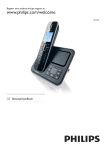
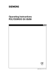
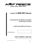
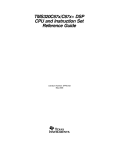
KEE HING CHEUNG KEE CO., LTD.
DLFTZ CHANG HING KEE
INT’L INDUSTRY & TRADE CO., LTD.
Medical Diagnostic X-Ray System
CONTENT
KHCK 001
KHCK 002
KHCK 003
KHCK 004
KHCK 005
KHCK 006
KHCK 007
KHCK 008
KHCK 009
KHCK 010
KHCK 011
KHCK 012
KHCK 013
KHCK 014
30ma X-Ray Mobile Machine............................................................................. 2
50ma X-Ray Mobile Machine............................................................................. 2
100ma X-Ray Mobile Machine........................................................................... 3
The medical diagnostic X-ray unit 200mA....................................................... 4
The medical diagnostic X-ray unit 300mA....................................................... 8
The medical diagnostic X-ray unit 500mA .................................................. 12
The High frequency radiography system 50kw-CE* ..................................... 17
The High frequency remote-controlled X-ray unit (80kW)............................ 19
The remote controlled diagnostic X-ray unit (500mA)-CE* .......................... 26
The Mobile series C-arm X-ray Unit ............................................................... 28
The high frequency mobile X-ray unit............................................................ 29
The new advanced digital radiography system(DR system) ....................... 30
Low-Intensity Portable X-Ray Imaging Scope............................................... 34
Portable & high frequency medical diagnosis X-ray machine..................... 39
KHCK1
KHCK2
For people use ........................................................................................ 39
For animal special use ............................................................................. 43
Automatic film processor-CE.................................................................................................. 45
KHCK 015
Dental x-ray unit............................................................................................... 47
KHCK1- Micro focus Dental x-ray unit........................................................................ 47
KHCK2-HIGH FREQUENCY DENTAL X-RAY UNIT .......................................................... 50
TABLE-TOP DARKROOM........................................................................................... 52
KHCK 016
ORAL PANORAMIC X-RAY UNIT..................................................................... 52
KHCK 017
Mammography machine.................................................................................. 54
Set of accessories for X-RAY room use ................................................................................. 56
X-RAY film illuminator ............................................................................................. 56
X-RAY RADIOGRAPHIC CASSETTE............................................................................. 57
Medical Computer Interphone System ....................................................................... 58
X-ray Film Hanger .................................................................................................. 58
X-ray Film Safety Light ........................................................................................... 59
Chart Light Box...................................................................................................... 59
Intensifying Screen ................................................................................................ 60
Container For Film Development............................................................................... 60
Radiation protection series..................................................................................................... 61
1
KHCK 001
30ma X-Ray Mobile Machine
Brief introduction
z
Used in ward and emergency treatment room for fluoroscopy and radiography
z
Combined X-RAY generator
z
Single focus, full ware rectification
z
It is precise, safe, reliable and flexile
z
Remote control device (control range≥5 m) is used
KHCK 002
50ma X-Ray Mobile Machine
Brief introduction
z
This X-ray apparatus features fullwave rectification and modular X-ray tube head.
z
The frame of the unit, of cantilever construction, is compact in design easy of movement.
z
The machine can be used for radiography in hospital wards or operating rooms.
Technical indexes
z
Maximum ratings
S
kv
m mA
z
z
z
0.08-1
2-2.5
3.2-4.0
5.0-6.3
30
90
90
90
90
40
90
90
90
/
50
90
80
/
/
Power requirements:
Voltage:180-240V Frequency:50Hz
Internal resistance: ≤1ΩRating:5k VA
Current:Maimum15A for radiography
Time:0.08 to 6.32 steps electronic
Maximum height from X-ray tube focus to floor>1820mm
2
z
z
z
z
z
z
z
Minimum height from X-ray tube focus to floor<550mm
Collimator:Maximum film size at 1000mm
Focal distance: 430×430mm
Maximumermote control distance:6m
X-ray tube focus:2.3×2.3
Unit moving force: <5kg
Net weight of the unit:90kg
KHCK 003
100ma X-Ray Mobile Machine
Brief introduction
z
100Ma X-Ray Mobile Machine
z
Take x-ray photography of the head, extremitiles, chest cavity and other parts of human
z
In the ward or in operation room.
Features
z
Single focus, full-wave rectifier, combined X-ray generator
z
SCM control (easy to maintain and repair)
z
High visual and operational console with LCD monitor
z
Prestore 8 kinds of photography parameters and select, modify, store the parameters under the condition
of choice
z
Power voltage (V), photograph kilovolt (KV) infinitely variable control
z
Premier high voltage with high power SCR zero control circuit
z
Function of load chains, exposure time control, auto alarm, preheat filament, subassemblies temperature
control, and so on Cantilever structure makes the small volume and easily moving
Technical indexes
z
Power supply:
Voltage: 180-240 V
Frequency: 50HZ
3
Internal resistivity <1.0Ω
Current 35A instant
Rating ≥7k VA
z
Photography:
Voltage: 50-100 kV
current 16ma. 32ma 63ma 100ma
time 0.08s ~6.3s
X-ray tube focus 4.3mm X4.3mm
z
Maximum remote control distance: 7m
z
Maximum height of focal spot from floor>1880 mm
z
Minimum height of focal spot from floor <520 mm
z
Columns turning angle:±45
z
Collimator: maximum film size at 650mm focal distance: 350 mm X 350 mm
z
The net weight of the unit: 150kg gross Weight: 240kg
z
Shipping volume: 150cm X 100cm X 150cm
Focus of X-ray tube
Photograph Current (mA)
Max photograph voltage (kvp)
Max allowable exposure time
16
90
6.3
32
90
6.3
63
85
4.2
100
80
3.0
Big focus
KHCK 004
The medical diagnostic X-ray unit 200mA
This unit can make fluoroscopy, routine radiography, spot film radiography, bucky
radiography and linear tomography.
4
Configuration:
Features:
z
z
z
z
z
z
z
·One-table, one-tube.
·Single-phase, full wave, high voltage rectification.
·Protective devices for X-ray tube capacity, anode -starting, and
exposure timing.
·External-balanced diagnostic table, motor driven table tilting with
spring-oscillating bucky under the table.
·Spot film device with self-centering holder for cassettes of different
sizes, electromagnetic locks.
·Ceiling-floor rail type tube stand with safety device.
·Linear tomographic attachment can be selected.
Technical specification:
Item
Fluoroscopy
Radiography
Contents
0.5 ~ 5mA, 45 ~ 90kV adjustable continuously
Tube voltage
50 ~ 100kV adjustable continuously
Tube current
30 ~ 200mA adjustable continuously
Exposure time
0.05 ~ 6s 23 steps
Table tilting
+90°~0°~ -5°motor-driven
Longitudinal
560mm
Movable
Transverse
80mm
range
Spotfilm
Compressing
300mm
device
Locking
Electro-magnetic lock
Film
127×178mm(5"×7")
254×305mm(10"×12")
size
203×254mm(8"×10")
280×356mm(11"×14")
Density
N28
Buck
Focus
f0100
(oscillating type)
Ratio
r8
Tabletop-film
60mm
distance
Collimator
Single-leaf manual
5
Tube stand
X-ray tube
assembly
Power
supply
Adjustable range between ceiling and floor:2820 ~ 3185mm
Sliding in longitudinal (along ceiling and floor): 1800mm
Transversely movement (along rotation arm):
660mm (from focus to center of stand)
Vertical movement (along stand):
295 ~ 1800 (from focus to the ground)
Model
XD51-20.40/100-T1A
Focus
1×1mm/ 2×2mm
Speed
2800r/min
Voltage
Single phase 380V/ 220V AC
Capacity
≥13kVA
Frequency
50Hz
Inner resistance
380V: 1Ω 220V: 0.3Ω
Item (optional)
Contents
Tomographic attachment
Exposure angle
30°~ 60°adjustable
Layer height
20 ~ 200mm manual
This unit can make fluoroscopy, routine radiography, spot film radiography, bucky
radiography and linear tomography.
Configuration:
Features:
z
z
z
z
z
z
z
z
·Two-table, two-tube.
·Single-phase, full wave, high voltage rectification.
·Protective devices for X-ray tube capacity, anode-starting
and exposure timing.
·External-balanced diagnostic table, motor driven table tilting.
·Spot film device with self-centering holder for cassettes of
different sizes, electromagnetic locks.
·Radiographic table with spring oscillating bucky.
6
z
z
·Ceiling-floor rail type tube stand with safety device.
·Linear tomographic attachment can be selected.
Technical specifications:
Item
Fluoroscopy
Radiography
Diagnostic
Tube stand
Radiographic
table
X-ray tube
assembly
Power
supply
Contents
0.5 ~ 5mA, 45 ~ 110kV adjustable continuously
Tube voltage
50 ~ 125kV adjustable continuously
Tube current
30 ~ 200mA 5 steps
Exposure time
0.01 ~ 5s 23 steps
Table tilting
+90°~0°~ -5°motor-driven
560mm
Spotfilm Movable Longitudinal
range
device
Transverse
80mm
Compressing
300mm
Locking Electro-magnetic lock
127×178mm(5"×7")
254×305mm(10"×12")
Film
size
203×254mm(8"×10")
280×356mm(11"×14")
Adjustable range between ceiling and floor:2820 ~ 3185mm
Sliding in longitudinal (along ceiling and floor): 1800mm
Transversely movement (along rotation arm):
700mm (from focus to center of stand)
Vertical movement (along stand):
60~ 1800 (from focus to the ground)
Tabletop
Longitudinal: Manual
Lock mode
Electron agnetic lock
Buck
Density: N28
(oscillating type)
Focus: f0100
Ratio: r8
Model
XD51-20.40/125
Focus
1×1mm/ 2×2mm
Speed
2800r/min
Voltage
Single phase 380V/ 220V AC
Frequency
50Hz
Inner resistance
380V: 0.9Ω
220V: 0.3Ω
7
KHCK 005
The medical diagnostic X-ray unit 300mA
This unit can make fluoroscopy, routine radiography, spot film radiography, bucky
radiography and linear tomography.
Configuration:
Features:
z
z
z
z
z
z
z
·Two-table, two-tube.
·Single-phase, full wave, high voltage rectification.
·SCR exposure switch.
·Protective devices for X-ray tube capacity, anode starting and exposure timing.
·Inner-balanced diagnostic table, motor driven table tilting and tabletop moving at head end electrically.
·Spot filming parameters preset on spot film device.
·Spot film device with self-centering holder for cassettes of different sizes, high density stationary grid and
electromagnetic locks.
8
z
z
z
z
z
·The tabletop of radiographic table can be moved in longitudinal and transverse direction with oscillating
bucky and electromagnetic locks.
·Ceiling-floor rail type tube stand, telescopic rotating tube arm, electromagnetic locks and double-cable
safety device.
·Motor-driven multi-leaf collimator for under-table tube and manual multi-leaf collimator with light beam
indicator for over-table tube.
·Motor-driven linear tomographic attachment can be selected, layer height adjusted electrically.
·Interface for TV system and vertical bucky stand.
Technical specifications:
Item
Fluoroscop
y
Contents
0.5~5mA, 45~110kV adjustable continuously
Radiograph Tube voltage
y
Tube current
Diagnostic
table
25~300mA adjustable continuously
Exposure time
0.02~5s adjustable by 23 steps
Table tilting
+90°~0°~- 15° motor-driven
Tabletop moving
500mm at head end
Spotfilm
device
Moveable
range
Locking
Bucky
Tube stand
assembly
50~125kV adjustable continuously
longitudina
1720mm
transverse
125mm
compressing
300mm
Electromagnetic lock
Stationary
Density: N40
Focus: f070
Ratio: r8
Film
127×178mm(5"×7")
254×305mm(10"×12")
size
203×254mm(8"×10")
280×356mm(11"×14")
Multi-leaf motor-driven
Collimator
Adjustable range between ceiling and floor:2650 ~ 3200mm
Sliding in longitudinal (along ceiling and floor): 2000mm
Sliding in transverse (along rotation arm):
900~1140mm (from focus to center of stand)
9
Up-down movement (along stand):
500~ 1800 (from focus to the ground)
Item
Contents
Radiographic
table
Moving range of tabletop: Longitudinal: 1200mm Transverse: 200mm
X-ray tube
assembly
Power
supply
Lock mode
Electromagnetic lock
Density:
N40
Bucky
Focus: f070
(Oscillating type)
Ratio: r8
Tabletop-film distance
≤80mm
Model
XD51-20.40/125
Focus
1mm/,2mm
Speed
2800r/min
Voltage
Single phase 380V/ 220V AC
Capacity
≥20kVA
Frequency
50Hz
Inner
380V: 0.75Ω
220V: 0.25Ω
resistance
Item (optional)
Contents
Tomographic
attachment
Exposure angle
10° 30° 50°three steps
Layer height
0 ~ 220mm motor-driven
Vertical bucky stand
Height adjustable
Bucky tiltable
KHCK 005
The medical diagnostic X-ray unit 300mA
·Computer controlled.
·Automatically distinguish line power and frequency, and auto. compensate power fluctuation.
·Assure output precision, improve image quality, simplify operating procession.
Specification:
Improve performance and stability
z
Simply operate and steadily output by adopted computerized control.
Self-Diagnosis and Error Code Display
z
The system has 23 error codes. Once failure occurs, error code is
automatically displayed on the control panel for maintenance.
Auto. Tube Capacity Protection
z
z
Besides 3-point control (kW, mA, Sec), exposure parameters can be set
with kV, mAs.
According to the maximum capacity of tube and minimum exposure time
can fix the upper limit of exposure mA and protect the tube.
Preset Exposure Parameters
z
The system provides 90 programs, which can be modified, corrected and stored according to exposure
10
position and patient size.
Technique Specification
Standard Composition:
Microprocessor controlled H.T. Generator FS202-3
Diagnostic Table ZC15X-2
Radiographic table SC3-2
X-ray tube stand ZZ-3
Specification:
Power supply: 380V AC±10% single phase
Frequency: 50Hz
Inner resistance: 0.3Ω
Setting range:
Radiography: Tube voltage: 50~125kV
Tube current: 30~300mA
Exposure time: 0.02~5s 23 steps
Fluoroscopy: Tube voltage: 40~110kV
Tube current: 0.5~5mA
Option:
TV system
Vertical bucky stand
11
KHCK 006
The medical diagnostic X-ray unit
500mA
Features:
z
One-table, one-tube. Single-phase, full-wave, high voltage rectification. High voltage switch on/off with
SCR. Protectors for X-ray capacity, anode starting and exposure time. Integrated radiographic table
without ceiling-floor railway, makes installation easier. The table top can move longitudinally and
transversely with electromagnetic Locks and foot switch controls. X-ray tube assembly can rotate freely
and stay at stated position. It can combine with vertical bucky stand
Application:
z
The product provided many kinds of selections for customers, It is radiography whichcan make routine
radiography, bucky radiography, linear tomography and tilting radiography.
12
13
14
15
16
KHCK 007
The High frequency radiography system 50kw-CE*
Features:
z
z
z
z
z
z
z
z
z
z
z
z
z
z
z
z
z
·Large LCD displays all radiographic modes.
·Friendly user interface and easy menu design.
·Normal radiography, bucky radiography, tilting radiography,
Ion chamber AEC and APR mode can be selected.
·Best radiographic mode ensures high quality image.
·Automatic heat capacity protection.
·The preset parameter can be modified and saved under
APR mode.
·New-designed table, displays SID and rotation angle
automatically.
·Multi-functional radiography meets clinical requirements.
·High frequency design (25kHz).
·Short exposure time (min 2ms).
·High kV (150kVp), high mA (630mA), small size focus
(1mm).
·AEC+APR function.
·Multi-style console installation.
·System upgrade.
Console on the table/wall or with pedestal
·Modularized design.
·Extensive configuration.
17
Standard Configuration:
z
z
z
z
z
z
H.T. generator
H.T. cable 75kV/16m
X-ray tube assembly Siemens RAY-12
Radiographic table (including Ion Chamber)
X-ray tube stand
Collimator
Option:
z
Vertical bucky stand LS-3
Technical Specifications:
Generator
Transformation
mode
kVp
mA
Time
mAs
Max. capacity
Power supply
X-ray tube
assembly
H.T. cable
H.F. inverter type
25kHz
1kVp/step
By step
By step
By step
40~150kVp±(5%+1kVp)
25~630mA±(5%+1mA)
2ms~5s±(1%+0.1ms)
1~500mAs±(5%+mAs)
50kW
<0.3Ω
3-phase 380V
50kVA
Inner-resistance
Voltage
Capacity
Siemens RAY-12
75kV/16m
Bucky
stand
Radiographic table
Table top
Collimator
Tube vertical movement 520~1760mm
Tube longitudinal movement along table body 2380mm
Tube assembly rotation around lateral arm-120°~+120°can be
fixed at any position
Tube stand rotation 0°~±90°, fixed position at 90°
Length 2100mm
Width 800mm
Height 690mm
Longitudinal movement 900mm
Transverse movement 250mm
Manual
18
KHCK 008
The High frequency remote-controlled X-ray unit
(80kW)
High frequency technology IGBT module
High Precision X-ray Control System
z
z
z
z
z
·Computer Controlled System.
·High frequency technology, constant DC
output, improved image quality, reduced
patient radiation exposure.
·IPM IGBT ensuring the reliability.
·Real-time, closed-loop control system ensures
the high precision of tube kV and mA, and
good repeatability.
·Precise exposure time as short as 1 ms.
AEC
z
z
z
z
z
z
z
z
z
z
z
z
·Multi-function AEC.
·Manual choosing of kVp, mA, and preparation
time,
system auto control exposure time, 3 steps
exposure doze.
·Many radiography kVp and fluoro kVp
formulas can
be chosen.
·Full-automatic mode.
·ABC system protect the patient and physician
from unnecessary X-ray radiation.
·kVp compensation function of auto-exposure
voltage
compensation system can acquire high clarity
images of any patient size.
·PMT/Ion chambers AEC.
·Bucky stand/RAD table can be integrated ion
chamber.
·The exposure dose can be automatic
controlled according to different imaging
Pulse Fluoro Function
·Pulse fluoro can meet the requirements
of digital image system. Lower dose,
higher image quality.
APR Function
·600 memory programs can be chosen and
the conditions can be spot defined. It is adjustable to
meet the different physician habits.
Safety-protection Function
z
z
19
·Computed-controlled X-ray tube permits
the load and heat capacity management.
media.
KHCK 008 The High frequency remote-controlled X-ray unit (80kW)
DIGITAL TOUCH-SCREEN DISPLAY
PERFECT WORK AND FUNCTIONS
SIMPLE AND EASY OPERATION
HIGHLY INCREASED EFFICIENCY
Digital Touch-screen Display
z
z
z
z
·LCD with special system support system makes big improvement in clinical operation. The operation
control system is developed to touch-screen.
·Radiography parameters can be set by pressing the keys or inputting directly on the screen.
·Patient manage functions, ID of the patient, diagnostic position, physician, time, date and notes etc
information can be stored.
·The X-ray tube heat-capacity diagram is showed directly.
Main Technical Specifications
z
·Generator power: 80kW.
20
z
z
z
z
·Rad kV:40~150kV, continuous adjustable, 111 steps.
·Rad mA: 10~800mA(1000mA can be chosen).
·Rad time: 1ms~10s.
·Fluoro kV: 40~120kV, continuous adjustable.
High Definition Image
z
z
z
z
·Integrated sensor system with high-assimilation rate.
·17" or 21" high resolution medical X-screen.
·Small focal spot, high power, high speed X-tube digitalcontrol stator.
·High resolution multi-field image intensifier.
Economic Table Simple configuration
Easy interface Superb Performance
Special attention is given to the interface of 80kW generator and diagnostic tables to ensure overall system
performance. The combined advantages of the generator and the table result in versatility and multiple
applications, including gastro-intestinal examination, endoscope bronchography, spinal cord catheterization,
angiography and interventional procedures.
Large range of spotfilm device
movement allows fluoroscopic
and radiographic exposures
of various positions without
patient movement.
21
Spotfilm device movement
72cm in longitudinal
Diagnostic table can tilt smoothly
from +90°~-25°.It is comfortable
for patient.
Table tilting at -25°
Table tilting at +90°
ZC25SY-2 diagnostic table can
do chest radiography , focal
distance 1.5m (option)
Integrative linear tomography
table SC5-1
Integrative
SC4-4
radiography
table
EXCELLENT OFFER LATEST DEVELOPED CASTELESS, CASSETTE REMOTE CONTROLLED TABLES
90°/-45°Table Tilting
Large negative tilting degree allows radiography
of stomach lining.
Fast Radiography Cassetteless Table
The film supply system and receiving system can
automatically carry out serial radiography without
cassette. 3 film sizes (10" ×12", 11"×14", 14"×
14") can be selected. The supply magazine can be
loaded up to 50 films. Patient name and number
can be printed on the film.
22
Convenient to Select Fluoro and Rad
Position
The radiographic system of such serial
spotfilm device can move more than 600cm
towards head-end without table top movement
to realize a complete radiography for gastro
-intestinal examination. Convenient for
endoscope bronchography examination
during fluoroscopy.
Swing Radiography
The X-ray tube assembly can swing from +30°~-30°,
satisfying special exposure requirement.
Table Top Movement Suitable for
Multipurpose Examination
Variable Focal Distance
The distance from X-ray tube focus to film can be
adjusted at 110cm, 120m, 130cm.
Such serial diagnostic table top can move
1,500mm longitudinally, suitable for multi-purpose
examination and diagnosis.
The digital image processing system
z
z
z
z
z
12bit Acquisition, Image Quality Is Similar to Film
CCD camera, 12 bit acquisition.
4096 grey level.
CCD camera (1k×1k), Gamma correction.
Image processing, display and saving.
Digital Spot-film Acquisition Speed
z
z
1024×1024×12 bit single frame.
1024×1024×12 bit 0.5-15fps.
Digital Fluoroscopy Acquisition Speed
23
z
z
z
z
1024×1024×12 bit single frame.
1024×1024×12 bit 1-30fps.
Support various width pulse fluoro.
128 frames fluoro loop.
Real-time Image Processing
z
z
z
z
z
z
z
z
z
z
z
4 level real-time edge enhancement, 7 level
Notation, 4/16frames on one screen.
Zooming and mapping.
Real-time review.
DICOM 3.0 interface (optional).
CD-R (optional).
Laser camera interface.
Road map display.
DSA (optional).
Mask re-setting, background holding.
(in DSA function optional).
On-site Service Function
z
z
z
The system with auto on-site service software.
The software can perform self-diagnosis on the whole
system or a single board or a single replaceable unit.
OPERATING ROOM DRAWING:
24
Layout: mm
Configuration:
25
KHCK 009
The remote controlled diagnostic X-ray unit
(500mA)-CE*
Microprocessor control More functions more flexibility
Anatomical exposure position selection
z
Exposure parameters can be set based on patient body size and
exposure positions. The parameters can also be corrected stored.
Safety function
z
z
z
The error self-diagnosis function can determine problems
quickly and accurately. It can execute protection
automatically and display error code for maintenance.
Transverse movement
APR function
z
z
Programmable functions, provides 90 memory programs
for radiography. The parameters can also be set manually.
X-ray tube protection
z
z
z
X-ray tube heat capacity and load capacity are managed
and protected by computer programs, to allow maximum
utilization of the tube capacity and exposure parameters.
Excellent image processing
z
z
z
z
z
z
z
Digital dynamic noise reduction, image positive/negative
reverse, transverse/vertical flip, black spot correction, LIH.
Diagnostic table, motor driven table tilting and
tabletop moving at head end electrically.
Combined with TV system to do the remote operation, can meet
requirements of TV fluoroscopy and spot film.
All electrical movement of spot film device make
“ corss” four division radiography.
26
Longitudinal movement
of spot film device
z
Frequency conversion technology is applied to the table tilting, makes
movement smooth and reduce system noise.
Compressing direction
movement of spot film device
Longitudinal movement of table top
Integrated Radiographic Table
No.-SC4-4
Carbon fiber table top, high
intensity low absorption
Remote Table No.-ZC15XY-1
Rotatable foot support (optional)
X-ray Generator
Microprocessor
controlled 500mA
Remote Table
No.-ZC15XY-1
Integrated Radiographic Table
No. SC4-4
Image Intensifier
9" Ⅰ.Ⅰ.
TV Monitor
14" Monitor
27
X-ray Tube Assembly
H.T. Cable
KHCK 010
The Mobile series C-arm X-ray Unit
Specification
Fluoroscopic Capacity
z
z
z
z
z
Max rated capacity:Tube Current 4mA, Tube Voltage 120kV
Automatic Fluoroscopy:Tube Voltage:40kV~120kV adjust automatically
Tube Current:0.3mA~4mA adjust automatically
Manual Fluoroscopy:Continuous tube Voltage:40kV~120kV; Continuous tube Current:0.3mA~4mA
Pulse Fluoroscopy:Continuous tube Voltage:40kV~120kV; Continuous tube Current:4.1mA~8Ma
Photography Capacity
z
z
z
Max rated capacity:5.0 KW
Tube voltage, mAs : 40kV~120kV
20~100mA
1~180mAs
Plateholder size:200mm×250mm(8″×10″)
or 250mm×300mm(10″×12″)
X-ray Tube
z
z
z
X-ray tube special for High frequency:
Fixed anode x-ray tube with 2 focus:
Large focus: 0.6, small focus: 0.3
Inverter Frequency: 40kHz
Thermal capacity: 150kJ (200kHU)
Video System
z
z
z
z
z
Image Intensifier:Image Intensifier made
by TOSHIBA (9″)
CCD vidicon:Imported CCD Vidicon with
ultra-low luminosity
Monitor:Horizontal 1000 lines and vertical 800 lines,
Bandwidth: 12.5MHz, Image/sec: 25
CCU (central control):Recursive filter: K=8, 7 images storage, image upright, image overturn, positive &
negative image; LIH(last image freeze), and OSD(monitor display)
28
Structure
z
z
z
z
z
z
Direction-wheel:±90°revolution, can change the moving direction of the unit.
Ascending & descending
range of pillar≥400mm
C-Arm:Forward and backward movement: 200mm
Revolution around horizontal axis: ±180°
Revolution around vertical axis:±15
Slip on orbit: 120°(+90°~ -30°)
KHCK 011
The high frequency mobile X-ray unit
Technical Specifications:
z
z
z
z
z
z
z
z
Output: 3.2kW
kV: 40~ 110kV
mA: 10~ 60mA
mAs: 1~ 250mAs
X-ray tube: 1.5mm focal spot
Power supply: 220V, single phase
Arm rotation: ±90 °
Vertical movement: ≥140cm
Features:
z
z
z
z
z
z
Suitable kinds of power supply conditions.
High frequency generator, higher image quality, shorter
exposure time, lower radiation.
Small rotation radius.
Stronger power, any position radiography.
X-ray tube can rotate ±90 in one horizontal plane, improve the patient flow rate.
X-ray tube, smaller focal spot to acquire better image.
Option:
z
remote hand switch.
29
KHCK 012
The new advanced digital radiography system(DR
system)
New advanced full body DR system
Introduction
z
The newest DR technology fulfils the dream of full digital of X-ray image. The excellent image quality
reaches the highest level. The fast-image ability minus diagnosis time in big scale and improves your
work-flow-rate. The requirements of overload clinical work can be reached.
High Quality Images
z
z
High quality image High resolution, high quality images
provide plenty details and diagnosis information.
Instant Imaging
z
z
The image can be displayed in 5 seconds after the
exposure, the film-developing is not needed any more.
Large Dynamic Range
z
z
z
The 14bit acquisition make sure that all information from
skin to the bones can be gained just in one exposure.
The clinical range of X-ray radiography is enlarged.
High MTF DQE
z
z
Image details contracts and SNR are much better than
traditional film; the image quality is much improved.
Wider Exposure Dynamic Range
z
z
The wide exposure dynamic range is the guarantee of
high quality image, which can be reached in low dose.
Digital Image
z
Digital image will not need paper documents any more.
30
z
And the image can be shared by internet very easily.
Easy Operation
z
z
z
The process of exposure, connecting, transmitting,
saving and printing can be automatically controlled,
and you will liberate from the complicated work.
Simple Service
z
z
z
The simple system is cost-effective. Digital applications
improve efficiency and reduce film costs as well as the
time associated with conventional cassette handing.
KHCK 012
The new advanced digital radiography system
Advanced System Design
System
Characters
Outstanding Image Functions
The fully application of
state of art technology
·Combining the state of art
technology of a-Si FPD
detector with special image
processing technology to
fulfill high quality digital
image.
·Open image workstation
makes operation very easy
and have strong processing
functions.
·System design and unit
integration ability guarantee
high quality image.
Multi-function design
·Multi-function in one
unit, according to the
different clinical
requirements, Optima
URS or table can be
chosen, one DR can
finish the test of whole
body.
·One panel with many
body parts, 17"×17" FPD
can fulfill the big part
such as breast test, the
FPD doesn't to be
rotated.
31
Module extendable design
·Based on the standard
composition, it can be
extended according to the
customers' requirements.
·Without HIS, worklist,
diagnosis workstation, laser
printer and CD-RW are
options.
·The fully solution from single
workstation to whole PACS
system.
Complete DICOM support
·The newest version DICOM
3.0(2000 version) and the
special requirements of DR
image is fully supported.
·It's compatible with lower
versions so that it protects
the original investment of
the hospital.
Work station
Software
Functions
Outstanding image
processing functions
·Automatic/Manual ROI
window level & window
width.
·Automatic/Manual electric
cut. Image flip, rotation,
mirror image.
·Continual zoom & roam.
·Image measure, notation &
enhancement.
System safety design
·The system safety
operation is guaranteed
to meet the daily
operation requirements.
·Special data base
design to avoid data lose
by improper operation.
The data safety is
guaranteed.
·At the high frequency of
exposure and
radiography, the system
can work in high
efficiency and stable to
meet the requirements of
high patient passing rate.
Database management
·Patient
information
(patient name, ID, date,
etc.) can be stored and
revised.
·Convenient for enquiry.
32
Complete DICOM 3.0 support
DICOM print support
DICOM archive support
DICOM transmission support
DICOM worklist support
DICOM 3.0 support (2000
edition)
The new advanced digital radiography system
z
z
z
z
Latest patent design.
Single panel, full function.
Can perform radiography to any body position, and oblique radiography.
Full-auto tracking and positioning.
Various System Configuration Meet Different Clinical Requirements
z
z
z
z
z
z
z
z
z
Multi-function Integrated Design
·Patent DR system.
·Bucky stand / radiography table design to meet the full
range requirements from chest to general radiography.
·X-tube and flat panel full automatic tracking functions to
improve the efficiency.
·The big range movement of Ceiling system with table
movement to meet the requirements of whole body
different position radiography.
33
KHCK 013
Low-Intensity Portable X-Ray Imaging Scope
Price:
NAME
Low-Intensity
Portable X-Ray
Imaging Scope
MODEL
KHCK-50A
KHCK-50B
KHCK-75A
KHCK-75B
NOTE
The model A just can be used for
fluoroscopy .The model B can be
used for both in fluoroscopy and
taking the photo (With film-making
function). The size of the photo: 5×7
inch
Note:
z
z
z
The film of the KHCK-75 is clearer than the KHCK-50, but tell the truth, the maintain rate also is
higher than the KHCK-50 MODEL, my suggestion is that you had better choose the KHCK-50
MODEL, they are used better in sudan market.They have been used for 2 years and there are
no machines need to be repaired. The quality is very good. Because the weather in your country
is very high,so all the units which are sold in your market need to be adjusted to meet your
weather needs.
Pls see the base of the machine, our base is made of the round and hardness figue, which is
burliness than the two feet figue.
Our product’s main parts(vacuum porcelain imaging strengthening machine) is made by our
own,so the price and the quality is the best.Most of the Chinese handhold X-RAY machine’s
imaging strengthening machines are bought from my manufactory.
34
Brief Introduction
z
z
z
z
Low-Intensity Portable X-Ray Imaging Scope (the
following in short X-Ray Imaging Scope), a new type of
X-Ray medical instruments in the world, consists of
main engine and power supply. From the appearance,
the main engine adopts the structure of C-Type-Arm,
which amplifies the radius of observation. At the same
time, the materials select ABS engineering plastics,
heightening the insulation capacity.
Because
of
adopting
MCP-X-Ray
imagine
strengthening device, having compact texture, high gain,
high resolution, the scope can produce clear real-time
images without darkroom. The advance technology of widening pulse is used to control electrical
appliance of X-Ray tube, which has many measures to be protected, so it has high working stability and
no noises.
In addition, the scope takes strict precautions, the leak of X-Ray reducing, and then the operators do not
need any protections. This scope applies to orthopedics and pediatrics in the hospitals and to diagnose
IUD for childbearing-age women in time. Especially, it applies to various field rescues in such as industry
materials and apparatus quality inspection, athletes, armies, field working and safety checking, and so on.
The scope belongs toⅠClass B Model Equipment.
Specifications and Conditions
1 Specifications:
z
z
z
z
z
z
z
z
z
z
z
Output image size:
Φ50mm/Φ75mm
Survey thickness:
≤300mm
X-Ray tube voltage: 35~70kv continual adjust
Voltage accuracy:
≤±1%
Tube current:
0.25~0.5mA continual adjust
Current accuracy:
≤±2%
Resolution:
≥40Ip/cm
Ratio :
≥4%
Gray level:
≥10 level
Main engine working frequency:
20KHz
Radiation leakage:
the corresponding National Standard GB9706.12
35
2 Conditions
z
z
Temperature:
Opposite humidity:
5oC~40oC
≤80%
3 Power supply
z
z
z
z
220V±10%
50Hz~60Hz
Power consumption:
80W
Resistance maximum:
0.5
Releasing current machine:
1A
4 Shape size and weight
z
z
z
Length × width × thickness:
Main engine: 3.6Kg
Gross weight: 9Kg
550×420×130
Guard: 0.61 Kg
Working principle
z
X-Ray Imaging Scope is made up of different parts in the pictureⅠ. When put through power supply, turn
on “start” button, and then the scope begins to work. The pulse signal from main engineⅠamplified
36
through efficiency reaches to X-Ray tube anode, while the pulse signal from main engineⅡ amplified
reaches to X-Ray tube wick, which releases X-Ray. Then the corresponding number KV/μA will come out
on the board. This moment the object is laid between X-Ray source and image strengthening machine, on
whose monitor we will see the clear image. In order to work stability, the system adopting the technology
of widening pulse, makes tube current and tube voltage stable, and the X-Ray tube works in good
condition. Due to the function of high voltage starting slowly, the X-Ray tube anode doesn’t have the
phenomenon of high voltage being too high. The main control machine with the mini-slice equipment
working at the frequency of the 20KHz, improves the entire system’s efficiency and clears away the noises,
and it supplies workers for quite environment with a smaller volume. The power supply of perspective
device adopts a switch with high frequency and high efficiency, having overall protections. To ensure the
safety of perspective device, the scope takes different measures, so the device is safe and reliable.
Service Manual of X-ray Detector
1. Detection Methods of General Failures of X-ray Detector:
X-ray detector is a kind of precision instruments and shall be repaired by
professional technicians. When repairing, firstly remove visible failures such as broken
wire, touched wire, key switch and fuse and do not disassemble the instrument and its
power switch.
z
z
z
z
z
z
If the digital display shows voltage and current of X-ray tube, these is x-ray; if the display is black, there is
usually a failure in image intensifier and it should be replaced.
If the digital display works but there are not voltage and current of X-ray tube, the power supply should be
normal and there may be a failure in mainboard or voltage doubler and please have it repaired.
After the instrument is electrified, if the panel shall display but cannot work normally and work indicator
light does not work, you should check: a. if driver inserter is fixed; b. if panel inserter is fixed; c. driving
tube is fixed.
After the instrument is electrified, if the over-temperature indicator light is on, you should check: a. if the
temperature of high-voltage X-ray source is too hot. If it is not too hot, please check if the inserter on base
of high-voltage X-ray source is fixed.
After the instrument is electrified, if the instrument does not work, you should check: a. 24V power supply;
b. if panel displays. If it does not, send it to manufacturer for repair; c. check DC/DC. If it is not normal,
send it to manufacturer for repair.
If intensifier or voltage doubler is broken, send it to manufacturer for repair.
2. Adjustment Method and Process of Image
z
Before turning on the instrument, minimize voltage and current. After turning on the instrument, adjust
voltage and current according to the sizes and thickness of perspective object until the image is clear. The
37
z
z
z
parameters for reference are as follows:
For limbs of adult: adjust current of display panel to 0.5mA and voltage to 65-90KV;
For limbs of child: adjust current of display panel to 0.3-0.5mA and voltage to 45-65KV;
For other objects: adjust them according to conditions.
3. Layout of Inner Components of X-ray Instrument
Image Intensifier
DC/DC Module
Driving
Tube Front Panel
Driving Board
Power Switch
24V Transformer
Voltage Doubler (High-voltage X-ray Source)
Base of High-voltage X-ray Source
Note: the above broken lines are the layout of main inner components of X-ray
instrument.
38
KHCK 014
Portable & high frequency medical diagnosis X-ray
machine
KHCK1
For people use
Advantages:
z
Integrated with high frequency and high voltage generator,
main control system, intelligent computer system, numeric
display and beam limited device, it is designed and
manufactured precisely and compactly, more flexibly and
widely used.
z
Using the technique of inverse converter for HF and
computer-controlled closed-loop adjusting System, stable high
voltage output is generated and high-quality images can be
obtained.
z
Using the buttons of KV and mAs to adjust the photographic
parameters, display them on high-brightness LEDs, interlock
protection conditions is determined by software, which is
convenient for doctors to operate.
z
X-ray Tube is placed in lead cylinder which can effectively shield the gamma leakage and protect
environment and operators’ safety.
z
z
20 radiographic conditions are preset and can be customized according to user requirements.
Photography Time is controlled precisely by computer, host will automatically delay 10 seconds at the end
of photography which can effectively control photography interval, avoid overloading and achieve
automatic protection.
z
z
Hand-controlled and remote-controlled photography function
Using computer to monitor fault sources, the computer will cut all outputs, display fault code and identify
the fault reasons if a fault appears.
z
Suitable for all kinds of cassettes.
Structural performance
z
z
Radiography can be launched after X-ray apparatus is suspended on the frame.
The fuselage of X-ray apparatus is suspended on the frame, can rotate left and right at larger than or
39
equal to ±90°, X-ray tube head can rotate at 90°which is easy to project in different angles.
z
z
The arm can move up and down, has Servo-balance and lock-up device.
The fuselage of X-ray apparatus weighs 19Kg, is more convenient to carry. The frame weighs 23 Kg, is
more flexible to move at different places.
z
The frame can be disassembled and folded quickly, then putted into trunk, which can make out-call more
convenient.
Technical Specification (there are three models)
Model 1:
z
z
z
z
z
z
z
z
z
z
z
z
z
z
z
Max. photograph tube voltage: 90kV/22mA
Max. photograph tube current: 50mA
Max. Output: 2.0kW
Exposal time: 0.02s ~ 4.0s
MAS scope: 0.5mAs ~ 160mAs with 46 alternations
Voltage scope: 40~90kV KV with 26 alternations
Main frequency: 30kHz
Max. light field: ≤35CMX35CM @65CM SID
Min. light field: ≤5CMX5CM @100CM SID
Focus size: 2.3mm
Power: 220V ± 10% single phase alternating current with the frequency of 50Hz ± 1Hz
Resistance: ≤ 1.0 Ω
Power capability: ≥ 2.5kVA
Electricity protection type: Class I, Type B
Weight: 19kg
Model 2:
z
z
z
z
z
z
z
z
z
z
z
z
z
Max. photograph tube voltage: 100kV/20mA
Max. photograph tube current: 60mA
Max. Output: 2.4kW
Exposal time: 0.008s ~ 3.33s
MAS scope: 0.5mAs ~ 160mAs with 46 alternations
Voltage scope: 40~100kV KV with 31 alternations
Main frequency: 30kHz
Max. light field: ≤35CMX35CM @65CM SID
Min. light field: ≤5CMX5CM @100CM SID
Focus size: 1.5mm
Power: 220V ± 10% single phase alternating current with the frequency of 50Hz ± 1Hz
Resistance: ≤ 1.0 Ω
Power capability: ≥ 3.5kVA
40
z
z
Electricity protection type: Class I, Type B
Weight: 19kg
Model 3:
z
z
z
z
z
z
z
z
z
z
z
z
z
z
z
Max. photograph tube voltage: 100kV/30mA
Max. photograph tube current: 70mA
Max. Output: 3.2kW
Exposal time: 0.008s ~ 3.33s
MAS scope: 0.5mAs ~ 200mAs with 48 alternations
Voltage scope: 40~100kV KV with 31 alternations
Main frequency: 30kHz
Max. light field: ≤35CMX35CM @65CM SID
Min. light field: ≤5CMX5CM @100CM SID
Focus size: 1.5mm
Power: 220V ± 10% single phase alternating current with the frequency of 50Hz ± 1Hz
Resistance: ≤ 1.0 Ω
Power capability: ≥ 4.5kVA
Electricity protection type: Class I, Type B
Weight: 19kg
The Container for X-ray machine, can be option as you like
Wall bucky for chest x-rays
Our bucky tray can move as your like.
Introduction:
The standing photographing shelf is suitable for x-ray photographing of abdomen
41
and above .it is made up of high quality rolled steel and the exterior surface is decorated with static spray
plastic ,which is elegant and durable .the photographing shelf is supported by a slanted-pole ,which can
ensure there is no shake while taking photograph and therefore make the film more clear.
Specification:
Fram:1140*750*310mm,23KG
Main unit:490*400*400mm,19KG
Solution of the problem of imaging fat people
The usage of Anti-scatter grid can settle
your problem successfully.
z
z
The grids have superior performance and offer less
absorption, which can greatly reduce the scattered radiator
producing clearer and diagnostically superior. It is widely
used in the Chest X-rays.
It is produced by Korea producer.
Specification:
z
z
Size: 15*18mm
focus ranges:1.8M
Another advantages of our X-ray machine:
1 The ordinary table or bed is OK for supine films, no special x-ray table needed.
42
KHCK2
For animal special use
Advantages:
z
z
z
z
z
z
z
z
z
Integrated with high frequency and high voltage generator, main
control system, intelligent computer system, numeric display and
beam limited device, it is designed and manufactured precisely
and compactly, more flexibly and widely used.
Using the technique of inverse converter for HF and
computer-controlled closed-loop adjusting System, stable high
voltage output is generated and high-quality images can be
obtained.
Using the buttons of KV and mAs to adjust the photographic
parameters, display them on high-brightness LEDs, interlock
protection conditions is determined by software, which is
convenient for doctors to operate.
X-ray Tube is placed in lead cylinder which can effectively shield
the gamma leakage and protect environment and operators’
safety.
20 radiographic conditions are preset and can be customized
according to user requirements.
Photography Time is controlled precisely by computer, host will automatically delay 10 seconds at the end
of photography which can effectively control photography interval, avoid overloading and achieve
automatic protection.
Hand-controlled and remote-controlled photography function
Using computer to monitor fault sources, the computer will cut all outputs, display fault code and identify
the fault reasons if a fault appears.
Suitable for all kinds of cassettes.
Structural performance:
z
z
z
z
Radiography can be launched after X-ray apparatus is
suspended on the frame.
The fuselage of X-ray apparatus is suspended on the frame,
can rotate left and right at larger than or equal to ±90°, X-ray
tube head can rotate at 90°which is easy to project in
different angles.
The arm can move up and down, has Servo-balance and
lock-up device.
The fuselage of X-ray apparatus weighs 19Kg, is more
convenient to carry. The frame weighs 23 Kg, Can be quickly moved to injured animals and launch
43
z
z
diagnosis and treatment.
The frame can be disassembled and folded quickly, then putted into trunk, which can make out-call more
convenient.
Providing great convenience for animal diagnosis and treatment.Cooperating with beds, it is more suitable
for animal hospitals.
Main specifications and parameters:
z
z
z
z
z
z
z
z
z
z
z
z
z
z
z
Max. photograph tube voltage: 90kV/22mA
Max. photograph tube current: 50mA
Max. Output: 2.0kW
Exposal time: 0.02s ~ 4.0s
MAS scope: 0.5mAs ~ 160mAs with 46 alternations
Voltage scope: 40~90kV KV with 26 alternations
Main frequency: 30kHz
Max. light field: ≤35CMX35CM @65CM SID
Min. light field: ≤5CMX5CM @100CM SID
Focus size: 2.3mm
Power: 220V ± 10% single phase alternating current with the
frequency of 50Hz ± 1Hz
Resistance: ≤ 1.0 Ω
Power capability: ≥ 2.5kVA
Electricity protection type: Class I, Type B
Weight: 19kg
Use:
z
animal hospital, equestrian club, animal quarantine, animal and veterinary, animal laboratory
44
Automatic film processor-CE
Product detail:
z
This desk-top small film processor with less expensive price is newly launched in market by CHINA
Imaging. It possesses only 0.36 ㎡ ground area with front and rear two output modes for selection.
Excellent performance plus affordable price, the machine can be widely applied in middle and small
hospital for mode of several machines in one hospital, several machines in one operation room and
processing mode instead of traditional manual operation. Its
sale price is much lower than similar product and its fix and
develop chemical volume is only 4.5L. It is specially designed
as a hi-efficiency, energy-saving medical film processing device
for user who has some volume of film processing everyday.
Max processing width is 14 inch and fastest whole process
needs only 90 sec.
Film Processor Features
Small Body
z
Ground occupied area is only 0.36 ㎡, which is probably suitable for any small dark room. You don't need
to worry about space issue.
High Auto Control
z
This product is an auto film processing device controlled by a micro computer. To easily set processing
data, adjusting of temperature and speed is set by two groups of potentiometer on control board.
Functions like chemical replenish, ready for film processing, solution low-level indicator and every type of
alarm, can be automatically realized through LED indicators of control panel. It is easy to understand and
operate. Indicators on control panel would let you know all working status at any time. When chemical
temperature reaches set value, what you do is only to put a film in and machine will automatically enter
working status and make the processing.
Save Chemicals
z
Develop and fix volume of this product is only 5 liters which may largely reduce new chemicals
replacement cycle and keep chemical effectiveness at a good status. To reduce air oxidation to chemicals,
a special designed anti-oxidation cover is placed in chemical tank. It largely reduces air oxidation to
chemical solution and meanwhile it reduces oxidized chemical’s pollution to air in working room.
Hi-efficient Dryer
z
There is a hot-air duct-mode dryer system of hi-efficiency and squeeze roller of hi-performance in this
45
product dryer section. This guarantees the high performance of dyer required by mass processing of film
in a high-temperature environment.
High Working Reliability
z
This machine has state of the art and matured design in film-transport rack’s structure, roller transport,
electronic control, hot-air dryer, chemical replenish and circulation. It has a long life period for usage. Film
jam, scratches and film not dried will not occur.
Film Output Selectable
z
Two modes are designed for this product’s film output way, they are:
Front film output mode: in this mode the entire machine is placed in dark room for processing
operation. Film will be out on the top of machine after processing.
Rear film output mode: what you do is only to adjust a group of guide in dryer exit to the status of
rear-film-output, and make an 8×48cm hole in the dark room and separation wall, mount the film
collector below the hole. Film output is collected in light room.
Fast Processing Speed
z
This product is designed according to hi-speed requirement on film processing. Fastest whole processing
time of machine can be at 90 sec, suitable for any urgent diagnosis or hospital that may require fast
processing.
Cost Effective
z
This product market sale price is lower. It applies for organization that processes 30~150 pieces everyday.
Chemical, power and water consumption of this machine is low. Usage cost of whole machine is 25%~
40% lower than similar processor, so it applies for the one with less film processing volume, one hospital
with several machines, and the hospital or department that one working room is configured with several
machines.
Easy Maintenance
z
Easy maintenance is well considered during this product’s design. Besides features of small machine
body, light weight, easy to move, the machine’s racks from develop to dryer can be all easily released out
and washed by water for maintenance. Electrical control part is enclosed and mounted below entrance
feeding box. By opening external cover of this E-box, you may do the maintenance easily on every
electronic component inside.
Energy Saving
z
Max power consumption of this product whole machine is only 2.2 KW, water consumption per minute is
only 2 liters which will be automatically opened when processing film. If no film processing, water flow and
dryer heating system, etc, will automatically enter standby and energy saving mode.
46
KHCK 015
Dental x-ray unit
KHCK1- Micro focus Dental x-ray unit
Type 1 mobile type
Product detail:
z
The product is designed successfully by our company recently with international
advanced level, which is a new type of dental x-ray unit. It must be used in
coordination with dental film with intensifying screen. The product is suitable for
filming oral cavity x-ray in every hospital, oral cavity outpatient service, private clinic
and clinical or teaching usage of relative medical college and research institution.
I. Features of the unit:
z
z
Filming in coordination with dental film with intensifying screen by making use of 1.5mA X-ray output
capacity. Compared with 7mA-10mA dental unit in the world, X-ray radiation dosage has reduced by
92%-98% to protect health of patients.
The scattering or leaking x-ray dosage in the air where one meter away from radius of shining center is
47
z
z
z
z
z
lower than 4% of defined limited value to protect health of doctors.
The clearest image can be acquired by making use of microfocus x-ray tube.
Using microcomputer controller, which is very convenient and accurate. Only according to relative
standard picture, the best exposure control can be reached.
It has infrared remote control and linear control and the direction and anti-interference of infrared remote
control are strong.
It has flexible and firm suspending arm to adjust ball tube freely to any angle or direction and balance in
any position.
It is equipped with firm and steady seat and back, and connected with machine to make patients feel
comfortable and safe. The height of seat can be adjusted according to different height of patients.
Technical parameter
z
z
z
z
z
z
z
z
z
z
Power requirement: 50Hz, 220V±10%
Peak point voltage: 65 kvp
Peak point current: 1.5mA/3mA/7mA/10/mA
Maximum power: ≤ 300w
Exposure time: 0.1-0.2s
Radiation diameter: 6cm
Focus spot: 0.6 mm×0.8 mm
X-ray tube longitudinal displacement:≥ 600mm
Vertical travel of rocker: ≥ 400mm
Vertical travel of seat: ≥ 200mm
Type 2 Wall mounted type
Product detail:
z
The product is designed successfully by our company recently with international
advanced level, which is a new type of dental x-ray unit. It must be used in
coordination with dental film with intensifying screen. The product is suitable for
filming oral cavity x-ray in every hospital, oral cavity outpatient service, private clinic
and clinical or teaching usage of relative medical college and research institution.
Features of the unit:
z
z
z
Filming in coordination with dental film with intensifying screen by making use of 1.5mA X-ray output
capacity. Compared with 7mA-10mA dental unit in the world, X-ray radiation dosage has reduced by
92%-98% to protect health of patients.
The scattering or leaking x-ray dosage in the air where one meter away from radius of shining center is
lower than 4% of defined limited value to protect health of doctors.
The clearest image can be acquired by making use of microfocus x-ray tube.
48
z
z
z
Using microcomputer controller, which is very convenient and accurate. Only according to relative
standard picture, the best exposure control can be reached.
It has infrared remote control and linear control and the direction and anti-interference of infrared remote
control are strong.
It has flexible and firm suspending arm to adjust ball tube freely to any angle or direction and balance in
any position.
Technical parameter
z
z
z
z
z
z
z
z
z
Power requirement: 50Hz, 220V±10%
Peak point voltage: 65 kvp
Peak point current: 1.5mA/3mA/7mA/10/mA
Maximum power: ≤ 300w
Exposure time: 0.1-0.2s
Radiation diameter: 6cm
Focus spot: 0.6 mm×0.8 mm
X-ray tube longitudinal displacement:≥ 1600mm
Vertical travel of rocker: ≥ 600mm
Type 3 fixed type
Product detail
z
The product is designed successfully by our company recently with
international advanced level, which is a new type of dental x-ray
unit. It must be used in coordination with dental film with
intensifying screen. The product is suitable for filming oral cavity
x-ray in every hospital, oral cavity outpatient service, private clinic
and clinical or teaching usage of relative medical college and
research institution.
Features of the unit:
z
z
z
z
z
Filming in coordination with dental film with intensifying screen by
making use of 1.5mA X-ray output capacity. Compared with
7mA-10mA dental unit in the world, X-ray radiation dosage has
reduced by 92%-98% to protect health of patients.
The scattering or leaking x-ray dosage in the air where one meter away from radius of shining center is
lower than 4% of defined limited value to protect health of doctors.
The clearest image can be acquired by making use of microfocus x-ray tube.
Using microcomputer controller, which is very convenient and accurate. Only according to relative
standard picture, the best exposure control can be reached.
It has infrared remote control and linear control and the direction and anti-interference of infrared remote
49
z
z
control are strong.
It has flexible and firm suspending arm to adjust ball tube freely to any angle or direction and balance in
any position.
It is equipped with firm and steady seat and back, and connected with machine to make patients feel
comfortable and safe. The height of seat can be adjusted according to different height of patients.
Technical parameter
z
z
z
z
z
z
z
z
z
z
Power requirement: 50Hz, 220V±10%
Peak point voltage: 70 kvp
Peak point current: 3mA/7mA/10/mA
Maximum power: ≥ 400w
Exposure time: 0.1-0.2s
Radiation diameter: 6cm
Focus spot: 0.6 mm×0.8 mm
X-ray tube longitudinal displacement:≥ 800mm
Vertical travel of rocker: ≥ 400mm
Vertical travel of seat: ≥ 200mm
z
Rotatory range of tube: 0-360℃
KHCK2-HIGH FREQUENCY DENTAL X-RAY UNIT
Type 1 fixed type
Product detail
The product is designed successfully by our company with international
advanced level, which is a new type of dental x-ray unit with few scatter &
leakage X-ray to image high definition film. This machine is suitable for all
levels hospital, oral cavity clinic, individual clinic and relevant medical
universities and colleges, the R&D institution.
Features of the unit:
The unit uses high frequency DC, lower grenz ray output. The advanced
unit is more flexible to operate.
Take a photograph adopting 3mA x-ray output and matching with our dental
film (packing with intensifying screen), reduce the radiation dosage than the
domestic and international machine, and protect the patient’s and doctor’s health.
The scatter & leakage x-ray is accord with GBZ130-2002 standard “Medical X-ray health diagnosis protection
standard” , to protect doctor’s health.
Micro size spot to ensure high image resolution. With the filament pre-heat, prolong the usage of X-ray tube.
50
It is convenient and accurate to adopt microcomputer controller, can achieve the best exposure condition, only
press the corresponding standard figure.
With good direction and jam resistance, the unit can be controlled by infrared remote control and wire control.
.It has two flexible and firm suspending arm to adjust ball tube freely to any angle or direction and balance in
any position. It also increases the length of arm.
It is equipped with firm and steady stand back and connected with machine to make patients feel comfortable
and safe. The chair can be adjusted according to different height of patients.
Technical parameter
Power requirement: 50Hz, 220V±10%
Peak point voltage: 70 kvp
Peak point current: 3mA/7mA/10/mA (DC)
X-ray frequency: 30 kHz
Maximum power: ≥ 400w
Exposure time: 0.1-0.2s
Radiation diameter: 6cm
Focus spot: 0.6 mm×0.8 mm
Horizon range of tube:≥ 2000mm
Vertical travel of tube: ≥ 1500mm
Type 2 wall mounted type
Product detail
z
The product is designed successfully by our company with
international advanced level, which is a new type of dental x-ray
unit with few scatter & leakage X-ray to image high definition film.
This machine is suitable for all levels hospital, oral cavity clinic,
individual clinic and relevant medical universities and colleges, the
R&D institution.
Features of the unit:
z
z
z
z
z
The unit uses high frequency DC, lower grenz ray output. The
advanced unit is more flexible to operate.
Take a photograph adopting 3mA x-ray output and matching with
our dental film (packing with intensifying screen), reduce the radiation dosage than the domestic and
international machine, and protect the patient’s and doctor’s health.
The scatter & leakage x-ray is accord with GBZ130-2002 standard “Medical X-ray health diagnosis
protection standard” , to protect doctor’s health.
Micro size spot to ensure high image resolution. With the filament pre-heat, prolong the usage of X-ray
tube.
It is convenient and accurate to adopt microcomputer controller, can achieve the best exposure condition,
51
z
z
only press the corresponding standard figure.
With good direction and jam resistance, the unit can be controlled by infrared remote control and wire
control.
It has two flexible and firm suspending arm to adjust ball tube freely to any angle or direction and balance
in any position. It also increases the length of arm.
Technical parameter
z
z
z
z
z
z
z
z
z
Power requirement: 50Hz, 220V±10%
Peak point voltage: 70 kvp
Peak point current: 3mA/7mA/10/mA (DC)
X-ray frequency: 30 kHz
Maximum power: ≥ 400w
Exposure time: 0.1-0.2s
Radiation diameter: 6cm
Focus spot: 0.6 mm×0.8 mm
Horizon range of tube:≥ 2000mm
z
Vertical travel of tube: ≥ 1500mm
TABLE-TOP DARKROOM
Unique design
z
z
z
z
Develop in lightroom: extricate you from darkroom
Simple and convenient: save time and gain benefit
Small volume: operate on the table, save space
Tooth image: it is clearer than any method of developing
KHCK 016
ORAL PANORAMIC X-RAY UNIT
Product characteristic
z
z
z
z
Revolutionary SCSRS imaging plates,the most abundant procedure can
provide more clinical diagnosis message and jaw image information.
The advanced scanning makes image reducing and definition reach a high
level.
Scan localization is more accurate,less image interference ,to get more
precise,truer clinical diagnosis message.
Operating environment is more sciencetific,rational,intuitive,simple and
52
z
convenient.
The appearance and structural design are succinct and smmoth.,it is a perfect combination of the
technology and design.
Technical parameter
z
z
z
z
z
z
z
z
z
z
z
z
z
z
z
z
z
z
input voltage
220V±10%
50HZ±1HZ
fuse≤1Ω
input power
Instantaneous loading:2500W
Standby mode:110W 0.5A
fuse
15A (ф6×30)
anode
voltage
auto
60KV~88KV ≤10%
auto set
manual
60kV~88kV ≤10%
anode current panoramic 12mA
±20%
jaw 12mA ±20%
focus spot size
0.5mm×0.5mm
zoom in 1.20-1.30
total filtering
2.5mm Al
exposure time
panoramic 14s
film size 150×300mm
x-ray
cassette
meisheng
150mm×300mm
x-ray intensifying screen
MEISHENG MSL-40
lift range of tube head 853 mm ~1643mm
853 mm ~1643mm
lifting device manual electromagnetism locked
net weight
160kg
53
KHCK 017
Mammography machine
Brief introduction
z
z
z
The mammography System is mainly used to acquire X-ray images of
human breasts for clinical diagnosis. It is a film-based mammography
system and proves to be an effective solution for breast diseases.
The mammography system consists of the main body unit and the control
console (see the picture of the system), including the key components of
dual-focus X-ray tube, collimator, rotating anode starter and protection
system, high-frequency and high voltage generator (frequency: 40KHz),
tube filament heating system, breast compression device, supporting arm
and other mechanical and electric devices.
X-ray tube is one of the major components which decide the breast image
quality. MCR-6000 system uses a high power rotating anode of dual-focus
tube with Mo target and Be window (bias dual-focus: 0.1mm/0.3mm) to ensure the clear images. The
carbon-based bucky features high transmission rate, and high efficiency in blocking the scattered X-ray,
which is also helpful for improving image quality. The automatic exposure control system is highly
sensitive and quickly reactive, so the exposure is automatically adjusted according to the factures of age
and thickness etc.
High standard and high resolution image
54
z
z
z
Spatial resolution: 20Lp/mm
Density resolution: 1.19mm
Min. Calcification resolution: 130um.
Features of the unit
z
The breast compression device is automatically controlled with three speeds. The ascending speed is
high to reduce operation time. The descending speed is 1/2 of the ascending speed to prepare for
pressing the breast. After getting in touch with the breast, it will automatically decrease its speed and
impose pressure. This way the patient feels more comfortable. When the pressure reaches the preset
level, it will automatically stop and the actual breast thickness and pressure will be displayed. If the
pressing conditions are not proper, the operator may change it on the way until desired conditions are
available.
Specification
z
z
z
z
z
z
z
Power 220VAC,30A
High voltage generator
High frequency constant voltage:50kHz
Voltage range:20---45KVp,1KV step
Max.Current:100mA
Max. Output power:5kW
Exposure condition control: Manual/Auto
X-ray tube
z
z
z
z
Dual focus:0.1mm/0.3mm
Anode heat capacity:150KHU
Anode type: Rotating Anode
Anode material: Mo
C--arm
z
z
z
z
z
z
z
Rotating angle: +180/-160
SID:65cm Fixed
Anti-scatter grid: Carbon based fine plate
Cassette size:18×24cm OR 24×30
Dimensions Gantry:1810mm×880mm×880mm
Console:960mm×480mm×500mm
C-arm vertical movement range:600mm
Overall weight:200Kg
Compressor
z
z
Operating mode: Auto(motor)
Compression force/method:Max.18Kg/Auto
Intelligent, flexible
55
z
z
fast pressure release
Compression force display: LED
Compressor paddle movement range:280mm
Note:
This equipment should be equipped with film processor to make the printing.
The film processor which you purchased from us is also suitable for this equipment.
Set of accessories for X-RAY room use
X-RAY film illuminator
Brief introduction
z
z
z
z
z
z
z
z
CE certified
The most advanced film clamping device assure easy film insertion, reliable film clamping and easy film
take-off.
Frame is made of highly strong aluminum alloy material which is highly rigid and not deformable.
Screen is made of imported organic glass panel which has no color fading for long-time use.
Electronic ballast has advantages of power saving, no flickering and easy switch-on under low voltage.
Frame and container body is connected by hinge, which assure easy replacement and maintenance of
fluorescent tube.
It is patent product of computer light-regulated illuminator, which has novel cosmetics and stable property.
Customization service is offered.
Model 1
Super-thin LCD
Standard type
Light-regulated
Model 2
Super-thin type
super-thin type
super-thin type computer light-regulated
56
Model 3
Luxury type
Luxury type
Luxuty type common light-regulated
Equipped with work table
Luxuty type computer light-regulated
equipped with mobile supporting trestle
Model 4
Wall built-in type
Wall built-in type Computer light-regulated
ordinary type
X-RAY RADIOGRAPHIC CASSETTE
Brief introduction
z
z
z
CE certified , Manufacture according to
international standard.
The XH series of x-ray radiographic cassette
include three types;XH -P is the standard type;
XH–PW has ID window;and XH-PM is a patent
product with label installation, the three all have
different specification in either metric or imperial
system.
The series are made of specified aluminium alloy
frame, specified engineering plastics and
57
z
high-stamina aluminium plate, they look good in shape and are convenient and durabe in use. You
wouldn’t worry about damage even in case it falls down from one meter high.
With a inner layer made out of high-stretch material, the X-ray film stick to the screen tightly and equally,
the photograph is clear and rich in levels. The inner layer is covered by cloth, so it is very convenient to
change the screen is and without damage to the inner layer.
Medical Computer Interphone System
Brief introduction
z
z
z
z
z
Medical computer interphone system has
adopted the most advanced microcomputer
special chip. The main line communication
preparation and ease lead to unending calling.
Its multiple functions and performances have
achieved the creation of this line in our country.
It is an easy way for the nurse to communicate
with the patient and know about the situation
in the sickroom so that the efficiency and the
grade of the hospital can be raised.
X-ray Film Hanger
58
X-ray Film Safety Light
Brief introduction
z
z
z
z
z
z
z
z
z
z
The X-ray film safety light has special light-filtered plate,
it is suitable for use in a darkroom during the
processing of
blue-sense and green-sense X-ray films.
The light is installed with a 15W common light
bulb, of which
the working voltage is 220V. The circuit is
finely-designed and
there are two types for your favor, one is the
standard type and
the other is light-adjustable. It has the
characteristics of
having a wide scope of application, and being
easy to install
and superb in design. It is an ideal illuminating device for a
darkroom during film processing.
Chart Light Box
Brief introduction
z
z
z
This product is suitable for the checking of myopia and hyperopia in school, hospital and other relevant
departments.
The front frame of the visual chart is made of
high-strength aluminum alloy, matched with
highly-clear board of the visual signs. The box
body is wholly made of high-quality steel plate
and is of static spray coating. It is superb in
design and durable to use. The visual signs are
in great contrast and highly clear, ensuring an
exact checking result.
A fluorescent lamp is the light source. The light
box is equipped with an electronic ballast. It can
function normarlly under low voltage, with
steady illumination and no flash. Maintenance is simple. By opening up the front frame, circuit checking
59
z
and fluorescent lamp changing can be done right away.
The light box has two specifications of testing distances: 2.5m and 5.0m.
Intensifying Screen
Container For Film Development
60
Radiation protection series
61