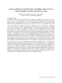
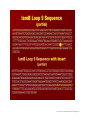
1
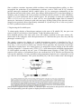
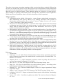
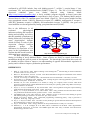
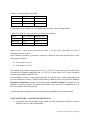
The disease is most severe in broiler breeders and brown egg layers. White layer lines are less
affected. The mortality is usually negligible. Birds infected vertically can remain asymptomatic
until the bird become sexually mature. The eggs from infected birds are mostly shell-less, thinshelled, discoloured or misshapen and have a poor internal quality. It take mostly 4-10 weeks till the
birds re-start to produce normal.
At necropsy there is no specific lesion, but a slight atrophy of ovary and oviduct can be observed.
Histopathological changes can be seen in the oviduct and uterus (shell gland). There may be severe
degeneration and desquamation of the epithelial cells, atrophy of the uterine glands, and infiltration
of heterophils, lymphocytes, and plasmacytes. Intranuclear inclusion bodies may be found in the
epithelial cells of the uterus, isthmus, and vaginal gland region (Adair and Smyth, 2008, Smyth and
McNulty, 2008).
Diagnosis of Adenovirus infections
Basically the diagnosis of poultry diseases based on case history, clinical signs and post-mortem
examination as important steps toward disease diagnosis, but it should not be the final step. In most
cases clinical signs and lesions of many diseases are similar and laboratory tests are required to
identify the specific cause (Fig. 1).
The laboratory diagnosis can be applied to direct detection as well as for isolation and identification
of the causative agent or indirectly to detect antibodies (Hafez and Hess, 1999).
The diagnosis of adenovirus infection in poultry is in most cases based on histological
investigations and detection of intranuclear inclusion bodies in hepatocytes or on detection of the
antigen or virus particles using Immunofluorescence test or electron microscopy. In the last years
several molecular biological tools such as PCR, Real-time PCR and REA were developed allow the
detection of the Virus – DNA as well as the further identification and typing of adenoviruses (Erny
et al., 1991; Raue and Hess, 1998, Hess et al., 1999, Raue et al., 1999, Hess, 2000, Lüschow et al.,
2007).
However the isolation of the aviadenoviruses using chicken embryo liver (CEL) cell culture and
chicken embryo fibroblast cell culture with further identification and determination of the
pathogenicity seems to be very important, since the pathogenicity of the isolates within the same
serotype can be widely differ. Cross neutralization tests are necessary to serotype the isolated virus
and to determine a new serotype.
EDSV can be isolated in embryonated duck or goose eggs, and in cell cultures. Susceptible cell
lines include duck and chick embryo liver, duck kidney, and fibroblast cells. The virus may be
isolated directly from the reproductive tract of affected hens. Alternatively, abnormal eggs may be
fed to naive hens; virus isolation is attempted from the shell gland of these hens when they produce
abnormal eggs.
The most common serologic test is the immunodiffusion test that detects the group specific antigen.
This test is not sensitive enough. A group specific ELISA and IIF tests are more sensitive. The
serum neutralization test has been used to detect serotype-specific antibody but is labour intensive
and expensive. In general the interpretation of serologic tests is difficult because antibodies against
AAVs can be found in both healthy and diseased birds.
2a reunion AECACEM Querétaro 2009 Pág. 85