1
G.
H.
I.
J.
K.
L.
M.
N.
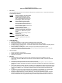
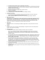
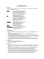
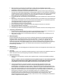
The patient should be advised of the use and limitations of this device.
Pre-operative planning and evaluation, surgical approaches and technique, and familiarity with the implant,
including its instrumentation and limitations are necessary components in achieving a good surgical result.
This device must never be reused.
This device must never be re-sterilized.
Appropriate instrumentation should be used to implant this device.
Never use a bent Drill Bit.
The Drill Bit is not intended for use as an implant.
Loading the Drill Bit in an off axis manner may result in breakage or bending.
5.
Packaging and Labeling:
A. Do not use this product if the packaging or labeling has been damaged, shows signs of exposure to
moisture or extreme temperature or has been altered in any way.
B. Please contact Parcus Medical Customer Service to report any package damage or alterations.
6.
Material Specifications:
The MiTi Suture Anchor is supplied with or without needles attached to the high-strength, braided, polyethylene
polyblend sutures. Contrasting suture colors are used in suture anchors supplied with multiple sutures. The
anchor material is Ti-6Al-4V ELI (ASTM F136). The Drill Bit is manufactured from a Stainless Steel Alloy. The
materials used in the manufacture of this device that are intended to be placed inside the body are radioopaque and can, therefore, be detected with conventional X-Ray or fluoroscopy.
7.
Sterilization:
The MiTi Suture Anchor, driver assembly and Drill Bit is supplied sterile. These products must never be resterilized.
8.
Storage:
Products must be stored in the original unopened package in a dry place and must not be used beyond the
expiration date indicated on the package.
9.
Directions for Use
A. Identify bone of sufficient quantity and quality into which the anchor is to be placed.
Note: In cases where bone quality is suspect and a MiTi Suture Anchor does not provide the
desired fixation, the larger diameter V-LoX Titanium or PEEK CF Suture Anchor may provide an
alternative solution.
B.
C.
D.
E.
F.
Place the Drill Bit into the chuck of a purpose designed drill or wire driver. Taking care to create an entry to
the desired placement site as close to perpendicular as possible, use the appropriate MiTi Suture Anchor
Drill Bit to prepare a hole in the bone. Insert the Drill Bit into the bone to the depth indicated by the laser
line on the distal end of the Drill Bit.
Place the MiTi Suture Anchor into the prepared site, as described above, and turn the driver handle
clockwise until the circumferential laser etched line on the distal end of the driver shaft is flush with the
surrounding bone. The orientation of the sutures passing through the implant may be visualized as they
are exterior to the shaft. Placing the sutures in a plane perpendicular to the targeted tissue offers the best
orientation to allow the suture to slide through the anchor.
One or two strands of polyethylene polyblend high strength suture either with or without needles can be
found attached to the anchor.
As applicable, remove needles from keeper and pass the sutures through the targeted tissue in the user
preferred manner.
Complete the tissue repair by tying knots of the surgeon preferred configuration and cutting the suture tails
above the knots.