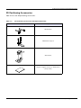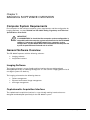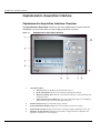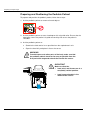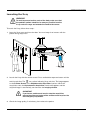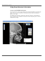
1
CS 9300C User Guide Notice Congratulations on your purchase of the CS 9300C. Thank you for your confidence in our products and we will do all in our power to ensure your complete satisfaction. The User Guide for the CS 9300C includes information on the cephalometric features. For the panoramic or the 3D features, see the CS 9300 (SM748) User Guide. We recommend that you thoroughly familiarize yourself with this Guide in order to make the most effective use of your system. The CS 9300C is intended to produce two-dimensional and three-dimensional digital x-ray images of the dento-maxillo-facial and ENT (Ear, Nose and Throat) areas to be used at the direction of healthcare professionals as diagnostic support for pediatric and adult patients. In addition, the CS 9300C is a system that is also intended to produce cephalometric images. This includes imaging the hand and wrist to obtain the carpus image for growth and maturity assessment. WARNING: We recommend that you consult the “Safety, Regulatory and the Technical Specification User Guide” before using the CS 9300C. WARNING: Do not use cone beam imaging for routine or screening examinations. Consider using other diagnostic tools. You must justify that the imaging method that you use to examine each patient demonstrates that the benefit outweights the risks. The information contained in this Guide may be subject to modification without notice, justification or notification to the persons concerned. No part of this Guide may be reproduced without the express permission of Carestream Health, Inc. The US Federal law restricts this device to sale by or on the order of a physician. This document is originally written in English. Manual Name: CS 9300C User Guide Part Number: SM750 Revision Number: 01 Print Date: 2011-09 The brand names and logos reproduced in this Guide are copyright. The CS 9300C, complies with Directive 93/42/EEC relating to medical equipment. 0086 Manufacturer Care stream Hea lth, Inc. 150 Verona Street Roche ster, NY 1 4 60 8, USA Authorized Representative in the European Community EC REP TROPHY 4, Rue F. Pelloutier, Croissy-Beaubourg 77435 Marne la Vallée Cedex 2, France Contents 1—About This Guide Conventions in this Guide . . . . . . . . . . . . . . . . . . . . . . . . . . . . . . . . . . . . . . . . . . . . . . . . . . . . . . . . . . . . . . . . . . . . . . . 1–1 2—CS 9300C OVERVIEW General Overview . . . . . . . . . . . . . . . . . . . . . . . . . . . . . . . . . . . . . . . . . . . . . . . . . . . . . . . . . . . . . . . . . . . . . . . . . . . . . 2–1 Mobile Components . . . . . . . . . . . . . . . . . . . . . . . . . . . . . . . . . . . . . . . . . . . . . . . . . . . . . . . . . . . . . . . . . . . . . . . . 2–2 General Functional Components . . . . . . . . . . . . . . . . . . . . . . . . . . . . . . . . . . . . . . . . . . . . . . . . . . . . . . . . . . . . . . 2–3 Digital Sensor Locations. . . . . . . . . . . . . . . . . . . . . . . . . . . . . . . . . . . . . . . . . . . . . . . . . . . . . . . . . . . . . . . . . . . . . 2–4 Laser Locations. . . . . . . . . . . . . . . . . . . . . . . . . . . . . . . . . . . . . . . . . . . . . . . . . . . . . . . . . . . . . . . . . . . . . . . . . . . . 2–5 Control Panel Overview . . . . . . . . . . . . . . . . . . . . . . . . . . . . . . . . . . . . . . . . . . . . . . . . . . . . . . . . . . . . . . . . . . . . . . . . 2–6 X-Ray Remote Control Overview. . . . . . . . . . . . . . . . . . . . . . . . . . . . . . . . . . . . . . . . . . . . . . . . . . . . . . . . . . . . . . . . . 2–7 Positioning Accessories and Replacement Parts. . . . . . . . . . . . . . . . . . . . . . . . . . . . . . . . . . . . . . . . . . . . . . . . . . . . . . 2–8 Panoramic Positioning Accessories . . . . . . . . . . . . . . . . . . . . . . . . . . . . . . . . . . . . . . . . . . . . . . . . . . . . . . . . . . . . 2–8 3D Positioning Accessories . . . . . . . . . . . . . . . . . . . . . . . . . . . . . . . . . . . . . . . . . . . . . . . . . . . . . . . . . . . . . . . . . . 2–9 3—IMAGING SOFTWARE OVERVIEW Computer System Requirements. . . . . . . . . . . . . . . . . . . . . . . . . . . . . . . . . . . . . . . . . . . . . . . . . . . . . . . . . . . . . . . . . . 3–1 General Software Overview . . . . . . . . . . . . . . . . . . . . . . . . . . . . . . . . . . . . . . . . . . . . . . . . . . . . . . . . . . . . . . . . . . . . . 3–1 Imaging Software . . . . . . . . . . . . . . . . . . . . . . . . . . . . . . . . . . . . . . . . . . . . . . . . . . . . . . . . . . . . . . . . . . . . . . . . . . 3–1 Cephalometric Acquisition Interface . . . . . . . . . . . . . . . . . . . . . . . . . . . . . . . . . . . . . . . . . . . . . . . . . . . . . . . . . . . 3–1 Cephalometric Acquisition Interface . . . . . . . . . . . . . . . . . . . . . . . . . . . . . . . . . . . . . . . . . . . . . . . . . . . . . . . . . . . . . . 3–2 Cephalometric Acquisition Interface Overview . . . . . . . . . . . . . . . . . . . . . . . . . . . . . . . . . . . . . . . . . . . . . . . . . . . 3–2 Cephalometric Program Pane. . . . . . . . . . . . . . . . . . . . . . . . . . . . . . . . . . . . . . . . . . . . . . . . . . . . . . . . . . . . . . 3–4 Cephalometric Patient Pane . . . . . . . . . . . . . . . . . . . . . . . . . . . . . . . . . . . . . . . . . . . . . . . . . . . . . . . . . . . . . . . 3–5 Cephalometric Parameter Pane . . . . . . . . . . . . . . . . . . . . . . . . . . . . . . . . . . . . . . . . . . . . . . . . . . . . . . . . . . . . 3–6 4—GETTING STARTED Switching On the Unit. . . . . . . . . . . . . . . . . . . . . . . . . . . . . . . . . . . . . . . . . . . . . . . . . . . . . . . . . . . . . . . . . . . . . . . . . . 4–1 Starting the Imaging Software. . . . . . . . . . . . . . . . . . . . . . . . . . . . . . . . . . . . . . . . . . . . . . . . . . . . . . . . . . . . . . . . . . . . 4–2 Starting the KODAK Dental Imaging Software . . . . . . . . . . . . . . . . . . . . . . . . . . . . . . . . . . . . . . . . . . . . . . . . . . . 4–2 Creating a Patient Record. . . . . . . . . . . . . . . . . . . . . . . . . . . . . . . . . . . . . . . . . . . . . . . . . . . . . . . . . . . . . . . . . 4–2 Accessing the Cephalometric Acquisition Interface . . . . . . . . . . . . . . . . . . . . . . . . . . . . . . . . . . . . . . . . . . . . 4–3 Starting the CS Imaging Software . . . . . . . . . . . . . . . . . . . . . . . . . . . . . . . . . . . . . . . . . . . . . . . . . . . . . . . . . . . . . 4–4 Creating a Patient Card . . . . . . . . . . . . . . . . . . . . . . . . . . . . . . . . . . . . . . . . . . . . . . . . . . . . . . . . . . . . . . . . . . 4–4 Accessing the Acquisition Interface. . . . . . . . . . . . . . . . . . . . . . . . . . . . . . . . . . . . . . . . . . . . . . . . . . . . . . . . . 4–5 5—ACQUIRING CEPHALOMETRIC IMAGES Acquiring a Lateral Image. . . . . . . . . . . . . . . . . . . . . . . . . . . . . . . . . . . . . . . . . . . . . . . . . . . . . . . . . . . . . . . . . . . . . . . 5–1 Preparing the Unit and Setting the Acquisition Parameters for the Adult Patient . . . . . . . . . . . . . . . . . . . . . . . . . 5–1 Preparing the Unit and Setting the Acquisition Parameters for the Pediatric Patient . . . . . . . . . . . . . . . . . . . . . .5–2 Preparing and Positioning the Adult Patient. . . . . . . . . . . . . . . . . . . . . . . . . . . . . . . . . . . . . . . . . . . . . . . . . . . . . . 5–3 Preparing and Positioning the Pediatric Patient . . . . . . . . . . . . . . . . . . . . . . . . . . . . . . . . . . . . . . . . . . . . . . . . . . . 5–5 Acquiring a Frontal AP or PA Image . . . . . . . . . . . . . . . . . . . . . . . . . . . . . . . . . . . . . . . . . . . . . . . . . . . . . . . . . . . . . . 5–8 Preparing the Unit and Setting the Acquisition Parameters for the Adult Patient . . . . . . . . . . . . . . . . . . . . . . . . . 5–8 CS 9300C User Guide (SM750)_Ed 01 iii Contents Preparing the Unit and Setting the Acquisition Parameters for the Pediatric Patient. . . . . . . . . . . . . . . . . . . . . . .5–9 Preparing and Positioning the Adult Patient . . . . . . . . . . . . . . . . . . . . . . . . . . . . . . . . . . . . . . . . . . . . . . . . . . . . .5–10 Preparing and Positioning the Pediatric Patient . . . . . . . . . . . . . . . . . . . . . . . . . . . . . . . . . . . . . . . . . . . . . . . . . .5–12 Acquiring an Oblique Image . . . . . . . . . . . . . . . . . . . . . . . . . . . . . . . . . . . . . . . . . . . . . . . . . . . . . . . . . . . . . . . . . . . .5–15 Preparing the Unit and Setting the Acquisition Parameters for the Adult Patient . . . . . . . . . . . . . . . . . . . . . . . .5–15 Preparing the Unit and Setting the Acquisition Parameters for the Pediatric Patient. . . . . . . . . . . . . . . . . . . . . .5–16 Preparing and Positioning the Adult Patient . . . . . . . . . . . . . . . . . . . . . . . . . . . . . . . . . . . . . . . . . . . . . . . . . . . . .5–17 Preparing and Positioning the Pediatric Patient . . . . . . . . . . . . . . . . . . . . . . . . . . . . . . . . . . . . . . . . . . . . . . . . . .5–18 Acquiring a Submento-Vertex Image . . . . . . . . . . . . . . . . . . . . . . . . . . . . . . . . . . . . . . . . . . . . . . . . . . . . . . . . . . . . .5–20 Preparing the Unit and Setting the Acquisition Parameters for the Adult Patient . . . . . . . . . . . . . . . . . . . . . . . .5–20 Preparing the Unit and Setting the Acquisition Parameters for the Pediatric Patient. . . . . . . . . . . . . . . . . . . . . .5–21 Preparing and Positioning the Adult Patient . . . . . . . . . . . . . . . . . . . . . . . . . . . . . . . . . . . . . . . . . . . . . . . . . . . . .5–22 Preparing and Positioning the Pediatric Patient . . . . . . . . . . . . . . . . . . . . . . . . . . . . . . . . . . . . . . . . . . . . . . . . . .5–23 Acquiring a Carpus Image . . . . . . . . . . . . . . . . . . . . . . . . . . . . . . . . . . . . . . . . . . . . . . . . . . . . . . . . . . . . . . . . . . . . . .5–27 Preparing the Unit and Setting the Acquisition Parameters for the Pediatric Patient. . . . . . . . . . . . . . . . . . . . . .5–27 Preparing and Positioning the Pediatric Patient . . . . . . . . . . . . . . . . . . . . . . . . . . . . . . . . . . . . . . . . . . . . . . . . . .5–28 X-Ray Dose Emission Information . . . . . . . . . . . . . . . . . . . . . . . . . . . . . . . . . . . . . . . . . . . . . . . . . . . . . . . . . . . . . . .5–30 6—MAINTENANCE Daily . . . . . . . . . . . . . . . . . . . . . . . . . . . . . . . . . . . . . . . . . . . . . . . . . . . . . . . . . . . . . . . . . . . . . . . . . . . . . . . . . . . . . . .6–1 Cleaning the CS 9300C unit . . . . . . . . . . . . . . . . . . . . . . . . . . . . . . . . . . . . . . . . . . . . . . . . . . . . . . . . . . . . . . . . . .6–1 Cleaning and disinfecting the Accessories . . . . . . . . . . . . . . . . . . . . . . . . . . . . . . . . . . . . . . . . . . . . . . . . . . . . . . .6–1 Cleaning and disinfecting the accessories that have contact with the mucous membranes . . . . . . . . . . . . . . .6–1 Cleaning and disinfecting the ear cones . . . . . . . . . . . . . . . . . . . . . . . . . . . . . . . . . . . . . . . . . . . . . . . . . . . . . .6–2 Cleaning and disinfecting the components that have skin contact . . . . . . . . . . . . . . . . . . . . . . . . . . . . . . . . . .6–2 Monthly . . . . . . . . . . . . . . . . . . . . . . . . . . . . . . . . . . . . . . . . . . . . . . . . . . . . . . . . . . . . . . . . . . . . . . . . . . . . . . . . . . . . .6–3 Annually . . . . . . . . . . . . . . . . . . . . . . . . . . . . . . . . . . . . . . . . . . . . . . . . . . . . . . . . . . . . . . . . . . . . . . . . . . . . . . . . . . . .6–3 7—TROUBLESHOOTING Quick Troubleshooting . . . . . . . . . . . . . . . . . . . . . . . . . . . . . . . . . . . . . . . . . . . . . . . . . . . . . . . . . . . . . . . . . . . . . . . . .7–1 iv Chapter 1 About This Guide Conventions in this Guide The following special messages emphasize information or indicate potential risk to personnel or equipment: WARNING Warns you to avoid injury to yourself or others by following the safety instructions precisely. CAUTION Alerts you to a condition that might cause serious damage. IMPORTANT Alerts you to a condition that might cause problems. NOTE Emphasizes important information. TIP Provides extra information and hints. CS 9300C User Guide (SM750)_Ed 01 1–1 Conventions in this Guide 1–2 About This Guide Chapter 2 CS 9300C OVERVIEW General Overview The CS 9300C is composed of the following functional components: The unit head that contains all the electronic control The rotative arm The fixed arm with a control panel The panoramic and 3D digital sensor The X-ray source assembly The X-ray remote control The chin rest base The panoramic chin rest and bite block The 3D bite block and support The 3D head rest The temple supports The hand grips The cephalostat arm The cephalostat head The head clamps and ear cones The nasion support The acquisition software (see “Imaging Software Overview”) The following figures illustrate the general overview of the CS 9300C system. CS 9300C User Guide (SM750)_Ed 01 2–1 General Overview Mobile Components Figure 2-1 illustrates the up and down movement of the CS 9300C mobile component and the 360° rotation of the rotative arm. Figure 2–1 2–2 CS 9300C OVERVIEW CS 9300C Mobile Components General Overview General Functional Components Figure 2-2 illustrates the general functional components of the CS 9300C. Figure 2–2 CS 9300C Functional Components 5b 9 11 1 5 12 7 6 8 14 5a 13 2 5 15 4 3 10 1 ON/OFF button 7 Sensor 2 Unit fixed arm 8 X-ray source assembly 3 Control panel 9 Unit rotative arm 4 Hand Grips 10 X-Ray remote control 5 Chin rest base 11 Cephalostat arm 5a Panoramic chin rest 12 Cephalostat head 5b 3D chin rest and bite block 13 Head clamps and ear cones 6 Temple supports 14 Nasion support 15 PC hosting the imaging and the acquisition software CS 9300C User Guide (SM750)_Ed 01 2–3 General Overview Digital Sensor Locations Figure 2-3 illustrates the locations of the following digital sensors: *Panoramic and 3D digital sensor Cephalometric digital sensor panoramic, 3D and cephalometric digital sensors of the CS 9300C. Figure 2–3 2–4 CS 9300C OVERVIEW CS 9300C System Digital Sensor Locations General Overview Laser Locations Figure 2-4 illustrates the location of the lasers of the CS 9300C. Figure 2–4 CS 9300C System Laser Positions 2 3 B 4 5 A 1 B A 3 5 4 1 2 2 1 1 3D central positioning laser beam 2 Mid-sagittal positioning laser beam 3 Horizontal positioning laser beam 4 3D Field of View (FoV) positioning laser beam 5 Cephalometric Frankfurt positioning laser beam CS 9300C User Guide (SM750)_Ed 01 2–5 Control Panel Overview Control Panel Overview The control panel is an alphanumeric, digital soft touch console. It allows the operator to control certain unit functions. It also displays the operating parameters and error messages. Figure 2–5 Unit Control Panel 8 kV mA 1 2 3 S mem 9 2–6 CS 9300C OVERVIEW 10 11 4 5 6 1 Height Adjustment button: Adjusts the height of the unit to the height of the patient. 2 3D Head Adjustment button: Adjusts the patient head to the X-ray beams. 3 3D Adjustment button (active in Technical Mode only): Adjusts the unit rotative arm movements. 4 Laser Beam button: Activates the beams to correctly position the patient. 5 3D Position Verification button: Positions the unit rotative arm at the selected or memorized 3D position. 6 Reset button: Resets the unit arm to the initial position to enable the patient to enter and exit the Unit. 7 3D Memorization button (active in Technical Mode only): Memorizes the 3D current positioning parameter settings that override the default parameters. 8 Display Screen: Displays the current acquisition parameters and the error messages. 9 Ready Indicator LED: Green, indicates the unit is ready for acquisition. 10 X-Ray Emission LED: Yellow, indicates the X-rays are being emitted. 11 System Status LED: Red, indicates the error alerts. 7 X-Ray Remote Control Overview X-Ray Remote Control Overview The x- ray remote control enables you to launch a radiological image acquisition via the exposure button from outside the X-ray room. You must press and hold the exposure button until the end of acquisition. WARNING Premature release of the exposure button will interrupt the acquisition. Figure 2–6 X-Ray Remote Control 1 1 Exposure button: launches image acquisition. CS 9300C User Guide (SM750)_Ed 01 2–7 Positioning Accessories and Replacement Parts Positioning Accessories and Replacement Parts The following accessories are used when positioning a patient. They are delivered with the CS 9300. Panoramic Positioning Accessories Table 2-1 lists the panoramic positioning accessories. Table 2–1 Panoramic Positioning Accessories and Replacement Parts Accessory Description Panoramic chin rest TMJ x2 Sinus TMJ x4 nose rest Panoramic standard bite block Bite block for edentulous patients Single use sheaths for bite blocks (500 pcs box) 2–8 CS 9300C OVERVIEW Positioning Accessories and Replacement Parts 3D Positioning Accessories Table 2-2 lists the 3D positioning accessories. Table 2–2 3D Positioning Accessories and Replacement Parts Accessory Description 3D bite block 3D bite block support 3D head rest Single use hygienic sleeves for 3D bite block (100 pcs box) CS 9300C User Guide (SM750)_Ed 01 2–9 Positioning Accessories and Replacement Parts 2–10 CS 9300C OVERVIEW Chapter 3 IMAGING SOFTWARE OVERVIEW Computer System Requirements For information on the minimum computer system requirements and the configuration for Imaging Software, see the CS 9300 and CS 9300C Safety, Regulatory and Technical Specifications User Guide. IMPORTANT It is MANDATORY to check that the computer system configuration is compatible with the computer system requirements for the CS 9300C software. If necessary you MUST update your computer system configuration. The CS 9300C MUST be connected to the computer via a point-to-point Ethernet link and not via a LAN. General Software Overview The CS 9300C operates with the following software: Imaging software Acquisition interface Imaging Software The imaging software is a user-friendly working interface that was designed and developed specifically for radiological diagnosis. It is the common imaging platform for all our digital systems for dentistry. The imaging software has the following features: Patient management Extraoral and intraoral image management 3D image management Cephalometric Acquisition Interface The cephalometric acquisition interface is a user-friendly working interface that was designed and developed specifically for the CS 9300C system. CS 9300C User Guide (SM750)_Ed 01 3–1 Cephalometric Acquisition Interface Cephalometric Acquisition Interface Cephalometric Acquisition Interface Overview The Cephalometric Acquisition interface is the main cephalometric interface with the CS 9300C system that provides you with imaging acquisition functions. Cephalometric Acquisition Interface Figure 3–1 1 Information button: About: Identifies the Software and the Firmware versions. 3–2 Reset of the Values: Resets to the manufacturing parameter settings. Memorize settings: Memorizes the user preference settings for each patient type (kV, mA and seconds). Save current mode as default: Sets the current selected program to be available by default each time you access the Acquisition interface. 2 Preview Screen: Displays the acquired image in real time. 3 Selected Parameter Display: Displays the current acquisition parameter settings. 4 System Status Screen: Displays various alert or warning messages originating from the unit. 5 Generator Cooling indicator: Indicates the automatic cooling time (mm:ss) required for the generator to reach 0 for a new acquisition. IMAGING SOFTWARE OVERVIEW Cephalometric Acquisition Interface 6 Stop button: Stops the unit rotative arm movement 7 Ready Indicator LED Green indicates the unit is ready to start acquisition. Black indicates the unit is not ready to start acquisition. 8 X-Ray Emission indicator: Yellow, indicates the X-ray emission status. 9 Exit button: Closes the Acquisition interface. 10 Selector Button: Selects different acquisition setting options. Click Program to select examination type options. Click Patient to select patient type parameters. Click Parameters to select exposure parameter options. CS 9300C User Guide (SM750)_Ed 01 3–3 Cephalometric Acquisition Interface Cephalometric Program Pane The cephalometric Program pane enables you to choose different radiological exams as well as different acquisition formats. Figure 3–2 1 Cephalometric Program Pane Radiological exam options: Click for a lateral exam. Click or for a frontal exam (AP and PA). Click 2 Click Click for a submento-vertex exam. for a carpus exam. for an oblique exam. Acquisition format options: Select for a 18 x 18 format. Select Select for a 18 x 24 format. Select for a 24 x 24 format. Select format. for a 30 x 30 for a 24 x 30 format. NOTE The above list of exam types are only a sample of exam options of the Program pane. 3–4 IMAGING SOFTWARE OVERVIEW Cephalometric Acquisition Interface Cephalometric Patient Pane The cephalometric Patient pane enables you to choose different patient parameters. The selection of the patient parameters influences the quality of the image. The selected parameters must be based on the patient age and morphology. Figure 3–3 Cephalometric Patient Pane Patient type parameters: Click if the patient is a child. Click if the patient is small. Click if the patient is medium. Click if the patient is large. CS 9300C User Guide (SM750)_Ed 01 3–5 Cephalometric Acquisition Interface Cephalometric Parameter Pane The cephalometric Parameter pane enables you to choose exposure parameters for the radiological image acquisition. If the default parameter setting is not adapted to your patient type, you can manually adapt the parameter settings to the patient type and save this setting as the default setting. Figure 3–4 1 Cephalometric Parameter Pane Radiation dose options: kV: Kilovolt mA: Milliampere S: Second 2 Fine-tuning buttons: Click 3–6 IMAGING SOFTWARE OVERVIEW to fine-tune the kV, mA and the second. Chapter 4 GETTING STARTED Switching On the Unit Before switching on the unit, check that: The installation of the unit is complete. The PC is switched ON. IMPORTANT You must switch On the PC and wait for it to be ready for connection before switching ON the unit. To switch on the unit, follow these steps: 1. On the unit column, press the ON button. 2. Switch on the unit and wait for a minute for the connection between the unit and the computer to be established. In the status bar, (red) must become (green). 3. If you start the imaging software before the connection is established an error message is displayed. Click OK, close the imaging software and wait for the connection to be established. 4. You can now proceed to start the imaging software. IMPORTANT To increase the operating life of the X-ray tube, if the unit has not been used for a month, you must follow the following procedures before use. 1. In the Panoramic Acquisition interface, select the Parameter pane. CS 9300C User Guide (SM750)_Ed 01 4–1 Starting the Imaging Software 2. Select the following series of parameter settings: 70 kV - 6.3 mA 80 kV - 10 mA 85 kV - 10 mA 3. Leave the X-ray room and close the door. For each parameter setting, from the X-ray remote control, press and hold the button to launch the X-ray. The unit is now ready to be used for acquisition. Starting the Imaging Software Depending on your particular configuration you may have either Kodak Dental Imaging Software or CS Imaging Software installed. For KODAK Dental Imaging Software see “Starting the KODAK Dental Imaging Software”. For CS Imaging Software see “Starting the CS Imaging Software”. Starting the KODAK Dental Imaging Software To start the imaging software, follow these steps: 1. On your desktop, double-click . OR From your computer, click Start > All Programs > Kodak > Kodak Dental Imaging Software. A blank Patient window is displayed. 2. Create or open an existing patient record. Creating a Patient Record To create a patient record, follow these steps: 4–2 GETTING STARTED Starting the Imaging Software 1. In the Patient window, from the toolbar, click OR From the menu bar, select Patient > New. 2. Enter the required patient information. The Last name, the First name and the Date of birth fields are required. 3. From the menu bar, select Picture > Insert Picture to add a *.tif or *.bmp picture of the patient to the record. Select the picture from your directory and click Open. 4. Click OK to save. The patient record is automatically assigned a 7-digit number starting with a letter (for example, M0000001). 5. Click to access the Imaging window. 6. Select an image acquisition. Accessing the Cephalometric Acquisition Interface To access the Acquisition interface, follow these steps: 1. In the Imaging window, from the toolbar, click Acquisition interface. to access the Cephalometric 2. Prepare the acquisition parameters and launch an acquisition. CS 9300C User Guide (SM750)_Ed 01 4–3 Starting the Imaging Software Starting the CS Imaging Software To start the CS Imaging Software, follow these steps: 1 On your desktop, double-click . The Patient Browser tab is displayed. 2 You must now create a patient card. Creating a Patient Card To create a patient card, follow these steps: 1 Click in the Patient Browser tab. The Patient Card window is displayed. 2 Enter the patient information in the Patient Card window. Note: The First Name and Last Name fields are mandatory. All other fields are optional. Shaded fields are filled automatically and are not editable. 3 When you have finished entering data, click OK. The Patient Card window closes, and the patient now appears in the patient list. 4–4 GETTING STARTED Starting the Imaging Software 4 You can now access the acquisition interface. Accessing the Acquisition Interface To access the Acquisition interface, follow these steps: 1 Select and double-click the patient card in the patient list. The Image Viewer tab is displayed. 2. In the Image Viewer tab, click interface. to access the Cephalometric Acquisition The acquisition interface is displayed. See the chapter “Acquiring an Image” to launch an acquisition. CS 9300C User Guide (SM750)_Ed 01 4–5 Starting the Imaging Software 4–6 GETTING STARTED Chapter 5 ACQUIRING CEPHALOMETRIC IMAGES Acquiring a Lateral Image Before acquiring a lateral image, check that you have: Reset the unit rotative arm to the start position for patient to enter the unit. Selected the patient record. Accessed the Imaging window. Accessed the Cephalometric Acquisition interface. Preparing the Unit and Setting the Acquisition Parameters for the Adult Patient To set the acquisition parameters, follow these steps: 1. Position the head clamps manually for the lateral exam. IMPORTANT You must position the head clamps manually because they are not positioned automatically from the Program pane exam type selection. In this case, the relevant exam type selection icon becomes active. 2. In the Cephalometric Acquisition interface, click the Program button to access the Program pane. In the Program pane: The for a lateral exam is active. Select the appropriate acquisition format option. 3. Click the Patient button to access the Patient pane. Select the adult patient type. 4. If the default parameter setting is not adapted to your adult patient type, click the Parameter button and select the appropriate parameters. To save the new parameter settings as the default settings, click and select Memorize settings. CS 9300C User Guide (SM750)_Ed 01 5–1 Acquiring a Lateral Image Preparing the Unit and Setting the Acquisition Parameters for the Pediatric Patient To set the acquisition parameters, follow these steps: 1. Position the head clamps manually for the lateral exam. IMPORTANT You must position the head clamps manually because they are not positioned automatically from the Program pane exam type selection. In this case, the relevant exam type selection icon becomes active. 2. In the Cephalometric Acquisition interface, click the Program button to access the Program pane. In the Program pane: The for a lateral exam is active. Select the appropriate acquisition format option. 3. Click the Patient button to access the Patient pane. Select the pediatric patient type. 4. If the default parameter setting is not adapted to your pediatric patient type, click the Parameter button and select the appropriate parameters. To save the new parameter settings as the default settings, click 5–2 ACQUIRING CEPHALOMETRIC IMAGES and select Memorize settings. Acquiring a Lateral Image Preparing and Positioning the Adult Patient To prepare and position the patient, follow these steps: 1. Ask the patient to remove all metal objects. 2. Ask the patient to wear a lead apron with a thyroid collar. Ensure that the apron lays flat across the patient’s shoulders. 3. Open the head clamps and ask the patient to stand up straight, in front of the cephalometric unit, in the appropriate position. 4. On the control panel, press and hold auditory canals. to level the ear cones to the patient’s 5. Insert gently one cone in the auditory canal of the patient. Turn gently the button to close the arms. Insert gently the second cone in the auditory canal of the patient. 6. On the control panel, click to turn ON the Frankfort laser positioning beam. Align the patient with the Frankfort laser beam. CS 9300C User Guide (SM750)_Ed 01 5–3 Acquiring a Lateral Image 7. Lower the nasion support to a vertical position. 5–4 ACQUIRING CEPHALOMETRIC IMAGES Acquiring a Lateral Image Preparing and Positioning the Pediatric Patient To prepare and position the pediatric patient, follow these steps: 1. Ask the pediatric patient to remove all metal objects. 2. Ask the pediatric patient to wear a lead apron with a thyroid collar. Ensure that the apron lays flat across the patient’s shoulders. 3. Open the head clamps and ask the pediatric patient to stand up straight, in front of the cephalometric unit, in the appropriate position. 4. On the control panel, press and hold patient’s auditory canals. to level the ear cones to the pediatric 5. Insert gently one cone in the auditory canal of the pediatric patient. Turn gently the button to close the arms. Insert gently the second cone in the auditory canal of the pediatric patient. 6. On the control panel, click to turn ON the Frankfort laser positioning beam. Align the pediatric patient with the Frankfort laser beam. CS 9300C User Guide (SM750)_Ed 01 5–5 Acquiring a Lateral Image 7. Lower the nasion support to a vertical position. 5–6 ACQUIRING CEPHALOMETRIC IMAGES Acquiring a Lateral Image Launching the X-ray To launch the X-ray, follow these steps: 1. Leave the X-ray room and close the door. You must keep visual contact with the patient during acquisition. IMPORTANT To stop the acquisition, if any problem, release the exposure button of the remote control or press the red emergency stop button. 2. Launch the X-ray with the remote control. Press and hold the exposure button until the end of acquisition. The turns yellow, indicating X-ray emission. The image appears on the Preview Screen of the Cephalometric Acquisition interface. When the acquisition ends, the Cephalometric Acquisition interface disappears and the acquired image is transferred automatically to the Imaging window. 3. Check the image quality, if satisfactory, remove the ear cones and the nasion support and release the patient. CS 9300C User Guide (SM750)_Ed 01 5–7 Acquiring a Frontal AP or PA Image Acquiring a Frontal AP or PA Image Before acquiring a frontal AP or PA image, check that you have: Reset the unit rotative arm to the start position for patient to enter the unit. Selected the patient record. Accessed the Imaging window. Accessed the Cephalometric Acquisition interface. Preparing the Unit and Setting the Acquisition Parameters for the Adult Patient To acquire a frontal AP or PA image, follow these steps: 1. Position the head clamps manually for the frontal AP or PA exam. IMPORTANT You must position the head clamps manually because they are not positioned automatically from the Program pane exam type selection. In this case, the relevant exam type selection icon becomes active. 2. In the Cephalometric Acquisition interface, click the Program button to access the Program pane. In the Program pane: The Click for a frontal PA exam. Select the appropriate acquisition format option. for a frontal AP exam is active. 3. Click the Patient button to access the Patient pane. Select the adult patient type. 4. If the default parameter setting is not adapted to your adult patient type, click the Parameter button and select the appropriate parameters. To save the new parameter settings as the default settings, click 5–8 ACQUIRING CEPHALOMETRIC IMAGES and select Memorize settings. Acquiring a Frontal AP or PA Image Preparing the Unit and Setting the Acquisition Parameters for the Pediatric Patient To acquire a frontal AP or PA image, follow these steps: 1. Position the head clamps manually for the frontal AP or PA exam. IMPORTANT You must position the head clamps manually because they are not positioned automatically from the Program pane exam type selection. In this case, the relevant exam type selection icon becomes active. 2. In the Cephalometric Acquisition interface, click the Program button to access the Program pane. In the Program pane: The Click for a frontal PA exam. Select the appropriate acquisition format option. for a frontal AP exam is active. 3. Click the Patient button to access the Patient pane. Select the pediatric patient type. 4. If the default parameter setting is not adapted to your pediatric patient type, click the Parameter button and select the appropriate parameters. To save the new parameter settings as the default settings, click and select Memorize settings. CS 9300C User Guide (SM750)_Ed 01 5–9 Acquiring a Frontal AP or PA Image Preparing and Positioning the Adult Patient To prepare and position the patient, follow these steps: 1. Ask the patient to remove all metal objects. 2. Ask the patient to wear a lead apron with a thyroid collar. Ensure that the apron lays flat across the patient’s shoulders. 3. Open and position the head clamps parallel to the cephalometric sensor. Ask the patient to stand up straight in front of the cephalometric unit in the following positions: For a frontal AP, facing the generator For a frontal PA, facing the cephalometric sensor 4. On the control panel, press and hold auditory canals. to level the ear cones to the patient’s 5. Insert gently one cone in the auditory canal of the patient. Turn gently the button to close the arms. Insert gently the second cone in the auditory canal of the patient. 6. On the control panel, click to turn ON the Frankfort laser positioning beam. Align the patient with the Frankfort laser beam for the frontal AP only. Figure 5–1 5–10 ACQUIRING CEPHALOMETRIC IMAGES Frontal AP Figure 5–2 Frontal PA Acquiring a Frontal AP or PA Image Figure 5–3 Frontal AP Figure 5–4 Frontal PA CS 9300C User Guide (SM750)_Ed 01 5–11 Acquiring a Frontal AP or PA Image Preparing and Positioning the Pediatric Patient To prepare and position the pediatric patient, follow these steps: 1. Ask the pediatric patient to remove all metal objects. 2. Ask the pediatric patient to wear a lead apron with a thyroid collar. Ensure that the apron lays flat across the pediatric patient’s shoulders. 3. Open and position the head clamps parallel to the cephalometric sensor. Ask the pediatric patient to stand up straight in front of the cephalometric unit in the following positions: For a frontal AP, facing the generator For a frontal PA, facing the cephalometric sensor 4. On the control panel, press and hold patient’s auditory canals. to level the ear cones to the pediatric 5. Insert gently one cone in the auditory canal of the pediatric patient. Turn gently the button to close the arms. Insert gently the second cone in the auditory canal of the pediatric patient. 6. On the control panel, click to turn ON the Frankfort laser positioning beam. Align the pediatric patient with the Frankfort laser beam for the frontal AP only. Figure 5–5 5–12 ACQUIRING CEPHALOMETRIC IMAGES Frontal AP Figure 5–6 Frontal PA Acquiring a Frontal AP or PA Image Figure 5–7 Frontal AP Figure 5–8 Frontal PA Launching the X-ray To launch the X-ray, follow these steps: 1. Leave the X-ray room and close the door. You must keep visual contact with the patient during acquisition. IMPORTANT To stop the acquisition, if any problem, release the exposure button of the remote control or press the red emergency stop button. CS 9300C User Guide (SM750)_Ed 01 5–13 Acquiring a Frontal AP or PA Image Figure 5–9 Frontal AP Figure 5–10 Frontal PA 2. Launch the X-ray with the remote control. Press and hold the exposure button until the end of acquisition. The turns yellow indicating X-ray emission. The image appears on the Preview Screen of the Cephalometric Acquisition interface. When the acquisition ends, the Cephalometric Acquisition interface disappears and the acquired image is automatically transferred to the Imaging window. 3. Check the image quality. If satisfactory, remove the ear cones and release the patient. 5–14 ACQUIRING CEPHALOMETRIC IMAGES Acquiring an Oblique Image Acquiring an Oblique Image Before acquiring an oblique image, check that you have: Reset the unit rotative arm to the start position for patient to enter the unit. Selected the patient record. Accessed the Imaging window. Accessed the Cephalometric Acquisition interface. Preparing the Unit and Setting the Acquisition Parameters for the Adult Patient To acquire an oblique image, follow these steps: 1. Position the head clamps manually for the oblique exam with the desired angle. IMPORTANT You must position the head clamps manually because they are not positioned automatically from the Program pane exam type selection. In this case, the relevant exam type selection icon becomes active. 2. In the Cephalometric Acquisition interface, click the Program button to access the Program pane. In the Program pane: The Click to select the desired angle. Select the appropriate acquisition format option. for an oblique exam is active. 3. Click the Patient button to access the Patient pane. Select the adult patient type. 4. If the default parameter setting is not adapted to your adult patient type, click the Parameter button and select the appropriate parameters. To save the new parameter settings as the default settings, click and select Memorize settings. CS 9300C User Guide (SM750)_Ed 01 5–15 Acquiring an Oblique Image Preparing the Unit and Setting the Acquisition Parameters for the Pediatric Patient To acquire an oblique image, follow these steps: 1. Position the head clamps manually for the oblique exam with the desired angle. IMPORTANT You must position the head clamps manually because they are not positioned automatically from the Program pane exam type selection. In this case, the relevant exam type selection icon becomes active. 2. In the Cephalometric Acquisition interface, click the Program button to access the Program pane. In the Program pane: The Click to select the desired angle. Select the appropriate acquisition format option. for an oblique exam is active. 3. Click the Patient button to access the Patient pane. Select the pediatric patient type. 4. If the default parameter setting is not adapted to your pediatric patient type, click the Parameter button and select the appropriate parameters. To save the new parameter settings as the default settings, click 5–16 ACQUIRING CEPHALOMETRIC IMAGES and select Memorize settings. Acquiring an Oblique Image Preparing and Positioning the Adult Patient To prepare and position the patient, follow these steps: 1. Ask the patient to remove all metal objects. 2. Ask the patient to wear a lead apron with a thyroid collar. Ensure that the apron lays flat across the patient’s shoulders. 3. Open the head clamps and ask the patient to stand up straight in front of the cephalometric unit, in the appropriate position. 4. On the control panel, press and hold auditory canals. to level the ear cones to the patient’s 5. Insert gently one cone in the auditory canal of the patient. Turn gently the button to close the arms. Insert gently the second cone in the auditory canal of the patient. 6. Lower the nasion support to a vertical position. CS 9300C User Guide (SM750)_Ed 01 5–17 Acquiring an Oblique Image Preparing and Positioning the Pediatric Patient To prepare and position the pediatric patient, follow these steps: 1. Ask the pediatric patient to remove all metal objects. 2. Ask the pediatric patient to wear a lead apron with a thyroid collar. Ensure that the apron lays flat across the pediatric patient’s shoulders. 3. Open the head clamps and ask the pediatric patient to stand up straight in front of the cephalometric unit, in the appropriate position. 4. On the control panel, press and hold patient’s auditory canals. to level the ear cones to the pediatric 5. Insert gently one cone in the auditory canal of the pediatric patient. Turn gently the button to close the arms. Insert gently the second cone in the auditory canal of the pediatric patient. 6. Lower the nasion support to a vertical position. 5–18 ACQUIRING CEPHALOMETRIC IMAGES Acquiring an Oblique Image Launching the X-ray To launch the X-ray, follow these steps: 1. Leave the X-ray room and close the door. You must keep visual contact with the patient during acquisition. IMPORTANT To stop the acquisition, if any problem, release the exposure button of the remote control or press the red emergency stop button. 2. Launch the X-ray with the remote control. Press and hold the exposure button until the end of acquisition. The turns yellow indicating X-ray emission. The image appears on the Preview Screen of the Cephalometric Acquisition window. When the acquisition ends, the Cephalometric Acquisition interface disappears and the acquired image is automatically transferred to the Imaging window. 3. Check the image quality. If satisfactory, remove the ear cones and the nasion support. Release the patient. CS 9300C User Guide (SM750)_Ed 01 5–19 Acquiring a Submento-Vertex Image Acquiring a Submento-Vertex Image Before acquiring a submento-vertex image, check that you have: Reset the unit rotative arm to the start position for patient to enter the unit. Selected the patient record. Accessed the Imaging window. Accessed the Cephalometric Acquisition interface. Preparing the Unit and Setting the Acquisition Parameters for the Adult Patient To acquire a submento-vertex image, follow these steps: 1. Position the head clamps manually for a frontal AP exam. IMPORTANT You must position the head clamps manually because they are not positioned automatically from the Program pane exam type selection. In this case, the relevant exam type selection icon becomes active. 1. In the Cephalometric Acquisition interface, click the Program button to access the Program pane. In the Program pane: The Click for a submento-vertex exam. Select the appropriate acquisition format option. for a frontal AP exam is active. 2. Click the Patient button to access the Patient pane. Select the adult patient type. 3. If the default parameter setting is not adapted to your adult patient type, click the Parameter button and select the appropriate parameters. To save the new parameter settings as the default settings, click 5–20 ACQUIRING CEPHALOMETRIC IMAGES and select Memorize settings. Acquiring a Submento-Vertex Image Preparing the Unit and Setting the Acquisition Parameters for the Pediatric Patient To acquire a submento-vertex image, follow these steps: 1. Position the head clamps manually for a frontal AP exam. IMPORTANT You must position the head clamps manually because they are not positioned automatically from the Program pane exam type selection. In this case, the relevant exam type selection icon becomes active. 1. In the Cephalometric Acquisition interface, click the Program button to access the Program pane. In the Program pane: The Click for a submento-vertex exam. Select the appropriate acquisition format option. for a frontal AP exam is active. 2. Click the Patient button to access the Patient pane. Select the pediatric patient type. 3. If the default parameter setting is not adapted to your pediatric patient type, click the Parameter button and select the appropriate parameters. To save the new parameter settings as the default settings, click and select Memorize settings. CS 9300C User Guide (SM750)_Ed 01 5–21 Acquiring a Submento-Vertex Image Preparing and Positioning the Adult Patient To prepare and position the patient, follow these steps: 1. Ask the patient to remove all metal objects. 2. Ask the patient to wear a lead apron with a thyroid collar. Ensure that the apron lays flat across the patient’s shoulders. 3. Open the head clamps and ask the patient to sit, for a more comfortable position, or stand up straight in front of the cephalometric unit, in the appropriate position. 4. On the control panel, press and hold auditory canals. to level the ear cones to the patient’s 5. Insert gently one cone in the auditory canal of the patient. Turn gently the button to close the arms. Insert gently the second cone in the auditory canal of the patient. 5–22 ACQUIRING CEPHALOMETRIC IMAGES Acquiring a Submento-Vertex Image Preparing and Positioning the Pediatric Patient To prepare and position the pediatric patient, follow these steps: 1. Ask the pediatric patient to remove all metal objects. 2. Ask the pediatric patient to wear a lead apron with a thyroid collar. Ensure that the apron lays flat across the pediatric patient’s shoulders. 3. Open the head clamps and ask the pediatric patient to sit, for a more comfortable position, or stand up straight in front of the cephalometric unit, in the appropriate position. 4. On the control panel, press and hold patient’s auditory canals. to level the ear cones to the pediatric CS 9300C User Guide (SM750)_Ed 01 5–23 Acquiring a Submento-Vertex Image 5. Insert gently one cone in the auditory canal of the pediatric patient. Turn gently the button to close the arms. Insert gently the second cone in the auditory canal of the pediatric patient. Launching the X-ray To launch the X-ray, follow these steps: 1. Leave the X-ray room and close the door. You must keep visual contact with the patient during acquisition. IMPORTANT To stop the acquisition, if any problem, release the exposure button of the remote control or press the red emergency stop button. 5–24 ACQUIRING CEPHALOMETRIC IMAGES Acquiring a Submento-Vertex Image CS 9300C User Guide (SM750)_Ed 01 5–25 Acquiring a Submento-Vertex Image 2. Launch the X-ray with the remote control. Press and hold the exposure button until the end of acquisition. The turns yellow indicating X-ray emission. The image appears on the Preview Screen of the Cephalometric Acquisition interface. When the acquisition ends, the Cephalometric Acquisition interface disappears and the acquired image is automatically transferred to the Imaging window. 3. Check the image quality. If satisfactory, remove the ear cones and release the patient. 5–26 ACQUIRING CEPHALOMETRIC IMAGES Acquiring a Carpus Image Acquiring a Carpus Image Through radiological analysis of ossification to the carpal bone, you can assess the growth or maturity rate of a pediatric patient. Before acquiring a carpus image, check that you have: Reset the unit rotative arm to the start position for patient to enter the unit. Selected the patient record. Accessed the Imaging window. Accessed the Cephalometric Acquisition interface. Preparing the Unit and Setting the Acquisition Parameters for the Pediatric Patient To acquire a carpus image, follow these steps: 1. Position the head clamps manually for a frontal AP exam. IMPORTANT You must position the head clamps manually because they are not positioned automatically from the Program pane exam type selection. In this case, the relevant exam type selection icon becomes active. 2. In the Cephalometric Acquisition interface, click the Program button to access the Program pane. In the Program pane: The Click for a carpus exam. Select the 30 x 30 acquisition format. for a frontal AP exam is active. 3. Click the Patient button to access the Patient pane. Select the pediatric patient type. 4. If the default parameter setting is not adapted to your pediatric patient type, click the Parameter button and select the appropriate parameters. To save the new parameter settings as the default settings, click and select Memorize settings. CS 9300C User Guide (SM750)_Ed 01 5–27 Acquiring a Carpus Image Preparing and Positioning the Pediatric Patient To prepare and position the pediatric patient, follow these steps: 1. Ask the pediatric patient to remove all metal objects. 2. Ask the pediatric patient to wear a lead apron with a thyroid collar. Ensure that the lead apron covers the patient’s thyroid area and lays flat across the patient’s shoulders. 3. Ask the pediatric patient to: Stand to the side and as far as possible from the cephalometric unit. Open the hand fully and place it flat on the sensor. WARNING To avoid exposure to other parts of the body, make sure that the pediatric patient stands as far away as possible from the X-ray emission range and extends the hand to the sensor. IMPORTANT Make sure that the: hand, wrist, and forearm are in a completely vertical position. 5–28 ACQUIRING CEPHALOMETRIC IMAGES hand is approximately in the middle of the sensor. Acquiring a Carpus Image Launching the X-ray WARNING To avoid exposure to other parts of the body, make sure that the pediatric patient stands as far away as possible from the X-ray emission range and extends the hand to the sensor. To launch the X-ray, follow these steps: 1. Leave the X-ray room and close the door. You must keep visual contact with the patient during acquisition. 2. Launch the X-ray with the remote control. Press and hold the exposure button until the end of acquisition. The turns yellow indicating X-ray emission. The image appears on the Preview Screen of the Cephalometric Acquisition interface. When the acquisition ends, the Cephalometric Acquisition interface disappears and the acquired image is automatically transferred to the Imaging window. IMPORTANT If you have a problem and need to stop the acquisition, release the exposure button of the remote control or press the red emergency stop button. 3. Check the image quality, if satisfactory, then release the patient. CS 9300C User Guide (SM750)_Ed 01 5–29 X-Ray Dose Emission Information X-Ray Dose Emission Information Compliance with EURATOM 97/43 Directive You can right-click on each image to display the estimated emitted dose received by the patient. You can use this information to calculate the effective dose received by the patient for the image. The radiation emission dose is expressed in mGy.cm2. This dose is measured at the primary collimator outlet. The dose is accurate to +/-30%. The primary slot is 17.6 mm wide and 17.6 mm high. 5–30 ACQUIRING CEPHALOMETRIC IMAGES Chapter 6 MAINTENANCE Perform the following maintenance activities on your CS 9300C unit. Daily Cleaning the CS 9300C unit 1. Switch off the unit. 2. Clean all accessible parts of the unit using low-level disinfectants that are U.S. Environmental Protection Agency (EPA)-registered or low-level disinfectants that are recognized by your Local Authority. An EPA-registered hospital disinfectant or any other low-level disinfectant must have clear label claims for intended use. You must follow the manufacturer instructions for use. CAUTION Avoid applying any cleaning liquid to the inside parts of the unit. Cleaning and disinfecting the Accessories CAUTION We recommend that you cover the accessories with FDA-cleared protective sheaths that are available from distributors. After you remove and discard the protective sheath, each accessory must be cleaned and disinfected between each patient use. Cleaning and disinfecting the accessories that have contact with the mucous membranes The accessories are delivered unsterilized. To ensure maximum hygienic safety for the patient, follow the following instructions carefully to prepare them for use: The following accessories must be heat-sterilized or disinfected with high-level disinfectant between each patient use: TMJx4 nose rest Panoramic standard bite block Bite block for edentulous patient 3D bite block To clean and disinfect the accessories: 1. Remove all visible soil by scrubbing with a surfactant, detergent, and water. CS 9300C User Guide (SM750)_Ed 01 6–1 Daily 2. Rinse with water to remove chemical or detergent residue. 3. Sterilize by steaming under pressure (autoclaving) for 18 mn at 134 °C, or use a high-level disinfectant (for example, Glutaraldehyde, glutaraldehyde with phenol, hydrogen peroxide, hydrogen peroxide with peracetic acid, ortho-phthalaldehyde). WARNING To sterilize with heat, you must use a medical sterilization equipment cleared by the FDA in the USA. You must always follow the sterilization operating parameters recommended by the manufacturer of the equipment. You must always follow the manufacturer instructions for use of high-level disinfectants. 4. Use immediately or within a short period of time. Cleaning and disinfecting the ear cones CAUTION Ear cones must be covered with a FDA-cleared protective sheath which is available from distributors. After use, remove and discard the protective sheath. You must clean and disinfect the ear cones between each patient use with an EPA-registered, intermediate-level hospital disinfectant with label claims of tuberculocidal activity (for example: a chlorine containing product, a quaternary ammonium compound with alcohol, a phenolics, an iodophors, an EPA-registered chlorine-base product). To clean and disinfect the ear cones: 1. Remove the protective sheath. 2. Clean and remove all visible soil by scrubbing with a surfactant, detergent, and water. 3. Use an intermediate-level disinfectant with tuberculocidal activity as identified above and as recommended by the manufacturer of the disinfectant. WARNING Do not rinse the ear cones. 4. Allow to dry in open air. Cleaning and disinfecting the components that have skin contact You must disinfect all components and the following accessories with a low-level disinfectant between each patient use: 6–2 MAINTENANCE Panoramic/ TMJx2/ Sinus chin rest 3D bite block support Monthly 3D head rest Nasion support To clean and disinfect the components: 1. Clean and remove all visible soil by scrubbing with a surfactant, detergent, and water. 2. Rinse with water to remove chemical or detergent residue. 3. Disinfect with an EPA-registered, hospital disinfectant for low-level activity (for example, quaternary ammonium compounds, some phenolics, and some iodophors). You must use the disinfectant according to the manufacturer instructions. CAUTION If there is a visible contamination with blood, you must clean the components and accessories with an EPA-registered intermediate-level hospital disinfectant that has a tuberculocidal claim. You must use the disinfectant according to the manufacturer instructions. Monthly Wipe the outer covers of the unit with a soft, dry cloth. Annually We recommend a general inspection of the unit carried out by an authorized service technician. CS 9300C User Guide (SM750)_Ed 01 6–3 Annually 6–4 MAINTENANCE Chapter 7 TROUBLESHOOTING Quick Troubleshooting Occasionally, malfunctions can occur during use in the event of an incorrect action. An information (I) error code is displayed on the Display Screen of the unit Control Panel and the message is displayed on the popup on the Acquisition interface System Status Screen. In some cases, an audible warning is also issued. The following table lists the information messages, their description and the action to take: IMPORTANT If an “E” message is displayed, the malfunction persists or more serious conditions occur, contact a qualified technician. When you call the qualified technician have the following information ready: Table 7–1 Model Number: CS 9300C Error Code Number: E xx Message displayed on the popup on the Acquisition interface. Quick Troubleshooting Information Error Code Information Message Description Action I1 X-ray tube cooling Cooling in progress. Wait until the Generator Cooling Indicator on the Acquisition interface reaches zero. I2 Thermal security Cooling in progress. Wait until the Generator Cooling Indicator on the Acquisition interface reaches zero. I3 Release handswitch The acquisition has ended. Release the exposure button of the X-ray remote control. I5 Head clamps position The head clamps position does Position manually the head clamps for the not match the selected exam selected exam. program. I9 Cooling Ceph sensor The Cephalometric sensor is in the cooling process. I 15 Interface inactive The Acquisition interface cannot be accessed. I 17 Sensor in movement Wait until the Ready Indicator LED is green. Check that the unit is switched ON. Wait for the connection between the unit and the PC. Check that the Acquisition interface is not masked by another application, in this case close the masking application. The sensor is in the positioning Wait for the end of the positioning process. process for either panoramic, 3D or Ceph mode. CS 9300C User Guide (SM750)_Ed 01 7–1 Quick Troubleshooting 7–2 TROUBLESHOOTING