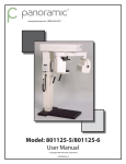
1
PC-1000/Laser 1000 User Manual
Panoramic Corporation Dental Panoramic/Cephalometric X-ray Machine
PMC6000
PMC6000
RevRev
D C
© Copyright 2007 Panoramic Corporation
Table of Contents
Table of Contents ............................................................................................................................ 2
Introduction .................................................................................................................................. 3-4
Purpose ....................................................................................................................... 3
Statement of Compatibility ........................................................................................... 3
Warning of Voltage Regulators ..................................................................................... 4
X-Ray Shielding Requirements ..................................................................................... 4
PC-1000 Components ..................................................................................................................... 5
PC-1000 Pre-Patient Setup .......................................................................................................... 6-8
PC-1000 Patient Positioning ...................................................................................................... 9-14
Prepare The Patient ................................................................................................ 9-10
Position The Patient ............................................................................................. 11-12
Set The kVp .............................................................................................................. 12
Recheck Patient Positioning ...................................................................................... 12
Take The Exposure .................................................................................................... 13
Release The Patient .................................................................................................. 13
Reset The Machine ............................................................................................... 13-14
Process The Film ...................................................................................................... 14
PC-1000 TMJ Pre-Patient Setup ............................................................................................... 15-17
PC-1000 TMJ Patient Positioning ............................................................................................. 18-25
Prepare The Patient .............................................................................................. 18-19
Position The Patient ............................................................................................. 20-21
Set The kVp .............................................................................................................. 21
Recheck Patient Positioning ...................................................................................... 21
Take The Exposure ............................................................................................... 22-24
Release The Patient .................................................................................................. 25
Process The Film ...................................................................................................... 25
Notes ............................................................................................................................................ 26
Laser 1000 Components ............................................................................................................... 27
Laser 1000 Pre-Patient Setup .................................................................................................. 28-32
Laser 1000 Patient Positioning ................................................................................................. 33-36
Cephalometric To Panoramic Setup ......................................................................................... 37-38
Panoramic Radiography ................................................................................................. Appendix A
Darkroom Procedures .................................................................................................... Appendix B
Manual Processing
Automatic Processing
Darkroom Light Leak Test
Loading The Panoramic Cassette
Loading The Cephalometric Cassette
Maintenance Schedule ................................................................................................... Appendix C
PC-1000/Laser 1000 Labeling ......................................................................................... Appendix D
PC-1000/Laser 1000 Specifications ................................................................................ Appendix E
PC-1000/Laser 1000 Space Requirements ...................................................................... Appendix F
2
© Copyright 2007 Panoramic Corporation
Introduction
Purpose
Panoramic Corporation provides this printed manual as a guide for the operation of the PC-1000 dental
panoramic X-ray machine and the PC-1000/Laser 1000 dental panoramic/cephalometric dental X-ray
machine.
The PC-1000 will enable the user to take panoramic X-ray images. The PC-1000/Laser 1000 will enable
the user to take panoramic X-ray images, as well as cephalometric X-ray images. A laser alignment
device is incorporated into the PC-1000/Laser 1000.
The information contained in this manual is not all inclusive and Panoramic Corporation should be
contacted for assistance and clarification when necessary.
It is imperative that this equipment be installed, serviced, and used by personnel familiar with the
precautions required to prevent excessive exposure to both primary and secondary radiation. This
equipment features protective designs for limiting both the primary and secondary radiation produced by
the X-ray beam. However, design features cannot prevent carelessness, negligence, or lack of
knowledge.
Only personnel authorized by Panoramic Corporation are qualified to install and service this equipment.
Any attempt to install or service this equipment by anyone not so authorized will void the warranty and
may create hazardous exposure to X-radiation.
Statement of Compatibility
January 1, 1988
Please address any comments/questions concerning this statement of compatibility to:
Panoramic Corporation • 4321 Goshen Road • Fort Wayne, IN 46818 USA • Attn: Director of Engineering
The only components compatible with the PC-1000 are those supplied with the machine.
Regardless of possible statements made by other manufacturers, no one is authorized or certified to make
additions or deletions to this machine. Only the combination of components delivered with the machine is
certified compatible by the manufacturer. As compatible accessories become available, Panoramic
Corporation will certify them as compatible and make them available to the user.
Statement of Compatibility Addendum
October 1, 1988
The Laser 1000 Cephalometric Attachment is certified by Panoramic Corporation to be compatible with the
PC-1000 dental X-ray machine, provided installation is performed by an authorized representative utilizing
specific installation instructions furnished by Panoramic Corporation.
Statement of Compatibility Addendum
October 1, 1995
Laser 1000 Cephalometric Attachments manufactured after October 1, 1995 are compatible only with
PC-1000 X-ray machines manufactured after October 1, 1995.
© Copyright 2007 Panoramic Corporation
3
Introduction
Voltage Regulator Warning
Do not plug this machine into ANY voltage regulating device. Contact Panoramic Corporation with any
questions regarding this.
X-ray Shielding Requirements
The requirements for panoramic and cephalometric shielding for building, operator, and patient, depend on
state and local regulations. Contact your state Department of Health for compliance information.
Compliance could involve a blueprint review, facility check, wall construction, film badge implementation,
remote switch installation, and/or a lead apron. It is beyond the scope of this manual to advise on these
regulations.
4
© Copyright 2007 Panoramic Corporation
PC-1000 Components
PC-1000
Top Cover
Rotating Arm
Temple Supports
Cassette Sleeve
Film Drum
Forehead Support
Mirror
Tubehead
kVp Setting Knob
Chinrest
Control Panel
Handles
Exposure Switch
Base
© Copyright 2007 Panoramic Corporation
5
PC-1000 Pre-Patient Setup
1. In a lighttight darkroom, load a panoramic film in
the cassette sleeve between the intensifying
screens.
2. Power the PC-1000 on by depressing the
POWER switch on the control panel.
3. Rotate the function switch on the control panel
to either PANORAMIC L or PANORAMIC R.
PANORAMIC L will cause the film drum to
start on the patient's left side, PANORAMIC R
will cause the film drum to start on the patient's
right side. The PC-1000 will automatically
rotate the film drum to the proper side, if the
function switch is changed.
4. If the film drum is not in it's "home" position
(patient's left side on PANORAMIC L, patient's
right side on PANORAMIC R), momentarily
depress the RESET switch on the control
panel. The PC-1000 will rotate the film drum
to the proper side.
5. Loosen the knob on the rear of the film drum.
6. Load a film cassette sleeve on the film drum:
a. Place the loaded cassette sleeve on the
film drum with the lower edge of the
cassette sleeve resting on the lower lip of
the film drum.
b. Attach the vertical velcro at the end of the
cassette sleeve to the mating velcro on the
end of the film drum. Ensure the cassette
sleeve is flat against the film drum.
6
© Copyright 2007 Panoramic Corporation
PC-1000 Pre-Patient Setup
c. Attach the velcro tab of the cassette sleeve
to the mating velcro on the center post of
the film drum.
d. The FUNCTION switch on the control panel
will determine on which side (patient's) the
film drum will reset and start. Align the
pointer on the rear of the film drum to the
appropriate mark. If the FUNCTION
switch is set to PANORAMIC L, align the
pointer to L1. If the FUNCTION switch is
set to PANORAMIC R, align the pointer to
R1.
e. Tighten the film drum knob on the rear of
the film drum.
Note: The film drum will rotate with increased
friction after the film drum knob is tightened.
Manual rotation, while the knob is tight, should
be avoided to prevent abnormal wear.
7. Using the knob at the rear of the forehead
support, adjust the forehead support toward
the mirror to allow room for positioning the
patient.
© Copyright 2007 Panoramic Corporation
7
PC-1000 Pre-Patient Setup
8. Slide the temple supports apart to allow room
for positioning the patient's head.
9. Using the UP/DOWN switch on the chinrest
arm, adjust the chinrest arm until it is slightly
higher than the patient's chin.
This will ensure that the patient stands up as
straight as possible.
10. If a stool is to be used, place the stool so that
the seat is centered under the chinrest.
This will help ensure that the patient's neck is
straight.
8
© Copyright 2007 Panoramic Corporation
PC-1000 Patient Positioning
Prepare The Patient
1. Ask the patient to remove any metal objects,
such as glasses, earrings, removable
dentures, hearing aids, hair pins, neck chains,
bib chains, and collar zippers from their head
and neck area.
These objects can prevent X-rays from
reaching the film, causing poor diagnosticquality images.
2. If a lead apron is used, and a panoramic
poncho is not available, place the lead apron
on the patient's back. Ensure that it does not
cover the back of the neck.
As the tubehead rotates around the patient,
the X-rays pass through the head at a slight
upward angle. This allows the X-ray beam to
pass through the skull more efficiently by
avoiding the denser area of the patient's skull.
3. Guide the patient into the PC-1000. Use the
UP/DOWN switch on the chinrest arm to adjust
the chinrest arm until it is slightly higher than
the patient's chin.
This will ensure that the patient is standing up
as straight as possible.
© Copyright 2007 Panoramic Corporation
9
PC-1000 Patient Positioning
4. Have the patient hold on to the handles and
move his/her feet under the chinrest.
This will help ensure that the patient's neck is
straight.
5. Place the appropriate chinrest on the
permanent, black chinrest on the chinrest arm:
a. Small child - removable, black plastic
chinrest
May be needed to allow the child's
forehead to reach the forehead support.
b. Adolescent and adult - no additional
chinrest
Majority of patients require no additional
chinrest.
c. Edentulous - removable, clear plastic
chinrest
Aids in centering and consistently
positioning the patient.
10
© Copyright 2007 Panoramic Corporation
PC-1000 Patient Positioning
6. If the patient is not edentulous, insert a
disposable bite-guide in the permanent, black
chinrest. Have the patient bite on the
disposable bite-guide. Ensure that the biteguide is centered between the central incisors.
For edentulous patients, have the patient bite
on a 1 1/2" cotton roll to keep the maxilla and
mandible separated.
Position The Patient
1. Place your hand at the base of the patient's
skull and apply pressure vertically to stretch
the neck while slowly lowering the machine
until the patient's Frankfort Plane is horizontal
(parallel to the red guidelines on the temple
supports).
The Frankfort Plane is the imaginary line from
the middle of the ear opening to the bottom of
the eye orbit. A level Frankfort Plane will
ensure that the upper and lower anterior teeth
are in one vertical plane and will help stretch
the patient's neck enough to allow X-rays to
pass between the vertebrae.
2. Using the knob on the rear of the forehead
support, adjust the forehead support until it
touches the patient's forehead.
3. Center the patient's head using the red line on
the forehead support. Turn the mirror to see
the patient to verify centering.
© Copyright 2007 Panoramic Corporation
11
PC-1000 Patient Positioning
4. Gently slide the temple supports against the
patient's head. These will help keep the
patient centered and measure the width of the
skull to determine the amount of kVp required
to penetrate the skull.
Set The kVp
1. Note which number the arrow points to on the
kVp scale decal on the outside of the temple
support rod.
2. Using the kVp setting knob on the control panel,
set the kVp meter to the number from the kVp
scale decal on the temple support rod
(previous step). This sets the penetrating
power of the X-rays.
Re-check Patient Positioning
1. Ensure that the:
a. patient's chin is firmly seated on the chinrest
b. patient's head is centered
c. patient's Frankfort Plane is horizontal
d. patient's neck is stretched
e. patient's forehead is resting on the forehead support
f. temple supports are against the patient's head
g. kVp meter is set to the number on the temple support rod
12
© Copyright 2007 Panoramic Corporation
PC-1000 Patient Positioning
Take The Exposure
1. Instruct the patient to close his/her lips around
the disposable bite-guide, swallow, place his/
her tongue on the roof of his/her mouth, and
remain still during the exposure.
This will help equalize tissue densities and
help prevent unwanted artifacts and blurring.
2. Depress and hold the exposure switch. The
arm will begin to rotate around the patient after
a second. After 12 seconds, the exposure will
automatically terminate.
To prematurely terminate an exposure, release
the exposure switch at anytime during the
exposure.
Release The Patient
1. Slide the temple supports away from the
patient's head to release the patient.
2. Instruct the patient to step out of the machine.
Reset The Machine
1. Discard the disposable bite-guide.
© Copyright 2007 Panoramic Corporation
13
PC-1000 Patient Positioning
2. Depress the RESET switch on the control panel
to reposition the film drum to its "home"
position.
Process The Film
1. Loosen the film drum knob on the rear of the
film drum and remove the cassette sleeve from
the film drum.
2. Under lighttight darkroom conditions, process
the film.
14
© Copyright 2007 Panoramic Corporation
PC-1000 TMJ Pre-Patient Setup
A TMJ series is simply four two-second panoramic
images exposed on one film. The TMJ film will
show the patient's right closed, right open, left
open, and left closed temporomandibular joints.
1. In a lighttight darkroom, load a panoramic film in
the cassette sleeve between the intensifying
screens.
Right
Closed
(R1)
Right
Open
(R2)
Left
Open
(L2)
Left
Closed
(L1)
2. Power the PC-1000 on by depressing the
POWER switch on the control panel.
3. Rotate the FUNCTION switch on the control
panel to TMJ L. The PC-1000 will
automatically rotate the film drum to the
patient's left side, if the function switch is
changed.
4. If the film drum is not in it's "home" position (left
side), momentarily depress the RESET switch
on the control panel. The PC-1000 will rotate
the film drum to the patient's left side.
© Copyright 2007 Panoramic Corporation
15
PC-1000 TMJ Pre-Patient Setup
5. Loosen the knob on the rear of the film drum.
6. Load a film cassette sleeve on the film drum:
a. Place the loaded cassette sleeve on the
film drum with the lower edge of the
cassette sleeve resting on the lower lip of
the film drum.
b. Attach the vertical velcro at the end of the
cassette sleeve to the mating velcro on the
end of the film drum. Ensure the cassette
sleeve is flat against the film drum.
c. Attach the velcro tab of the cassette sleeve
to the mating velcro on the center post of
the film drum.
d. Align the pointer on the rear of the film drum
to L1.
e. Tighten the film drum knob on the rear of
the film drum.
Note: The film drum will rotate with increased
friction after the film drum knob is tightened.
Manual rotation, while the knob is tight, should
be avoided to prevent abnormal wear
16
© Copyright 2007 Panoramic Corporation
PC-1000 TMJ Pre-Patient Setup
7. Using the knob at the rear of the forehead
support, adjust the forehead support toward
the mirror to allow room for positioning the
patient.
8. Slide the temple supports apart to allow room
for positioning the patient's head.
9. Using the UP/DOWN switch on the chinrest
arm, adjust the chinrest arm until it is slightly
higher than the patient's chin.
This will ensure that the patient stands up as
straight as possible.
10. If a stool is to be used, place the stool so that
the seat is centered under the chinrest.
This will help ensure that the patient's neck is
straight.
© Copyright 2007 Panoramic Corporation
17
PC-1000 TMJ Patient Positioning
Prepare The Patient
1. Ask the patient to remove any metal objects,
such as glasses, earrings, removable
dentures, hearing aids, hair pins, neck chains,
bib chains, and collar zippers from their head
and neck area.
These objects can prevent X-rays from
reaching the film, causing poor diagnosticquality images.
2. If a lead apron is used, and a panoramic
poncho is not available, place the lead apron
on the patient's back. Ensure that it does not
cover the back of the neck.
As the tubehead rotates around the patient,
the X-rays pass through the head at a slight
upward angle. This allows the X-ray beam to
pass through the skull more efficiently by
avoiding the denser area of the patient's skull.
3. Guide the patient into the PC-1000. Use the
UP/DOWN switch on the chinrest arm to adjust
the chinrest arm until it is slightly higher than
the patient's chin.
This will ensure that the patient is standing up
as straight as possible.
18
© Copyright 2007 Panoramic Corporation
PC-1000 TMJ Patient Positioning
4. Have the patient hold on to the handles and
move his/her feet under the chinrest.
This will help ensure that the patient's neck is
straight.
5. Place the appropriate chinrest on the
permanent, black chinrest on the chinrest arm:
a. Small child - removable, clear plastic
chinrest
Aids in centering and consistently
positioning the patient.
b. Adult - removable, clear plastic chinrest
Aids in centering and consistently
positioning the patient.
c. Edentulous - removable, clear plastic
chinrest
Aids in centering and consistently
positioning the patient.
© Copyright 2007 Panoramic Corporation
19
PC-1000 TMJ Patient Positioning
Position The Patient
1. Place your hand at the base of the patient's
skull and apply pressure vertically to stretch
the neck while slowly lowering the machine
until the patient's Frankfort Plane is horizontal
(parallel to the red guidelines on the temple
supports).
The Frankfort Plane is the imaginary line from
the middle of the ear opening to the bottom of
the eye orbit. A level Frankfort Plane will help
ensure that the temporomandibular joints are
consistently vertical.
2. Using the knob on the rear of the forehead
support, adjust the forehead support until it
touches the patient's forehead.
3. Center the patient's head using the red line on
the forehead support. Turn the mirror to see
the patient to verify centering.
20
© Copyright 2007 Panoramic Corporation
PC-1000 TMJ Patient Positioning
4. Gently slide the temple supports against the
patient's head.
These will help keep the patient centered and
measure the width of the skull to determine the
amount of kVp required to penetrate the skull.
Set The kVp
1. Note which number the arrow points to on the
kVp scale decal on the outside of the temple
support rod.
2. Using the kVp setting knob on the control panel,
set the kVp meter to the number from the kVp
scale decal on the temple support rod (previous
step).
This sets the penetrating power of the X-rays.
Re-check Patient Positioning
1. Ensure that the:
a. patient's chin is firmly seated on the chinrest
b. patient's head is centered
c. patient's Frankfort Plane is horizontal
d. patient's neck is stretched
e. patient's forehead is resting on the forehead support
f. temple supports are against the patient's head
g. kVp meter is set to the number on the temple support rod
© Copyright 2007 Panoramic Corporation
21
PC-1000 TMJ Patient Positioning
Take The Exposure
1. Instruct the patient to close his/her lips,
swallow, place his/her tongue on the roof of
his/her mouth, and remain still during the four
exposures.
This will help equalize tissue densities and
help prevent unwanted artifacts and blurring.
2. Depress and hold the exposure switch.
The arm will begin to rotate around the patient
after a second. After about 2 seconds, the
exposure will automatically terminate. When
the exposure switch is released, the film drum
will reset to its "home" position on the patient's
left side.
3. Loosen the knob on the rear of the film drum.
4. Align the pointer on the rear of the film drum to
L2 and tighten the film drum knob.
5. Instruct the patient to open his/her mouth.
This will place the temporomandibular joints in
their open positions. (A surgical bite block
may be used to help the patient keep his/her
mouth open).
22
© Copyright 2007 Panoramic Corporation
PC-1000 TMJ Patient Positioning
6. Depress and hold the exposure switch.
The arm will begin to rotate around the patient
after a second. After about 2 seconds, the
exposure will automatically terminate. When
the exposure switch is released, the film drum
will reset to its "home" position on the patient's
left side.
7. Rotate the function switch on the control panel
to TMJ R.
The PC-1000 will automatically rotate the film
drum to the patient's right side.
8. Align the pointer on the rear of the film drum to
R2 and tighten the film drum knob.
9. Instruct the patient to remain still with his/her
mouth open.
© Copyright 2007 Panoramic Corporation
23
PC-1000 TMJ Patient Positioning
10. Depress and hold the exposure switch.
The arm will begin to rotate around the patient
after a second. After about 2 seconds, the
exposure will automatically terminate. When
the exposure switch is released, the film drum
will reset to its "home" position on the patient's
right side.
11. Allow the patient to close his/her mouth.
12. Align the pointer on the rear of the film drum to
R1 and tighten the film drum knob.
13. Depress and hold the exposure switch.
The arm will begin to rotate around the patient
after a second. After about 2 seconds, the
exposure will automatically terminate. When
the exposure switch is released, the film drum
will reset to its "home" position on the patient's
right side.
24
© Copyright 2007 Panoramic Corporation
PC-1000 TMJ Patient Positioning
Release The Patient
1. Slide the temple supports away from the
patient's head to release the patient.
2. Instruct the patient to step out of the machine.
Process The Film
1. Loosen the film drum knob on the rear of the
film drum and remove the cassette sleeve from
the film drum.
P0047
2. Under lighttight darkroom conditions, process
the film.
© Copyright 2007 Panoramic Corporation
25
Notes
26
© Copyright 2007 Panoramic Corporation
PC-1000/Laser 1000 Components
Horizontal Locking Knob
Vertical Locking Knob
Mid-Sagittal to Film Scale
Laser Switch
Cephalostat Locking Knob
Collimator Lever
Suggested Settings Decal
PC-1000
Cephalostat Arm
Cephalometric
Exposure Time Chart
Panoramic Corporation ®
Seconds
90 kVP 10 mA
Rare Earth Screens And Film
LASER 1000
Patient Size:
Small Child
Large Child
Average Adult
Large Adult
Lateral
.5-1.0
.6-1.5
.8-2.0
1.0-2.5
AP-PA
.8-1.5
1.0-2.5
1.5-3.5
2.5-4.0
Ear Posts
Nasion Support
Ear Rings
Ear Rods
Film Cassette
© Copyright 2007 Panoramic Corporation
27
Laser 1000 Pre-Patient Setup
CAUTION
• Do not look into the laser beam when operating.
Laser light can cause permanent eye damage.
• Safe operating instructions must be observed to prevent
an optical hazard to the patient and/or operator.
1. In a lighttight darkroom, load a cephalometric
film in the film cassette between the
intensifying screens.
2. Power the PC-1000/Laser 1000 on by
depressing the POWER switch on the control
panel.
3. Rotate the FUNCTION switch on the control
panel to one of the CEPHALOMETRIC
SECONDS settings.
This will set the duration of the X-ray exposure.
Suggested settings can be found on a decal on
the cephalometric arm.
If the tubehead is not in it's "home" position
(on the opposite side of the cephalometric
arm), the PC-1000/Laser 1000 will rotate the
tubehead to the proper side.
28
© Copyright 2007 Panoramic Corporation
Laser 1000 Pre-Patient Setup
4. Loosen both of the locking knobs on the rear of
the cassette holder at the end of the
cephalometric arm.
5. Align the top edge of the sliding portion of the
cassette holder to the appropriate mark on the
decal on the rear of the fixed portion of
cassette holder. Use the upper mark,
LATERAL, if the film cassette will be oriented
horizontally for lateral views.
Use the lower mark, AP-PA, if the film cassette
will be oriented vertically for all other views.
6. Tighten the lower knob.
© Copyright 2007 Panoramic Corporation
29
Laser 1000 Pre-Patient Setup
6. Place the film cassette into the cassette holder
and center it horizontally. Tighten the upper
knob.
7. Remove the locking pin from the top of the
cephalometric head positioner and use the ear
posts' metal supports only, to temporarily
rotate the head positioner to a 45º position to
allow the laser alignment beam to strike the
film cassette target.
Note: Never use the wooden ear posts to rotate
the head positioner or misalignment can result.
8. Slide the lever in the collimator assembly on the
front of the tubehead to select the appropriate
collimator based on the orientation of the film
cassette. Select the LATERAL position for a
cassette oriented horizontally. Select the
AP-PA position for a cassette oriented vertically.
NOTE: If the collimator is NOT firmly seated in its
locking notch:
a. the alignment laser will not fire
b. when the exposure switch is depressed, the
machine will beep rapidly, but no radiation
will be emitted
30
© Copyright 2007 Panoramic Corporation
Laser 1000 Pre-Patient Setup
9. Loosen the two locking knobs on the tubehead
assembly and turn the tubehead towards the
cephalometric film cassette.
10. Depress the momentary toggle switch on the
right side of the tubehead to activate the laser.
CAUTION: Do not look into the laser beam
because laser light can cause permanent eye
damage.
11. Move the tubehead until the laser alignment
beam strikes the upright face target on the film
cassette.
12. Tighten both horizontal and vertical tubehead
locking knobs.
13. Activate the laser again to verify proper
alignment.
© Copyright 2007 Panoramic Corporation
31
Laser 1000 Pre-Patient Setup
14. Remove the locking pin in the top of the
cephalostat head positioner. Without using the
wooden ear posts, rotate the head positioner
into its proper position for the appropriate view:
lateral,
anterior to posterior,
posterior to anterior,
or any 45º increment.
15. Reinstall the locking pin in the top of the head
positioner. Ensure that it is fully seated by
gently rotating the head positioner back and
forth.
32
© Copyright 2007 Panoramic Corporation
Laser 1000 Patient Positioning
1. Verify the setting of the CEPHALOMETRIC
SECONDS on the function switch on the
control panel.
Suggested settings can be found on a decal on
the cephalometric arm.
2. Using the kVp setting knob on the control panel,
adjust the kVp to the appropriate setting.
Suggested settings can be found on a decal on
the cephalometric arm. This sets the
penetrating power of the X-rays.
3. Open the ear posts on the cephalostat head
positioner as far as possible by using the lever
on the rear of the head positioner.
© Copyright 2007 Panoramic Corporation
33
Laser 1000 Patient Positioning
4. Using the UP/DOWN switch on the chinrest
arm, adjust the height of the ear posts to
approximately the height of the patient's ear
canals.
5. Have the patient stand directly under the head
positioner looking away from the cephalometric
arm. Use the UP/DOWN switch on the
chinrest arm until the ear rods can be guided
gently into the patient's ear canals with the
lever on the rear of the head positioner. Have
the patient attempt to turn his/her head back
and forth slightly and continue to guide the ear
rods until there is minimal head movement.
6. Adjust the patient's head so that the Frankfort
Plane is horizontal (level with the floor).
The Frankfort Plane is the imaginary line from
the middle of the ear opening to the bottom of
the eye orbit.
7. Lower and tighten the nasion support over the
bridge of the patient's nose to help the patient
retain this position.
34
© Copyright 2007 Panoramic Corporation
Laser 1000 Patient Positioning
8. Slide the cephalometric film holder and cassette
towards the patient's head as close as
possible.
The less distance between the film and the
patient results in less magnification.
For reproducibility, record the number at the red
dot on the midsagittal to film scale.
9. If a recording of magnification is desired, place
the correctional scale in its slot in the nasion
support.
The scale markings will appear on the film
after processing to allow for magnification
ratios to be determined.
10. Have the patient close his/her mouth and lips
in normal occlusion and remain still.
© Copyright 2007 Panoramic Corporation
35
Laser 1000 Patient Positioning
11. Depress the exposure button until the machine
automatically terminates the exposure.
12. Open the ear posts by using the lever on the
rear of the cephalostat head positioner or their
metal supports to allow the patient to exit the
machine.
Note: Do not pull or push the wooden ear posts
directly or misalignment can result.
13. Remove the cephalometric film cassette from
the cassette holder.
14. Under lighttight darkroom conditions, process
the film.
36
© Copyright 2007 Panoramic Corporation
Cephalometric to Panoramic Setup
CAUTION
• Do not look into the laser beam when operating.
Laser light can cause permanent eye damage.
• Safe operating instructions must be observed to prevent
an optical hazard to the patient and/or operator.
1. Rotate the function switch on the control panel
to PANORAMIC L or PANORAMIC R.
PANORAMIC L will cause the film drum to
start on the patient's left side, PANORAMIC R
will cause the film drum to start on the patient's
right side. The PC-1000 will automatically
rotate the film drum to the proper side.
2. Slide the lever in the collimator assembly on the
front of the tubehead to select the
PANORAMIC position.
NOTE: If the collimator is NOT firmly seated in its
locking notch:
a. the alignment laser will not fire
b. when the exposure switch is depressed, the
machine will beep rapidly, but no radiation
will be emitted
3. Loosen the two locking knobs on the tubehead
assembly and turn the tubehead towards the
panoramic film drum.
© Copyright 2007 Panoramic Corporation
37
Cephalometric to Panoramic Setup
4. Depress the momentary toggle switch on the
right side of the tubehead to activate the laser.
CAUTION: Do not look into the laser beam
because laser light can cause permanent eye
damage.
5. Move the tubehead until the laser alignment
beam strikes the cross-hair target on the film
mask in front of the film drum.
6. Tighten both horizontal and vertical tubehead
locking knobs.
7. Activate the laser again to verify proper
alignment.
38
© Copyright 2007 Panoramic Corporation
Panoramic Radiography
Panoramic Radiography has been in use for over 30
years. In panoramic radiography, the X-ray source
and film rotate around the patient's head at the
same speed. Simultaneously, the film rotates
about its own axis.
X-rays
X-rays are emitted from the tubehead in a very
narrow vertical band, pass through the patient's
head (where some are absorbed), and strike the
film cassette sleeve. Intensifying screens are used
inside the film cassette sleeve. The intensifying
screens glow whenever X-rays strike them, the
more X-rays striking the screen, the brighter the
glow. Film, which is sensitive to light, is placed
between the intensifying screens. The more light
that is exposed to the film, the darker the film is.
Since the patient is between the X-ray source and
the film, the amount of X-rays that reach the film
will vary depending on the density of the patient's
anatomy. Dense matter, such as bone, will absorb
more of the X-rays than less dense matter, such as
tissue. Less X-rays reach the film when striking
the teeth, causing them to appear on the film as
lighter areas. More X-rays reach the film when
striking tissue, causing it to appear on the film as
darker areas.
Film Drum
Tubehead
Film
Cassette
Sleeve
In order to pass as many X-rays through the
patient's head as possible, the tubehead is tilted at
a slight upward angle to:
1. move the dense portion of the skull out of
the path of the X-rays
2. cause the upper and lower anterior root tips
to be aligned vertically
3. stretch the vertebrae in the neck to allow
the X-rays to pass more efficiently through
the vertebrae to expose the anterior teeth
As the tubehead and film rotate around the patient,
the film is gradually exposed by a narrow vertical
band. It is imperative that the film is aligned to
start at the correct position and that nothing stops
the film drum or tubehead from moving while the
exposure is being taken.
© Copyright 2007 Panoramic Corporation
Appendix
A
Darkroom Procedures
The darkroom must be lighttight. Extraoral (panoramic/cephalometric) film is more
sensitive than intraoral (bite-wings) film to light, and processing time and temperature.
Manual Processing
• Lighttight darkroom
• Dip tanks
• Timer
• Thermometer
• Developer and fixer solutions • Film Hanger
• Water supply and drain
• Safelight (GBX-2 filter or equivalent, 15 W bulb or less and at least 4' from film)
1. Prepare developer and fixer solutions according to the solution's directions.
2. Verify developer temperature.
3. Under safelight conditions, remove the exposed film from the cassette sleeve and attach it to a film
hanger.
4. Set the timer based on the developer temperature and the processing chart.
5. Immerse the film quickly into the developer and agitate it vigorously for only 5 seconds to dislodge any
air bubbles.
6. When the timer sounds, remove the film from the developer and immediately rinse it with water for 30
seconds while agitating it. Do not allow the excess developer to drain back into the developer tank.
7. Immerse the film into the fixer and agitate it for 5 seconds every 30 seconds. Allow the excess fixer to
drain back into the fixer tank.
8. Immerse the film in the water wash tank and rinse it thoroughly.
9. Dry the film at room temperature or in a drying cabinet.
Automatic Processing
A thermometer should be present to periodically verify the temperature. It is imperative that the processor's
maintenance schedule is followed thoroughly.
Manual Processing
Film Type
Developer
Temperature Time
Rinse
Automatic Processing
Fixer
T-MAT
68º F 20.0º C 8 min
30 sec
2-4 min
(for use72º F 22.0º C 7 min 60º F 15.5º C
60º F 15.5º C
with Lanex 76º F 24.5º C 5 min
to
to
screens)
80º F 26.5º C 4 min 85º F 29.5º C 85º F 29.5º C
Wash
5 min
60º F 15.5º C
to
85º F 29.5º C
82º F 28.0º C 5.5 min
83º F 28.5º C 4.5 min
85º F 29.5º C 4 min
Note: Extraoral film requires more frequent solution replenishment than intraoral film. One ounce of
chemicals are typically required for replenishment for every 75 intraoral, 3 panoramic, or 2 cephalometric
films.
Appendix
B
© Copyright 2007 Panoramic Corporation
Darkroom Procedures
Darkroom Light Leak Test
Extraoral film is more sensitive to light than intraoral film. The purpose of the intensifying screens inside of
the cassette sleeve is to convert the X-ray energy into light, thus exposing the film. While the light
sensitivity of the film allows a very small amount of radiation to expose the film, it also can pose a problem if
the darkroom is not completely lighttight. Small light leaks can cause fogging of the film while handling and
processing the film in the darkroom.
The following test should be performed in the darkroom under safelight conditions to ensure it is lighttight:
1. Remove one sheet of extraoral film from the box.
2. Lay it on the counter in the darkroom under normal darkroom conditions.
3. Place a couple of coins, a pair of scissors, or any other opaque object on top of the film.
4. Wait for two minutes.
5. Process the film as usual.
The processed film should be clear. None of the objects should be visible on the film.
If any of the objects can be seen on the processed film, there is a light leak or other light source in the
darkroom. The light leak fogs the test film, everywhere except where the opaque objects are blocking the
light. To find the light leak, turn all of the lights off in the darkroom and inspect the darkroom for cracks
around the door and ceiling tiles. Indicator lights on equipment, such as stereos, and improper safelights
can also cause fogging. Turn off all unnecessary equipment and the safelight and try this test again.
© Copyright 2007 Panoramic Corporation
Appendix
B
Darkroom Procedures
It is recommended that the panoramic and
cephalometric cassettes be loaded with film just
prior to use. Do not leave a film loaded in the
cassettes for an extended period of time. This will
prevent background radiation from prematurely
exposing the film. The film should be stored in a
cool and dark place.
Loading The Panoramic Cassette
In a lighttight darkroom, open the flexible,
panoramic cassette sleeve and slowly remove the
intensifying screens. Open the screens on the
counter and place a sheet of panoramic film on top
of one of the screens. Close the screens and
slowly slide them back into the cassette sleeve.
Ensure that the hinged end of the screens is placed
into the cassette sleeve first and the "TUBESIDE"
decal is facing the same direction as the writing on
the outside of the cassette sleeve. Ensure that all
excess air is expelled from the cassette sleeve.
Loading The Cephalometric Cassette
In a lighttight darkroom, unlock and open the rigid,
cephalometric cassette. Place a sheet of
cephalometric film on top of one of the screens.
Close and lock the cassette.
Note: Remove and discard the protective sheet
from between new intensifying screens before
their first use.
Appendix
B
© Copyright 2007 Panoramic Corporation
Maintenance Schedule
Panoramic Corporation strongly recommends a preventive maintenance be performed on your equipment
at least every two years. All service requests must be submitted through Panoramic Corporation’s Service
Department by calling our toll-free number at (800) 654-2027.
Panoramic has an extensive network of independent installation and service organizations throughout the
U.S. and Canada to install and service our products. The Independent Representatives have been
specifically trained by our organization in the service and installation of Panoramic products. We
strongly recommend that you use one of our Independent Representatives to service Panoramic products.
To the extent you use third parties other than Independent Representatives to service Panoramic products,
we cannot accept responsibility or liability for any work performed by those third parties and any
resulting damages or liability attributable thereto. In no event shall Panoramic be liable to you or any
other third party for any direct, indirect, punitive, incidental, consequential or special damages or lost
profits arising from, relating to or connected with, the installation of or repair of a Panoramic product by
someone other than an Independent Representative.
Always refer to your state and local regulations to determine how often to perform a preventive
maintenance on your equipment as the regulations may supersede manufacturers’ recommendation.
Owners of Panoramic Corporation X -Ray machines must call Panoramic Corporation Service Department
for all reasons listed below but not limited to:
‐
‐
‐
‐
‐
‐
‐
Preventive maintenance at least every two years
Physical relocation of machine
Changing the power source to a different power source from original installation
Questions/Help related to compliance with your state, and local regulations regarding
radiological equipment
Corrective Maintenance
Physical damage that may affect radiation safety
Interrupted movement, unusual noises, leaks, etc.
To schedule a preventive maintenance on your equipment contact the Service Department by dialing our
toll-free number at (800) 654-2027.
© Copyright 2007 Panoramic Corporation
Appendix
C
PC-1000 Labeling
Panoramic Corporation PC-1000
Fort Wayne, Indiana U.S.A.
Date
Serial #
X-RAY CONTROL
MODEL PC-1000
Complies with: DHHS
21 CFR Subchapter J
CAN CSA Std 22.2
No. 114-M90 LR 96864-1
kVp
Panoramic
70 - 90 kVp, 6 mA
12 Sec.
R
Cephalometric
70 - 90 kVp, 10 mA
0.4 - 2.0 Sec.
L
R
L
115V 50/60Hz
0.5A Continuous
10.0A Momentary
10 A
RADIATION
RESET
POWER
1A
WARNING: THIS X-RAY UNIT MAY BE DANGEROUS TO
PATIENT AND OPERATOR UNLESS SAFE EXPOSURE
AND OPERATING INSTRUCTIONS ARE OBSERVED
Control Panel
Appendix
D
© Copyright 2007 Panoramic Corporation
®
!
Laser 1000 Labeling
Panoramic Corporation PC-1000
Fort Wayne, Indiana U.S.A.
Date
Serial #
X-RAY CONTROL
MODEL PC-1000
Complies with: DHHS
21 CFR Subchapter J
kVp
Panoramic
70 - 90 kVp, 6 mA
12 Sec.
Cephalometric
70 - 90 kVp, 10 mA
0.4 - 2.0 Sec.
Rear Tubehead Cover
VISIBLE LASER LIGHT EMITTED FROM APERATURE ABOVE - AVOID EXPOSURE
AP-PA
Panoramic Corporation
Fort Wayne, Indiana USA
R
L
!""" #
2.0
0.4
1.5
0.5
1.2
LASER 1000
Cephalometric: SID 1720mm, SIZE 203mm X 254mm
Panoramic:
SID 460mm,
SIZE 6 x 150mm
Front Tubehead Cover
0.6
1.0
115V 50/60Hz
0.5A Continuous
10.0A Momentary
PAN
OFF
R
L
LATERAL
0.8
RADIATION
RESET
POWER
CEPHALOMETRIC EXPOSURE
TIME & KVP CHART 10mA
Recommended Times for Rare Earth Screens & Film
10 A
Panoramic Corporation ®
1A
WARNING:
THIS X-RAY UNIT MAY BE DANGEROUS TO
PATIENT AND OPERATOR UNLESS SAFE EXPOSURE
AND OPERATING INSTRUCTIONS ARE OBSERVED
Control Panel
!
LASER 1000
May use temple support KVP settings from Panoramic
Patient Size
KVP
SECONDS
LATERAL AP-PA
0.8
Child
75
0.5
Adolescent
80
0.6
1.0
Average Adult
85
0.8
1.2
Large Adult
90
1.0
1.5
Cephalometric Arm
© Copyright 2007 Panoramic Corporation
Appendix
D
PC-1000/Laser 1000 Specifications
Tube Housing Thermal Characteristics
Tube Maximum Current
350
25
60 kV
70 kV
210
140
50 kV
90 kV
100 kV
15
10
70
0
80 kV
20
Tube current in mA
Heat in kJ
280
5
0
2
4
6
8
0
0.1
10
0.2 0.3
0.5 0.7 1.0
2.0 3.0 5.0 7.0 10.0
Time in seconds
Time in hours
1 kJ=1400 H.U.
1 Watt=1.4 H.U./sec
Radiation Scatter Survey
Anode Thermal Characteristics
180˚
35
0.00
315 W
30
225˚
225 W
175 W
Heat storage in kJ
25
135˚
0.00
0.00
0.00
0.00
0.00
20
0.03
0.03
15
270˚
0.00
0.00
0.03
0.03
10
0.00
90˚
0.00
PC-1000
0.03
0.03
5
0.00
16" 0.04
0.00
0
0
2
4
6
Time in minutes
8
10
Heating
Cooling
Self-rectified
0.00
24" 0.00
0.00
45˚
315˚
36" 0.00
Focal spot: 0.5 mm
0
Technique Factors:
Values in mR / 14 second exposure
Tube Current: 6.0 mA
Tube Voltage: 90 kVp
Exposure Duration: 14 seconds
Method:
Survey meter (Nuclear Associtates Model 06-107) at level of
phantom skull at each position for duration of exposure.
Appendix
E
© Copyright 2007 Panoramic Corporation
PC-1000/Laser 1000 Specifications
Power Requirements The PC-1000 and PC-1000/Laser 1000 requires line voltage from 105 to 125 VAC,
no load at 5% or better line regulation and draws 12 A under worst case
conditions. The total power required is 1.5 kVa.
Generator Type
Single-phase, half-wave, self-rectified, center-grounded.
Duty Cycle
At 90 kVp/6 mA - One 12 second exposure every 5 minutes to a maximum of 30
exposures.
Tubehead Assembly X-ray Tube
Rated Tube Potential Peak
Leakage Technique Factors
Inherent Filtration
Added Aluminum Filtration
Total Filtration
Peak Tube Potential at which Aluminum
Equivalent was Obtained
X-ray Tube
Manufacturer
Type
Brand X-Ray or Superior or K-Alpha
100 kVp
90 kVp/6 mA
1 mm
1.8 mm
2.8 mm
90 kVp/6 mA
Focal Spot
Maximum Peak Voltage
Anode Heat Dissipation Rate
Anode Heat Storage Capacity
Brand X-Ray or Superior or K-Alpha
BX-4P0.5 or SXR-100-R-5P or
KAX-90-10-P
.5 mm x .5 mm
100 kVp
250 Watts
1 Watt=1.4 H.U./sec.
35 kJ
1 kJ=1400 H.U.
Statement of
Deviation
Peak Tube Potential
Tube Current
Exposure Time
± 12% over range of rated line voltage
± 10% over line voltage
± 10% over line voltage
Measurement
Techniques
Exposure Time
Measured with Engineered Systems &
Design Model XR201MS pulse
counter.
Measured directly with a DC mA meter
having a basic accuracy of no less
than ± 3%.
Measured using a computerized kVp
measurement system.
System accuracy is ± 3% exclusive of
waveform, inherent filtration,
and reproducibility.
Machine set at 90 kVp/6 mA
Tube Current
Peak Tube Potential
Maximum Line Current
Screen/Film Type
Kodak Lanex Regular with Kodak T-MAT G (green/400 speed) or equivalent
© Copyright 2007 Panoramic Corporation
Appendix
E
Laser 1000 Specifications
Cephalometric Attachment
Film Size
8" x 10"
Source to Image Distance (SID)
1624 mm - 1724 mm (63" - 68")
Exposure Times
0.4, 0.5, 0.6, 0.8, 1.0, 1.2, 1.5, 2.0 seconds
Tube Voltage
70 - 90 kVp ± 12%
Tube Current
10 mA ± 10%
Laser
Class
II
Type
Laser Diode, Toshiba TOLD9442M
Wavelength
650 nm
Average Operating Current
34 mA
Average Operating Voltage
0.75 VDC
Maximum Average Radiant Power
0.2 mW
Maximum Peak Radiant Power
< 1 mW
Beam Divergence
0.9 mRadian
Beam Diameter
1.8 mm
Emission Duration
Operator-controlled by a momentary switch
Appendix
E
© Copyright 2007 Panoramic Corporation
PC-1000 Space Requirements
35"
23"
Physical Dimensions
35" W x 42" D x 91" H
Minimum Working Space
48" W x 48" D x 91" H
42"
32"
PC-1000
PC-1000
91" Maximum
58" Minimum
The PC-1000 weighs approximately 485 pounds
and is freestanding, requiring no extra support in
the wall or floor.
The factory configuration is shipped with the
control panel mounted on the patient's left side,
unless specified by the customer prior to
shipping. The control panel can be easily
relocated to the right side at the time of
installation.
Note: The FDA requires that the technique
factors (kVp meter) be viewable during the
exposure.
© Copyright 2007 Panoramic Corporation
Appendix
F
PC-1000/Laser 1000 Space Requirements
72"
35"
23"
44"
Physical Dimensions
72" W x 44" D x 91" H
33"
PC-1000
Minimum Working Space
84" W x 48" D x 91" H
PC-1000
Cephalometric
Exposure Time Chart
Panoramic Corporation ®
Seconds
90 kVP 10 mA
Rare Earth Screens And Film
LASER 1000
Patient Size:
Small Child
Large Child
Average Adult
Large Adult
91" Maximum
58" Minimum
Lateral
.5-1.0
.6-1.5
.8-2.0
1.0-2.5
AP-PA
.8-1.5
1.0-2.5
1.5-3.5
2.5-4.0
The PC-1000/Laser 1000 weighs approximately
515 pounds and is freestanding, requiring no
extra support in the wall or floor.
The factory configuration is shipped with the
control panel mounted on the patient's left side
and the cephalometric arm mounted on the
patient's right side, unless specified by the
customer prior to shipping. The control panel
can be easily relocated to the right side at the
time of installation.
Note: The FDA requires that the technique
factors (kVp meter) be viewable during the
exposure.
Appendix
F
© Copyright 2007 Panoramic Corporation