1
Sample Preparation Using Supported Liquid Extraction
Supported Liquid Extraction
Mechanism of Action
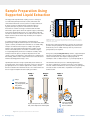
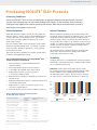
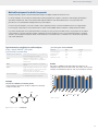
ISOLUTE® SLE+ products contain a modified form of
diatomaceous earth, which provides a support for the liquidliquid extraction process to occur, but does not interact
chemically with the aqueous sample.
Application of the sample to the column results in the aqueous
sample spreading over the surface of the material, forming an
immobilized layer of small droplets held in place by a network of
pores (Figure 2).
Simple sample preparation techniques such as ‘dilute
and shoot’
and protein precipitation do not significantly
PL
Lyso PL
reduce8.19E+07
phospholipid concentration in sample extracts,
5.87E+07
5.37E+04
4.60E+03
meaning
analytical sensitivity and extract cleanliness can be
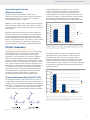
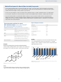
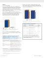
compromised. Figure 4 compares the levels of phospholipids
(PL) and lysophospholipids (lysoPL) in sample extracts
prepared using protein precipitation (PPT) and ISOLUTE SLE+.
9.00E+07
8.00E+07
7.00E+07
6.00E+07
When the water immiscible extraction solvent is applied for
the elution step, it flows over the aqueous droplets allowing
efficient analyte partitioning. The large surface area of the
extraction interface and flow through nature of the technique
leads to a very efficient extraction procedure, because analytes
come into contact with fresh solvent as the organic phase
travels through the bed, mimicking a repeat LLE mechanism. PPT
PPT
5.00E+07
ISOLUTE SLE+
4.00E+07
3.00E+07
2.00E+07
1.00E+07
Lyso PL
PL
Lyso PL
PL
Lyso PL
5.87E+07 8.19E+07
Depletion
'P'5.02E+05 4.34E+04
PPT
Phree
5.02E+05 4.34E+04
Depletion 'O'2.15E+05 1.59E+04
ISOLUTE SLE+
Figure 4. Comparison of phospholipids and lysophospholipids present in
Ostro
2.15E+05 1.59E+04
ISOLUTE SLE+5.37E+04 4.60E+03
sample extracts prepared using protein precipitation (PPT, 100µL human
ISOLUTE SLE+
5.37E+04 4.60E+03
plasma precipitated with 300µL acetonitrile) and ISOLUTE SLE+ (100 µL
Extract Cleanliness
Excellent removal of phospholipids and proteins
Although supported liquid extraction is a very simple sample
preparation technique, it provides much cleaner extracts
than techniques such as ‘dilute and shoot’ and protein
precipitation. Extract cleanliness can often be comparable
with more selective SPE approaches. In supported liquid
extraction, analytes are extracted by partitioning into a water
immiscible organic solvent. Endogenous components such as
salts, proteins peptides and phospholipids (present in blood
related matrices) which cause matrix effects in LC-MS/MS are
not soluble in these solvents. As a result they remain in the
aqueous phase on the sorbent and are therefore eliminated
from the final extract. High analyte recoveries along with
low matrix components and reduced ion suppression
result in better quantitation and method performance.
Phospholipid Removal Using ISOLUTE SLE+
0.00E+00
PL
human plasma extracted using MTBE).
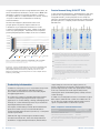
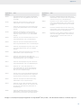
Phospholipid depletion products are designed to remove
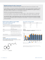
from sample extracts. Figure 5 compares
phospholipid
(PL
and lysoPL) levels in sample extracts
5.00E+07
PL
prepared using two popular phospholipid depletion products
Lyso PL
0.00E+00
with those in ISOLUTE SLE+ extracts. Using the fast, simple
load-wait-elute procedure, ISOLUTE SLE+ reduces phospholipid
concentration to significantly lower levels than phospholipid
depletion products.
1.00E+08
phospholipids
6.00E+05
5.00E+05
®
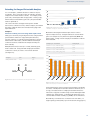
Phospholipids are matrix components found in blood based
biological fluids such as plasma and serum. Their presence in
sample extracts to be analyzed by LC-MS/MS is problematic as
they often co-elute with analytes of interest causing significant
ion suppression. Elimination of phospholipids from sample
extracts is therefore essential for reliable analyte quantitation.
4.00E+05
PL
3.00E+05
Lyso PL
2.00E+05
1.00E+05
0.00E+00
Depletion
'P' ‘P’
Depletion
Depletion
'O' ‘0’
Depletion
ISOLUTE
SLE+ SLE+
ISOLUTE
Figure 5. Comparison of phospholipids and lysophospholipids present in
sample extracts prepared using phospholipid depletion (100 µL human
plasma prepared using manufacturer’s instructions) and ISOLUTE SLE+
(100 µL human plasma extracted using MTBE).
Figure 3. Examples of phospholipid structures.
5