1
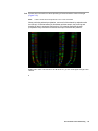
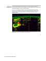
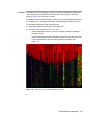
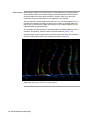
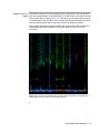
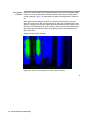
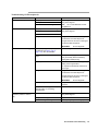
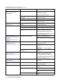
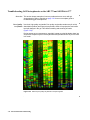
Troubleshooting Gel Electrophoresis on the ABI 373 and ABI PRISM 377
Overview This section shows examples of common problems that can occur with gel
electrophoresis. Refer to the table on page 7-53 for a more complete guide to
troubleshooting gel electrophoresis.
Poor-Quality Use fresh, high-quality acrylamide. Poor quality acrylamide contains acrylic acid (a
Acrylamide deamidation product) and linear polyacrylamide, which will copolymerize and cause
local pH changes in the gel. This causes streaking and smearing of bands
(Figure 7-50).
During storage at room temperature, especially in water, acrylamide breaks down into
acrylic acid. Prepare only as much acrylamide-bisacrylamide solution as you will need
in a month.
Figure 7-50 Effect of poor quality acrylamide on sequencing data
7-44 Data Evaluation and Troubleshooting
Salt Excess salt in the wells can cause pinching of lanes toward the center of the gel
(Figure 7-51).
Note
Lanes 3–8 are short PCR products. Lane 11 was not loaded.
When performing ethanol precipitation, remove all of the ethanol by aspiration after
the first spin. If residual ethanol is dried down with the sample, the pinching and
bending of lanes is worsened. Performing a 70% ethanol wash after ethanol
precipitation of dye terminator reactions also helps to alleviate this problem.
Figure 7-51 Effect of excess salt on an ABI PRISM 377 gel. The overall signal strength is also
lowered.
Data Evaluation and Troubleshooting 7-45
Fluorescent Contaminating fluorescent species can obscure sequencing data completely. A
Contaminants common cause of fluorescent contamination is ink from marker pens. Do not write on
the gel plates, spacers, combs, or buffer chambers.
In Figure 7-52, a large green band is seen shortly after the first fragments are
detected. The band is from a marking pen that was used to label a spacer. Even
though the spacer was cleaned before use, enough ink remained to ruin the
sequencing data.
Figure 7-52 Fluorescent contamination from a marking pen
7-46 Data Evaluation and Troubleshooting
Buffer Leaks If buffer spills or leaks onto the read region of the gel plates, it can cause blue or green
artifacts on the gel image (Figure 7-53).
To avoid buffer leaks, make sure that the plates are clamped correctly and that the
upper buffer chamber gasket makes a proper seal. Do not spill buffer behind the upper
buffer chamber, as wicking can occur.
Figure 7-53 Buffer leak in the read region of the plates
Buffer leaks or evaporation also can cause electrophoresis failure if there is not
enough buffer for electrophoresis. Note that electrophoresis fails at the same point in
each sample (Figure 7-54 on page 7-48), causing diffuse bands to appear throughout
the rest of the run.
Data Evaluation and Troubleshooting 7-47
Figure 7-54 Electrophoresis failure caused by a buffer leak
To prevent this from happening:
♦
Clean the front plate well, so the gasket will make a good seal.
♦
Use the lid on the upper buffer chamber.
♦
Take care when filling the upper buffer chamber not to spill buffer behind it.
♦
Do not fill the upper buffer chamber to the top because buffer will wick over the
ears of the notched plate and run down the sides or back of the gel plates.
♦
Check gasket for leaks before starting the run.
7-48 Data Evaluation and Troubleshooting
Red Rain Gel destruction in the read region of the gel can cause red streaks in the data, often
near the end of the run (and therefore near the top of the gel image). This effect,
shown in Figure 7-55, is known as “red rain.”
Gel destruction often results from drying out of the gel, and is exacerbated by extreme
run conditions, e.g., high voltage, high power, high temperature, and long run times.
The following can be used to help prevent red rain:
♦
Wrap the gel plates to prevent the gel from drying out.
♦
Lower the run temperature from 51 °C to 48 °C.
–
A lower temperature results in a slower run speed. Less data is collected in
the same run time.
–
A lower temperature also means less denaturing power in the gel, which can
lead to more extension product secondary structure in the gel. This can result
in more compressions, particularly with dye primer chemistries (see
page 7-31).
Figure 7-55 “Red rain” on a 48-cm, 36-lane ABI PRISM 377 gel
Data Evaluation and Troubleshooting 7-49
Gel Extrusion When voltage is applied on the ABI PRISM 377 DNA Sequencer, the polyacrylamide
gel sometimes moves from between the glass gel plates toward the cathode (upper
electrode) and into the upper buffer chamber. In extreme cases, up to about five
centimeters of gel in a folded sheet can be deposited in the chamber.
This “gel extrusion” usually begins at the start of a run or even during the prerun. It is
believed to be caused by a buildup of charge on the surface of the glass plate such
that the gel is not bound to the plate after pouring. As the voltage is applied, the gel
migrates toward the upper electrode.
The gel image can show a variety of anomalous effects, including catastrophic loss of
resolution, lane splitting, extreme band tilt, and band distortion (Figure 7-56).
Almost all known cases of gel extrusion have been resolved by either acid washing or
alcoholic KOH washing. Refer to the cleaning procedures on page 4-9.
Figure 7-56 Effect of gel extrusion on sample migration
7-50 Data Evaluation and Troubleshooting
Temporary Loss of This problem usually occurs at the beginning of the gel (between 150 and 250 base
Signal pairs in the analyzed data). It manifests itself as a band of little or no signal across the
entire width of the gel image (Figure 7-57). Temporary loss of signal has been traced
to contaminants on the gel plates. These contaminants include surfactants, fatty acids,
and long chain polymers that are not removed when the plates are washed.
Rinsing glass plates with hot deionized water (90 °C) has been found to remove the
contaminants that cause temporary loss of signal. Refer to page 4-9 for more
information.
Figure 7-57 Portion of an ABI PRISM 377 gel image showing temporary loss of signal. The
vertical blue line is from dust in the read region of the gel.
Data Evaluation and Troubleshooting 7-51
Poor-Quality Plates from vendors other than PE Applied Biosystems may not have adequate quality
Gel Plates control for use on our instruments. Several problems result from poor quality plates,
including warping. Figure 7-58 shows data from plates that warped after 6 months of
use.
When plates become warped, the laser is no longer focused correctly on the gel.
When this occurs on the ABI 373 DNA Sequencer, laser light is scattered back to the
detector, causing the gel image to appear blue and green and obscuring data. On the
ABI PRISM 377 DNA Sequencer, there is a filter to keep out most of the scattered laser
light. Data quality still suffers because the scattering results in less excitation of the
dyes by the laser.
Always use high-quality gel plates.
Figure 7-58 Effect of warped gel plates on ABI 373 sequencing data
7-52 Data Evaluation and Troubleshooting
Troubleshooting Gel Electrophoresis
Observation
Gel runs too quickly
Possible Causes
Recommended Actions
Total polymer concentration too low
Check reagents. Prepare new solutions
using fresh reagents.
Bisacrylamide concentration too low
Gel runs too slowly
Buffer concentration too high
Note
Do not use TBE buffer if it has
precipitate in it.
Total polymer concentration too high
Check reagents. Prepare new solutions
using fresh reagents.
Bisacrylamide concentration too high
Buffer concentration too low
Old gel
Use gels within 2–6 hours of casting for
the ABI PRISM 377 DNA Sequencer.
Use gels within 18–24 hours of casting for
the ABI 373 DNA Sequencer.
IMPORTANT
Poor resolution caused by gel
Do not refrigerate.
Poor quality reagents, especially
acrylamide (see Figure 7-50 on
page 7-44), APS, and TEMED
Use fresh reagents from a reliable source.
Small bubble between load and read
region
Clean plates thoroughly.
Well shape not flat
Assure that no air bubbles are trapped by
casting comb at gel surface.
Cast gel carefully. Remove bubble by
tapping plates while pouring.
Do not push the sharktooth comb too far
into the gel.
Old gel
Use gels within 2–6 hours of casting for
the ABI PRISM 377 DNA Sequencer.
Use gels within 18–24 hours of casting for
the ABI 373 DNA Sequencer.
IMPORTANT
Visible non-homogeneity
(Schlieren pattern or “swirl” in
gel)
Do not refrigerate.
Variation in spacers
Use spacers and comb sets that are equal
thickness.
Temperature of room, gel solution, or
glass too warm or cool during
polymerization
20–23 °C is optimal.
Excessive TEMED or APS
Check reagents. Prepare new solutions
using fresh reagents.
Temperature too high
Polymerize at 20–23 °C.
Insufficient reagent mixing
Mix reagents gently, but thoroughly.
Data Evaluation and Troubleshooting 7-53
Troubleshooting Gel Electrophoresis
(continued)
Observation
Possible Causes
Recommended Actions
Polymerization too slow (gels
should polymerize within
15–20 minutes)
Excessive dissolved oxygen
Keep vacuum filter strength/time constant.
Stir and pour gel gently.
Filter and pour gel at 20–23 °C.
Not enough TEMED or APS (or
degraded)
Check reagents. Prepare new solutions
using fresh reagents.
Temperature too low during casting
Polymerize at 20–23 °C.
Did not use deionized water
Use only deionized or distilled water for
making all solutions.
Gel loses signal around 200 bp
(see page 7-51)
Contaminant polymers on plate
surface
Wash plates with mild detergent and hot
deionized water rinses.
Lanes appear as smears
Impure or degraded TEMED or APS
Use fresh reagents.
Samples are overloaded
Follow loading procedure.
Electrophoresis failure due to buffer
leak (see Figure 7-54 on page 7-48)
Make sure that the plates are clamped
correctly, and that the upper buffer
chamber gasket makes a proper seal.
Do not spill buffer behind the upper buffer
chamber, as wicking can occur.
Gel image contains vertical red
streaks near end of run (top of
gel image)
(“red rain,” see Figure 7-55 on
page 7-49
Gel destruction in read region
Wrap the gel to prevent drying.
Gel image contains green/blue
streaks throughout run
(see Figure 7-52 on page 7-46)
Fluorescent contaminant in gel
Vacuum filter solution. Cast gel in
dust-free environment.
Urea crystals present in gel
Use room temperature reagents. Pour at
20–23 °C.
Run at a lower temperature or voltage.
IMPORTANT
Blue or green streaks (“curtain”)
at top of gel image
(see Figure 7-53 on page 7-47)
Do not refrigerate.
Particles on outer surface of plates in
read region
Wipe read region with damp lint-free
KimWipe
Buffer leak
Make sure that the plates are clamped
correctly, and that the upper buffer
chamber gasket makes a proper seal.
Do not spill buffer behind the upper buffer
chamber, as wicking can occur.
Blue or green curtain obscuring
entire gel image
(see Figure 7-58 on page 7-52)
Warped gel plate
Use gel plates from PE Applied
Biosystems.
Green streak through entire gel
lane
Protein in template
Clean up the template before performing
sequencing reactions.
Greenish-yellow haze
Poor gel plate alignment
Remove the gel plates and realign them
correctly.
Fluorescent contaminant in gel
Use fresh reagents.
Do not write on the gel plates with marking
pens.
Residual detergent on plates
7-54 Data Evaluation and Troubleshooting
Rinse plates thoroughly with hot deionized
water.