Download BM5 User Manual
Transcript
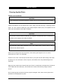
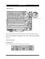
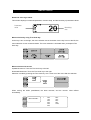
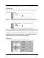
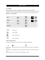
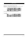
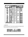
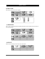
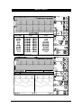
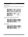
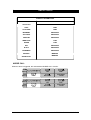
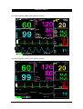
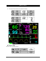
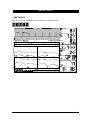
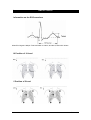
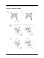
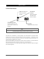
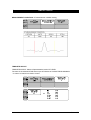
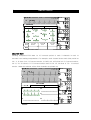
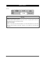
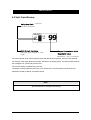
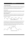
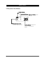
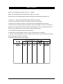
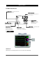
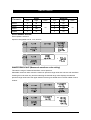
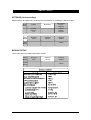
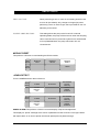
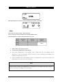
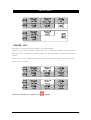
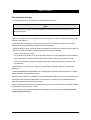
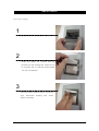
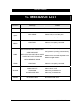
BM5 User’s Manual Signal and Data Validity It is extremely important to determine that the probe is attached to the patient correctly and the data is verifiable. To make this determination, three indications from the monitor are of assistance—signal strength bar, quality of the SPO2 waveform, and the stability of the SPO2 values. It is critical to observe all three indications simultaneously when ascertaining signal and data validity. Signal Strength Bar The signal strength bar is displayed within the SPO2 values window. This bar consists of 20 blocks set depending on the strength of the signal. Proper environmental conditions and probe attachment will help to ensure a strong signal. Quality of SPO2 Waveform Under normal conditions, the SPO2 waveform corresponds to (but is not proportional to) the arterial pressure waveform. The typical SPO2 waveform indicates not only a good waveform, but helps the user find a probe placement with the least noise spikes present. The figure below represents an SPO2 waveform of good quality. Good Quality SPO2 Waveform If noise (artifact) is seen on the waveform because of poor probe placement, the photodetector may not be flush with the tissue. Check that the probe is secured and the tissue sample is not too thick. Pulse rate is determined from the SPO2 waveform which can be disrupted by a cough or other hemodynamic pressure disturbances. Motion at the probe site is indicated by noise spikes in the normal waveform. (See the figure below.) It has been noted that letting the patient view the SPO2 waveform enables them to assist in reducing motion artifact. SPO2 Waveform with Artifact Rev. 3.0 6.SpO2 114