1
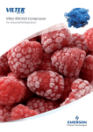
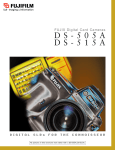
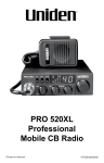
Specifications and Applications
Image Capturing Unit (Camera Head and Controller)
CCD chip
Number of pixels
Pixel size
Fujifilm Super CCD Area Type chip
3.2M pixels
10.75 x 10.75µm
Cooling
Two-stage thermoelectric module with air circulation
Cooling temperature
-30°C (when room is below 28°C)
Focusing
Power focusing; remote and preset control
Exposure time
1/100 second to 30 hrs (2 hrs to 30 hrs to be set manually)
Dynamic range
Four orders of magnitude
Gradation
16 bits (65,536)
Image size
12.6MB Max.; 49.2KB Min.
Maximum sample size
25 x 25cm (wide angle lens); 14 x 21cm (Fujinon VRF43LMD lens)
Binning
1 x 2, 2 x 4, 4 x 8, and 8 x 16 pixels
Interface
USB1.1
Software
Fujifilm Image Reader (Mac and Windows )
Image analysis
Fujifilm Image Gauge (Mac™); Fujifilm MultiGauge (Windows®)
®
Dimensions and Weights
Camera head
180 (W) x 170 (H) x 250mm (D)
3.4kg
Dark Box IV
510 (W) x 730 (H) x 480mm (D)
49.0kg
Chemiluminescence
Set a tray
Windows® XP or Mac™ OS 9 & X
Applicable Reagents and/or Samples
Chemiluminescence
CDP-Star®, ECL™, ECL Plus™, SuperSignal, ImmunoStar, CSPD®
Fluorescence
EtBr (W/UV light), SYBR® Green I & II, SYPRO® Orange,
GFP, DY-458XL (blue LED)
Sample
Chemiluminescence Mode
Pro-Q™ Diamond, Cy™3, RFP, DY-520XL (green LED), Alexa Fluor 633,
Cy™5, DY-647 (red LED)
Chemifluorescence
*AttoPhos™
Digitization
CBB-stained gel, Silver stained gel (Transilluminator)
NBT/BCIP-stained membrane. MW marker (Epi-illuminator)
Remote controlled
focusing
EPI-illuminator for fluorescence
blue LED (470nm), green LED (520nm), red LED (630nm)
EPI-illuminator for digitization
White-light source
Transilluminator - for digitization of
stained gels and autoradiographic films
White LED
UV transilluminator**
UV-light source (312nm)
Filter turret
Five positions
Fluorescence
Chemifluorescence
UV transilluminator
Intelligent Dark Box IV
Exposure
Filter
blue LED
green LED
red LED
Fluorescence
Chemifluorescence Mode
Digitization
White light
epi-illuminator
White transilluminator
Printers
Pictrography 3500
Lens
Remote Controlled
Sample is placed on a tray
Analyzing Unit
Operating system
Super CCD
The binning mode of the LAS-3000 allows researchers to select from four
binning settings to enhance both imaging sensitivity and image resolution.
Image capture
™
Analysis
Lens
High-sensitivity lens (FUJINON VRF43LMD)
Wide-view lens
F-number
0.85
F-number
Focal length
43mm
Focal length
24mm
Focus
Remote power focusing
Focus
Manual
Mount
Bayonet
Mount
Adapter to Nikon F mount
Digitize Mode
2.0
Science Imaging System
The image analysis process captures images in three modes:
chemiluminescence, fluorescence/chemifluorescence and digitization.
Fuji Photo Film Co., Ltd. 26-30, Nishiazabu 2-Chome, Minato-ku, Tokyo 106-8620, Japan, Tel: +81-3-3406-2201, Fax: +81-3-3406-2158 • http://lifescience.fujifilm.com • E-mail: [email protected]
Fujifilm Medical Systems U.S.A., Inc. 419 West Avenue, Stamford, CT 06902, U.S.A., Tel: +1-203-324-2000 ext. 6112 (1-800-431-1850 ext. 6112 in the U.S.), Fax: +1-203-351-4713 • http://www.fujimed.com • E-mail: [email protected]
Fuji Photo Film (Europe) GmbH, Heesenstr. 31, 40549 Düsseldorf, Germany, Tel: +49-211-5089-174, Fax: +49-211-5089-139 • http://www.fujifilm.de • E-mail: [email protected]
Specifications and system configuration subject to change for improvement without notice. All other product names mentioned herein are the trademarks of their respective owners.
Ref. No. BB-204E (04,09)
*No license is granted for use of AttoPhos™ to detect nucleic acid on a nylon membrane.
**No license is granted for pre-labeling gel electrophoresis method with the UV transilluminator.
LAS-3OOO
Luminescent image analyzer (LAS) for applications requiring high
sensitivity and a wide range of fluorescent capabilities
GFP expression in tobacco leaf
Detection of EPO as doping substance in sports
Isoelectric profiles of natural EPO found in normal
human urine (B) and the bands found in urine after
injections of epoetin or darbepoetin.
Data courtesy of Dr. Françoise Lasne.
Laboratoire National de Dépistage du Dopage,
Châtenay-Malabry, France
The LAS-3000 imaging system combines
Fujifilm’s high-sensitivity Super CCD
camera technology with the added versatility of white, blue, green and red EPI
illuminators. The Super CCD imaging
chip, binning mode and specially designed
camera lens allow researchers to capture
faint-light luminescent images with
unprecedented sensitivity and resolution.
Multicolor illuminator options enlarge
application area in fluorescent imaging.
More sensitivity for
chemiluminescent detection
More versatility for
fluorescent detection
Western blotting, Southern blotting and
Northern blotting detection by chemiluminescence is a widely accepted method. The
use of a cooled CCD camera system enables
the generation of a digital image and quantitative analysis of the image’s signal strength.
More and more fluorescent dyes are introduced into the biochemistry and molecular
biology fields for staining proteins and
nucleic acids in gels, membranes and well
plates. Expression analysis using green fluorescent protein (GFP) and red fluorescent
protein (RFP) can be detected by blue excitation or green excitation respectively.
Several state-of-the-art technologies were
incorporated to make the LAS-3000 system
as sensitive as the conventional film method:
• The newly designed F0.85 high-sensitivity Fujinon lens gathers as much light as
possible onto the CCD.
• The CCD is a 3.2M-pixel Super CCD
having large octagonal-shaped CCD pixels
10.75 x 10.75µm in size.
• The CCD is cooled to -30°C by peltier
when the environment is below 28°C.
• Pixel binning in the LAS-3000 electronically increases the pixel area to increase
sensitivity. The binning mode includes
four binning levels: 1 x 2 (Standard),
2 x 4 (High), 4 x 8 (Super) and 8 x 16
(Ultra). In the Ultra mode, the exposure
time is nearly 60 times faster than
Standard. An image smoothing function
increases the pixels to make the image
size the same as the Standard.
• Long exposure times are required to
capture images of extremely low-light
samples. The low-noise design of the
LAS-3000 system includes default settings
for a two-hour exposure and a 15-hour
exposure in addition to manual settings
for exposures as long as 30 hours.
Fujifilm Super CCD Area
Type imaging chip
Users are able to control the LAS-3000
with the Image Reader software in either
the user-friendly Lite mode or the more
advanced Pro mode. The modes are
selected with the click of a mouse.
Super CCD - By rotating pixels 45 degrees
to form an interwoven layout, the Super
CCD’s pixel pitch in the horizontal and
vertical directions is narrower than in the
diagonal direction, achieving higher horizontal and vertical resolution.
The five-position filter turret holds 77mm
filters, such as Y515 (general purpose for
blue LED excitation), 510DF10 (GFP, etc.),
575DF20 (RFP, Cy™3, Pro-Q™Diamond,
etc.), R670 (Cy™5, ALEXA Fluor® 633, etc.)
and 605DF40 (EtBr, etc.).
Quantitative image
analysis function
The 16-bit signal accumulated on each
CCD pixel is processed through an A/D
converter to form a digital image. The
image data obtained at the closed shutter
condition is the dark frame. The image
data of an even, flat sample is the flat
frame. To analyze the image data, the dark
frame and flat frame data are subtracted
from the original image. This correction function is included in the Image
Reader software. MultiGauge software
for Windows® and ImageGauge software
for Mac™ are the standard image analysis
software included in the ScienceLab
software provided by Fujifilm.
Digitizing functions
Capturing images as we see them is called
digitizing. For CBB-stained gel or silverstained gel, use of the white LED DIAilluminator at iris f2.8 without any filter is
recommended. For digitizing the molecular
weight standard on blotted membrane, white
light epi-illuminator, which is located in the
center part of the blue, green and red LED
epi-illuminator unit, is used.
FUJINON lens VRF43LMD and
five-position filter turret
Applications
Fluorescence/Chemifluorescence
Blue:
SYBR® Green I & II
SYPRO® Ruby
SYPRO® Tangerine
FAM™
ECFP
DY-458XL
SYBR® Gold
SYPRO® Orange
FITC
EGFP
AttoPhos™
Green:
SYPRO® Red
TAMRA™
HEX™
Alexa Fluor® 546
Pro-Q™ Diamond
BODIPY®576/589
R-phycoerythrin
HNPP
DY-547
Cy™3
ROX™
Alexa Fluor® 532
Deep Purple
Rhodamine Red™
NED™
RFP
DY-520XL
Red:
Alexa Fluor® 633
Alexa Fluor® 647
BODIPY®650/665
TOTO®-3
DY-647
Alexa Fluor® 635
Cy™5
DiD
DDAO phosphate
UV:
Ethidium Bromide
SYPRO® Rose
White:
Silver stained
NBT / BCIP
CBB
X-ray film
Chemiluminecence
NP Tray
•
Samples are placed on the appropriate tray
and placed in the intelligent dark box.
After closing the door, all the functions are
controlled remotely from the Image Reader
software. The software automatically recognizes the type of illuminator installed or
changed during operation.
The LAS-3000 includes multiple LED
options for various fluorescence applications,
including blue LED (470nm), green LED
(520nm) and red LED (630nm) for epi-illumination and a UV (312nm) light source for
DIA-illumination.
• A non-parallax (NP) tray is used to eliminate the parallax effect when imaging
chemiluminescence in the 96-well plate.
Up to two plates can be placed.
C H E M I L U M I N E S C E N C E
Easy-to-use operation
The five filter options available
for the LAS-3000.
F L U O R E S C E N C E / C H E M I F L U O R E S C E N C E
•
LED units
D I G I T I Z A T I O N
ECL™
ECL Advance™
Super Signal®
CSPD®
Bright-Star™
ECL Plus™
Lumi-Light Plus
CDP-Star™
Renaissance™