1
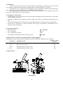
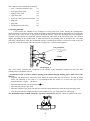
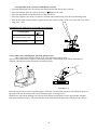
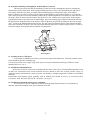
accessories - Cat. No.: 16610 user manual - Cat. No.: 20610.ENG 2013-09-23 USER MANUAL Patent Office: P – 301 657 International Patent: CT/PL94/00006 CYTO INSERT FOR ROTORS: 12452, 12271 Read this before use! MPW MED. INSTRUMENTS, 46 Boremlowska Street, 04-347 Warsaw/Poland tel. +48 22 610 81 07 (service), fax +48 22 610 55 36 [email protected], www.mpw.pl WARNING SIGNS WARNING! Warning of potential injury or health risk. This manual was prepared with special care. MPW MED. INSTRUMENTS may change the manual at any time and without notice because of improvements, typographical errors, inaccuracies of current information or improvements to facilities. 2 Contents 1. INTENDED USE. 4 2. ADVANTAGES OF CYTO INSERT. 4 3. OPERATION PARAMETERS. 4 4. SPECIFICATION OF CYTO INSERT (CATALOGUE NUMBER 16610) EQUIPMENT. 4 5. OUTFIT CONTENTS. 4 6. WORKING PRINCIPLE. 5 7. PREPARATION OF THE CYTO INSERT WITH THE PASSING OF THE FILTRATE THROUGH BLOTTING PAPER (WITH A LOSS OF THE FILTRATE). 5 8. PREPARATION OF THE CYTO INSERT WITH FILTRATE RETRIEVAL. 6 DIAMETER OF OPENINGS IN FILTRATION PAPER 6 9. PROCEDURE AFTER CENTRIFUGATION, WHEN THE FILTRATE IS LOST. 6 10. PROCEDURE FOLLOWING CENTRIFUGATION, WITH THE FILTRATE RETRIEVED. 7 11. CLEANING OF THE CYTO INSERT. 7 12. STERILIZATION AND DISINFECTION. 7 13. STERILIZATION AND DISINFECTIONS PROCESS VALIDATION. 7 14. MANUFACTURER’S DATA. 8 15. DISTRIBUTOR INFORMATION. 8 3 1. Intended use. The CYTO insert is the non sterile insert assigned for centrifuging in cytological rotors 12452 and 12271 applied to MPW-350/R/RH, MPW 351/R/RH, MPW 352/R/RH and MPW 223c spinners. Is intended for in vitro diagnostics (IVD) equipped; used to centrifugal separation of natural biological fluids, suspensions in isotonic solutions in order to obtain deposits and filtrates. The cytological insert get into the set of the rotor is essential in medicine and veterinary science, and may also be used in industry as well as technology. 2. Advantages of CYTO insert. fast deposition of cells on the microscope slide through centrifugation and the penetration of the filtrate into the blotting/ filter paper, possibility of filtrate recovery after the cells have settled on the slide by its automatic collection into a test tube, securing the fluid against its escaping into the centrifugation chamber (avoiding air-born contamination), inserts used as disposable non sterile equipment will help prevent infections, contamination of lab personnel or the environment, 3. Operation parameters. Rotational speed 100 ÷ 2500 rpm max. acceleration 750 x g max. centrifugation capacity 2 ml. area diameter of settling cells on the microscope slide 8,7 mm 4. Specification of CYTO insert (catalogue number 16610) equipment. Depending on the type of the purchased centrifuge, the below-mentioned equipment may be ordered. Number of piece Catalog No. Equipment name in the set 15123 Decantation test tube 2,2 ml with cap 100 16614 Basic microscope slide 100 16616 Blotting/ filtration card 9,5 mm 100 Blotting/ filtration card 12,5 mm 100 or 16617 5. Outfit contents. 6 8 7 4 5 3 2 1 Drawing No.1 4 The complete insert contains the following: base – collection chamber (PP) (1), basic microscope slide (2), blotting/ filter card (3), cyto insert (PS) (4), seal 8,7 mm (60 mm2) (Silicone) (5), plug (PE) (6), plug (PE) (7), decantation test tube (PP) (8), 6. Working principle. CYTO inserts are adapted to use in hangers of 12452 and 12271 rotors. During the centrifugation, insert turns into a horizontal position, which positioning is then blocked with snap fastening units located in the side arms of the rotor in case of the need to decant the filtrate. Under the centrifugal force the morphotic elements (cell precipitate) are separated from the suspension and settled on the microscope slide. The cell-free filtrate, depending on the method used, is either absorbed by the blotting paper or decanted into a test tube following centrifugation. After the decantation test tube containing the filtrate is removed in order to dry the precipitate on the microscope slide, the test tube is subjected to a second centrifugation. Cell precipitate CYTO Filtrate SUPERNATANT Drawing No. 2 The CYTO insert, following the completion of the settling of the morphotic elements on the basic slide centrifugation with filtrate retrieval. 7. Preparation of the cyto insert with the passing of the filtrate through blotting paper (with a loss of the filtrate). Prepared and labeled basic microscope slide should be inserted into the base-collector. In order to obtain better cell adhesion to the slide it is recommended that the slides be covered with poly-L-lysine, manufactured by “Sigma” – Chemical Place the blotting card with an opening of 9,5. Place the plugs on the cyto insert. Place the complete cyto insert onto the base-collector and symmetrically close the snap-fastening catch. Pour the prepared fluid sample into the central cylinder of the cyto insert and close with a plug. The maximum amount of fluid, which the cyto insert can hold is 2 ml. That volume is marked with a line. Drawing No. 3 5 8. Preparation of the cyto insert with filtrate retrieval. Prepared and labeled basic microscope slide should be inserted into the base-collector. Place the blotting/ filter card with an opening of 12,5 over the slide. Place the plug and the decantation tube over the cyto insert. Place the complete cyto insert over the base-collector and symmetrically close the snap-fastening catch. Pour in the tested biological fluid, suspension into the central cylinder of the cyto insert and close with a plug (max. 2 ml). DIAMETER OF OPENINGS IN FILTRATION PAPER Centrifugation Seal 8,7 With a loss of the filtrate into the filtration paper (card) 9,5 With filtrate retrieval into decantation test tube 12,5 Drawing No. 4 9. Procedure after centrifugation, when the filtrate is lost. Take out the insert from the hanger of the rotor in the horizontal position. The cyto insert should not be tipped, to avoid the possibility of the spillage of the filtrate from the basecollector, which has not been absorbed by the blotting paper. 9 10 Drawing No. 5 Place the cyto insert on a piece of blotting paper, which has one foiled side, and open it by lifting the snaps of the catch. Take the microscope slide out by lifting the insert at an angle of 45o. With forceps, take off the blotting card. Dry the tested filtrate, permeabilize in 96% ethyl alcohol, and then stain with a method of choice. The used elements of the insert should be placed in a sanitary bag for plastics. 6 10. Procedure following centrifugation, with the filtrate retrieved. Take the cyto insert from the blocked hanger of the rotor in the centrifugation position, keeping the decantation test tube tilted down. With a gentle twisting motion we remove the decantation tube and close it with a plug. Place the cyto insert on single-side foiled blotting paper and open the snaps of the catches, Remove just the cyto insert by lifting it up and place another blotting paper with an opening of 12.5 onto the slide, while trying not to move the remaining sample drop with the suspension. Put on the cyto insert and close the snaps. Then, in order to dry the sample, spin again for 3 min. with the rpm set previously. Following the centrifugation remove the cyto insert from the hanger, place onto blotting paper with one side foiled and open the the insert by lifting the snaps of the catches. Take out the microscope slide by tilting the insert at an angle of 45o.. Remove the filtration paper using forceps. Dry the tested precipitant with 96% ethyl alcohol and then stain with a technique of choice. The used cyto insert should be placed in a sanitary bag for plastics, in order to reduce the danger of personnel and environment contamination. Drawing No. 6 11. Cleaning of the CYTO insert. It is possible to depending on needs to use the insert many times till destroy. The used elements of the insert should be placed in a sanitary bag. It is necessary before every single usage of the insert to clean her and then dried using soft fabric or in the chamber drier at ca. 50 C. 12. Sterilization and disinfection. In the case of need it is necessary to disinfection the insert. One can use all standard disinfectants. Parts of insert are constructed from various materials and one should to take into account possible variety of materials. During sterilization by means of steam one should to consider temperature resistance of individual materials. Disinfectants and cleaning agents generally used in medical care should be used (e.g. Aerodesina-2000, Lysoformin 3000, Melseptol, Melsept SF, Sanepidex, Cutasept F). 13. Sterilization and disinfections process validation. It is necessary to execute validation of inserts cleaning and disinfection process in accordance to domestic instructions binding in the given laboratory tests unit. 7 14. Manufacturer’s data. MPW MED. INSTRUMENTS Boremlowska 46 Street 04-347 Warsaw +48 22 610 56 67 22 610 81 07 22 610 55 36 http:// www.mpw.pl e-mail: [email protected] 15. Distributor information. DISTRIBUTOR: 8 sales department service fax DECLARATION OF CONFORMITY Product CYTO insert cytology rotor Product classification on the basis of the Directive 98/79/EC Non classified to list A or B and not for self-testing Product complies with the requirements: · Directive 98/79/EC (IVD), including the requirements of harmonised standards: PN-EN ISO 13485:2012 PN-EN ISO 18113-3:2011 PN-EN ISO 13485:2012/AC:2013-03 PN-EN 61010-1:2011 PN-EN 13612:2006 PN-EN 61010-2-101:2005 PN-EN ISO 14971:2012 PN-EN ISO 62366:2008 · standard PN-EN ISO 15223-1:2012 „MPW MED. INSTRUMENTS” SPÓŁDZIELNIA PRACY Warsaw, 46 Boremlowska Street Quality policy in line with ISO 9001:2008 Certifying authority Warsaw, 07.12.2014 nr 10.610.03