1
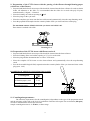
accessories type: CYTOSET: - set 12452c for MPW-350/R/RH, MPW-351/R/RH, MPW-352/R/RH user manual - Cat. No.: - set 12271c for MPW-223c 20452.ENG 2013-09-23 USER MANUAL Patent Office: P – 301 657 International Patent: CT/PL94/00006 CYTOLOGY SET CYTOSET Read this before use! MPW MED. INSTRUMENTS, 46 Boremlowska Street, 04-347 Warsaw/Poland tel. +48 22 610 81 07 (service), fax +48 22 610 55 36 [email protected], www.mpw.pl WARNING SIGNS WARNING! Warning of potential injury or health risk. This manual was prepared with special care. MPW MED. INSTRUMENTS may change the manual at any time and without notice because of improvements, typographical errors, inaccuracies of current information or improvements to facilities. 2 Contents 1. INTENDED USE. 4 2. ADVANTAGES OF "CYTOSET". 4 3. OPERATION PARAMETERS. 4 4. SPECIFICATION OF EQUIPMENT. 5 5. CYTOLOGICAL OUTFIT CONTENTS. 5 6. WORKING PRINCIPLE. 6 7. PREPARATION OF SAMPLES FOR CENTRIFUGATION. 6 8. TABLE OF DILUTIONS IN HEMATOLOGY. 6 9. PREPARATION OF THE CYTO INSERT WITH THE PASSING OF THE FILTRATE THROUGH BLOTTING PAPER (WITH A LOSS OF THE FILTRATE). 7 10. PREPARATION OF THE CYTO INSERT WITH FILTRATE RETRIEVAL. 7 DIAMETER OF OPENINGS IN FILTRATION PAPER 7 11. CENTRIFUGATION PARAMETERS. 7 12. STEPS IN THE USE OF CENTRIFUGE WITH THE "CYTOSET". 8 13. PROCEDURE AFTER CENTRIFUGATION, WHEN THE FILTRATE IS LOST. 9 14. PROCEDURE FOLLOWING CENTRIFUGATION, WITH THE FILTRATE RETRIEVED. 9 15. MAIN VIEW. 10 16. CLEANING, DISINFECTION, MAINTENANCE. 10 16.1. CLEANING OF THE ACCESSORIES. 16.2. CLEANING OF THE CYTO INSERT. 16.3. LUBRICATION. 16.4. STERILIZATION AND DISINFECTIONS OF THE ACCESSORIES. 16.5. STERILIZATION AND DISINFECTIONS PROCESS VALIDATION. 10 10 10 11 11 17. MANUFACTURER’S DATA. 11 18. DISTRIBUTOR INFORMATION. 11 3 1. Intended use. The “CYTOSET” cytological outfit is intended for in vitro diagnostics (IVD) equipped; used to centrifugal separation of body fluids into fine subconstituent elements from the filtrate. The fine elements are deposited directly on basic microscope slides, while the filtrate is accumulated in special tubes, following the settling of the particulate matter. The tested physiological fluids constitute a suspension of fine elements in the fluid fraction. Among them are the following: a) natural biological fluids including: cerebrospinal fluid, urines, effusions, serum leaks, joint fluid, leukorrhoea, pus and related others, b) suspensions in isotonic smear solutions, tissue punctuates, sputum, rinsings form the bronchi and respiratory pathways and related others. These suspensions may be tested in order to: 1) obtain precipitates of fine matter and the filtrate from the same sample of tested biological suspension, 2) obtain just a precipitate of the fine matter through passing of the filtrate into a blotting paper The cytological outfit is essential in medicine and veterinary science, and may also be used in industry as well as technology. 2. Advantages of "CYTOSET". fast deposition of cells on the microscope slide through centrifugation and the penetration of the filtrate into the blotting/ filter paper, possibility of filtrate recovery after the cells have settled on the slide by its automatic collection into a test tube, securing the fluid against its escaping into the centrifugation chamber (avoiding air-born contamination), inserts used as disposable equipment will help prevent infections, contamination of lab personnel or the environment, 3. Operation parameters. Rotational speed 100 ÷ 2500 rpm max. acceleration 750 x g max. centrifugation capacity 2 ml. area diameter of settling cells on the microscope slide 8,7 mm 4 4. Specification of equipment. Depending on the type of the purchased centrifuge, the below-mentioned “CYTOSET” cytological outfit may be ordered. Catalog No. Cytology rotor for centrifuges MPW-350/R/RH, MPW-351/R/RH, MPW352/R/RH Cytology rotor for centrifuge MPW-223c 12452 or 12271 Max rpm RCF Number of piece in the set 4 seats 2500 750 1 4 seats 2500 750 1 Equipment name 13606 Hanger 15123 Decantation test tube 2,2 ml with cap 100 16610 Cyto insert 100 16614 Basic microscope slide 100 16616 Blotting/ filtration card 9,5 mm 100 Blotting/ filtration card 12,5 mm 100 or 16617 4 5. Cytological outfit contents. The outfit contains a swing-out four-seats rotor with hangers, into which the complete cyto insert is 6 8 7 4 5 3 2 1 placed. Drawing No.1 The complete insert contains the following: base – collection chamber (1), basic microscope slide (2), blotting/ filter card (3), cyto insert (4), seal 8,7 mm (60 mm2) (5), plug (6), plug (7), decantation test tube (8), 5 6. Working principle. During the centrifugation, the cyto insert turns into a horizontal position, which positioning is then blocked with snap fastening units located in the side arms of the rotor in case of the need to decant the filtrate. Under the centrifugal force the morphotic elements (cell precipitate) are separated from the suspension and settled on the microscope slide. The cell-free filtrate, depending on the method used, is either absorbed by the blotting paper or decanted into a test tube following centrifugation. After the decantation test tube containing the filtrate is removed in order to dry the precipitate on the microscope slide, the test tube is subjected to a second centrifugation. Cell precipitate CYTO Filtrate SUPERNATANT Drawing No. 2 The CYTO insert, following the completion of the settling of the morphotic elements on the basic slide centrifugation with filtrate retrieval. 7. Preparation of samples for centrifugation. The amount of fluid used in centrifugation depends on its contents of cells, as well as its availability. If the fluid is cell-dense in order to avoid layering of the cells on the slide or their settling too densely, the fluid should be diluted with 0.9% NaCl solution or PBS. 8. Table of dilutions in hematology. Number of white cells Dilution factor Sample drops Saline drops 101 - 300 301 - 700 701 - 1500 1501 - 3000 1:2 1:5 1 : 10 20 10 4 2 0 10 16 18 When a cell-poor fluid with a low protein content (about 0.2 mg/ml) is tested there exists a danger, that the centrifuged cells on the slide may be fragmented, as well as the quality of the preparation is often not satisfactory. For that reason, if the filtrate is not needed for further tests, it is recommended that a few drops of blood serum or albumin solutions be added. Samples destined for cyto-centrifugation should be fresh in order to maximize the possibility of getting intact cells. 6 9. Preparation of the CYTO insert with the passing of the filtrate through blotting paper (with a loss of the filtrate). Prepared and labeled basic microscope slide should be inserted into the base-collector. In order to obtain better cell adhesion to the slide it is recommended that the slides be covered with poly-L-lysine, manufactured by “Sigma” – Chemical Place the blotting card with an opening of 9,5. Place the plugs on the cyto insert. Place the complete cyto insert onto the base-collector and symmetrically close the snap-fastening catch. Pour the prepared fluid sample into the central cylinder of the cyto insert and close with a plug. The maximum amount of fluid, which the cyto insert can hold is 2 ml. That volume is marked with a line. Drawing No. 3 10. Preparation of the CYTO insert with filtrate retrieval. Prepared and labeled basic microscope slide should be inserted into the base-collector. Place the blotting/ filter card with an opening of 12,5 over the slide. Place the plug and the decantation tube over the CYTO insert. Place the complete CYTO insert over the base-collector and symmetrically close the snap-fastening catch. Pour in the tested biological fluid, suspension into the central cylinder of the cyto insert and close with a plug (max. 2 ml). DIAMETER OF OPENINGS IN FILTRATION PAPER Centrifugation Seal 8,7 With a loss of the filtrate into the filtration paper (card) 9,5 With filtrate retrieval into decantation test tube 12,5 Drawing No. 4 11. Centrifugation parameters. The selection of parameters for the centrifugation is dependent on the type of the preparation tested and the resistance of the cells to the level of acceleration. Delicate cells require low acceleration (500 rpm), in order to maintain their microscopic structure. Sample centrifugation time is 3 –15 min., on the average. 7 12. Steps in the use of centrifuge with the "CYTOSET". place the centrifuge at the work station, plug into the power supply (socket with grounding), press the POWER switch, open the lid by pressing the lid-opening key, place the „ CYTOSET” rotor onto the sleeve and with a six-angle wrench turn the clamp until it will not turn any more place the hungers (9) into the rotor in the suitable position for the method used: a) when losing the filtrate into the blotting paper put the hanger (9) with the peg (10) from the rotor arm side without the snap (on the drawing No. 5, rotor pocket 2 and 4), b) when recovering the filtrate from the decantation tube, place the hanger (9) with the peg (10) at the rotor arm side with the snap (11) (on the drawing No. 5, rotor pocket 1 and 3), place the complete CYTO inserts filled with fluid into the hangers (minimum of two inserts in opposite pockets) in position like on the drawing No.8., close the lid of the centrifuge, set the centrifugation parameters (time, speed) and start the centrifuge according to the instruction manual, the timer will stop the centrifuge after the required time has elapsed, an earlier stop may be done by pressing STOP key. 10 11 10 9 Drawing No. 5 When the centrifuge has signalled the end of centrifuging – open the lid. Remove the insert in the position, in which it is located in the rotor, pay particular attention to the inserts with the decantation tube, which should be in the centrifugation position with the test tube facing down. 8 13. Procedure after centrifugation, when the filtrate is lost. Take out the insert from the hanger of the rotor in the horizontal position. The CYTO insert should not be tipped, to avoid the possibility of the spillage of the filtrate from the basecollector, which has not been absorbed by the blotting paper. 9 10 Drawing No. 6 Place the CYTO insert on a piece of blotting paper, which has one foiled side, and open it by lifting the snaps of the catch. Take the microscope slide out by lifting the insert at an angle of 45o. With forceps, take off the blotting card. Dry the tested filtrate, permeabilize in 96% ethyl alcohol, and then stain with a method of choice. The used elements of the insert should be placed in a sanitary bag for plastics. 14. Procedure following centrifugation, with the filtrate retrieved. Take the CYTO insert from the blocked hanger of the rotor in the centrifugation position, keeping the decantation test tube tilted down. With a gentle twisting motion we remove the decantation tube and close it with a plug. Place the CYTO insert on single-side foiled blotting paper and open the snaps of the catches, Remove just the CYTO insert by lifting it up and place another blotting paper with an opening of 12.5 onto the slide, while trying not to move the remaining sample drop with the suspension. Put on the CYTO insert and close the snaps. Then, in order to dry the sample, spin again for 3 min. with the rpm set previously. Following the centrifugation remove the CYTO insert from the hanger, place onto blotting paper with one side foiled and open the the insert by lifting the snaps of the catches. Take out the microscope slide by tilting the insert at an angle of 45o.. Remove the filtration paper using forceps. Dry the tested precipitant with 96% ethyl alcohol and then stain with a technique of choice. The used CYTO insert should be placed in a sanitary bag for plastics, in order to reduce the danger of personnel and environment contamination. Drawing No. 7 9 15. Main view. Drawing No. 8 Main view of “CYTOSET” cytological outfit. 16. Cleaning, disinfection, maintenance. CAUTION! It is necessary to use protective gloves during following work. 16.1. Cleaning of the accessories. In order to ensure safe operation one shall carry out in regular way periodical maintenance of the accessories. Manufactured rotors, hangers and round carriers have to withstand steady high stresses originating. Chemical reactions as well as corrosion (combination of variable pressure and chemical reactions) can cause corrosion or destruction of metals. Hard to observe surface cracks increase gradually and weaken material without visible symptoms. In the case of observation of surface damage, crevice or other change, as well as the corrosion, the given part (rotor, hanger, etc.) shall be immediately replaced. In order to prevent corrosion one has to clean regularly the rotor with the complete clamp and hangers. Cleaning of the accessories shall be carried out outside of the centrifuge once every week or still better after each use. Then, those parts shall be dried using soft fabric or in the chamber drier at ca. 50 C. Especially prone to the corrosion are parts made of aluminum. For cleaning them one should use neutral agent of pH value from 6 to 8. It is forbidden to use alkaline agent of pH above 8. In this way, the useful service life of the device is substantially increased and susceptibility to corrosion is diminished. Accurate maintenance increases the service life as well and protects against premature rotor failures. Corrosion and damages resulting from insufficient maintenance could not be subject of claims lodged against the manufacturer. 16.2. Cleaning of the CYTO insert. It is possible to depending on needs to use the insert many times till destroy. The used elements of the insert should be placed in a sanitary bag. It is necessary before every single usage of the insert to clean her and then dried using soft fabric or in the chamber drier at ca. 50 C. 16.3. Lubrication. The rotor pins shall be always lubricated with technical petroleum jelly. In this way, the uniform deflection of the buckets and quiet centrifuge operation is ensured. 10 16.4. Sterilization and disinfections of the accessories. One can use all standard disinfectants. Accessories are constructed from various materials and one should to take into account possible variety of materials. During sterilization by means of steam one should to consider temperature resistance of individual materials. Rotors and hangers can be sterilized in autoclave with temperature 121o – 124o C and pressure 215 kPa during 20 min. In the centrifuge, disinfectants and cleaning agents generally used in medical care should be used (e.g. Aerodesina-2000, Lysoformin 3000, Melseptol, Melsept SF, Sanepidex, Cutasept F). CYTO inserts aren't able to be sterilized in autoclave!!! During the above mentioned works one must wear safety gloves. 16.5. Sterilization and disinfections process validation. It is necessary to execute validation of inserts cleaning and disinfection process in accordance to domestic instructions binding in the given laboratory tests unit. 17. Manufacturer’s data. MPW MED. INSTRUMENTS Boremlowska 46 Street 04-347 Warsaw +48 22 610 56 67 22 610 81 07 22 610 55 36 www.mpw.pl e-mail: [email protected] http:// 18. Distributor information. DISTRIBUTOR: 11 sales department service fax