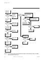
1
ABI 173A MicroBlotter User’s Manual © Copyright 2001, Applied Biosystems ABI PRISM and the ABI PRISM design, Applied Biosystems, Aquapore, Brownlee, GeneScan, INHERIT, Masterpiece, MicroCoat, MPLC, NEWGUARD, OPC, POLYPORE, Precipitette, ProBlott, PROCISE, ProSort, ProSpin, SeqEd, SPHERI10, SPHERI5, SynthAssist, and VeloSep are registered trademarks of Applera Corporation or its subsidiaries in the U.S. and certain other countries. ABI, Amplicover, Anitron, AutoAssembler, BaseSprinter, Biobytes, CATALYST, FastPhoramidite, GeneAssist, Genotyper, HLP, Hot Start, MicroBlotter, ONESTEP, PCR-MATE, PDQ, Phosphalink, ProFocus, ProSorb, Sequence Navigator, StockMarks, Stretch, and Synergy are trademarks of Applera Corporation or its subsidiaries in the U.S. and certain other countries. All other trademarks are the sole property of their respective owners. Applied Biosystems Contents 1 Introduction About This Manual System Description Optional Equipment User Attention Words Safety Information To Get Started Quickly Technical Support Contacting Technical Support To Contact Technical Support by E-Mail Hours for Telephone Technical Support To Contact Technical Support by Telephone or Fax To Reach Technical Support Through the Internet To Obtain Documents on Demand 2 Unpacking and Installation Introduction Preparing the Site Unpacking the System Components Description of the MicroBlotter Front Panel Rear Panel Operation Configuring the Voltage and Installing Fuses MicroBlotter Fuse Installation Chart Recorder Voltage Configuration and Fuse Installation Preparing the 140D Preparing the 785A Installing the Electrical Connections Installing the System Plumbing Assembling the Fittings Connecting Tubing with Teflon Couplers Configuring the 140D Priming the System Preparing the Solvents Purging the System Testing the System System Installation Test Program Verifying the System Test Performing Additional System Tests February 2001 1-1 1-3 1-4 1-5 1-6 1-7 1-8 1-9 1-9 1-9 1-9 1-10 1-13 1-13 2-1 2-3 2-4 2-5 2-7 2-7 2-8 2-8 2-9 2-9 2-10 2-11 2-14 2-15 2-17 2-17 2-24 2-27 2-28 2-28 2-28 2-32 2-36 2-40 2-42 iii Applied Biosystems Verifying the System Test 3 Operation Introduction Materials Required Materials Supplied with the 173A System Materials Required but not Supplied Dye Mark II cLC Staining Dye Operating the 173A System Preparing the Instruments for a Run Programming the 140D Preparing and Injecting Your Sample Executing a Programmed Gradient Run Processing Your Sample After a Run Excising Peaks of Interest From the PVDF Membrane Staining Blotted Proteins with the cLC Staining Dye Preparing Peptides for Sequencing Storing Excised Peaks of Interest Cleaning Blotter Tray Covers 4 Maintenance and Troubleshooting Introduction Maintenance System Storage Troubleshooting 173A General System Maintenance Maintaining the Column Flushing the Column Maintaining the MicroBlotter Adjusting the Tubing Running Through the Solenoid Replacing the Tubing That Runs Through the Solenoid Connecting Tubing with Teflon Couplers Maintaining the 140D Maintaining the 112A Maintaining the 785A Troubleshooting the 173A System 173A System Troubleshooting Guide Locating and Removing a Blockage Using the cLC Protein Standard Reconstituting the cLC Protein Standard Evaluating a cLC Protein Standard Run Storing the 173A System iv 2-44 3-1 3-3 3-4 3-4 3-4 3-5 3-5 3-7 3-7 3-13 3-22 3-23 3-26 3-26 3-27 3-28 3-28 3-28 4-1 4-3 4-3 4-3 4-3 4-4 4-5 4-5 4-6 4-6 4-6 4-8 4-9 4-10 4-11 4-12 4-12 4-18 4-21 4-21 4-21 4-23 February 2001 Applied Biosystems A Limited System Warranty A-1 B Sample Preparation Guidelines B-1 Sample Preparation Guidelines Prior to Injection onto the 173A System Protein Digestion Protocols Sample Preparation Recommendations Prior to Sequencing Recommendations for Sequencing Peptides C Plumbing Parts and Connections 140D Plumbing Diagram ABI 173A MicroBlotter Capillary HPLC System Plumbing Diagram D Accessory and Installation Kits February 2001 B-1 B-1 B-3 B-3 C-1 C-1 C-2 D-1 v Applied Biosystems 1 Introduction Contents About This Manual System Description Optional Equipment User Attention Words Safety Information To Get Started Quickly Technical Support Contacting Technical Support To Contact Technical Support by E-Mail Hours for Telephone Technical Support To Contact Technical Support by Telephone or Fax To Reach Technical Support Through the Internet To Obtain Documents on Demand April 2002 1 Introduction 1-3 1-4 1-5 1-6 1-7 1-8 1-9 1-9 1-9 1-9 1-10 1-13 1-13 1-1 Applied Biosystems 1-2 1 Introduction April 2002 Applied Biosystems About This Manual This user’s manual provides detailed information on the installation and use of the ABI 173A MicroBlotterTM Capillary HPLC System (173A system). The manual is organized into the following sections. 1 Introduction Includes a system description; important safety information; recommendations for getting started quickly; and information on contactingApplied Biosystems for technical assistance with this product. 2 Unpacking and Installation Describes how to unpack, install, configure, program and test the instruments in the 173A system. 3 Operation Provides detailed instructions on how to operate the 173A system. Instrument setup prior to a run; a standard protein/peptide gradient program for use as a learning tool and as a template for designing your own programs; and instructions on sample handling for subsequent analysis are included. 4 Maintenance and Troubleshooting Provides general maintenance procedures and recommendations that will help you keep the 173A system performing optimally. Also included are maintenance procedures for the column, and a troubleshooting guide. Appendixes Includes our warranty statement; sample preparation techniques including protocols for protein digests; recommendations for sample treatment prior to sequencing; and plumbing diagrams. Index A comprehensive index designed to help you locate the information you need quickly and easily. In addition, the ABI 173A MicroBlotter Capillary HPLC System Safety Summary (P/N 903918) is included in the front pocket of this binder. It contains important information on the safe installation, operation, and maintenance of the instruments in this system. The Safety Summary also contains Material Safety Data Sheets for the chemistry provided with this system. April 2002 1 Introduction 1-3 Applied Biosystems System Description The ABI 173A MicroBlotter Capillary HPLC System (173A system) is a capillary liquid chromatography sample preparation system. It provides users with an integrated protein/peptide mapping system for research requiring high sensitivity and high recoveries, especially for protein sequence analysis. The 173A system separates and prepares very small amounts of sample in one step with no significant sample loss. The eluted protein/peptide fractions at picomole or subpicomole levels are collected onto a PVDF membrane. The surface area onto which the fractions are collected is very small—2 to 3 mm o.d. spots. This eliminates the need for further concentration or transfer steps, thus minimizing possible sample loss. Once your sample has been separated and blotted onto a PVDF membrane, you simply line up the membrane with the corresponding chromatogram, and excise the fractions (peaks) you wish to analyze. The excised membranes can then be directly loaded into an automated protein/peptide sequencer for analysis. 173A system components have been selected and designed to ensure maximum reproducibility, sensitivity and sample recovery. System components include: • The ABI 140D Microgradient Delivery System (P/N 140D-01) The 140D accurately forms gradients at very low flow rates, typically 3 to 10 µL/min. It eliminates the need for using a split technique which is required by other HPLC systems to achieve similar flow rates. • The ABI 112A Oven/Injector (P/N 0650-0016) The 112A is a column injection oven which loads samples onto a capillary peptide column for separation and analysis. • The ABI 785A Programmable Absorbance Detector (P/N 604050) The 785A monitors the elution of the sample analytes (proteins/ peptides) as they pass through the capillary flowcell. • The ABI 173 MicroBlotter (P/N 173-0) The MicroBlotter is a membrane-based sample collector which eliminates manual fraction collection. Samples are collected directly onto a PVDF membrane after separation via a push-pull solenoid. Activated by the 140D, the solenoid gently contacts the terminus of the capillary tubing to the membrane as samples are eluted from the column. 1-4 1 Introduction April 2002 Applied Biosystems A user’s manual for each instrument, and installation/accessory kits are also included with this system. The kits include: • Consumables including solvents, MicroBlotter trays, a set of PVDF membranes and filters, the cLC Dye Mark, cLC Staining Dye, and a cLC Protein Standard. • A 0.5 mm x 15 cm capillary peptide column. • A capillary U-shaped flowcell Appendix D contains a complete listing of the contents of each installation and accessory kit shipped with the instruments in this system. Optional Equipment • The Kipp & Zonen Dual-Pen Strip Chart Recorder (P/N 400268) The 173A system was optimized using a chart recorder of this type; therefore, we recommend the use of the Kipp & Zonen Dual-Pen Strip Chart Recorder (P/N 400268). Throughout this user’s manual, generalized chart recorder instructions are listed along with specific instructions for the Kipp & Zonen chart recorder. April 2002 1 Introduction 1-5 Applied Biosystems User Attention Words Four user attention words appear throughout this manual. Each word implies a particular level of observation or action. 1-6 Note This word is used to call attention to information. IMPORTANT This information is given because it is necessary for correct operation of the instrument. Caution This word informs the user that damage to the instrument could occur if the user does not comply with this information. It also indicates a potentially hazardous situation which could result in minor or moderate injury to the user. WARNING Serious physical injury to the user or other persons could occur if these required precautions are not taken. 1 Introduction April 2002 Applied Biosystems Safety Information Safety warnings addressing chemical and high voltage issues appear throughout this user’s manual and the ABI 173A MicroBlotter Capillary HPLC System Safety Summary (P/N 903918). The Safety Summary is a stand-alone document located in the front pocket of this binder. It contains detailed information provided to ensure the safe installation, operation and maintenance of the 173A system. The Safety Summary also contains Material Safety Data Sheets (MSDSs) for the chemicals that are shipped with this system. The safety warnings listed throughout this manual appear in the following formats. April 2002 WARNING ELECTRICAL SHOCK HAZARD. The ABI 173A MicroBlotter Capillary HPLC System is comprised of instruments containing high voltage power supplies. Always disconnect the instrument(s) from their power source before changing a fuse, adjusting the voltage selection, or opening the instrument for maintenance or any other reason. WARNING CHEMICAL HAZARD. Some chemicals used with this instrument are considered hazardous. Hazard warnings are prominently displayed on the bottle labels of all hazardous chemicals. The Safety Summary for this manual contains material safety data sheets (MSDSs) that provide information about the physical characteristics, health hazards, safety precautions, first aid, spill cleanup and proper disposal procedures. Familiarize yourself with the information contained in the MSDSs before operating the instrument. Additional copies of the MSDSs are available from Applied Biosystems at no charge. 1 Introduction 1-7 Applied Biosystems To Get Started Quickly Once your 173A system has been installed, the 140D must be configured and primed, and the entire system must be tested for proper performance. These procedures are provided in section 2, "Unpacking and Installation". IMPORTANT Read the Safety Summary before installing, operating or performing maintenance on any of the instruments in this system. If you are not familiar with how to operate the 140D, we recommend you read Sections 2 and 3 of the 140D user’s manual to familiarize yourself with the instrument’s control panel and operation. Once your system has been configured, primed and tested, we recommend you perform at least one additional test run using a standard. This will help you familiarize yourself with setting up the instruments, and with handling the PVDF membrane before and after separation. If desired, the cLC Protein Standard supplied with this system can be used for the additional test. Instructions for preparing and executing a run with this standard are provided in section 2 on page 2-42. The standard protein/peptide gradient program provided in section 3 on page 3-14 can be used as the basis for your programmed gradient runs. The conditions listed for peptides are adequate for most enzymatic digests and peptide mixtures. The conditions listed for proteins can be used for most protein mixtures. Optimization of this program will be required, however, for your particular applications and samples. IMPORTANT 1-8 1 Introduction To avoid blocking the column and system tubing, samples must be as free of particulates as possible prior to injection. Refer to Appendix B, “Sample Preparation Guidelines” for more information on sample preparation and purification. April 2002 Applied Biosystems Technical Support Contacting Technical Support You can contact Applied Biosystems for technical support by telephone or fax, by e-mail, or through the Internet. You can order Applied Biosystems user documents, MSDSs, certificates of analysis, and other related documents 24 hours a day. In addition, you can download documents in PDF format from the Applied Biosystems Web site (please see the section “To Obtain Documents on Demand” following the telephone information below). To Contact Technical Support by E-Mail Contact technical support by e-mail for help in the following product areas: Product Area E-mail address Genetic Analysis (DNA Sequencing) [email protected] Sequence Detection Systems and PCR [email protected] Protein Sequencing, Peptide and DNA Synthesis [email protected] Biochromatography, PerSeptive DNA, PNA and Peptide Synthesis systems, CytoFluor®, FMAT™, Voyager™, and Mariner™ Mass Spectrometers [email protected] Applied Biosystems/MDS Sciex [email protected] Chemiluminescence (Tropix) [email protected] Hours for Telephone Technical Support In the United States and Canada, technical support is available at the following times: April 2002 Product Hours Chemiluminescence 8:30 a.m. to 5:30 p.m. Eastern Time Framingham support 8:00 a.m. to 6:00 p.m. Eastern Time All Other Products 5:30 a.m. to 5:00 p.m. Pacific Time 1 Introduction 1-9 Applied Biosystems To Contact Technical Support by Telephone or Fax In North America To contact Applied Biosystems Technical Support, use the telephone or fax numbers given below. (To open a service call for other support needs, or in case of an emergency, dial 1-800-831-6844 and press 1.) Product or Product Area Telephone Dial... Fax Dial... ABI PRISM® 3700 DNA Analyzer 1-800-831-6844, then press 8 1-650-638-5981 DNA Synthesis 1-800-831-6844, then press 21 1-650-638-5981 Fluorescent DNA Sequencing 1-800-831-6844, then press 22 1-650-638-5981 Fluorescent Fragment Analysis (includes GeneScan® applications) 1-800-831-6844, then press 23 1-650-638-5981 Integrated Thermal Cyclers (ABI PRISM ® 877 and Catalyst 800 instruments) 1-800-831-6844, then press 24 1-650-638-5981 ABI PRISM ® 3100 Genetic Analyzer 1-800-831-6844, then press 26 1-650-638-5981 BioInformatics (includes BioLIMS™, BioMerge™, and SQL GT™ applications) 1-800-831-6844, then press 25 1-505-982-7690 Peptide Synthesis (433 and 43X Systems) 1-800-831-6844, then press 31 1-650-638-5981 Protein Sequencing (Procise Protein Sequencing Systems) 1-800-831-6844, then press 32 1-650-638-5981 PCR and Sequence Detection 1-800-762-4001, then press 1 for PCR, 2 for the 7700 or 1-240-453-4613 5700, 6 for the 6700 or dial 1-800-831-6844, then press 5 1-10 Voyager MALDI-TOF Biospectrometry and Mariner ESI-TOF Mass Spectrometry Workstations 1-800-899-5858, then press 13 1-508-383-7855 Biochromatography (BioCAD Workstations and Poros Perfusion Chromatography Products) 1-800-899-5858, then press 14 1-508-383-7855 Expedite Nucleic acid Synthesis Systems 1-800-899-5858, then press 15 1-508-383-7855 Peptide Synthesis (Pioneer and 9050 Plus Peptide Synthesizers) 1-800-899-5858, then press 15 1-508-383-7855 1 Introduction April 2002 Applied Biosystems Product or Product Area PNA Custom and Synthesis Telephone Dial... Fax Dial... 1-800-899-5858, 1-508-383-7855 then press 15 FMAT 8100 HTS System and Cytofluor 4000 Fluorescence Plate Reader 1-800-899-5858, then press 16 1-508-383-7855 Chemiluminescence (Tropix) 1-800-542-2369 (U.S. 1-781-275-8581 only), or 1-781-271-0045 Applied Biosystems/MDS Sciex 1-800-952-4716 1-650-638-6223 Telephone Dial... Fax Dial... Outside North America Region Africa and the Middle East Africa (English Speaking) and West Asia (Fairlands, South Africa) 27 11 478 0411 27 11 478 0349 South Africa (Johannesburg) 27 11 478 0411 27 11 478 0349 Middle Eastern Countries and North Africa (Monza, Italia) 39 (0)39 8389 481 39 (0)39 8389 493 Eastern Asia, China, Oceania Australia (Scoresby, Victoria) 61 3 9730 8600 61 3 9730 8799 China (Beijing) 86 10 64106608 86 10 64106617 Hong Kong 852 2756 6928 852 2756 6968 Korea (Seoul) 82 2 593 6470/6471 82 2 593 6472 Malaysia (Petaling Jaya) 60 3 758 8268 60 3 754 9043 Singapore 65 896 2168 65 896 2147 Taiwan (Taipei Hsien) 886 2 22358 2838 886 2 2358 2839 Thailand (Bangkok) 66 2 719 6405 66 2 319 9788 Europe April 2002 Austria (Wien) 43 (0)1 867 35 75 0 43 (0)1 867 35 75 11 Belgium 32 (0)2 712 5555 32 (0)2 712 5516 Czech Republic and Slovakia (Praha) 420 2 61 222 164 420 2 61 222 168 Denmark (Naerum) 45 45 58 60 00 45 45 58 60 01 Finland (Espoo) 358 (0)9 251 24 250 358 (0)9 251 24 243 France (Paris) 33 (0)1 69 59 85 85 33 (0)1 69 59 85 00 Germany (Weiterstadt) 49 (0) 6150 101 0 49 (0) 6150 101 101 Hungary (Budapest) 36 (0)1 270 8398 36 (0)1 270 8288 Italy (Milano) 39 (0)39 83891 39 (0)39 838 9492 Norway (Oslo) 47 23 12 06 05 47 23 12 05 75 Poland, Lithuania, Latvia, and Estonia (Warszawa) 48 (22) 866 40 10 48 (22) 866 40 20 1 Introduction 1-11 Applied Biosystems Region Telephone Dial... Fax Dial... Portugal (Lisboa) 351 (0)22 605 33 14 351 (0)22 605 33 15 Russia (Moskva) 7 095 935 8888 7 095 564 8787 South East Europe (Zagreb, Croatia) 385 1 34 91 927 385 1 34 91 840 Spain (Tres Cantos) 34 (0)91 806 1210 34 (0)91 806 1206 Sweden (Stockholm) 46 (0)8 619 4400 46 (0)8 619 4401 Switzerland (Rotkreuz) 41 (0)41 799 7777 41 (0)41 790 0676 The Netherlands (Nieuwerkerk a/d IJssel) 31 (0)180 331400 31 (0)180 331409 United Kingdom (Warrington, Cheshire) 44 (0)1925 825650 44 (0)1925 282502 All other countries not listed (Warrington, UK) 44 (0)1925 282481 44 (0)1925 282509 Japan Japan (Hacchobori, Chuo-Ku, Tokyo) 81 3 5566 6230 81 3 5566 6507 Latin America Del.A. Obregon, Mexico 1-12 1 Introduction 305-670-4350 305-670-4349 April 2002 Applied Biosystems To Reach Technical Support Through the Internet We strongly encourage you to visit our Web site for answers to frequently asked questions and for more information about our products. You can also order technical documents or an index of available documents and have them faxed or e-mailed to you through our site. The Applied Biosystems Web site address is http://www.appliedbiosystems.com/techsupp To submit technical questions from North America or Europe: Step Action 1 Access the Applied Biosystems Technical Support Web site. 2 Under the Troubleshooting heading, click Support Request Forms, then select the relevant support region for the product area of interest. 3 Enter the requested information and your question in the displayed form, then click Ask Us RIGHT NOW (blue button with yellow text). 4 Enter the required information in the next form (if you have not already done so), then click Ask Us RIGHT NOW. You will receive an e-mail reply to your question from one of our technical experts within 24 to 48 hours. To Obtain Documents on Demand Free, 24-hour access to Applied Biosystems technical documents, including MSDSs, is available by fax or e-mail or by download from our Web site. To order documents... Then... by index number a. Access the Applied Biosystems Technical Support Web site at http://www.appliedbiosystems.com/techsupp b. Click the Index link for the document type you want, then find the document you want and record the index number. c. Use the index number when requesting documents following the procedures below. by phone for fax delivery a. From the U.S. or Canada, call 1-800-487-6809, or from outside the U.S. and Canada, call 1-858-712-0317. b. Follow the voice instructions to order the documents you want. Note April 2002 1 Introduction There is a limit of five documents per request. 1-13 Applied Biosystems To order documents... through the Internet for fax or e-mail delivery Then... a. Access the Applied Biosystems Technical Support Web site at http://www.appliedbiosystems.com/techsupp b. Under Resource Libraries, click the type of document you want. c. Enter or select the requested information in the displayed form, then click Search. d. In the displayed search results, select a check box for the method of delivery for each document that matches your criteria, then click Deliver Selected Documents Now (or click the PDF icon for the document to download it immediately). e. Fill in the information form (if you have not previously done so), then click Deliver Selected Documents Now to submit your order. Note There is a limit of five documents per request for fax delivery but no limit on the number of documents you can order for e-mail delivery. 1-14 1 Introduction April 2002 Applied Biosystems 2 Unpacking and Installation Contents Introduction Preparing the Site Unpacking the System Components Description of the MicroBlotter Front Panel Rear Panel Operation Configuring the Voltage and Installing Fuses MicroBlotter Fuse Installation Chart Recorder Voltage Configuration and Fuse Installation Preparing the 140D Preparing the 785A Installing the Electrical Connections Installing the System Plumbing Assembling the Fittings Connecting Tubing with Teflon Couplers Configuring the 140D Priming the System Preparing the Solvents Purging the System Testing the System System Installation Test Program Verifying the System Test Performing Additional System Tests Verifying the System Test April 2002 2 Unpacking and Installation 2-3 2-4 2-5 2-7 2-7 2-8 2-8 2-9 2-9 2-10 2-11 2-14 2-15 2-17 2-17 2-24 2-27 2-28 2-28 2-28 2-32 2-35 2-39 2-42 2-44 2-1 Applied Biosystems 2-2 2 Unpacking and Installation April 2002 Applied Biosystems Introduction This section describes how to install the ABI 173A MicroBlotter Capillary HPLC System (173A system) and prepare it for operation. The 173A system consists of five instruments. • ABI 140D Microgradient Delivery System (140D) • ABI 112A Oven/Injector (112A) • ABI 785A UV/VIS Detector (785A) • ABI 173 MicroBlotter (MicroBlotter) • Optional Kipp & Zonen Strip Chart Recorder (Chart Recorder) IMPORTANT A chart recorder must be installed with the 173A system, which was developed using a Kipp & Zonen Strip Chart Recorder. We recommend the use of the Kipp & Zonen Dual-Pen Strip Chart Recorder, P/N 400268, with this system. Each instrument has its own user’s manual except for the MicroBlotter which is covered in this manual. We recommend you review the installation and operation sections of the user’s manual for each instrument before proceeding with these instructions. At various points throughout this procedure, you will be referred to other user’s manuals for more detailed instructions. The steps required to install the 173A system are: April 2002 • Preparing the site • Unpacking the instruments • Installing the electrical connections • Installing the plumbing, capillary column and flowcell • Configuring the 140D • Priming the system • Configuring each instrument for operation • Testing the installation 2 Unpacking and Installation 2-3 Applied Biosystems Preparing the Site Site preparation requirements are covered in detail in the ABI 173A MicroBlotter Capillary HPLC System Pre-installation Manual (P/N 903829). Figure 2-1 illustrates the suggested instrument configuration for the 173A system. This configuration requires a clear table or bench area at least 6 ft long and 2.5 ft wide. In addition, 2 power outlets—one for the MicroBlotter and one for the power strip—are required. The MicroBlotter should always be placed on the bench or table surface, never on top of another instrument. Placing the MicroBlotter on top of another instrument requires longer tubing. This can adversely effect the precise timing required between sample placement onto the PVDF membrane in relation to the signals sent to the chart recorder to produce the corresponding chromatograph. 112A 140D Strip Chart Recorder 785A MicroBlotter Solvent and waste reservoirs Figure 2-1. Recommended configuration for the 173A system 2-4 2 Unpacking and Installation April 2002 Applied Biosystems Unpacking the System Components The hardware for the 173A system is shipped in six cartons. The Chemical Installation Kit (P/N 402066) is shipped separately to arrive before the hardware. To unpack the 173A system components and accessories: 1. Check that the Chemical Installation Kit has arrived, and that the contents were stored as directed. WARNING BACK INJURY HAZARD. Do not lift the 140D directly out of the carton. Unpacking the 140D requires two people. Use proper lifting procedures to avoid back injury. 2. Set the carton with the 140D on the floor. 3. Fold the flaps back tightly against the sides of the carton, and carefully roll the box upside down. 4. Lift the carton off the instrument and packing materials. 5. Carefully roll the 140D right side up. 6. Remove the foam and packing materials. 7. Unpack the remaining instruments one at a time. 8. Carefully inspect the instruments and the boxes they were shipped in for signs of damage. If any damage has occurred, save all the packaging materials for inspection and contact the shipper. 9. Verify that you have received the tubing and accessory kits listed below. Check the contents of each kit against the corresponding packing list. Packing lists are also included in Appendix D. If any parts are missing, contact your Applied Biosystems representative or Applied Biosystems technical support immediately. Refer to section 1, "Technical Support", on page 1-9 for the technical support number. Instrument 140D Accessory Kit 785A Accessory Kit 112A Accessory Kit 173A Voltage/Accessory Kit 173A System Tubing Kit April 2002 2 Unpacking and Installation Part Number P/N 603973 P/N 604091 P/N 0602-0081 P/N 604030 P/N 604066 (inside kit 604030) 2-5 Applied Biosystems 10. Place each instrument on the table or bench where the system will be used. Figure 2-1 on page 2-4 illustrates the recommended configuration. Allow 1 to 2 in. between each instrument (except those that are stacked), and at least 5 in. clearance behind the instruments to provide unobstructed air flow for ventilation. 2-6 2 Unpacking and Installation April 2002 Applied Biosystems Description of the MicroBlotter Front Panel The front panel of the MicroBlotter is illustrated in Figure 2-2. • Speed Toggle Switch and Indicators The speed at which the solenoid moves across the PVDF membrane in the blotter tray. The speed is set to 1 or 2 mm/min by pressing the toggle switch marked SPEED. The corresponding speed indicator LED will light. • Solenoid The mechanism by which the sample is transferred from the flowcell to the PVDF membrane. • Blotter Tray Holder Holds the blotter tray during a run. The holder and the blotter tray are keyed, so the tray fits into the housing one way only. • Reset Button Resets the solenoid to the home position by inserting the end of a paper clip into the hole marked reset. Home position Reset button Speed indicators Speed toggle switch Solenoid in the start position End position Blotter tray housing Figure 2-2. Front panel of MicroBlotter April 2002 2 Unpacking and Installation 2-7 Applied Biosystems Rear Panel Power switch Start signal connection Power cord connector Fuse holder Figure 2-3. Rear panel of the MicroBlotter Operation The MicroBlotter is activated by the 140D via a connection from the rear panel of the MicroBlotter to an event relay on the rear panel of the 140D. During a programmed run, the MicroBlotter and chart recorder are typically set to begin operation simultaneously at the end of equilibration. The solenoid on the MicroBlotter is programmed to travel across the membrane to the end position (Figure 2-2 on page 2-7) before returning automatically to the home position. The travel time is 200 min. If your run is less than 200 min, you can either press the reset button on the MicroBlotter control panel, turn the MicroBlotter power off and on, or allow the solenoid to finish traveling across the membrane and return to the home position automatically. 2-8 2 Unpacking and Installation April 2002 Applied Biosystems Configuring the Voltage and Installing Fuses The 140D, 112A, 785A and chart recorder are shipped with the voltage configured for 120 V. Prepare each of these instruments for connection to a power source per the installation instructions provided in each instrument user’s manual. The steps required for each instrument include: 1. Checking the voltage setting. 2. Reconfiguring the voltage if necessary. 3. Installing the appropriate fuse(s). Instructions for configuring the voltage on the 140D, 112A and 785A are also provided in the 173A Voltage/Accessory Kit. The part numbers for the instruction sheets are 0478-0006 (140D), 0478-0004 (785A), and 0478-0009 (112A). Fuses are also included in this kit. Instructions for the MicroBlotter and chart recorder and are provided below. MicroBlotter Fuse Installation The MicroBlotter contains a universal power supply, and requires one 0.5 A fuse to operate. Make sure the fuse is installed by following the instructions listed below. WARNING ELECTRICAL FIRE HAZARD. Improper fuses or power input can damage the wiring system and cause a fire. Before turning on the MicroBlotter, verify that the proper fuse is installed, and that the voltage setting on the instrument matches the voltage in your laboratory. WARNING ELECTRICAL SHOCK HAZARD. Disconnect the power cord before resetting the line voltage or changing the fuse. To install the fuse: 1. Insert the blade of a small screwdriver into the slot on the left side of the fuse holder cover (Figure 2-4 on page 2-10) on the rear panel of the MicroBlotter. 2. Pry the cover forward until it snaps out. April 2002 2 Unpacking and Installation 2-9 Applied Biosystems Slide screwdriver blade under cover here and lift up to remove fuse holder cover Figure 2-4. MicroBlotter fuse installation 3. If not already installed, insert a 0.5 A fuse (P/N 0117-0024 in the 173A Voltage/Accessories Kit) into the holder located on the cover. 4. Snap the fuse holder cover back in place. Chart Recorder Voltage Configuration and Fuse Installation Configure the chart recorder for the local voltage and install the appropriate fuses. Instructions for voltage configuration and fuse installation for the optional Kipp & Zonen chart recorder are listed below. To configure the voltage and install the fuses for the optional Kipp & Zonen Chart Recorder: 1. Move the red power selection switch on the back of the instrument to the appropriate position. 2. Remove the appropriate voltage kit from the 173A Voltage/Accessory Kit and install the fuses into the holders on the left-hand side of the instrument (250 mA for 115 V; 125 mA for 230 V). 2-10 2 Unpacking and Installation April 2002 Applied Biosystems Preparing the 140D Four solvent transfer lines must be installed on the 140D: three on the solvent manifold, and one on the solvent outlet port of the mixing tee. The transfer line installed on the mixing tee, with a static mixer in line, will be used to connect the 140D to port 3 on the injector valve in the 112A, and will be installed later in the system set up (see page 2-19). All of the parts listed in the following instructions (except the glass reservoirs and secondary reservoir container) are in the 140D Accessories Kit. Instructions for installing the solvent transfer lines are also listed in section 2, “Unpacking and Installation”, of the 140D User’s Manual. To prepare the 140D: 1. Place the two bottles of solvent supplied in the 173A Chemical Installation and Accessory Kit into a secondary container, and set them to the right of the 140D. Place an appropriate purge waste container in the secondary container as well. Tubing Ferrule Bushing Figure 2-5. Compression fitting and solvent manifold on the 140D 2. Locate the 60-in. length of 1/8-in. o.d. Teflon tubing (P/N 3100-0167 in the 140D Accessory Kit). This will be used for the purge waste line. 3. Slip one compression nut (P/N 0403-0063) and one ferrule (P/N 0403-0064) onto the waste line, and cut the tubing to the desired length. 4. Connect the tubing to waste outlet port. 5. Remove the two bottle cap assemblies from the 140D Accessory Kit (P/N 604218). 6. Following the line from the filter, cut the tubing 2 ft from the solvent cap (Figure 2-6 on page 2-12). Keep the stainless steel bushing and ferrule on the line with the filter. April 2002 2 Unpacking and Installation 2-11 Applied Biosystems Cut tubing here Stainless steel bushing and ferrule Plastic bushing and ferrule 2 ft of tubing Solvent cap Pull tubing through cap to this length and install a filter for helium sparge Filter Figure 2-6. Bottle cap assembly 2-12 2 Unpacking and Installation April 2002 Applied Biosystems 7. The remaining portion of tubing which has no filter can be configured for helium sparging. Simply pull the tubing down through the bottle cap until the length that will be inside the bottle matches that of the tubing with the filter (Figure 2-6). Attach one of the solvent inlet filters from the 140D Accessory Kit (P/N 200270) to the end of the tubing. Otherwise, remove this line including the plastic fitting. 8. Connect one bottle cap delivery line to port A on the 140D solvent manifold, the other to port B. 9. Screw the bottle cap assemblies onto the appropriate solvent bottles. April 2002 2 Unpacking and Installation 2-13 Applied Biosystems Preparing the 785A A dry cell and capillary U-shaped flowcell must be installed in the 785A. To prepare the 785A: 1. Open the front panel of the instrument. 2. If not already installed, install one of the dry cells (P/N 0403-0174 from the 173A Voltage/Accessory Kit) on the left side of the detector monochromator. Tape the spare dry cell to the inside of the front panel. 3. Install the capillary U-shaped flowcell (P/N 005935 in the 173A Accessory Kit) in the right side of the detector monochromator. IMPORTANT Handle the flowcell along the outer edge only. Touching or placing pressure on the exit capillary tubing in the flowcell housing can cause it to break. See Figure 2-7 below. 4. Route the tubing through the slot in the detector head top plate. Be careful not to break the capillary tubing when closing the compartment door. 5. Label the capillary closest to the front of the detector as the outlet. 6. Close the front panel of the detector. Exit capillary tubing Avoid pressing or touching the front of the flowcell housing and exit capillary tubing Handle flowcell on outer edge only Figure 2-7. Capillary U-shaped flowcell 2-14 2 Unpacking and Installation April 2002 Applied Biosystems Installing the Electrical Connections The electrical connections (cables) are in the 173A Voltage/Accessory Kit unless otherwise noted. Refer to Figure 2-8 while installing these cables. Terminal block Figure 2-8. 173A electrical connections To install the electrical connections: 1. Remove the terminal block (P/N 0301-0006) from the 140D Accessory Kit. 2. Using a small screwdriver, attach the 112A control cable (9-pin D-connector, P/N 6000-0590) to terminals 1 and 2 (Event 1) of the terminal block. Attach the D-connector to the 112A remote connection. 3. Attach the 785A control cable (25-pin D-connector, P/N 0502-0077) to terminals 3 and 4 (Event 2) by connecting both the white and red leads to terminal 3, and the black lead to terminal 4. Attach the D-connector to the 785A remote connection. 4. Attach the twisted pair cable (P/N 604103) to terminals 5 and 6 (Event 3). Attach the connector on the other end of the cable to the start signal connection on the back of the MicroBlotter (Figure 2-3 on page 2-8). 5. If you are using the Kipp & Zonen chart recorder (P/N 400268), attach the chart recorder cable (15-pin D-connector, P/N 604151) to terminals 7 and 8 (Event 4). Attach the D-connector to the chart recorder. If you are using a different chart recorder, connect the appropriate cable to terminals 7 and 8 (Event 4). April 2002 2 Unpacking and Installation 2-15 Applied Biosystems 6. Attach the 20 GA wire (P/N 6000-0112 in the 140D Accessory Kit) to terminals 11 and 12 (Inject) of the terminal block. 7. Plug the terminal block into the back of the 140D. 8. If you are using the Kipp & Zonen chart recorder, remove one of the detector signal cables (P/N 6000-0186) from the 785A accessory kit. Push the jackplug into the Record Out connection on the back of the 785A. Attach the leads at the other end of the cable to the +ve and -ve inputs for the small pen on the back of the chart recorder. Or, use the cables supplied with the Kipp & Zonen chart recorder. If you are using a different chart recorder, connect it appropriately to the Record Out connection on the back of the 785A. 9. If a data system will be used to collect data, connect it to the Computer output connection on the back of the 785A detector. 10. Place the four-way power strip (Socket Strip; P/N 603237) behind the 173A system. 11. Using the jumper cords (P/N 0192-0007), connect the chart recorder, 140D, 785A, and 112A to the power strip. 12. Install the appropriate power cord for the MicroBlotter, and plug the cord directly into a laboratory power source. Caution If power is being supplied via an uninterruptable power supply (UPS) or a voltage stabilizer, connect the power strip and all other system devices to this power source. 13. Power up all the instruments and check for obvious malfunctions. 14. Power down all the instruments before proceeding with the next section, "Installing the System Plumbing" on page 2-17. 2-16 2 Unpacking and Installation April 2002 Applied Biosystems Installing the System Plumbing The following plumbing procedure assumes that the system is configured using the side-by-side configuration illustrated in Figure 2-1 on page 2-4. To install the system plumbing you will need: • The 173A Tubing Kit (P/N 604066) • Two wrenches: a 1/4-in. wrench and a 7/16-in. wrench (both are in the 140D Accessory Kit) • Capillary cutters (P/N 0658-0001) Extra Rheodyne fittings are in the 112A Accessory Kit (P/N 0602-0081). Refer to Figure 2-10 on page 2-19 during this procedure. All capillary tubing lengths specified allow for trimming. IMPORTANT Always use one of the capillary cutters provided in the 173A Tubing Kit to cut the capillary tubing. Caution Wear safety glasses when working with capillary tubing to avoid eye injury. The compression-type fittings used to connect the capillary tubing to the instruments must be carefully made to prevent blockages. Instructions for the proper assembly and connection of these fittings is as follows. Assembling the Fittings To assemble and connect fittings with PEEK sleeves: 1. Slide the PEEK sleeve, bushing and ferrule over the end of the tubing until the tubing protrudes slightly out the end of the sleeve (Figure 2-9 on page 2-18). 2. Holding both the larger end of the PEEK sleeve and the tubing so they do not move, insert the fitting into the port as far as possible. 3. Holding the sleeve and tubing in place, use the 1/4-in. wrench to slowly tighten the bushing until the ferrule starts to grip the PEEK sleeve. At this point, the sleeve will not pull out of the port, but the tubing will still move within the sleeve. 4. Loosen the bushing and remove the fitting from the port. 5. Push the tubing through the sleeve. April 2002 2 Unpacking and Installation 2-17 Applied Biosystems Ferrule Bushing P/N 200248 P/N 200249 Cut tubing here PEEK sleeve P/N 225123 Tubing After sliding the tubing through the PEEK sleeve, cut 1/2 in. off the tubing. Then ease the tubing back into the fitting until it is flush with the front end of the PEEK sleeve. Figure 2-9. Compression-type fitting assembly using a PEEK sleeve 6. Cut 1/2-in. off the end of the tubing to remove any particles from the bore. 7. A. For the mixing-T connection in the 140D, it is critical that the tubing not bottom against the back of the mixing channel. Blockages can result if this occurs. (Refer to step 13 on page 2-20.) To properly position the tubing, pull it back through the PEEK sleeve until it is flush with the end of the sleeve. Re-install the fitting into the mixing-T outlet port. This connection must be very tight. To test the connection, tug on the tubing. If it slips out of the port, repeat this procedure starting from step 4. B. For all other connections, insert the fitting back into the port and maneuver the capillary until it bottoms in the port. Tighten the bushing. To test the connection, tug on the tubing. If it slips out of the port, repeat this procedure starting from step 4. 2-18 2 Unpacking and Installation April 2002 Applied Biosystems Figure 2-10. Plumbing diagram for the 173A system. The tubing lengths suggested above are for the side-by-side configuration illustrated in Figure 2-1 on page 2-4. To connect the 140D to the 112A: If not already installed, replace the 20 µL sample loop in the 112A with the 5 µL loop (P/N 201433 in the 173A Voltage/Accessory Kit). Install the 5 µL loop between ports 1 and 4 of the injection valve. 8. Twist together the pair of waste lines connected to ports 5 and 6 of the injector, and route them via the 140D to the purge waste bottle. 9. Cut an 8-in. length of 50 µm capillary tubing (P/N 0323-0003 in the 173A Tubing Kit). April 2002 2 Unpacking and Installation 2-19 Applied Biosystems 10. Connect the tubing to port 3 of the injector using a bushing (P/N 200249), PEEK sleeve (P/N 225123), and ferrule (P/N 200247). Follow the procedure listed under "Assembling the Fittings" on page 2-17 to assemble and connect this fitting. Pay particular attention to step 7B on page 2-18. 11. Connect the static mixer assembly (P/N 604268) to the 140D mixing tee using the PEEK tubing and fittings provided in the assembly. The larger portion of the static mixer body is the inlet, and an arrow indicating the flow direction is engraved in the body. 12. Cut a 24-in. length of 50 µM i.d. capillary tubing (P/N 0323-0003). 13. Connect this tubing to the static mixer outlet using a short bushing (P/N 200249), ferrule (P/N 200248), and PEEK sleeve (P/N 225123). Follow the procedure listed under "Assembling the Fittings" on page 2-17 to assemble and connect this fitting. Pay particular attention to step 7A on page 2-18. IMPORTANT Follow the instructions listed on page 2-17 when assembling and connecting fittings which use PEEK sleeves. Always trim 1/ 2 to 1 in. off the tubing after sliding it through the sleeve. This will remove particles which could permanently block the column. 14. Cover all but the last 3 in. of the tubing connected to the 140D mixing-T with a red Teflon sleeve (P/N 225060). 15. Route the tubing through the slotted trim of the 140D to the 112A (Figure 2-14 on page 2-25). 16. Connect the tubing to port 2 of the 112A injector (Figure 2-11) using a long bushing (P/N 200234), ferrule (P/N 200248), and PEEK sleeve (P/N 225123). Follow the procedure listed under "Assembling the Fittings" on page 2-17 to assemble and connect this fitting. 2-20 2 Unpacking and Installation April 2002 Applied Biosystems Figure 2-11. 112A injector valve To install the capillary column and connect the 112A to the 785A: 1. Cut a 15-in. length of 50 µm capillary tubing P/N 0325-0003. Connect the tubing to the capillary column (P/N 5100-0005) outlet using a bushing (P/N 200249), ferrule (P/N 200248), and PEEK sleeve (P/N 225123). The outlet is the end furthest from label. Follow the procedure listed under "Assembling the Fittings" on page 2-17 to assemble and connect this fitting. 2. Cover all but the last 3 inches of the tubing with a red Teflon sleeve (P/N 225060). 3. Referring to Figure 2-15 on page 2-26, place the column inside the 112A and connect the column inlet to the 8-in. capillary in the oven coming from port 3 on the injector. Orient the column so the label faces the back of the instrument. 4. Route the tubing from the column outlet port to the 785A, and couple it to the inlet tube of the U-cell by following the procedure "Connecting Tubing with Teflon Couplers" on page 2-24. By convention, the inlet tubing enters the rear of the cell. April 2002 2 Unpacking and Installation 2-21 Applied Biosystems To assemble the solenoid: 1. Cut a 15-in. piece of 30 µM i.d. capillary tubing (P/N 225118 in the 173A Tubing Kit). 2. Unplug the solenoid electrical connector, and unscrew the solenoid assembly from the drive arm. 3. Unscrew the solenoid. 4. Thread the tubing through the sleeve inside the solenoid core until approximately 2 in. of tubing protrudes from the blunt end of the core (Figure 2-12 on page 2-23). 5. Re-assemble the solenoid. 6. Cut a 1/4-in. length of 0.012 x 0.009 Teflon tube (P/N 225119 in the 173A Tubing Kit). This will be used as the Teflon foot shown in Figure 2-12. 7. Push the Teflon foot onto the end of the capillary protruding from the bottom of the solenoid assembly until it is flush with the end of the tubing. IMPORTANT The Teflon foot must be flush with the end of the capillary to ensure proper blotting of samples onto the PVDF membrane. 8. Adjust the tubing so the Teflon foot extends 2 mm from the bottom of the assembly. 9. Screw the solenoid assembly back onto the drive arm, and plug in the electrical connector. 10. Cut an 8-in. piece of red Teflon tubing (P/N 225060 in the 173A Tubing Kit), and slide it onto the end of the tubing to be connected to the flowcell outlet. 11. Route the tubing through the 112A so it protrudes vertically from the left-hand trim strip (Figure 2-14 on page 2-25). 12. Following the instructions listed under "Connecting Tubing with Teflon Couplers" on page 2-24, couple the tubing to the output end of the flowcell. 2-22 2 Unpacking and Installation April 2002 Applied Biosystems WARNING The tubing that connects the solenoid and flowcell can be cut and rejoined using a Teflon coupler. This allows you to bypass the MicroBlotter and collect your sample in a container. It is critical you do not cut and couple the tubing directly above the solenoid. If the coupler leaks, the liquid will drip into the solenoid. This will damage and possibly short the solenoid. Position the connection closer to the 785A. Figure 2-12. Solenoid assembly diagram April 2002 2 Unpacking and Installation 2-23 Applied Biosystems Connecting Tubing with Teflon Couplers To make coupling connections using Teflon couplers: 1. Remove the 0.012 X 0.062 Teflon tubing (P/N 225004) from the 173A Tubing Kit. 2. Using a razor blade, cut a 3/4-in. length. 3. Use a pointed object, such as a thumb tack, to slightly enlarge both ends of the tube. 4. Push the capillary tubing all the way through the coupler until approximately 1-2” protrudes. Trim 1/2 - 1” of the tubing from the protruding end, and then pull back into the coupler until the end is halfway through it. Then push the other piece of capillary to be joined into the coupler. Try to make the two capillaries touch, as shown in figure 2-13. Note When making connections with the flowcell tubing, start with the line being joined to that of the flowcell, so that the length of the flowcell tubing is not decreased. Figure 2-13. Capillary tubing inside a Teflon coupler 2-24 2 Unpacking and Installation April 2002 Figure 2-14. Routing diagram for capillary tubing. Use the clips (Elbows, P/N 201513 and 201514 in the 173A Tubing Kit) to hold the tubing in place. Applied Biosystems April 2002 2 Unpacking and Installation 2-25 Applied Biosystems Column inlet Column outlet Figure 2-15. Routing diagram for capillary tubing inside the 112A 2-26 2 Unpacking and Installation April 2002 Applied Biosystems Configuring the 140D The 173A system is operated via the software located on the 140D. During normal operation, the Configure function is used to program the 140D for a variety of HPLC applications. Prior to normal operation, however, the 140D must be properly configured, and the system must be primed. The configuration procedure consists of setting various parameters such as the pressure units and fill rate. To configure the 140D, follow the configuration procedure listed in section 2 of the 140D User’s Manual. If you are not familiar with how to operate and program the 140D, we strongly recommend you also review section 2,“Unpacking and Installation”, and section 5, “Software Reference”, of the 140D user’s manual. These sections contain detailed information on the front panel control keys, and the functions of the various screens used to program and operate the 140D. April 2002 2 Unpacking and Installation 2-27 Applied Biosystems Priming the System Priming the system involves purging the solvent transfer lines of air and particulates from the 140D to the column in the 112A. Trapped air can cause chromatographic anomalies; particulates can block the tubing or column. Once the system is operational, purges are also performed to replace old solvent with new solvent. The 140D must be primed • As part of the installation procedure • Whenever a solvent change is made • Each time a new run is started To purge the system, first prepare solvents A and B if necessary, then follow the procedures listed under "Purging the System". Preparing the Solvents Solvent purity is essential for the optimum operation of any LC system. Impurities from dirty glassware can cause baseline anomalies. Prepare solvents A and B in the following concentrations. WARNING CHEMICAL HAZARD! TFA is extremely dangerous and can cause severe acid burns upon contact. Secondary containment of solvents and waste bottles should be provided at all times. • Solvent A: 0.1% TFA/H2O • Solvent B: 0.085% TFA/acetonitrile Before adding TFA, degas solvents by helium sparge or sonication. The relative concentrations of TFA should produce a flat baseline. Purging the System For more information on the 140D control panel and the purge procedure, refer to section 2 of the 140D user’s manual. Note 2-28 Commands executed by pressing soft keys (F1, F2, F3 and F4) are shown in boldface type and are followed by a “>” (PURGE>, for example). Press the Stop key at any time to halt this procedure and return to the Ready screen. 2 Unpacking and Installation April 2002 Applied Biosystems To remove air from the 140D syringes: 1. Place the inlet and waste lines on the solvent manifold of the 140D into the appropriate reservoirs. 2. From the Ready Screen which is shown in Figure 2-16, press the PURGE> soft key to display the purge screen (Figure 2-17). 140D x.xx cLC FILL> PRESS EVENTS: PURGE> CAP A CAP B VALVE> UTILITY> Figure 2-16. Ready Screen. This is also referred to as the main menu. Note The values you change or enter are shown in this manual in bold face in the following screens. 3. Enter 15 for # OF PURGES; BOTH for SYRINGE?; and 100 for % OF SYRINGE. PURGE RATE? 2,500 SYRINGE? BOTH BEGIN> # OF PURGES? 15 % OF SYRINGE? 100.0 PURGE NO. Figure 2-17. Purge Screen 4. Press the BEGIN> soft key. There will be a slight delay before the pump responds and begins the purge procedure. Once the purge is complete, the Ready screen is displayed. To remove air from the injector: 1. Disconnect the tubing from the column inlet, and place the tubing in a small waste container such as a beaker. 2. Press FILL>, then BEGIN>. Wait for the syringes to refill. 3. Press MANUAL to place the pump in manual mode and display the Manual Status screen (Figure 2-18). April 2002 2 Unpacking and Installation 2-29 Applied Biosystems TIME FLOW PRESS EVENTS: CAP A CAP B %B FLOW> %B> PRESS> REFILL> Figure 2-18. Manual Status Screen Note Allow a few seconds for the screen to respond to commands when in manual mode. 4. Press the PRESS> soft key; type in 1500 for NEW MAX PRESSURE?; then press ENTER to set the maximum pressure to 1500 psi. Caution Pressures greater than 3500 psi may cause fittings to pop apart and fail. 5. Press the %B> soft key; type in 50 for NEW %B; then press Enter. 6. Press the FLOW> soft key; type in 100 for NEW FLOW RATE?; then press Enter to set the flow rate to 100 µL/min. 7. Switch the injector to the Inject position. 8. When air bubbles no longer elute and solvent flows continuously from the tubing, press STOP to halt the flow and return to the Ready screen. Note If the system pressure exceeds 1500 psi during this procedure, a blockage is present somewhere in the plumbing. Locate and remove the blockage before proceeding. For guidelines, refer to "Locating and Removing a Blockage" in section 4. 9. Reconnect the tubing to the column inlet. To remove air from the column and injector loop: 1. Disconnect the tubing from the column outlet and allow the effluent to run into a small waste container. 2. Refill the syringes by pressing the FILL> soft key, then the BEGIN> soft key. 3. Press MANUAL to place the 140D in manual mode, display the Manual Status screen, and begin the purge. 4. Observe the pressure increase. When the pressure rises above 1000 psi, press the FLOW> soft key. Type in 5 for NEW FLOW RATE?; then press ENTER to change the flow rate to 5 µL/min. 2-30 2 Unpacking and Installation April 2002 Applied Biosystems 5. After the pressure has stabilized, continue to flush the column at 50 %B for 20 min. 6. Press the %B> soft key; type in 95 for NEW %B; then press ENTER. Flush the column and injection loop at 95 %B for 20 min. 7. Check all the fittings for leaks. If you find a leak, tighten or replace the fitting as appropriate. 8. Reconnect the tubing to the column outlet, and allow the flow to continue for 10 more min. Again, the pressure should not exceed 1500 psi. Continue checking the entire system for leaks. 9. Press Stop to halt the flow and return to the Ready screen. You are now ready to test the installation. April 2002 2 Unpacking and Installation 2-31 Applied Biosystems Testing the System Testing the system consists of executing a programmed gradient run using the Dye Mark II supplied in the 173A Chemical Installation and Accessory Kit. The Dye Mark II is primarily used to help align the PVDF membrane with the chromatogram after a run. For this use, it is added to your sample prior to a run. You can run the dye mark alone, however, to verify system operation. Two dye marks will be visible on the membrane. These dye marks must correspond to two major peaks in the chromatogram. You will program the 140D with the "System Installation Test Program" on page 2-36. An explanation of the different phases of a programmed run— prepressurization, equilibration and gradient—is provided in the Operation section of the 140D user’s manual. Thoroughly review this section before proceeding. Note We strongly recommend you perform at least one more test using a standard before loading samples for further analysis on this system. Refer to page 2-42 for additional information. To test the 173A system installation: 1. Make sure you have all the materials required for the run. These are listed in section 3 on page 3-4. 2. Set the 112A oven temperature to 37 ˚C by pressing the Increment button on the inner panel until the correct temperature is displayed. Then set the 112A to the load position. WARNING ULTRAVIOLET LIGHT HAZARD. Exposure to ultraviolet radiation can cause blindness or permanent eye damage. To prevent eye injury, adjust the detector sensitivity from the ultraviolet to the visible range (500 nm) before beginning any detector maintenance procedures. Always wear protective UV-absorbing glasses when looking into the detector. 3. Set the 785A wavelength by pressing WAVE>, then 210, then Enter. 4. Set the 785A range by pressing RANGE>, then 0.2, then Enter. 5. Set the 785A rise time by pressing RTIME>, then 1.0, then Enter. 6. Prepare the MicroBlotter by following the instructions listed in section 3, "Preparing the MicroBlotter for a Run", on page 3-8. 7. Prepare the chart recorder by following the instructions listed in section 3, "Preparing the Chart Recorder for a Run", on page 3-12. 2-32 2 Unpacking and Installation April 2002 Applied Biosystems 8. Program the 140D per the instructions listed under "System Installation Test Program" on page 2-36. Note This must be done prior to step 12 below. 9. Prepare a solution of Dye Mark II by adding 38 µL of 0.1% TFA in DI water to a clean Eppendorf tube. Then add 1 µL of each Dye Mark, and vortex the solution for 30 sec. 10. Load the Dye Mark solution into the 25 µL syringe provided with this system. Insert the needle of the syringe into the 112A. 11. Ensure the 112A is in load position. 12. Begin execution of the test program by pressing Run on the 140D. The Run Select Screen (Figure 2-19) will be displayed. PROG 1. 9 #RUNS 1 PROG # RUNS BEGIN> 4. 2. 5. 3. 6. DELETE> MANUAL? N Figure 2-19. RUN SELECT Screen 13. Position the cursor at 1. under PROG. Enter 9 (the program number), and 1 for #RUNS (number of runs). 14. Press the BEGIN> soft key to start the program. The syringes fill automatically, and the RUN STATUS Screen (Figure 2-20) is displayed. TIME: FLOW %B PRESS EVENTS: PROG NO. CAP A CAP B STEP NO. RUN Figure 2-20. RUN STATUS Screen 15. Watch the 140D during the prepressurization phase. The pump should reach 800 psi in less than 1 min. If it does not, check the system for leaks, or purge the 140D again as appropriate. April 2002 2 Unpacking and Installation 2-33 Applied Biosystems 16. Inject the cLC Dye Mark solution during equilibration. Make sure the 112A is in the load position before injecting the dye. 17. Once the solenoid on the MicroBlotter begins the blotting action, adjust the height of the Teflon foot by raising or lowering it through the solenoid assembly as appropriate. When the solenoid is at its lowest point, the foot should slightly touch the membrane, and the effluent from the tubing will immediately be absorbed by the membrane. 18. Record the starting pressure of the column and check it weekly. A gradual increase in pressure indicates the start of a blockage. If this occurs, flush the column per the instructions listed in section 4, "Maintaining the Column", on page 4-5. The following is a brief summary of what you will see during the different phases of this run. In addition to the standard status information, various status messages such as PRESSURIZE, FLOWING, RUNNING, and EQUILIBRATE appear along the bottom of the liquid crystal display (LCD) on the 140D throughout the run. The system prepressurizes to the target pressure. As this occurs, the PRESSURIZE status message is displayed. When the target pressure is reached, the pump ramps to the target composition, during which time the TIME/TARGET status message is displayed. Note Even if the target pressure is not reached within 2 min, the program still begins at time-zero conditions. Composition changes are displayed until the specified time-zero value is reached. At the end of the target time, the status message changes to EQUILIBRATE. The cLC Dye Mark should be injected into the 112A during the equilibration phase. When the equilibration phase completes, the following should occur. • The 112A will move to the inject position. • The 785A will autozero. • The chart recorder will start to feed paper. • The MicroBlotter solenoid will move to the start position over the membrane and begin the blotting action. 2-34 2 Unpacking and Installation April 2002 Applied Biosystems At the end of the run, the display on the 140D automatically returns to the Ready Screen. The MicroBlotter has been designed to return to the home position after 200 min has elapsed. If you wish to stop the MicroBlotter when the run is finished, press the Reset button, or cycle the MicroBlotter power off and on. April 2002 2 Unpacking and Installation 2-35 Applied Biosystems System Installation Test Program The following program is used with the cLC Dye Mark to test the installation of the 173A system. To program the 140D: 1. Press the Edit key on the 140D to display the Edit Select Screen (Figure 2-21). 2. Enter 9 for PROG NO?, then press the ERASE> soft key. PROG NO? 9 EDIT> STEP NO? ERASE> Figure 2-21. Edit Select Screen 3. Use the Prev./Next keys to select Y (yes), then press Enter. Edit Screen 1 (Figure 2-22) is displayed. The parameters for the prepressurization phase of the program are set using this screen. Edit Screen 1 is identified by the word PRESSURIZE on the top line of the screen. PROG 9 PRESSURIZE %B? 5 0 FLOW? 1 0 0 NEXT STEP> MAX PRESS? 1 5 0 0 TARGET PRESS? 8 0 0 EXIT> Figure 2-22. Edit Screen 1 IMPORTANT Do not change the default value of 50% B for this test. 4. Type in 100 for FLOW?; 1500 for MAX PRESS? (the maximum pressure in psi); and 800 for TARGET PRESS?. Caution 2-36 Pressures greater than 3500 psi may cause the tubing to pop apart or fittings to fail. 2 Unpacking and Installation April 2002 Applied Biosystems 5. Press the NEXT STEP> soft key to display Edit Screen 2 (Figure 2-23). This screen continues entry of the prepressurization parameters and establishes time-zero (target time) conditions. 6. Enter 1.0 for TARGET TIME?; 25 for %B?; and 5 for FLOW?. PROG 9 TARGET TIME? 1 . 0 NEXT STEP> %B? 2 5 FLOW? 5 PREV STEP> MAX PRESS? MIN PRESS? EXIT> Figure 2-23. Edit Screen 2 Note Refer to the 140D user’s manual for more information on prepressurization and target time. 7. Press the NEXT STEP> soft key to display Edit Screen 3 (Figure 2-24). PROG 9 NEXT STEP> EQUILIBRATE TIME?3 0 PREV STEP> EVENTS? 1: 2: 3: 4: EXIT> Figure 2-24. Edit Screen 3 8. Enter 30 for EQUILIBRATE TIME? The equilibration conditions defined in Edit Screen 3 are typically equivalent to the time-zero %B composition and flow rate at which the separation will begin. Events are also set on this screen. 9. Move the cursor to EVENTS? 10. Use the Prev./Next keys to change Events 1, 2, 3 and 4 from O (open) to C (closed). Steps 11 and 12 designate an equilibration time of 30 min, and set the event relays to be activated at the end of equilibration (time-zero). The events activated at time-zero are as follows: Event 1: Switch sample valve to inject position Event 2: Autozero the detector Event 3: Activate the MicroBlotter Event 4: Start strip chart recorder feed April 2002 2 Unpacking and Installation 2-37 Applied Biosystems Note Events in Edit Screen 3 occur at the end of equilibration. Events designated in gradient steps occur at the beginning of the step. 11. Press the NEXT STEP> soft key to display Edit Screen 4 (Figure 2-25) and begin entry of the gradient phase of the program. The gradient phase consists of a variable number of steps, all of which use the same screen—Edit Screen 4. Typically, a different value for %B and Time is entered in each step. PROG 9 STEP %B? FLOW? TIME? 0.1 NEXT STEP> PREV STEP> MAX PRESS? MIN PRESS? EVENTS? 1:C 2:O 3:O 4:C ONLY? Y DELETE> EXIT> Figure 2-25. Edit Screen 4—Gradient Step 1 Step 1 in this program resets the detector autozero command (Event 2), and resets the MicroBlotter start command (Event 3). The ONLY? command allows for activation of events without affecting the %B or flow rate. 12. Enter 0.1 for TIME?, and Y for ONLY?. 13. Move cursor to EVENTS?. Use the Prev./Next keys to specify O (open) for Events 2 and 3. 14. Press the NEXT STEP> soft key to display another Edit Screen 4 and enter gradient step 2. 15. Enter 30 for TIME?; and 55 for %B? PROG 9 STEP %B?5 5 FLOW? 5 TIME? 30 NEXT STEP> PREV STEP> MAX PRESS? MIN PRESS? EVENTS? 1:C 2:O 3:O 4:C ONLY? Y DELETE> EXIT> Figure 2-26. Edit Screen 4—Gradient Step 2 16. Press the NEXT STEP> soft key to display another Edit Screen 4 and enter gradient step 3 (Figure 2-27). 2-38 2 Unpacking and Installation April 2002 Applied Biosystems PROG 9 STEP TIME? 60 %B?5 5 FLOW? 5 NEXT STEP> PREV STEP> MAX PRESS? 1 5 0 0 MIN PRESS?0 EVENTS? 1:C 2:O 3:O 4:C ONLY? N DELETE> EXIT> Figure 2-27. Edit Screen 4—Gradient Step 3 17. Enter 60 for TIME?; and 55 for %B? 18. Press the EXIT> soft key to display the SAVE Screen (Figure 2-28). 19. Press the PERM> soft key to store the program as program number 9. The values displayed for the A and B VOLUMEs is the volume in microliters required for each syringe for one run of program 9. Note The SAVE Screen is displayed for a few seconds, after which the Ready Screen is displayed automatically. SAVE AS PROG? 9 A VOLUME PERM> B VOLUME TEMP> Figure 2-28. Save Screen April 2002 2 Unpacking and Installation 2-39 Applied Biosystems Verifying the System Test When the run is finished, there should be three major peaks on the chromatogram and three corresponding dye marks on the membrane. If necessary, move the solenoid to the home position by pressing the Reset button, or by turning the MicroBlotter power switch off and on. To verify the system test: 1. Remove the blotter tray. 2. Gently remove the PVDF membrane with tweezers, and place it on an absorbent tissue. Discard the bottom filter. 3. Line up the membrane along the bottom of the chromatogram, and tape it in place. Three separate peaks on the chromatogram, and three separate, corresponding dye marks on the PVDF membrane indicate a successful installation. Figure 2-29 on page 2-41 illustrates the dye marks and chromatographic results of a successful run using the cLC Dye Mark Note We strongly recommend you perform at least one more run using a standard before you begin normal operation of the 173A system. Guidelines for executing a run using the cLC Protein Standard supplied with this system are on page 2-42. If the chromatogram or the PVDF membrane is not satisfactory, check the pump for a delivery problem. Refer to the troubleshooting guide provided in section 4, "Maintenance and Troubleshooting". If you require assistance, call Applied Biosystems Technical Support. Information on Technical Support is provided in section 1, "Technical Support", on page 1-9. 2-40 2 Unpacking and Installation April 2002 Applied Biosystems Chromatogram with peaks corresponding to the cLC Dye Mark cLC Dye Marks on the PVDF membrane Figure 2-29. Results of a successful run using the Dye Mark (not to scale). April 2002 2 Unpacking and Installation 2-41 Applied Biosystems Performing Additional System Tests Before normal operation, we strongly recommend you conduct at least one additional test using a standard to • Reverify that your system is functioning properly. • Gain experience working with the instruments, setting up the MicroBlotter, and handling samples after a run. The cLC Protein Standard supplied with this system can be used to perform additional tests. Instructions are provided below. You will program the 140D using the standard protein/peptide gradient program provided in section 3, "Operation". Again, an explanation of the different phases of a programmed run— prepressurization, equilibration and gradient—is provided in the Operation section of the 140D user’s manual. We recommend you thoroughly review these sections before proceeding with this test. To test the 173A system using the cLC Protein Standard: 1. Make sure you have all the materials required for a run. These are listed in section 3 on page 3-4. 2. Setup the instruments per the instructions listed under "Preparing the Instruments for a Run" (section 3, page 3-7 through page 3-13). 3. Program the 140D per the instructions listed under "Programming the 140D" (section 3, page 3-13 through page 3-22). Be sure to use the values specified for proteins. 4. Prepare the cLC Protein Standard per the instructions listed under "Using the cLC Protein Standard" in section 4, page 4-21. 5. Load 5 µL of the cLC Protein Standard into the syringe provided with this system. 6. Run the standard gradient program per the instructions listed under "Executing a Programmed Gradient Run" (section 3, page 3-23). Be sure the 112A is set to the load position before injecting the standard. 7. Watch the 140D during prepressurization. The pump should reach 800 psi in less than 1 min. If it does not, check the system for leaks, or purge the 140D again as appropriate. 8. Once the solenoid on the MicroBlotter begins the blotting action, adjust the height of the Teflon foot by raising or lowering the tubing which runs through the solenoid as appropriate. 2-42 2 Unpacking and Installation April 2002 Applied Biosystems When adjusted correctly, the foot lightly touches the membrane at its lowest point, and the effluent from the tubing is immediately absorbed by the membrane. The following is a brief summary of what you will see during the different phases of this run. In addition to the standard status information, various status messages such as PRESSURIZE, FLOWING, RUNNING, and EQUILIBRATE appear along the bottom of the liquid crystal display (LCD) on the 140D throughout the run. The system prepressurizes to the target pressure. As this occurs, the PRESSURIZE status message is displayed. When the target pressure is reached, the pump ramps to the target composition, and the TIME/ TARGET status message is displayed. Note Even if the target pressure is not reached within 2 min, the program still begins at time-zero conditions. Composition changes are displayed until the specified time-zero value is reached. At the end of the target time, the status message changes to EQUILIBRATE. The cLC Protein Standard should be injected into the 112A during the equilibration phase. When the equilibration phase completes, the following should occur: • The 112A will move to the inject position. • The 785A will autozero. • The chart recorder will start to feed paper. • The MicroBlotter solenoid will move to the start position over the membrane and begin the blotting action. At the end of the run, the display on the 140D will automatically return to the Ready Screen. The MicroBlotter has been designed to return to the home position after 200 min has elapsed. If you wish to stop the MicroBlotter when the run is finished, either press the Reset button or cycle the power off and on. April 2002 2 Unpacking and Installation 2-43 Applied Biosystems Verifying the System Test 1. Remove the blotter tray. 2. Gently remove the PVDF membrane with tweezers, and place it on an absorbent tissue. Discard the bottom filter. 3. Stain the PVDF membrane by following the instructions listed in section 3, "Staining Blotted Proteins with the cLC Staining Dye", on page 3-27. The cLC Protein Standard consists of 3 proteins. The chromatogram (Figure 2-30 on page 2-45) should show three distinct peaks, all baseline resolved. In addition, the marks from the staining dye should be welldefined, and should not exceed the width of the corresponding peak. If the chromatogram is satisfactory, your 173A system is performing correctly. If the chromatogram is not satisfactory, check the pump for a delivery problem. Refer to the troubleshooting guide provided in section 4, "Maintenance and Troubleshooting". If you require assistance, call Applied Biosystems Technical Support. Information on Technical Support is provided in section 1, "Technical Support", on page 1-9. Your 173A system is now ready for operation. 2-44 2 Unpacking and Installation April 2002 Applied Biosystems 1 2 3 1 = Ribonuclease A 2 = Lysozyme 3 = Apomyoglobin Sequence of Ribonuclease A: KETAAAKFGRQHMDSSTSAASSSNY Sequence of Lysozyme: KVFGRCELAAAMKRHGLDNYRGYSL Sequence of Apomyoglobin: GLSDGEWQQVLMVWGKVEADIAGHG Figure 2-30. cLC Protein Standard chromatogram and sequences April 2002 2 Unpacking and Installation 2-45 Applied Biosystems 3 Operation Contents Introduction Materials Required Materials Supplied with the 173A System Materials Required but not Supplied Dye Mark II cLC Staining Dye Operating the 173A System Preparing the Instruments for a Run Preparing the 140D for a Run Preparing the 785A for a Run Preparing the 112A for a Run Preparing the MicroBlotter for a Run Preparing the Chart Recorder for a Run Programming the 140D Entering a Gradient Program Preparing and Injecting Your Sample Executing a Programmed Gradient Run Processing Your Sample After a Run Excising Peaks of Interest From the PVDF Membrane Staining Blotted Proteins with the cLC Staining Dye Preparing Peptides for Sequencing Storing Excised Peaks of Interest Cleaning Blotter Tray Covers April 2002 3 Operation 3-3 3-4 3-4 3-4 3-5 3-5 3-7 3-7 3-7 3-7 3-8 3-8 3-12 3-13 3-14 3-22 3-23 3-26 3-26 3-27 3-28 3-28 3-28 3-1 Applied Biosystems 3-2 3 Operation April 2002 Applied Biosystems Introduction This section describes how to use the ABI 173A MicroBlotter Capillary HPLC System (173A system) to separate and prepare protein and peptide samples for analysis. Because cLC separations often require the use of solvent gradient programming for sample elution, a standard protein/ peptide gradient program has been included for your use. The gradient conditions listed for peptides are adequate for most enzymatic digests and peptide mixtures. The conditions listed for proteins can be used for most protein mixtures. Optimization of this program may be required, however, for your particular application or sample. Note The standard protein/peptide gradient program provided in this section may require optimization for your particular application or sample. Step-by-step instructions are provided for setting up the instruments, programming the 140D, and preparing your sample for injection onto the column. Because the i.d. of the tubing in this system is so small, particulates in your sample, or precipitation occurring inside the system, can block the tubing or column. IMPORTANT To avoid blocking the column and tubing, samples must be as free of particulates as possible prior to injection. Refer to Appendix B, "Sample Preparation Guidelines". Recommendations for sample preparation prior to injection are detailed in Appendix B, "Sample Preparation Guidelines". Protein digestion protocols are included. April 2002 3 Operation 3-3 Applied Biosystems Materials Required The following materials are required for most runs on the 173A system. Materials Supplied with the 173A System – PVDF membranes and filters (P/N 402010) – Blotter trays (P/N 402009) – Blotter tray covers (P/N 402140) – Dye MarkII (P/N 402256) – Blotter staining tray (P/N 402155) – cLC Staining Dye (P/N 402063) These supplies are included in the 173A Chemical Installation and Accessory Kit (P/N 402066) – Tweezers (P/N 402011) – Tweezers (P/N 100071) – Acetonitrile (P/N 400313) – Trifluoroacetic acid (TFA; P/N 400445) Materials Required but not Supplied – Disposable gloves – Bench-top microfuge – Single edge razor blades – Methanol These supplies can be purchased from a major laboratory supplier. – Distilled, deionized water (DI water) – Absorbent tissue or paper – Eppendorf tubes – Biobrene Plus (recommended for sample preparation prior to sequencing) 3-4 3 Operation Available from Applied Biosystems (P/N 400385) April 2002 Applied Biosystems Dye Mark II The Dye Mark II is recommended for use with peptides only since most peptides elute earlier than the dye mark. The dye mark is added to your sample prior to injection. During the run, two major dye marks are blotted onto the membrane. These marks coincide with two major peaks in the chromatogram. When using the standard protein/peptide gradient program provided in this section, the dye marks are typically visible towards the end of the run (Figure 3-16 on page 3-27). Note The cLC Dye Mark is recommended for use with peptide samples only, since most peptides elute earlier than the dye mark. Because the dye mark may co-elute with larger proteinaceous material, the cLC Staining Dye is recommended for use with protein samples. Once your run is finished, the dye marks are used in conjunction with the pencil mark made on the PVDF membrane before the start of the run (Figure 3-3 and 15 on page 3-10) to align the PVDF membrane with the chromatogram. The stock solution of cLC Dye Mark is 1 mg in 10 mL of 0.1% TFA. WARNING CHEMICAL HAZARD. The cLC Dye Mark solution contains hazardous chemicals and therefore requires special handling. Do not store, handle, or work with this solution unless you have received appropriate safety training and have read and understood all related Material Safety Data Sheets (MSDSs). Comply with all federal, state and local laws related to chemical storage, handling and disposal. The MSDS for this solution is located in the Safety Summary (P/N 903918) for this manual. cLC Staining Dye The cLC Staining Dye is recommended for use with protein samples only since most peptides are not stainable. Use the staining dye when you wish to see the location of protein fractions on the PVDF membrane. At least 200 ng of protein per spot must be present for the stain to appear and be of use. Note April 2002 3 Operation The cLC Staining Dye is recommended for use with protein samples only, since most peptides are not stainable. The cLC Dye Mark is recommended for use with peptide samples. 3-5 Applied Biosystems The cLC Staining Dye stock solution is 0.05% dye in 0.1% TFA (weight/ volume). Stained membranes can be stored wet for up to 2 months at 4 ˚C. WARNING 3-6 3 Operation CHEMICAL HAZARD. The cLC Staining Dye contains hazardous chemicals and therefore requires special handling. Do not store, handle, or work with this solution unless you have received appropriate safety training and have read and understood all related Material Safety Data Sheets (MSDSs). Comply with all federal, state and local laws related to chemical storage, handling and disposal. The MSDS for this dye is located in the Safety Summary (P/N 903918) for this manual. April 2002 Applied Biosystems Operating the 173A System The 173A system is easy to use. The steps typically involved in a run are: 1. Preparing the instruments for a run. 2. Programming the 140D. 3. Preparing and injecting your sample. 4. Executing the run. 5. Excising peaks of interest from the PVDF membrane. 6. Preparing the excised peaks for further analysis or storage. Preparing the Instruments for a Run Preparing the 140D for a Run The 140D must be configured and primed with the appropriate solvents before a run. If you are not familiar with these procedures, refer to Section 2, “Unpacking and Installation”, of the 140D user’s manual (P/N 903586). A brief description of the 140D control panel, how to move the cursor, and how to move from one screen to another is also provided in this section. To prepare the 140D for a run: 1. Ensure adequate quantities of solvents A and B are present. 2. Check the solvent lines for proper immersion in each solution. 3. Ensure that all the air has been purged from the 140D by priming the system 15 times at 100% for both syringes. Instructions for priming the 140D are provided in the 140D user’s manual. 4. If the run is to be automated, enter your program now. Total run time should not exceed 200 min. The standard protein/peptide gradient program with entry instructions begins on page 3-15. Preparing the 785A for a Run Set the detection wavelength as desired. For the standard protein/peptide gradient program, select one of the following settings. For proteins: 210 nm/1.5 Absorbance Units Full Scale (AUFS) with a 1 sec rise time For peptides: 210 nm/0.1 Absorbence Units Full Scale (AUFS) with a 1 sec rise time April 2002 3 Operation 3-7 Applied Biosystems Preparing the 112A for a Run To prepare the 112A for a run: 1. Set the 112A to the load position. This instrument resets to the load position automatically after every run. 2. Set the temperature to 37 ˚C. IMPORTANT If a power failure occurs, or if the power cord is disconnected, the 112A will default to the inject position when power is returned. If this occurs, you must reset the instrument to the load position; otherwise, your sample will be lost. Preparing the MicroBlotter for a Run A blotter tray cover is placed over the PVDF membrane to keep it from drying out during the run. These covers can be reused until they crack, break or become too contaminated for satisfactory sequencing results. Cleaning instructions for the blotter tray covers are listed under "Cleaning Blotter Tray Covers" on page 3-28. To prepare the MicroBlotter for a run: 1. Ensure the solenoid is in the home position—as far left as possible in the recess behind the MicroBlotter control panel (Figure 3-1). If it is not, insert the end of a paper clip into the hole marked Reset, and press the reset button. 2. Make sure the end of the teflon foot and the end of the tubing (Figure 3-1) are flush with each other. This is critical for proper blotting action. End position Home position Solenoid in the start position Control panel Teflon foot Blotter tray The end of the Teflon foot must be flush with the end of the tubing. Figure 3-1. Front panel of MicroBlotter 3-8 3 Operation April 2002 Applied Biosystems 3. Wearing gloves, remove one PVDF membrane with filters from the box (Figure 3-2). The filters have a tendency to adhere to the membrane, giving the appearance that one or both filters may be missing. Note To prevent sample contamination, always wear gloves and use tweezers when handling filters and PVDF membranes. Filters PVDF membrane Figure 3-2. PVDF membrane and filter sets. Each membrane is sandwiched by a pair of filters. Filters tend to adhere to the membrane. 4. Using tweezers, remove one of the filters, and position it in the blotter tray as shown in Figure 3-3. 5. Wet the filter thoroughly with DI water. 6. Tilt the blotter tray, and allow the excess water to drain onto an absorbent tissue. 7. Separate the PVDF membrane from the second filter. 8. Wet the membrane thoroughly with 100% methanol. 9. Place the membrane directly on top of the filter in the blotter tray. 10. Place the second filter directly on top of the membrane. April 2002 3 Operation 3-9 Applied Biosystems Chromatogram start position mark Tray positioning notch Pencil mark on PVDF membrane Solenoid tracking references Figure 3-3. MicroBlotter tray—top view. The solenoid moves across the membrane between the solenoid tracking references, depositing the sample and creating a slight impression in the membrane. 11. Starting at one end, run your gloved finger evenly along the top filter, pressing down firmly at the same time. This will force out air bubbles and excess fluid, and will cause the bottom filter and membrane to adhere to each other. 12. Gently lift off the top filter and discard. 13. Place a clean blotter tray cover on top of the PVDF membrane as illustrated in Figure 3-4. Pressing down firmly, run your gloved finger evenly across the length of the cover to create a seal. Note If a blotter tray cover is not used, the membrane could dry out before the end of the run. Tray covers must be thoroughly cleaned before reuse. See "Cleaning Blotter Tray Covers" on page 3-28. Blotter tray cover Figure 3-4. Blotter tray cover positioned on top of the PVDF membrane and filter 14. Place the blotter tray into the housing on the MicroBlotter. The tray positioning notch (Figure 3-3) ensures correct placement of the tray. 15. Make a small mark on the PVDF membrane with a pencil directly below the chromatogram start position mark on the blotter tray (Figure 3-3). 16. Set the MicroBlotter speed to 1 or 2 mm/min by pressing the toggle button on the control panel (Figure 3-5). 3-10 3 Operation April 2002 Applied Biosystems Reset button Speed indicator LEDs Toggle button for the speed setting Figure 3-5. MicroBlotter control panel April 2002 3 Operation 3-11 Applied Biosystems Preparing the Chart Recorder for a Run Two sets of instructions are listed below. One set is written specifically for the Kipp & Zonen Dual-Pen Strip Chart Recorder (P/N 400268). The other set is generalized to accommodate most other chart recorders. If you are using a chart recorder other than the Kipp & Zonen, you may need to refer to the user’s manual supplied with your recorder for further instructions. To prepare the Kipp & Zonen chart recorder (P/N 400268) for a run: 1. Load the chart recorder with paper. 2. Check and replace the pen if it has dried out. 3. Set the baseline to start on the first major grid by resetting the pen position. Press the zero button, turn the zero adjust potentiometer, and press the zero button again(Figure 3-6 on page 3-13). 4. Set the range selector to 10 mV; the record button to on; the int./ext. button to the int position; and the cal./var. button to cal. 5. Set the paper speed to the same speed as the MicroBlotter by setting the mm/s–mm/min button to mm/min. Then turn the paper speed control dial to 1 or 2 as appropriate. To prepare other chart recorders for a run: 1. Load the chart recorder with paper. 2. Check and replace the pen if it has dried out. 3. Set the baseline per the instructions listed in your chart recorder user’s manual. 4. Set the range to 10 mV, and turn the record control on. 5. Set your chart recorder so the source of the pulses that moves the paper is internal. 6. If applicable, set your chart recorder so that an input signal equal to the selected range will result in a full scale deflection. 7. Set the paper speed to the same speed as the MicroBlotter (either 1 mm/min or 2 mm/min). 3-12 3 Operation April 2002 Applied Biosystems Int/ext button Range selector Cal./var. button Paper speed controls Zero button Zero adjust potentiometer Record button Figure 3-6. Optional Kipp & Zonen chart recorder control panel Programming the 140D This section describes how to program the 140D using the standard protein/peptide gradient program. The conditions listed for peptides are adequate for most enzymatic digests and peptide mixtures. The conditions listed for proteins can be used for most protein mixtures. Optimization of this program may be required, however, for your particular application or sample. Note April 2002 3 Operation The standard protein/peptide gradient program may require optimization for your particular application or sample. 3-13 Applied Biosystems The cLC conditions for the standard protein/peptide gradient program are as follows. • Solvent A composition is 0.1% TFA/H2O • Solvent B composition is 0.085% TFA/Acetonitrile • Flow rate is 5 µL/min • Detection wavelength for proteins is 210 mm/1.5 AUFS with a 1 sec rise time; for peptides use 210 mm/0.1 AUFS with a 1 sec rise time • %B gradient consists of 4 steps: %B Gradient for Proteins %B Gradient for Peptides 1. Reset events 2 and 3 while holding at 15%B for 0.1 min 1. Reset events 2 and 3 while holding at 5%B for 0.1 min 2. Ramp to 65%B for 75 min 2. Ramp to 45%B for 140 min 3. Hold at 65%B for 10 min 3. Hold at 45%B for 40 min 4. Return to 15%B for 5 min 4. Return to 5%B for 5 min Total run time is 90 min Total run time is 185 min Protein separations can begin with a higher percentage of acetonitrile because they tend to be more hydrophobic than peptides. The values for the parameters in this program are entered into a series of screens. Unless specifically requested in the display, the Enter key does not have to be pressed each time an entry is made. If an improper entry is made, the message INVALID! will be displayed, and the program will wait for you to enter another value. If you enter a value incorrectly, press the Clear key, then enter a new value. Use the right and left arrow keys to move the cursor from field to field on each screen. The Prev. and Next keys are used to display, select or change the value of a parameter with a choice field. To move from one screen to another, press the NEXT STEP> or PREV STEP> soft key. Note For detailed information on programming the 140D, refer to sections 2, 3 and 4 of the 140D user’s manual (P/N 903586). Entering a Gradient Program Although the following instructions are based on entering the standard protein/peptide gradient program (described on page 3-13), they can be used as the basis for entering most gradient programs. In addition, the standard gradient program can be used as a template for creating your own programs. 3-14 3 Operation April 2002 Applied Biosystems To enter the standard protein/peptide gradient program: 1. From the Ready Screen (Figure 3-7), press Edit to display the Edit Select Screen (Figure 3-8). 140D x.xx cLC FILL> PRESS EVENTS: PURGE> CAP A CAP B VALVE> UTILITY> Figure 3-7. Ready Screen 2. Enter a 1 in the program number field (PROG NO). Leave the step number field (STEP NO) blank. PROG NO? 1 EDIT> STEP NO? ERASE> Figure 3-8. Edit Select Screen 3. Press the EDIT> soft key to display Edit Screen 1 (Figure 3-9). The parameters for the prepressurization phase of the program are set using this screen. Edit Screen 1 is identified by the word PRESSURIZE on the top line of the screen. Note The star symbol (✴) in a figure such as Figure 3-9 indicates the parameter can have more than one value for this particular program. Refer to the corresponding table for guidelines on appropriate values. PROG: 1 PRESSURIZE %B? 50 FLOW? 100 MAX PRESS? ✴ NEXT STEP> TARGET PRESS? 800 EXIT> Figure 3-9. Edit Screen 1—Prepressurization Phase April 2002 3 Operation 3-15 Applied Biosystems 4. Modify the values of each parameter to those shown in Table 3-1. Table 3-1. Edit Screen 1—Prepressurization Parameter %B Choices 1-99% Setting 50% Flow 0-1000 µL/min 100 µL/min ✴Max Press 0-3,500 psi Your choice Target Press 0-3,500 psi Your choice Comments The starting point for prepressurization. For most applications, this value should be 50%. The flow rate at which the pump prepressurizes. In general, 100 µL/min is adequate for most liquids. Select an upper pressure limit compatible with your column. For cLC operation, a target pressure of 800 psi is recommended for both protein and peptide samples. 5. Press the NEXT STEP> soft key to display Edit Screen 2 (Figure 3-10). This screen continues entry of the prepressurization parameters and establishes time-zero (target time) conditions. 6. Set the values in Edit Screen 2 to those shown in Table 3-2. PROG: 1 %B? ✴ MAX PRESS? ✴ TARGET TIME? 1 NEXT STEP> FLOW? 5 PREV STEP> MIN PRESS? ✴ EXIT> Figure 3-10. Edit Screen 2 Table 3-2. Edit Screen 2—Target Time Parameter Target Time Choices 0-546 min Setting 1.0 min ✴%B 1-99% 15% for proteins 5% for peptides Flow 0-500 µL/min 5 µL/min ✴Max Press 0-3,500 psi Your choice ✴Min Press 0-3,500 psi Your choice Comments The time the 140D will take to ramp from the prepressurization %B to the time-zero %B. The time-zero composition (the composition desired for the first step of the gradient). The time-zero flow rate (the flow desired for the first step of the gradient). Select an upper pressure limit compatible with your column. Typically set to zero. 7. Press the NEXT STEP> soft key to advance to Edit Screen 3 (Figure 3-11). 3-16 3 Operation April 2002 Applied Biosystems PROG 1 NEXT STEP> EQUILIBRATE TIME? 3 0 PREV STEP> EVENTS? 1:C 2:C 3:C 4:C EXIT> Figure 3-11. Edit Screen 3 8. Set the values in Edit Screen 3 to those shown in Table 3-3. The equilibration conditions defined in Edit Screen 3 are typically equivalent to the time-zero composition and flow rate at which the gradient will begin. During equilibration, time-zero conditions are held to achieve a steady-state before beginning the gradient. Equilibration times will differ based on the column, sample type and solvents being used; and must be adjusted to ensure the most reproducible retention times possible throughout a multigradient run. Events are also set on this screen. For the standard protein/peptide gradient program, all the events are set to Closed (C). This instructs the 140D to start the 112A, 785A, MicroBlotter and chart recorder. Note Events in Edit Screen 3 occur at the end of equilibration. Events designated in gradient steps occur at the beginning of the step. Table 3-3. Edit Screen 3—Equilibration Parameter Choices Setting Equilibrate Time 0-546 min 30 min for both proteins and peptides Events (1, 2, 3, 4) O for Open C for Closed 1-C, 2-C, 3-C, 4-C Event 1: Injector (112A) position Event 2: UV/VIS Detector (785A) auto zero Event 3: MicroBlotter feed Event 4: Chart recorder paper feed Events set in Edit Screen 3 occur at the end of equilibration, immediately before Step 1 of the gradient phase. 9. Press the NEXT STEP> soft key to display Edit Screen 4 (Figure 3-12), and begin entry of the gradient phase of the program. The gradient phase consists of a variable number of steps, all of which use the same screen—Edit Screen 4. Typically, a different value for %B and TIME is entered in each step. For the standard protein/peptide gradient program, the gradient phase consists of four steps. April 2002 3 Operation 3-17 Applied Biosystems Note When a step is created, the default values displayed are those from the previous step. PROG: 1 %B? ✴ STEP: 1 TIME? 0.1 FLOW? 5 NEXT STEP> PREV STEP> MAX PRESS? 2000 MIN PRESS? 0 EVENTS? 1:C 2:O 3:O 4:C DELETE> ONLY? N EXIT> Figure 3-12. Edit Screen 4—Gradient Step 1 10. Enter the values for each parameter as shown in Table 3-4, Edit Screen 4—Gradient Step 1. Table 3-4. Edit Screen 4—Gradient Step 1 Parameter Choices Setting Comments Step 1-150 Time 0-546 min 0.1 min Isocratic hold. ✴%B 1-99% 15% for proteins 5% for peptides Composition at the end of the step. Flow 0-500 µL/min 5 µL/min Flow rate at the end of the step. Max Press 0-3,500 psi 2,000 psi Min Press 0-3,500 psi 0 psi Only Y, N N Events (1, 2, 3, 4) O for Open C for Closed 1-C, 2-O, 3-O, 4-C The purpose of this step is to reset events 2 and 3 to open. Events defined in Table 3-3 on page 3-17. Events set in each step of the gradient occur at the beginning of the step. This is converse to the equilibration phase. 11. Press the NEXT STEP> soft key to proceed to Step 2. Enter the values shown in Table 3-5 on page 3-19. 3-18 3 Operation April 2002 Applied Biosystems Table 3-5. Edit Screen 4—Gradient Step 2 Parameter Choices Setting Comments Step 1-150 Time 0-546 75 min for proteins 140 min for peptides The values for Time are cumulative. ✴%B 1-99 65% for proteins 45% for peptides Composition at the end of the step. Flow 0-500 5 µL/min Same as step 1. Max Press 0-3,500 psi 2,000 psi Same as step 1. Min Press 0-3,500 psi 0 psi Same as step 1. Only Y, N N Same as step 1. Events (1, 2, 3, 4) O for Open C for Closed 1-C, 2-O, 3-O, 4-C Same as Step 1. Events defined in Table 3-3 on page 3-17. Events set in each step of the gradient occur at the beginning of the step. This is converse to the equilibration phase. Note The values entered in the TIME parameter for the gradient phase are cumulative. The values entered in the TIME parameter for the gradient phase are cumulative. For example, the duration of Step 2 above is 75 min for proteins; 140 min for peptides. The duration of Step 3 is 10 min for proteins and 40 min for peptides. However, the value that must be entered for the TIME in Step 3 is 85 min for proteins (75 min + 10 min) or 180 min for peptides (140 min + 40 min). 12. Press the NEXT STEP> soft key to proceed to Step 3. Enter the values shown in Table 3-6 on page 3-20. April 2002 3 Operation 3-19 Applied Biosystems Table 3-6. Edit Screen 4—Gradient Step 3 Parameter Choices Setting Comments Step 1-150 Time 0-546 85 min for proteins 180 min for peptides 10 min isocratic hold. 40 min isocratic hold. The values for Time are cumulative. ✴%B 1-99 65% for proteins 45% for peptides Composition during the entire step. Flow 0-500 5 µL/min Same as step 1. Max Press 0-3,500 psi 2,000 psi Same as step 1. Min Press 0-3,500 psi 0 psi Same as step 1. Only Y, N N Same as step 1. Events (1, 2, 3, 4) O for Open C for Closed 1-C, 2-O, 3-O, 4-C Same as Step 1. Events defined in Table 3-3 on page 3-17. Events set in each step of the gradient occur at the beginning of the step. This is converse to the equilibration phase. 13. Press the NEXT STEP> soft key to proceed to Step 4. Enter the values shown in Table 3-7 on page 3-21. 3-20 3 Operation April 2002 Applied Biosystems Table 3-7. Edit Screen 4—Gradient Step 4 Parameter Choices Setting Comments Step 1-150 Time 0-546 90 min for proteins 185 min for peptides 5 min gradient to return to the %B composition specified in step 1. The values for Time are cumulative. ✴%B 1-99 15% for proteins 5% for peptides Composition at the end of the step. Flow 0-500 5 µL/min Same as step 1. Max Press 0-3,500 psi 2,000 psi Same as step 1. Min Press 0-3,500 psi 0 psi Same as step 1. Only Y, N N Same as step 1. Events (1, 2, 3, 4) O for Open C for Closed 1-C, 2-O, 3-O, 4-C Same as Step 1. Events defined in Table 3-3 on page 3-17. Events set in each step of the gradient occur at the beginning of the step. This is converse to the equilibration phase. 14. Press the EXIT> soft key to display the Save Screen (Figure 3-13). The program can be assigned a different program number at this time, or abandoned without saving it by pressing the Stop key. For this program, make sure that 1 is displayed in the SAVE AS PROG field. 15. Press the PERM> soft key to save the program. The volume of solvents A and B required will be displayed with the status message SAVING... SAVE AS PROG? 1 A VOLUME PERM> B VOLUME TEMP> Figure 3-13. Save Screen Once the program has been saved, the display will automatically return to the Ready Screen. April 2002 3 Operation 3-21 Applied Biosystems Preparing and Injecting Your Sample Because the i.d. of the capillary tubing is so small, samples must be as free of particulates as possible before injecting them onto the column. In addition to removing particulates, we recommend you avoid using reagents which may interact with solvents or react to temperature changes and cause precipitation. Precipitation occurring after sample injection can also block the tubing or column. IMPORTANT Never inject a sample which contains visible particulates. This can block the column and damage it beyond repair. Refer to Appendix B for sample preparation recommendations. When preparing your samples, we strongly recommend you • Use only the highest quality reagents for digestions, etc. • Minimize the content of reagents in your samples which could precipitate in the system. • Centrifuge every sample for at least 5 min before injection. Sample preparation recommendations including protein digest protocols are provided in Appendix B, “Sample Preparation Guidelines”. To prepare your sample: 1. For peptide solutions only, Add the Dye Mark II [dyes A and B] to the sample solution such that 125-200 nL of each are injected. The amount to be added to your sample will depend upon the volume of the sample loop used with the injector. For example: with the 5 µL loop installed, add 1-2 µL of each dye [A+B} to the sample solution, bringing the final volume to 40 µL, and using 0.1% TFA in water for any additional volume necessary to reach 40 µL. When injected, this will load 125-250 nL of each dye. Larger loops will require additional dilution of the dye in the solution to be injected. Do not add the dye mark to protein solutions; a dye mark component may coelute with a peak of interest. 2. Centrifuge the sample for at least 5 min in a bench-top microfuge. 3. Remove your sample from the tube using the 25 µL syringe provided with the system. Once you have loaded your sample into the syringe, continue to "Executing a Programmed Gradient Run" on page 3-23. 3-22 3 Operation April 2002 Applied Biosystems Executing a Programmed Gradient Run The following instructions are based on the execution of the standard protein/peptide gradient program. As a reminder, runs can be stopped one of two ways. 1. Press Manual to enter manual mode operation. Set the flow rate to zero by pressing the FLOW> soft key, entering the value zero, and pressing the Enter key. Or, 2. Press Stop. This will terminate the programmed or manual run, and return you to the Ready screen. Note Whenever a programmed run is stopped, the MicroBlotter must be manually reset to the home position. Refer to page 3-25 for instructions on resetting the MicroBlotter. To run the standard protein/peptide gradient program: 1. Press Run to display the Run Select Screen (Figure 3-14). Position the cursor at 1. under PROG. Enter 1 (the program number) and 1 for #RUNS (number of runs). PROG 1. 1 #RUNS 1 PROG # RUNS BEGIN> 4. 2. 5. 3. 6. DELETE> MANUAL? N Figure 3-14. Run Select Screen 2. Press the BEGIN> soft key to start the program. The syringes fill automatically, and the Run Status Screen (Figure 3-15) is displayed. TIME: FLOW %B PRESS EVENTS:0000 PROG NO. CAP A CAP B STEP NO. RUN Figure 3-15. Run Status Screen April 2002 3 Operation 3-23 Applied Biosystems The following status information is displayed during each run. • TIME—elapsed time • FLOW—current flow rate • %B—composition of %B solvent • PRESS—current pressure • EVENTS—current status of events • CAP A and CAP B—remaining syringe capacities for solvents A and B • PROG NO—number of the program being executed • STEP NO—step number currently being executed Note Syringe capacities and the correct pressure are displayed after the syringes have been filled. In addition, various status messages such as PRESSURIZE, FLOWING, RUNNING, and EQUILIBRATE appear along the bottom of the screen throughout the program. The system will prepressurize to the target pressure. As this occurs, the PRESSURIZE status message is displayed. Once the target pressure is reached, the 140D ramps to the target composition. The TIME/ TARGET status message is displayed as this occurs. Note If the target pressure is not reached within 2 min, the program will still begin at time-zero conditions. Composition changes are displayed until the specified time-zero value is reached. At the end of the target time, the status message changes to EQUILIBRATE, and the equilibration phase begins. 3. During equilibration, inject your sample into the 112A after first making sure the 112A is set to the load position. At the end of equilibration, the 785A is autozeroed, the MicroBlotter and chart recorder are activated, and the gradient phase begins. 4. Check the blotting action of the MicroBlotter. The tubing should lightly touch the membrane at the end of each downward stroke, leaving a slight impression in the membrane. If the membrane is being damaged, adjust the position of the tubing by gently pulling it up. 3-24 3 Operation April 2002 Applied Biosystems IMPORTANT Record the starting pressure of each new column, once it is completely hydrated. (This can take 24 to 36 h of constant flow at 5 µL/min. Until then, the pressure will be lower than normal.) Then, check the pressure weekly. A gradual increase in pressure indicates the start of a blockage. If this occurs, flush the column per the instructions listed in section 4, See Maintaining the Column on page 4-5. At the end of the run, the screen on the 140D automatically returns to the Ready Screen. After 200 min, the MicroBlotter automatically resets the solenoid to the home position. Note April 2002 3 Operation The solenoid on the MicroBlotter will continue traveling across the membrane to the end position before returning automatically to the home position. Travel time is 200 min. If the length of your run is less than 200 min, you can either press the reset button on the MicroBlotter control panel, turn the MicroBlotter power off and on, or allow the solenoid to finish traveling across the membrane and return to the home position on its own. 3-25 Applied Biosystems Processing Your Sample After a Run Once your sample has been separated and blotted onto the PVDF membrane, the membrane is lined up with the chromatogram, and the peaks of interest are excised for further analysis. Follow the procedure listed below for removing the PVDF membrane from the MicroBlotter tray and excising the peaks of interest. Before sequencing peptides, we recommend the addition of Biobrene Plus to your sample. Instructions are provided under "Preparing Peptides for Sequencing" on page 3-28. Excising Peaks of Interest From the PVDF Membrane Note To prevent contamination, always wear gloves and use tweezers when handling filters and PVDF membranes. To excise peaks of interest from the PVDF membrane: 1. Remove the tray from the MicroBlotter. 2. Using tweezers, gently remove the blotter tray cover. Set it aside for cleaning and inspection (procedure on page 3-28). 3. Again using tweezers, gently remove the PVDF membrane, and place it on absorbent tissue. 4. Discard the bottom filter. 5. Allow the membrane to dry completely. 6. If desired, follow the instructions on page 3-27 to stain the PVDF membrane with the cLC Staining Dye. Remember, the cLC Staining Dye is recommended for use with proteins only. 7. Referring to Figure 3-16 on page 3-27, position the membrane along the bottom of the chromatogram. Use the pencil mark made on the membrane at the beginning of the run and the 3 dye marks (if the cLC Dye Mark was used) or staining dye marks as guides. 8. Tape the membrane to the chromatogram. Apply the tape carefully to the outer edges of the membrane where no sample is present. 9. Using a razor blade, excise each peak of interest. Excise the stained area (if the cLC Staining Dye was used), or a piece of membrane the width of the peak. 10. Place each piece of membrane in an Eppendorf tube, one piece per tube. Cap and label each tube. 3-26 3 Operation April 2002 Applied Biosystems Dye Mark peaks Chromatogram start point PVDF membrane Pencil mark made at start of run Pieces of PVDF excised for sequencing Dye Marks on the PVDF Figure 3-16. Positioning the PVDF membrane with the chromatogram of a peptide map. Numbered peaks are the peaks of interest excised from the PVDF membrane for sequencing. Staining Blotted Proteins with the cLC Staining Dye To stain the PVDF membrane: 1. Wet the membrane with 100% methanol for 3 to 5 sec. 2. Rinse with DI water for 1 min. 3. Transfer the membrane to the Blotter staining tray, and add enough cLC Staining Dye to cover the entire membrane. Soak the membrane for 2 min. 4. Wash the membrane 4 times with 50 mL of DI water each time. 5. Discard the stain. Fresh stain should be used for every membrane. Stained membranes can be stored wet for up to 2 months at 4 ˚C. April 2002 3 Operation 3-27 Applied Biosystems This stain will not produce additional junk peaks when the sample is sequenced. However, you may experience lower yields of histidine and arginine residues. Preparing Peptides for Sequencing Before sequencing peptides, we recommend you add Biobrene Plus to your sample. Note Biobrene Plus is available from Applied Biosystems. Ask for P/N 400385. To prepare peptides for sequencing: 1. Mix 1 vol Biobrene Plus stock solution with 1 vol 0.1% TFA and 2 vol MeOH. The final concentration is 25 µg of Biobrene Plus. 2. Apply 2 µL of this solution to each piece of excised PVDF membrane. 3. Allow the pieces to dry completely. Once dry, your samples are ready for sequencing. Storing Excised Peaks of Interest For samples that will not be subjected to further analysis the same day they are excised, cap the tubes and store them at 4 ˚C. Cleaning Blotter Tray Covers The blotter tray covers supplied with this system can be reused until they crack, break or become contaminated. To clean and inspect blotter tray covers: 1. Wash the cover with 100% methanol. 2. Wipe the methanol and contaminants off the cover with an absorbent tissue. 3. Inspect the cover for damage and contaminants. If damaged or still contaminated, discard the cover. 4. If the cover is in good condition, allow it to air dry before reuse. 3-28 3 Operation April 2002 Applied Biosystems 4 Maintenance and Troubleshooting Contents Introduction Maintenance System Storage Troubleshooting 173A General System Maintenance Maintaining the Column Flushing the Column Maintaining the MicroBlotter Adjusting the Tubing Running Through the Solenoid Replacing the Tubing That Runs Through the Solenoid Connecting Tubing with Teflon Couplers Maintaining the 140D Maintaining the 112A Maintaining the 785A Troubleshooting the 173A System 173A System Troubleshooting Guide Locating and Removing a Blockage Using the cLC Protein Standard Reconstituting the cLC Protein Standard Evaluating a cLC Protein Standard Run Storing the 173A System April 2002 4 Maintenance and Troubleshooting 4-3 4-3 4-3 4-3 4-4 4-5 4-5 4-6 4-6 4-6 4-8 4-9 4-10 4-11 4-12 4-12 4-18 4-21 4-21 4-21 4-23 4-1 Applied Biosystems 4-2 4 Maintenance and Troubleshooting April 2002 Applied Biosystems Introduction This section of the 173A user’s manual describes how to maintain optimal performance of the ABI 173A MicroBlotter Capillary HPLC System; how to properly store the system during idle periods; and how to troubleshoot most of the problems you may encounter while using the system. Maintenance General system maintenance is described under "173A General System Maintenance" on page 4-4. Included are recommendations for ensuring consistent results from run to run, and tips for avoiding column and tubing blockages. The required maintenance for each major system component is listed under the component name, for example, "Maintaining the 140D" on page 4-9. System Storage If the system will be idle for one or more weeks, the instruments and column should be properly stored. Proper storage of system components will help ensure their continued optimal performance. Refer to "Storing the 173A System" on page 4-23. Troubleshooting The troubleshooting portion of this section includes a troubleshooting guide; instructions for locating and removing blockages; and instructions for using the cLC Protein Standard. April 2002 4 Maintenance and Troubleshooting 4-3 Applied Biosystems 173A General System Maintenance • All samples must be as particulate free as possible before loading them onto the system. Therefore, we recommend you use only the highest quality reagents and solvents when preparing your samples. Sample preparation guidelines including protein digestion protocols are provided in Appendix B. In addition, centrifuge all samples for at least 5 min before injection. • To help extend the life of the column, flush the column after every two weeks of continuous use. Instructions for this procedure are listed under "Maintaining the Column" on page 4-5. • Prepare fresh solvents bi-weekly to ensure consistent resolution, retention times, and baseline profiles with each run. • Monitor system pressure routinely. Investigate sudden drops or increases in pressure greater than 100 psi. Refer to "Troubleshooting the 173A System" on page 4-12 for assistance. • Record the starting pressure of each new column once it is completely hydrated. (This can take 24 to 36 h of constant flow at 5 µL/min. Until then, the pressure will be lower than normal.) Check the pressure weekly. A gradual increase in pressure indicates the start of a blockage. If this occurs, flush the column per the instructions listed on page 4-5. • Periodically check the degree of solenoid contact on the PVDF membrane. The Teflon foot on the end of the tubing should lightly touch the membrane, creating a slight impression in the membrane as it moves along its path. Along with the cLC Staining Dye, the cLC Dye Mark, and the notches on the MicroBlotter tray, this impression will help you locate the sample on the membrane after blotting is complete. If adjustment is required, gently pull up on or push down on the tubing. Also, it is critical the end of the capillary tubing be flush with the end of the Teflon foot that touches the membrane. Perform a run using the cLC Protein Standard and the sample gradient program to test any adjustments. Instructions for using the protein standard are listed on page 4-21; the sample protein/peptide gradient program is listed in section 3 on page 3-14. 4-4 • Keep the system dust free. • If the system will be idle for one or more weeks, store the components according to the procedure listed under "Storing the 173A System" on page 4-23. This will ensure continued optimal system performance. 4 Maintenance and Troubleshooting April 2002 Applied Biosystems Maintaining the Column Each time a new column is installed, we recommend your record the column starting pressure once it is completely hydrated. This can take 24 to 36 h of constant flow at 5 µL/min. Until then, the pressure will be lower than normal. Check the pressure weekly. A gradual increase in pressure indicates the start of a blockage. If this occurs, flush the column per the instructions listed below. Regular flushing can help extend the life of the column by removing particulates which may be accumulating at the frit. As mentioned throughout this manual, sample purity is critical when working with the capillary column and tubing used in this system. Samples must be as particulate free as possible before injection. Therefore, we recommend you use only the highest quality reagents and solvents. Also, centrifuge your samples for at least 5 min before injection. Sample preparation guidelines including protein digestion protocols are provided in Appendix B. IMPORTANT Never inject a sample containing visible particulates. Particulates can block the column beyond repair. Flushing the Column To flush the column: 1. Disconnect the tubing from the column inlet and outlet ports. 2. Remove the column, flip it end-to-end, and place it back in the 112A. 3. Reconnect the tubing from port 3 of the injector to the column outlet port (the port furthest from the label on the column). 4. Place the column inlet port on absorbent tissue. 5. Run 80% solvent B at 5 µL/min for 1 h through the column to flush out particles accumulating on the frit. 6. Remove the column from the 112A, flip it end-to-end, and reinstall it in the proper position. 7. Reconnect the tubing to the column inlet and outlet ports. If the column was flushed to remove a blockage, run the cLC Protein Standard using the sample gradient program to test the effectiveness of this procedure. Instructions for using the protein standard are listed on page 4-21. The sample protein/peptide gradient program is listed in Section 3, “Operation”. If performance remains poor, replace the column. April 2002 4 Maintenance and Troubleshooting 4-5 Applied Biosystems Maintaining the MicroBlotter The Teflon foot and coupler (Figure 4-1 on page 4-7) should be replaced every 3 months. The tubing that runs through the solenoid may require periodic adjustment or replacement. Adjusting the Tubing Running Through the Solenoid If adjustment is required, gently pull up on or push down on the tubing. Perform a run using the cLC Protein Standard and the sample protein/ peptide gradient program to test the adjustment. Instructions for using the protein standard are listed on page 4-21; the sample protein/peptide gradient program is listed in section 3 on page 3-14. Replacing the Tubing That Runs Through the Solenoid IMPORTANT Always turn the instrument power off before replacing the tubing. To replace the tubing that runs through the solenoid: 1. Disconnect the tubing from the 112A to the MicroBlotter by pulling it apart at the Teflon coupler between the two instruments. 2. Unplug the solenoid electrical connector, and unscrew the solenoid assembly from the drive arm. 3. Unscrew the solenoid and remove the old tubing and Teflon foot. 4. Cut a new 15-in. piece of 30 µM i.d. capillary tubing (P/N 225118 in the 173A Tubing Kit). 5. Thread the tubing through the sleeve inside the solenoid core until approximately 2 in. of tubing protrudes from the blunt end of the core (Figure 4-1 on page 4-7). 6. Re-assemble the solenoid. 7. Cut a new 1/4-in. length of 0.012 x 0.009 Teflon tube (P/N 225119 in the 173A Tubing Kit). This will be used as the Teflon foot. 8. Push the Teflon foot onto the end of the capillary protruding from the bottom of the solenoid assembly until it is flush with the end of the tubing. IMPORTANT 4-6 The Teflon foot must be flush with the end of the capillary to ensure proper blotting of samples onto the PVDF membrane. 4 Maintenance and Troubleshooting April 2002 Applied Biosystems Figure 4-1. Solenoid assembly diagram 9. Adjust the tubing so the Teflon foot extends 2 mm from the bottom of the assembly. 10. Screw the solenoid assembly back onto the drive arm, and plug in the electrical connector. 11. Cut an 8-in. piece of red Teflon tubing (P/N 225060 in the 173A Tubing Kit), and slide it onto the end of the tubing to be connected to the flowcell outlet. 12. Insert the tubing from the flowcell outlet into the Teflon coupler following the instructions listed below under "Connecting Tubing with Teflon Couplers" on page 4-8. WARNING April 2002 The tubing that connects the solenoid and flowcell can be cut and rejoined using a Teflon coupler. This allows you to bypass the MicroBlotter and collect your sample in a container. It is critical you do not cut and couple the tubing directly above the solenoid. If the coupler leaks, the liquid will drip into the solenoid. This will damage and possibly short the solenoid. Position the connection closer to the 785A. 4 Maintenance and Troubleshooting 4-7 Applied Biosystems Connecting Tubing with Teflon Couplers To make coupling connections using Teflon couplers: 1. Remove the 0.012 X 0.062 tubing (P/N 225004) from the tubing kit. 2. Using a razor blade, cut a 3/4-in. length. 3. Using a pointed object, such as a thumb tack, slightly enlarge each end of the tube. 4. Push the capillary tubing all the way through the coupler until approximately 1-2” protrudes. Trim 1/2-1” of the tubing from the protruding end, and then pull back into the coupler until the end is halfway through it. Then push the other piece of capillary to be joined into the coupler. Try to make the two capillaries touch as shown in figure 4-2. Note When making connections with the flowcell tubing, start with the line being joined to that of the flowcell, so that the length of the flowcell tubing is not decreased. Figure 4-2. Capillary tubing inside a Teflon coupler 4-8 4 Maintenance and Troubleshooting April 2002 Applied Biosystems Maintaining the 140D The 140D requires the following routine maintenance. • Replace the piston and head seals every 6 months. • Replace the rheodyne valve rotor seals after every 6 to 24 months of continuous use. Refer to the maintenance section of the 140D user’s manual for instructions on replacing these seals. April 2002 4 Maintenance and Troubleshooting 4-9 Applied Biosystems Maintaining the 112A • The capillary column can be flushed regularly to help prevent blockages from occurring. Instructions for flushing the column are provided under "Maintaining the Column" on page 4-5. • Each time a new column is installed, record its starting pressure once the column is completely hydrated. This can take 24 to 36 h of constant flow at 5 µL/min. Until then, the pressure will be lower than normal. A gradual increase in pressure indicates the start of a blockage. If this occurs, flush the column per the instructions listed on page 4-5. 4-10 • Check the oven performance every 3 months. Refer to the maintenance section of 112A user’s manual for further instructions. • Check for solvent leaks on a weekly basis. Refer to the maintenance section of 112A user’s manual for further instructions. • Change the rotor seal when leakage is observed from the valve, or when cross-port contamination occurs. Refer to the maintenance section of 112A user’s manual for further instructions. 4 Maintenance and Troubleshooting April 2002 Applied Biosystems Maintaining the 785A Replace the lamp after every 1500 to 2000 h of normal use. Refer to the 785A user’s manual for instructions on how to test and replace the lamp. April 2002 WARNING ULTRAVIOLET LIGHT HAZARD. Exposure to ultraviolet radiation can cause blindness or permanent eye damage. To prevent eye injury, adjust the detector sensitivity from the ultraviolet to the visible range (500 nm) before beginning any detector maintenance procedures. Always wear protective UV-absorbing glasses when looking into the detector. WARNING PHYSICAL INJURY HAZARD. The lamp can become very hot while in use. Turn off the power to the lamp and allow it to cool before removing it from the fixture. Always wear protective gloves when handling the lamp. 4 Maintenance and Troubleshooting 4-11 Applied Biosystems Troubleshooting the 173A System The following troubleshooting guide provides detailed information on most of the problems you may encounter while using the 173A system. Additional troubleshooting information can also be found in the user’s manual for each instrument. If you require further assistance, contact the Technical Assistance Center at Applied Biosystems. The telephone number for the center is listed under "Technical Support" in section 1of this user’s manual. 173A System Troubleshooting Guide Problem Possible Causes No chromatogram; Improper injection. late chromatogram. Recommended Actions Improper injection occurs when the 112A is set to the inject rather than the load position when injecting the sample. If the 112A is set to inject, the sample will be lost. Be sure the 112A is set to the load position before injecting your sample. Injector completely or partially blocked. This is usually accompanied by a sudden increase in operating pressure and smaller peaks. Bypass the injector. If the pressure drops back to the normal, the injector is blocked. Remove the blockage per the instructions provided in the 112A user’s manual. Use the cLC Protein Standard to retest the system after making the repair. The normal initial operating pressure of every system will vary. Your system’s pressure should fall between 550 to 900 psi when using the standard protein/peptide gradient program conditions listed in section 3, "Operation". The head or piston seals in the 140D are leaking, resulting in lower flow rates than expected. Check the head and piston seal leak points. Replace the seals if necessary. Refer to the 140D user’s manual for instructions on checking the leak points and replacing these seals. Leakage present between the column and detector. Visually check for leaks between the column and detector. If leakage is found, repair as appropriate. The detector is not on, or the lamp has burned out. First make sure the detector is turned on. If so, run the diagnostics listed in the 785A user’s manual to test the lamp. Replace the lamp if necessary. WARNING ULTRAVIOLET LIGHT HAZARD. Exposure to ultraviolet radiation can cause blindness or permanent eye damage. To prevent eye injury, adjust the detector sensitivity from the ultraviolet to the visible range (500 nm) before beginning any detector maintenance procedures. Always wear protective UV-absorbing glasses when looking into the detector. Turn off the power to the lamp and allow it to cool before removing it. Bad electrical connection between the chart recorder and detector. 4-12 Check to see if the chart recorder responds to detector chart mark activation. If not, check the cable connection. Adjust or replace if necessary. 4 Maintenance and Troubleshooting April 2002 Applied Biosystems Problem Possible Causes Recommended Actions Irreproducible chromatograms. Leaks. Inspect all fittings for leakage. If no leaks are apparent, perform the static pressure test provided in the 140D user’s manual. Tighten or replace fittings and seals as required. Instructions for replacing seals in the 140D are provided in the 140D user’s manual. Inadequate target time or equilibration time. Increase the target and/or equilibration time. Inadequate target pressure. Increase the target pressure. PVDF membrane not lined up correctly with chromatogram, or a piece of PVDF membrane with no sample was excised. Check membrane alignment with the chromatogram (Figure 3-16 in section 3 on page 3-27). A slight impression across the membrane indicates the path along which the sample has been blotted. If the sample is a protein, stain the membrane with cLC Staining Dye. Refer to "Processing Your Sample After a Run" in section 3 on page 3-26 for guidelines. Add the cLC Dye Mark to your peptide samples before injection. Leak from detector to blotter. Visually check for leaks and fix accordingly. Teflon foot on the MicroBlotter not touching the PVDF membrane; sample diffused. Adjust the height of the Teflon foot by following the instructions listed on page 4-6. Ensure no stress develops on the tubing between the detector and the MicroBlotter as the solenoid travels across the membrane. Low sample recoveries. Teflon foot on MicroBlotter not installed and/or positioned correctly. Check the installation and positioning of the Teflon foot on the end of the tubing that runs through the MicroBlotter solenoid. The end of the foot must be flush with the end of the tubing. The Teflon foot should lightly touch the PVDF membrane each time it is lowered, creating a slight impression in the membrane as it moves along its path. Adjust the tubing/Teflon foot by following the instructions on page 4-6. Baseline abnormalities and shifts. Problem with a fitting on the 140D mixing tee; the column injector valve; the column inlet port; or the column outlet port. Turn the flow off. If the problem continues, proceed to the next possible cause—the detector lamp. Otherwise, visually check for leakage at all the fittings. If leakage is not apparent, remove each fitting one at a time and inspect them. On the mixing tee, the end of the tubing must be flush with the end of the PEEK sleeve. Adjust the position of the tubing and the sleeve as necessary. Remember to trim the end of any tubing you push through PEEK sleeves to remove particulates which could block the system. Reinstall the fittings. Bad or improperly seated detector lamp. If the lamp is still good, reseat it if necessary. Replace the lamp if it has expired. Be sure the new lamp is properly seated. No sequencing results; good chromatogram. April 2002 4 Maintenance and Troubleshooting 4-13 Applied Biosystems Problem Possible Causes Recommended Actions Drifting baseline either up or down during a run. Solvents improperly balanced. Replace solvents A and B with fresh solutions. Make sure the proper amount of TFA has been added to both solvents. Solvents aged or poorly prepared. Replace solvents with fresh solutions and degas. Residual air in solvents. Degas solvents by helium sparge or sonication. Significant drop in A fitting has popped apart. system pressure of Possible causes include 200 psi or more. blockage, tubing accidentally pulled apart, or too high a flow rate. Loss of signal-tonoise resulting in smaller peaks or Increased baseline noise. 4-14 1. Check the flow rate. Lower the rate if it is too high. Monitor the system pressure. If the problem persists, continue to step 2. 2. Visually check all fittings and connection points along the tubing route. If a fitting has popped apart, replace the coupling sleeve, or the PEEK sleeve and fitting as appropriate. Reconnect the tubing. Monitor the system pressure. If the problem persists, a blockage is most likely present at some point after the fitting that popped apart. Track the blockage and replace the appropriate part per the instructions listed under "Locating and Removing a Blockage" on page 4-18. Leakage in a system component or somewhere along the capillary tube route. Visually check the entire system for leaks. If leakage is spotted at a coupling sleeve, push the tubing together if it has pulled apart. If leakage continues, replace the coupling sleeve. If leakage is spotted inside an instrument at a seal or other connection, replace the seal or PEEK sleeve and fitting. Refer to the appropriate user’s manual for replacement instructions. New column not completely hydrated. New columns can take 24 to 36 h of constant flow at 5 µL/min to completely hydrate. Until then, the system pressure will be lower than normal. Leakage in system, or Faulty detector lamp or electronics. First, follow the instructions listed to the right to determine whether the problem is being caused by leakage or the detector. To determine the cause of a noisy baseline, set the flow rate to 0 and monitor the ABS trace on the chart recorder. If the noise stops, there is probably a leak in the system that is causing pressure problems. If the problem remains, the detector may not be functioning properly. Each possible cause and the recommended action(s) are listed below individually. Leaks. Inspect all fittings for leaks. If no leaks are apparent, perform the static pressure test provided in the 140D user’s manual. Tighten or replace fittings and seals as required. Instructions for replacing seals in the 140D are listed in the 140D user’s manual. 4 Maintenance and Troubleshooting April 2002 Applied Biosystems Problem Possible Causes Recommended Actions Detector lamp going bad. Check the reference energy values for the detector. If low (– .15 or higher), replace the lamp. Refer to the 785A user’s manual for replacement instructions. WARNING ULTRAVIOLET LIGHT HAZARD. Exposure to ultraviolet radiation can cause blindness or permanent eye damage. To prevent eye injury, adjust the detector sensitivity from the ultraviolet to the visible range (500 nm) before beginning any detector maintenance procedures. Always wear protective UV-absorbing glasses when looking into the detector. Turn off the power to the lamp and allow it to cool before removing it from its fixture. Detector lamp set at low power. Set lamp power to high. Detector not warmed up. Be sure the detector is warmed up before use. Detector lamp not seated properly. Reseat the lamp. Detector wavelength setting incorrect. For most applications, the wavelength should be set at 210 nM. Wearing the proper protective glasses, verify the detector is emitting the proper wavelengths (green light at 555 nM; fairly bright ruby red light at 656-657 nM). WARNING ULTRAVIOLET LIGHT HAZARD. Exposure to ultraviolet radiation can cause blindness or permanent eye damage. To prevent eye injury, adjust the detector sensitivity from the ultraviolet to the visible range (500 nm) before beginning any detector maintenance procedures. Always wear protective UV-absorbing glasses when looking into the detector. Turn off the power to the lamp and allow it to cool before removing it from its fixture. April 2002 Air bubbles, dirt or leaks in flowcell. Check the flowcell for air bubbles, dirt or leaks. Clean the flowcell with 100% methanol. Column is deteriorating. This can be accompanied by an increase or decrease in system pressure. Remove the column and flush it per the procedure listed on page 4-5. Reinstall the column. If the pressure is normal, run the cLC Protein Standard per the instructions listed on page 4-21. If the chromatogram is not correct, the column is bad. Replace the column. Remember that new columns can take 24 to 36 h of constant flow at 5 µL/min to completely hydrate. Until then, the system pressure will be lower than normal. If the operating pressure remains low, check the entire system for leaks and repair them accordingly. If the operating pressure remains high, a blockage may be present. Follow the procedure listed on page 4-18 to locate and remove the blockage. 4 Maintenance and Troubleshooting 4-15 Applied Biosystems Problem Sample beads up on the PVDF membrane towards the end of the run. Possible Causes Recommended Actions Injector is partially blocked. This is typically accompanied by smaller peaks, and a sudden increase in operating pressure. Bypass the injector. If the pressure returns to normal, the injector is blocked. Follow the instructions listed in the 112A user’s manual to remove the blockage. The 140D is not delivering solvent at the proper flow rates. Visually inspect the 140D for leaks. The rotor and/or piston seals may have deteriorated and need to be replaced. If a visual inspection reveals no leakage, follow the leak test procedure in the troubleshooting section of the 140D user’s manual to further check for leaks. The maintenance section of the 140D user’s manual includes instructions for replacing the rotor and piston seals. Perform the flow calibration procedure listed in the maintenance section of the 140D user’s manual to make sure the syringes are delivering flow at the proper rates. Injector rotor seal is leaking. Check the injector rotor seal for leakage; replace if necessary per the instructions provided in the 112A user’s manual. The membrane is drying out before the run is finished. This can occur when runs are longer than 150 min. Inspect the membrane. A dried out membrane will change color, becoming more white than translucent. First, make sure blotter tray covers are being properly installed before starting each run. The cover must be firmly pressed onto the PVDF membrane to create a seal. Review the instructions listed under "Preparing the MicroBlotter for a Run" on page 3-8 in Section 3, “Operation”. If the membrane is still drying out, check it halfway through an extended run. Cautiously add small amounts of DI water dropwise by pipette or squirt bottle to the right-most corner of the membrane. A blotter tray cover was either not used or not properly installed over the PVDF membrane. Membrane damaged. MicroBlotter tubing positioned too low. 112A injector valve Power resets of the 785A and 140D activate the injector activates out of valve. sequence or without programmed direction. Chart recorder or MicroBlotter not tracking at the proper speed. 4-16 Speed on the chart recorder or MicroBlotter not set correctly. Raise the tubing which runs through the MicroBlotter solenoid by hand. The Teflon foot should contact the PVDF membrane just enough to make a slight impression. Check for loose line connections on all devices. Check the polarity of the control cables between the 140D and other devices. Make sure the injector is set to the load position before starting each run. Check the speed setting on the chart recorder and the MicroBlotter. They should be identical. Check the voltage setting on the chart recorder. 4 Maintenance and Troubleshooting April 2002 Applied Biosystems Problem 140D will not run. April 2002 Possible Causes Recommended Actions The speed of the MicroBlotter or chart recorder is off. Check the calibration of the chart recorder and MicroBlotter by making a mark on the PVDF membrane and chart recorder paper at 10, 20 and 30 min. Measure the distance between the marks to make sure both instruments are travelling at the designated speed. Call Applied Biosystems if either instrument is not tracking properly. No line power. Make sure the 140D power switch is turned on. Check that the 140D and other instruments in the system are properly plugged into the power mains. Make sure the voltage of the power mains is correct. Blown fuse. Replace the fuse in the 140D with another of the proper rating. Information on replacing the fuse is provided in the installation section of the 140D user’s manual. Pressure limit exceeded. Check the minimum and maximum pressure settings on the 140D. If necessary, reconfigure these parameters for your particular application. 4 Maintenance and Troubleshooting 4-17 Applied Biosystems Locating and Removing a Blockage The following instructions will help you determine where a blockage is located, and how to remove it. The procedure begins by having you disconnect the tubing from the column inlet port. If the pressure drops, the blockage is somewhere from the column to the MicroBlotter. If the pressure remains high, the blockage is somewhere between the 140D and the column inlet port. In addition to these instructions, a flow chart illustrating this procedure is provided on page 4-20. To locate and remove a blockage: References to the various parts of Figure 4-3 are given throughout this procedure. References are indicated by a number placed in parenthesis such as (1) or a letter. Note When replacing fittings, always cut one half to one inch off the capillary tubing after feeding it through the PEEK sleeve. Particles from the PEEK sleeve can block the column at the frit. Once the column is blocked, it must be replaced. 1. Disconnect the fitting from the column inlet (1). If the pressure drops, the blockage is somewhere between the column and the MicroBlotter. Proceed to step 5. If the pressure remains high, the blockage is between the 140D and the column inlet. Proceed to step 2. Blockage between the 140D and column inlet. 2. Disconnect the tubing from port 3 of the injector (2). If the pressure remains high, continue to step 3. If the pressure drops, replace the tubing from the injector to the column. The blockage should no longer be present. 3. Switch the 112A from the inject to the load position. If the pressure remains high, switch the 112A back to the inject position, and continue to step 4. If the pressure drops, the injector loop is blocked. Replace the injector loop. 4. Replace the tubing from 140D static mixer to the 112A (3). If the pressure remains high, check for a blockage inside the 140D from the syringes to the static mixer, or in the static mixer itself. 4-18 4 Maintenance and Troubleshooting April 2002 Applied Biosystems Blockage between column inlet and the MicroBlotter. 5. Reconnect the tubing to the column inlet. 3 140D 2 Injector 1 4 Column 112A A 5 785A flowcell B MicroBlotter Figure 4-3. Blockage tracking guide 6. Disconnect the tubing from the column outlet. If the pressure drops, continue to step 7. If the pressure remains high, the column is blocked. Flush the column per the procedure listed on page 4-5. Then, reinstall the column. If the pressure remains high, the column is still blocked. Replace the column. The system pressure should return to the normal range. 7. Reconnect the tubing to the column outlet. Then disconnect the tubing at the coupling sleeve between the column and the 785A (4). If the pressure drops, reconnect the tubing and continue to step 8. If the pressure remains high, replace tubing A, and reinstall the coupling sleeve. The blockage should no longer be present. 8. Disconnect the tubing from the flowcell outlet (5). If the pressure drops, continue to step 9. If the pressure remains high, the flowcell is blocked. Reverse the flow direction to dislodge the blockage. If the pressure remains high, replace the flowcell. The blockage should no longer be present. 9. Replace tubing B. The blockage should no longer be present. April 2002 4 Maintenance and Troubleshooting 4-19 Applied Biosystems Disconnect fitting from column inlet Still blocked? N Y Reconnect column inlet fitting and disconnect column outlet fitting N Still blocked? Reconnect column outlet fitting and disconnect tubing from flowcell inlet coupler (4) Y Still blocked? Disconnect fitting from port 3 of injector N Reconnect coupler to flowcell and disconnect flowcell outlet coupler (5) Y N Still blocked? Replace tubing between injector and column inlet Replace tubing A between column and flowcell inlet Y Still blocked? Switch injector to load position N Replace tubing B Y Still blocked? N Y Disconnect fitting from inlet of injector, port 2 Still blocked? N Back flush or replace flowcell Replace the injection loop Wash column Flush or replace injector Y N N Blockage repaired Y Replace the column Disconnect fitting from 140D mixing tee outlet Still blocked? Still blocked? Replace tubing between 140D and injector Y Locate blockage in 140D Figure 4-4. Flow chart for locating a blockage. References to Figure 4-3 are indicated by a letter, or a number placed in parenthesis such as (1). 4-20 4 Maintenance and Troubleshooting April 2002 Applied Biosystems Using the cLC Protein Standard The cLC Protein Standard consists of 25 µg each Ribonuclease A, Lysozyme, and Apomyoglobin. Reconstitute the protein standard before use as follows. Reconstituting the cLC Protein Standard To reconstitute the cLC Protein Standard: 1. Uncap the cLC Protein Standard vial. Add 250 µL of 0.1% trifluoroacetic acid (TFA) solution to the vial. WARNING CHEMICAL HAZARD! Although dilute solutions are not hazardous, undiluted TFA is an extremely dangerous and corrosive liquid. Always wear gloves, a lab coat and eye protection when handling TFA. 2. Blanket the vial with inert gas. 3. Cap the vial and vortex thoroughly. Allow 20 min for the contents to dissolve, mixing several times during this period. 4. Store at –15 to –25 ˚C. The yield is 0.1 µg of each protein/1 µL of stock solution. To use the cLC Protein Standard: 1. Perform a run using the standard protein/peptide gradient program provided in Section 3, “Operation”. 2. Inject 5 µL of the protein standard onto the column at the appropriate point in the protocol. 3. Stain the PVDF membrane as described under "Staining Blotted Proteins with the cLC Staining Dye" on page 3-27 in section 3. Evaluating a cLC Protein Standard Run The cLC Protein Standard consists of 3 proteins. Three distinct peaks, all baseline resolved, should appear in the chromatogram (Figure 4-5 on page 4-22). In addition, the marks from the staining dye should be well-defined, and should not exceed the width of the corresponding peak. April 2002 4 Maintenance and Troubleshooting 4-21 Applied Biosystems If the chromatogram is satisfactory, the 173A system is performing correctly. If the chromatogram is not satisfactory, perform the following checks. 1. Check the pump for a delivery problem. Refer to the troubleshooting guide on page 4-12. 2. Check the column for deterioration by replacing it with a new column and running the protein standard. 1 2 3 1 = Ribonuclease A 2 = Lysozyme 3 = Apomyoglobin Sequence of Ribonuclease A: KETAAAKFGRQHMDSSTSAASSSNY Sequence of Lysozyme: KVFGRCELAAAMKRHGLDNYRGYSL Sequence of Apomyoglobin: GLSDGEWQQVLMVWGKVEADIAGHG Figure 4-5. cLC Protein Standard chromatogram and sequences 4-22 4 Maintenance and Troubleshooting April 2002 Applied Biosystems Storing the 173A System If your system will be idle for one or more weeks, it is important to store the instruments and column properly. We recommend the column be stored separately with 90% B, and the 140D be purged of solvents. To store the 173A system: 1. Run the 140D at 5 µL/min for 30 min at 90% B. 2. Stop the 140D. 3. Remove the column from the 112A. 4. Cap both ends of the column and place it in an appropriate storage area. 5. Remove the 140D solvent inlet lines from the buffer reservoirs. 6. Loosely cover the inlet filters to keep them free of contaminants. 7. From the Ready Screen, press the PURGE> soft key to display the Purge Screen (Figure 4-6). PURGE RATE? 2,500 SYRINGE? BOTH BEGIN> # OF PURGES? 7 % OF SYRINGE? 100.0 PURGE NO. Figure 4-6. Purge Screen 8. Set the parameters as follows. Purge Rate: 2,500 Syringe: Both # of purges: 7 % of syringe: 100 9. Press the BEGIN> soft key. 10. When the purge is finished, turn the power switches off on all the instruments. 11. Cover the instruments to protect them from dust. IMPORTANT April 2002 When you are ready to use the system again, remember to reconfigure and prime the 140D, and reinstall the column. 4 Maintenance and Troubleshooting 4-23 Applied Biosystems Appendix A Limited System Warranty Applied Biosystems warrants to the customer that, for a period ending on the earlier of one year from completion of installation or fifteen (15) months from the date of shipment to the customer (the “Warranty Period”), the ABI 173A MicroBlotter Capillary HPLC System purchased by the customer (the “Instrument”) will be free from defects in material and workmanship, and will perform in accordance with the minimum performance specifications set forth in the Instrument User’s Manual and/ or the Instrument’s Product Specification Sheet (the “Specifications”). During the Warranty Period, if the Instrument’s hardware becomes damaged or contaminated or if the Instrument otherwise fails to meet the Specifications, Applied Biosystems will repair or replace the Instrument so that it meets the Specifications, at Applied Biosystems expense. However, if the valves or reagent lines become damaged or contaminated, or if the chemical performance of the Instrument otherwise deteriorates due to solvents and/or reagents other than those supplied or expressly recommended by Applied Biosystems, Applied Biosystems will return the Instrument to Specification at the customer’s request and at the customer’s expense. After this service is performed, coverage of the parts repaired or replaced will be restored thereafter for the remainder of the Warranty Period. This Warranty does not extend to any Instrument on which any part (1) has been the subject of misuse, neglect or accident, (2) has been modified or repaired by any party other than Applied Biosystems, or (3) has been used in a manner not in accordance with the instructions contained in the Instrument User’s Manual. This Warranty does not cover the customerinstallable accessories or customer-installable consumable parts for the Instrument that are listed in the Instrument User’s Manual. Those items will be subject to separate warranties, if any. Applied Biosystems obligation under this Warranty is limited to repairs or replacements that Applied Biosystems deems necessary to correct those failures by the Instrument to meet the Specifications of which Applied Biosystems is notified prior to expiration of the Warranty Period. All repairs and replacements under this Warranty shall be performed by Applied Biosystems on-site at the Customer’s location at Applied Biosystems expense. No agent, employee, or representative of Applied Biosystems has any authority to bind Applied Biosystems to any affirmation, representation, or warranty concerning the Instrument that is not contained in the printed product literature or this Warranty Statement. Any such affirmation, representation or warranty made by any agent, employee, or representative of Applied Biosystems shall not be binding on Applied Biosystems. February 2001 Limited System Warranty A-1 Applied Biosystems Applied Biosystems shall not be liable for any incidental, special, or consequential loss, damage or expense directly or indirectly arising from the purchase or use of the Instrument. Applied Biosystems makes no warranty whatsoever with regard to products or parts furnished by third parties; such products or parts will be subject to the warranties, if any, of their respective manufacturers. This Warranty is limited to the original location of installation and electrical power connection and is not transferable. THIS WARRANTY IS THE SOLE AND EXCLUSIVE WARRANTY AS TO THE INSTRUMENT AND IS IN LIEU OF ANY OTHER EXPRESS OR IMPLIED WARRANTIES, INCLUDING, WITHOUT LIMITATION, ANY IMPLIED WARRANTY OF MERCHANTABILITY OR FITNESS FOR A PARTICULAR PURPOSE KNOWN TO THE SELLER AND OF ANY OTHER OBLIGATION ON THE PART OF APPLIED BIOSYSTEMS. A-2 Limited System Warranty February 2001 Applied Biosystems Appendix B Sample Preparation Guidelines Sample Preparation Guidelines Prior to Injection onto the 173A System To prevent blockages, samples must be as particulate free as possible before injecting them onto the capillary column in the 173A system. When preparing your samples, we strongly recommend you • Use only the highest quality reagents for digestions, etc. • Minimize the content of reagents in your samples which may interact with solvents or react to temperature changes and precipitate inside the system. Precipitation occurring after sample injection can block the capillary tubing or column. • Never inject samples which contain visible particulates. • Centrifuge every sample for at least 5 min immediately prior to injection. Protein Digestion Protocols To minimize the occurrence of blockages, we recommend you use one of the following protein digestion protocols to prepare your samples. The use of volatile digestion buffers minimizes the possible occurrence of blockage due to salt elimination. For Large Proteins with Multiple Disulfide Linkages Example: Bovine Serum Albumin To prepare a 500 ng to 100 microgram solution: 1. In an Eppendorf tube, dissolve the protein to be analyzed in a 100 to 200 µL solution of 250 mM Tris/HCl (pH 8.0) containing 2 M Guanidine HCl. 2. Add 10 µL of 10% b-mercaptoethanol or DTT. 3. Flush the tube with argon for 1 min. 4. Incubate the solution for 2 h at room temperature in the dark. 5. Add 2 µL of 4-vinylpyridine. 6. Incubate the solution for an additional 2 h at room temperature in the dark. February 2001 Sample Preparation Guidelines B-1 Applied Biosystems 7. Dialyze against DI water using a microdialysis technique1,2,3 for 4 h to remove excess reagents and salts. 8. Add 1 M NH4HCO3 to bring the final concentration to 200 mM NH4HCO3 (pH 8.0). 9. Add trypsin or Lys-C in an enzyme/substrate ratio of 1:30 (w/w). 10. Incubate the solution at 37 ˚C for 20 h in the dark. 11. Centrifuge the sample for at least 5 min immediately prior to injection onto the 173A system. For Small Proteins with Few or no Disulfide Bridges Example: Apomyoglobin In an Eppendorf tube: 1. Prepare a 10 µg solution of protein in approximately 50 µL of a 200 mM solution of NH4HCO3 containing 10% acetonitrile and 1% hydrogenated Triton X-100 (pH 8.0). 2. Add trypsin or Lys-C in a 1:20 w/w ratio of enzyme/substrate. 3. Incubate the solution at 37 ˚C for 20 h in the dark. 4. Centrifuge the sample for at least 5 min immediately prior to injection onto the 173A system. 1. Ausebel, F., Brent, R., Kingston, R.E., Moore, D.D., Smith, J.A., Seidman, J.G. and Struhl, K. (Eds.) 1994. Current Protocols in Molecular Biology. John Wiley and Sons, New York. 2. Lau, K.L., and Fujitaki, J.M. 1981. An improved method of microdialysis. Anal. Biochem. 110:144-145. 3. Orr, A., Ivanova, S., and Bonner, W.M. 1995. “Waterbug” Dialysis. BioTechniques 19:204-206. B-2 Sample Preparation Guidelines February 2001 Applied Biosystems Sample Preparation Recommendations Prior to Sequencing Recommendations for Sequencing Peptides Once your peptide has been separated and blotted by the 173A system, we recommend adding Biobrene Plus to the excised peaks of interest before sequencing. Note Biobrene Plus is available from Applied Biosystems (P/N 400385). To prepare peptides for sequencing: 1. Mix 1 vol Biobrene Plus stock solution with 1 vol 0.1% TFA and 2 vol MeOH. The final concentration is 25 µg of Biobrene Plus/µL of 50% MeOH. 2. Apply 2 µL of this solution to each piece of PVDF membrane. 3. Allow the pieces of membrane to dry completely. Once completely dry, your samples are ready for sequencing February 2001 Sample Preparation Guidelines B-3 Applied Biosystems Appendix C Plumbing Parts and Connections 140D Plumbing Diagram February 2001 Plumbing Parts and Connections C-1 Applied Biosystems ABI 173A MicroBlotter Capillary HPLC System Plumbing Diagram C-2 Plumbing Parts and Connections February 2001 Applied Biosystems Appendix D Accessory and Installation Kits 173A Voltage/Accessory Kit (P/N 604030) February 2001 Part Number Quantity 0117-0003 2 Fuse, 1 A, 220/240V 0117-0004 2 Fuse, 2 A, 250 V, 3 AG, Slo Blo 0117-0019 2 Fuse, 1.25 A, 250 V, SB, 1-1/4 x 1-1/4 0117-0020 4 Fuse, 0.6 A, SB, 5 x 20 mm 0117-0024 8 Fuse, 0.5 A, SB, 5 x 20 mm 0117-0025 4 Fuse, 1 A, SB, 5 x 20 mm 0192-0005 1 Cord, Power, 15 A, IEC320-Japan 0192-0006 1 Cord, Power, 10 A/240, IEC320-Austr 0192-0007 4 Power cord jumper 0478-0004 1 Instr, Volt Selection, 785,759 0478-0006 1 Instr, Volt Selection, 140B/D 0478-0009 1 Instr, Volt Selection, 112A 0502-0077 1 Cable Asy, Interconnect 2900-0078 2 Aperture, Dry Cell 5100-0005 1 Fuse, 1 A, 250 V, FB 1-1/4 x 1/4 6000-0074 1 Power Cord, US/Canada, 120 VAC/10 A 6000-0250 1 Power Cord, UK, 240 VAC/10 A 6000-0251 1 Power Cord, Ger/France, 220 V/10A 6000-0252 1 Power Cord, Swiss 6000-0590 1 Control cable, 172 251378 1 Connector, Start Signal 401966 1 Capillary column, 0.5 x 150 mm 603237 1 Socket Strip Asy Accessory and Installation Kits Description D-1 Applied Biosystems 604066 1 Kit, Tubing (see ABI 173A Tubing Kit below) 604103 2 Asy, Cable, Pump/Rec, twisted pair 903836 1 Manual, Users MicroBlotter 903886 1 Instr, Pkg/Chklst 173A Volt Kit 904069 1 Customer Letter, CE Emission/Immunity 173A Tubing Kit (P/N 604066)—packed inside the 173A P/N 604030 D-2 Part Number Quantity 5935 1 Capillary Flowcell 903887 1 Instr, Pkg/Chklst 173A Tube Kit 0323-0003 3m 0658-0001 1 pkg 200247 4 Ferrule, 1/16 SS Tube Rheodyne 200248 6 Ferrule, 1/16 in. Tube SST 200249 6 Bushing, 1/16 Tube Short SST 200234 4 Bushing, 1/16 in. SS long 201433 1 5 µL Sample Loop 201513 4 Elbow, 1/16 in. tubing, 90˚ 201514 3 Elbow, 1/16 in. tubing, 180˚ 201551 1 Frits, Static Mixer 201553 1 Loop, 50µL Rheodyne 8125 225004 1 ft Tube, TFE .012 x .062 225060 10 ft Tubing, red Teflon .020 x .062 225118 2m Tubing, 30 µm x 375 µm o.d. Silica 225119 2 in. Tube, TFE .012 x .009 Wall Nat 225123 10 Sleeve, .016 o.d. PEEK tubing 604268 1 Asy, Static Mixer 3µL Accessory and Installation Kits Description Tubing, Silica 50 µm x 375 µm o.d. Pkg, Capillary Cutters February 2001 Applied Biosystems 173A Chemical Installation and Accessory Kit (P/N 402066) Part Number Quantity Description 400313 1 Acetonitrile, Solvent B (120-1 L) 400445 1 Trifluoroacetic Acid (10 mL) 400905 1 Low pH Perf Eval Std I 402009 2 Blotter Tray 402010 7 sets 402011 1 Tweezers 402057 1 cLC Blotter Protein Standard 402063 1 cLC Staining Dye (40 mL) 402140 10 Blotter Tray Covers 402155 1 Blotter Staining Tray 402256 1 Dye Mark II 630118 2 Solvent bottles, 200 mL with 28-400 caps Blotter Paper/Membrane Sets 785A Accessory Kit (P/N 604091) February 2001 Part Number Quantity 0162-0050 1 Conn, D25, Male, Solder Cup 1000-0547 1 Manual, Operating 785A 1000-0555 1 Procedure, Bumpon installation 2500-0968 4 Bumpon, .81 x .52 2800-0997 1 Tool, Wrench, hex, 3/32, L type 3700-0072 1 Windows, extra set 6000-0186 2 Cable Asy, Analog output 6 ft. Pigt Accessory and Installation Kits Description D-3 Applied Biosystems 140D Accessory Kit (P/N 603973) Part Number Quantity Description 0301-0006 1 Terminal Block, 14 Position F 0381-0015 3 Ferrule, 1/8 in. SST 0392-0010 1 Tool, Wrench, 7/16 in. Combo 0403-0063 2 Bushing, 1/16 in. SS 0403-0064 2 Ferrule, 1/16 in. SS 0403-0161 1 Plug, 1/16 in. Shut Off 200371 2 Bushing, 1/8 in. SS 3100-0167 120 in. 5400-0010 2 6000-0112 6 in. Wire, Bus, 20 GA 604218 2 Assy, Bottle Cap 903585 1 List, w/ Instructions 140D 903586 1 Manual, Users 140D T-6134 1 Tool, Install Back-up Ring Tubing, Teflon, 1/16 x 1/8 Tool, Wrench Open End 1/4 x 5/16 112A Accessory Kit (P/N 0602-0081) D-4 Part Number Quantity 200235 2 Bushing, Ex long 200315 1 Syringe, 100 µL 904069 1 Customer Letter, CE Emission/Immunity 0540-0009 1 Manual, Users, 112A Oven/Inj 0954-0160 1 Inst, Packaging, 112A Accy Kit 0162-0049 1 Connector, D9, Fem, Solder Cup 0171-007 2 Ferrule, 1/16 in. Tube Rheodyne 2500-0985 2 pkg 5400-0010 1 Accessory and Installation Kits Description Fitting, Nut Ferrule PEEK Tool, Wrench Open End 1/4 × 5/16 February 2001 Applied Biosystems Index %B parameter 3-24 785A accessory kit D-3 description of 1-4 lamp replacement 4-11 maintenance of 4-11 preparation for a run 3-7 wavelength settings 3-7 Symbols 112A accessory kit D-4 activates on its own 4-16 activates out of sequence 416 checking the oven performance 4-10 description of 1-4 inject position default 3-8 maintenance of 4-10 preparation for a run 3-8 temperature setting 3-8 140D accessory kit D-4 description of 1-4 entering a gradient program 3-14 how to store 4-23 maintenance of 4-9 plumbing diagram C-1 preparation for a run 3-7 programming 3-13-to-3-21 seal replacement 4-9 status messages displayed during a run 3-24 will not run 4-17 173 MicroBlotter see MicroBlotter 173A system chemical and accessory kit D-3 description of 1-4 locating and removing blockages 4-18-to-4-20 plumbing diagram C-2 storage conditions 4-23 storage guidelines 4-4 troubleshooting guide 4-12 tubing kit D-2 use after storage 4-23 voltage/accessory kit D-1 warranty A-1 February 2001 chart recorder control panel 3-13 improper tracking speed 416 pen positioning 3-12 preparation for a run 3-12 speed selection 3-12 chemical warnings 1-7 chemicals acetonitrile 3-4 accessory kits biobrene plus 3-4 112A D-4 chemical installation kit D-3 140D D-4 dye mark warning 3-5 173A chemical/accessory D3 staining dye warning 3-6 173A tubing D-2 trifluoroacetic acid 3-4 173A voltage/accessory D-1 warnings, see safety chromatograms 785A D-3 baseline position 3-12 acetonitrile 3-4 how to line up with see also safety membranes 3-26 problems with 4-12 baseline start position 3-10 abnormalities and shifts 4-13 cLC dye mark see dye mark drifting 4-14 cLC Protein Standard increased noise 4-14 see protein standard baseline, chromatogram cLC staining dye see chromatograms see staining dye biobrene plus 3-4, 3-28, B-3 column 1-5 blockages blockage indicator 3-25 how to avoid 4-5 blockages 4-5 in the column 4-5 flushing 4-4 locating and removing 4-18how to flush 4-5 to-4-20 how to store 4-23 blotter tray covers hydration of 3-25, 4-5 see tray covers maintenance of 4-5 monitoring column pressure 4-5 CAP A parameter 3-24 recording the starting CAP B parameter 3-24 pressure of new caution columns 3-25, 4-4 description of 1-6 replacement of 4-5 centrifugation couplers recommendations 3-22 teflon 4-8 A B C Index 1 Applied Biosystems D description of 173A system 1-4 dye mark description of 3-5 see safety stock solution 3-5 instrument setup for a run 3-7-to-3- MicroBlotter 12 adjusting the tubing 4-6 112A 3-8 automatic reset to home 140 3-7 position 3-25 cleaning blotter tray 785A 3-7 covers 3-28 chart recorder 3-12 control panel description 3MicroBlotter 3-8 E L electrical warnings 1-7 end position, MicroBlotter 3-8 EVENTS parameter 3-24 lamp F M 11 description of 1-4 end position 3-8 handling samples after separation 3-26 home position 3-8 improper tracking speed 4- 785A, replacement of 4-11 785A, testing of 4-11 maintenance flushing the column 4-5 how to install in tray 3-9 for the 173A system 4-4 filters and membranes 3-9 of the 112A 4-10 FLOW parameter 3-24 of the 140D 4-9 flow rate of the 785A 4-11 too low 4-12 of the column 4-5 flushing the column 4-4, 4-5 of the MicroBlotter 4-6 foot material safety data sheets 1-6 adjusting the Teflon foot 4-4 membranes see also MicroBlotter see PVDF membrane membranes and filters 3-9 16 filters G N noise increased baseline noise 4-14 loss of signal to noise 4-14 gradient run see programming the 140D see runs see standard gradient program note description of 1-6 O H operation materials required 3-4 overview 3-3 see instrument setup for a run see materials required help how to contact Applied Biosystems 1-9 home position automatic reset 3-25 see MicroBlotter P I peaks too small 4-12, 4-14 plumbing 140D C-1 173A system C-2 important description of 1-6 injection of samples 3-22 when to inject samples 3-24 injector blockages 4-12 2 maintenance of 4-6 preparation for a run 3-8 proper blotting action 3-24 replacing the tubing 4-6 speed settings 3-10 start position 3-8 Teflon foot adjustment 4-6 Teflon foot replacement 4-6 Index February 2001 Applied Biosystems preparation runs of samples 3-22 of samples for sequencing 3- specifications 173A system 1-4 staining dye 3-26 28 description of 3-5 PRESS parameter 3-24 how to use 3-27 pressure see safety drop 4-14 safety stock solution 3-6 gradual increase of 4-10 dye mark warning 3-5 storage conditions 3-6 monitoring system general safety information 1storing stained 7 pressure 4-4 membranes 3-27 material safety data sheets 1- standard gradient program 1-8 monitoring the column 7 pressure 4-4 conditions for peptides 3-14 safety summary 1-7 sudden increase of 4-12 conditions for proteins 3-14 staining dye warning 3-6 pressure increases equilibration 3-17 safety summary 1-7 see blockages execution of 3-23-to-3-25 sample PROG NO parameter 3-24 gradient step 1 3-18 beading on membrane 4-16 programming the 140D 3-13-to-3-21 gradient step 2 3-19 how to prepare 3-22 entering a gradient gradient step 3 3-20 program 3-14 injection 3-22 gradient step 4 3-21 programming timed steps 2-38 low recoveries 4-13 prepressurization 3-15 protein standard preparation for summary of gradient steps 3evaluating a protein standard sequencing B-3 14 run 4-21 preparation guidelines B-1 target time 3-16 how to reconstitute 4-21 preparation of 4-4 standard protein/peptide gradient how to use 4-21 prepration for sequencing 3program 3-13-to-3-21 protocols 28 see standard gradient protein digestion B-1 processing after a run 3-26program to-3-28 purge 4-23 start position, MicroBlotter 3-8 PVDF membrane 3-9 when to inject 3-24 STEP NO parameter 3-24 sample preparation damaged 4-16 storage conditions prior to injection 3-3 how to line up with for excised peaks of seal replacement chromatograms 3-26 interest 3-28 112A 4-10 how to stain 3-27 for stained membranes 3-6 140D 4-9 processing after a run 3-26for the 173A system 4-4, 4-23 sequencing to-3-28 syringe no results 4-13 sample beading 4-16 capacities displayed on preparing samples for 3-28 storing excised peaks of 140D 3-24 sample preparation interest 3-28 system description 1-4 guidelines B-3 storing stained service membranes 3-27 how to contact Applied Teflon couplers 4-8 Biosystems 1-9 Teflon foot solenoid run see also foot and adjust of 4-4 evaluating a protein standard MicroBlotter solvent A composition 3-14 run 4-21 temperature solvent B composition 3-14 see 112A solvents testing the system 4-4 preparation of 4-4 TIME parameter 3-24 execution of 3-23-to-3-25 status messages displayed during a run 3-24 S T R 3 Index February 2001 Applied Biosystems time parameter explanation of 3-19 timed steps, programming 2-38 tray MicroBlotter 3-9 positioning notch 3-10 tray cover 3-10 tray covers how to clean and inspect 3- wavelength settings for proteins and peptides 3-7 settings for standard gradient program 3-14 28 how to install 3-8 trifluoroacetic acid 3-4 see safety troubleshooting chromatographic problems 4-12 irreproducible chromatograms 4-13 see also blockages see also help troubleshooting guide 4-12-to-4-17 tubing MicroBlotter solenoid, replacement of 4-6 MicroBlotter tube adjustment 4-4 Teflon couplers 4-8 tubing kit D-2 U user attention words caution 1-6 description of 1-6 important 1-6 note 1-6 warning 1-6 W warning description of 1-6 warnings chemical 1-7 electrical 1-7 see also safety warranty for the 173A system A-1 4 Index February 2001 Headquarters 850 Lincoln Centre Drive Foster City, CA 94404 USA Phone: +1 650.638.5800 Toll Free: +1 800.345.5224 Fax: +1 650.638.5884 Worldwide Sales Offices Applied Biosystems vast distribution and service network, composed of highly trained support and applications personnel, reaches into 150 countries on six continents. For international office locations, please call our local office or refer to our web site at www.appliedbiosystems.com. www.appliedbiosystems.com Applera Corporation is committed to providing the world’s leading technology and information for life scientists. Applera Corporation consists of the Applied Biosystems and Celera Genomics businesses. Printed in the USA, 02/2001 Part Number 903836D an Applera business