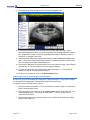
1
XVLITE™ Apteryx Inc. 313 S. High St. Suite 200 Akron, OH 44308 330-376-0889 voice 330-376-0788 fax [email protected] www.apteryx.com XVLITE™ User Manual Abstract Abstract Abstract This document contains work instructions related to utilizing the dental imaging application, XVLite™ version 3.12 and above. Copyright © 2011 Apteryx, Inc. Apteryx® is a registered trademark of Apteryx, Inc. All rights reserved. All other trademarks and tradenames are the property of their respective owners. This a controlled document. Printed or downloaded copies are considered uncontrolled and for reference only. Apteryx®, Inc. QMS_TFD_TS_XVLite_UserManual Page 1 of 99 August 22, 2011 XVLite™ TABLE OF CONTENTS TABLE OF CONTENTS ....................................................................................... 2 REVISION HISTORY ............................................................................................ 5 THE USER INTERFACE ...................................................................................... 6 GENERAL OVERVIEW ............................................................................................................... 6 MANAGING PATIENTS ....................................................................................... 7 OPEN/CREATE PATIENTS USING A PRACTICE MANAGEMENT BRIDGE ........................... 7 ADDING/CREATING A NEW PATIENT ...................................................................................... 7 EDITING PATIENT INFORMATION ............................................................................................ 8 DELETING A PATIENT ............................................................................................................... 8 VIEWING AN EXISTING PATIENT‟S IMAGES ........................................................................... 9 VIEWING A GROUP/SERIES OF IMAGES .............................................................................. 12 CAPTURING IMAGES FROM A X-RAY SENSOR/UNIT ................................... 13 CAPTURING SINGLE IMAGES FROM A X-RAY SENSOR/UNIT ............................................ 13 CAPTURING A GROUP/LAYOUT IMAGES FROM AN X-RAY SENSOR/UNIT ...................... 16 CAPTURING IMAGES FROM SCANNERS ....................................................... 19 GENERAL INFORMATION........................................................................................................ 19 CAPTURING SINGLE IMAGES FROM A SCANNER ............................................................... 19 CAPTURING GROUP/LAYOUT IMAGES FROM SCANNER ................................................... 21 CAPTURING IMAGES FROM AN INTRA-ORAL CAMERA .............................. 25 GENERAL INFORMATION........................................................................................................ 25 CAPTURING SINGLE IMAGES FROM AN IO CAMERA ......................................................... 25 CAPTURING GROUP/LAYOUT IMAGES FROM AN IO CAMERA .......................................... 28 IMAGE ENHANCEMENT ................................................................................... 32 GENERAL INFORMATION........................................................................................................ 32 ACCESSING IMAGE ENHANCEMENT OPTIONS ................................................................... 32 BRIGHTNESS/CONTRAST ....................................................................................................... 33 SHARPENING ........................................................................................................................... 35 NOISE REMOVAL ..................................................................................................................... 37 ADVANCED IMAGE ENHANCEMENT ALGORITHMS ............................................................ 38 LABELLING IMAGES ........................................................................................ 40 GENERAL INFORMATION........................................................................................................ 40 ADDING AND MODIFYING LABELS, MARKERS AND ANNOTATIONS ................................ 40 CALIBRATING IMAGES AND ADDING MEASUREMENT LABELS ........................................ 42 DISTRIBUTING IMAGES – TRANSIT™ ADD-ON COMPONENTS .................. 47 Apteryx®, Inc. Page 2 of 99 August 22, 2011 XVLite™ GENERAL INFORMATION........................................................................................................ 47 TRANSIT™ GENERAL EMAILER CREATOR .......................................................................... 47 TRANSIT™ EMAILER CREATOR............................................................................................. 51 FASTATTACH BY NEA ............................................................................................................. 52 XRAYVIEW™ E-MAILER CREATOR ........................................................................................ 57 XRAYVIEW™ CREATOR .......................................................................................................... 62 PRINTING IMAGES AND IMAGE-RELATED DOCUMENTS ............................ 68 GENERAL INFORMATION........................................................................................................ 68 PRINTING A SINGLE IMAGE.................................................................................................... 68 PRINTING MULTIPLE IMAGES ................................................................................................ 68 PRINTING IMAGES IN A LAYOUT/GROUP/SERIES ............................................................... 70 DOCUMENT TEMPLATES ........................................................................................................ 73 APPENDIX A: COMMON FUNCTIONS AND UTILITIES................................... 78 IMPORTING IMAGES FROM AN EXTERNAL SOURCE ......................................................... 78 DELETING A PATIENT‟S IMAGES ........................................................................................... 80 FINDING IMAGES BY DATE ..................................................................................................... 81 APPENDIX B: CUSTOMIZING LAYOUT DEFINITIONS ................................... 84 GENERAL INFORMATION........................................................................................................ 84 OVERVIEW OF THE „LAYOUT DEFINITION‟ DIALOG BOX ................................................... 84 COMMON LAYOUT TASKS: CHANGING THE CAPTURE ORDER ........................................ 86 COMMON LAYOUT TASKS: CHANGING THE CAPTURE SOURCE ..................................... 86 COMMON LAYOUT TASKS: ADDING TILES ........................................................................... 87 COMMON LAYOUT TASKS: CREATING NEW LAYOUTS BASED ON EXISTING LAYOUTS ................................................................................................................................................... 88 APPENDIX C: CONFIGURING DOCUMENT HANDLING OPTIONS ................ 89 OVERVIEW ................................................................................................................................ 89 CONFIGURING DOCUMENT TEMPLATE OPTIONS .............................................................. 89 APPENDIX D: CONFIGURING PREFERENCES............................................... 91 GENERAL INFORMATION........................................................................................................ 91 GENERAL PREFERENCES ...................................................................................................... 91 INTERFACE PREFERENCES................................................................................................... 92 SENSOR PREFERENCES ........................................................................................................ 93 IMAGING OPTIONS AND PREFERENCES ............................................................................. 95 PRINT PREFERENCES ............................................................................................................ 95 DATABASE PREFERENCES .................................................................................................... 96 ADVANCED PREFERENCES ................................................................................................... 97 Apteryx®, Inc. Page 3 of 99 August 22, 2011 Apteryx®, Inc. XVLite™ Page 4 of 99 XVLite™ August 22, 2011 REVISION HISTORY Revision Level Revision Date Description of Revision Revision Author DRAFT 6/10/2010 Draft SN 1.0 8/22/2011 Initial Release SN Apteryx®, Inc. Page 5 of 99 XVLite™ August 22, 2011 THE USER INTERFACE General Overview The imaging application provides a wizard-driven interface designed to make the image capturing and viewing process intuitive and simple. 1. Main Menu Bar: The main navigation menu. Provides wizard-driven access to functions such as: viewing images; taking X-rays from sensors; or acquiring images from devices such as flatbed: scanners; intra-oral camera; and panoramic/cephalometric systems. 2. Existing Patients List: Displays the current list of patients that exist in the patient database. 3. Patient Search Entry Form: Allows users to search the patient database. 4. Touch screen Keyboard (Optional): If selected, a keyboard displays for touch screen monitors typically used in dental operatories. Apteryx®, Inc. Page 6 of 99 XVLite™ August 22, 2011 MANAGING PATIENTS Open/Create Patients Using a Practice Management Bridge The imaging application provides users numerous methods to create or open patient records. Although patient records can be manually created, the most common method employed by dental practices is through integration referred to as a Practice Management (PM) Bridge. PM Bridging automates the patient creation process by “carrying over” patient (first name, last name, id number, SS number) and demographic (birth date, gender, etc.) information from common, third-party PM application to the imaging application. PM bridging assures that patient information in the imaging application matches the information contained in the PM application. A PM bridge can be established using the following methods: NameGrabber™ - Apteryx‟s patented applet. When installed, NameGrabber runs in the Windows System Tray. Users simply open a patient‟s record in the PM application by and then click on the NameGrabber icon in the system tray. The imaging application is automatically launched, the patient‟s information is carried over, and the patient‟s record is opened or created. Visit www.apteryx.com for more information. PM DataDrills – Thru the use of Apteryx‟s PM data drills, the imaging application can manually “pull” patient and related demographic information directly from a third-party PM database and use it to create a patient. DataDrills are typically used in conjunction with NameGrabber to fill in any missing information when pulling patient data from an application. Visit www.apteryx.com for more information. Integrated PM Bridge / Command Line Options – The imaging application offers many options to open patients using command line arguments. Parameters can be “sent” with the application‟s executable file to open/create patients. Command line options are useful to developers for creating a button within a third-party PM application that automates the process of opening or creating the related patients image file. For more information refer to the following online knowledgebase article: OVERVIEW: Command Line Parameters. In many circumstances, before a PM bridge is implemented, a dental practice manually creates patients in the software. As a result, patient information may not exactly match the information being passed via the PM bridge. After the PM bridge is implemented, the user may be presented with a „Patient Does Not Exist‟ dialog box when attempting to open a patient that DOES exist in the imaging application if there is a mismatch with the patient information passed. Refer to the following online knowledgebase article for instructions to update the existing patient: HELP: Error "Patient Does Not Exist". Adding/Creating a New Patient Manually Creating a Patient To manually create a new patient record: 1. Enter in the required patient information in the „Search Criteria‟ fields. The following restrictions are placed on the following entries: „SS Number‟ Maximum of 16 characters Can contain characters, digits or symbols „ID Number‟ Apteryx®, Inc. Maximum of 64 characters Can contain characters, digits or symbols Page 7 of 99 XVLite™ August 22, 2011 2. Scroll down on the main navigation if necessary and select Create Patient. 3. The „Existing Patients‟ list will refresh to display the new patient record. Editing Patient Information To modify an existing patient‟s record: 1. Enter the patient‟s information in one or more fields of the search utility or scroll down to find the desired patient record. 2. Select the patient‟s record and then select Advanced Patient Options > Edit Patient Information on the menu bar. 3. Add/Modify any of the provided fields as desired and then click on OK to save the changes. 4. Deleting a Patient Users can delete a patient from the database using the „Delete Patient‟ function. Only one patient can be deleted at a time using this functionality. To delete a patient: 1. Enter the patient‟s information in one or more fields of the search utility or scroll down to find the desired patient record. 2. Select the patient‟s record and then select Advanced Patient Options > Delete This Patient on the menu bar. 3. The following warning messages display. If sure, select Yes on each to continue with the deletion. Apteryx®, Inc. Page 8 of 99 XVLite™ August 22, 2011 a. b. c. Viewing an Existing Patient’s Images To view an existing patient‟s images: 1. Enter the patient‟s information in one or more fields of the search utility or scroll down to find the desired patient record. 2. Select the patient record and click on the View Images button on the menu bar. 3. The „View Images for which teeth?‟ screen displays. Apteryx®, Inc. Page 9 of 99 XVLite™ August 22, 2011 Use one of the following options to preview images of related teeth: By tooth number: Click on a tooth number to view thumbnails of all images related to that tooth. Teeth with related images are displayed in white. To view thumbnails of images associated to multiple teeth, left-click, hold down, and drag the cursor to select the desired teeth. Click on „Right‟ or „Left‟ to display thumbnails of all images related to a particular side of the patient‟s mouth. Apteryx®, Inc. By taken date: Select View Images By Date. The wizard will progress to the „View the images for which date?‟ screen. An „All Images‟ collapsible list is made available to allow users to filter images by year, month, or day to view thumbnails of related images. Page 10 of 99 XVLite™ August 22, 2011 View All Images: Select View Images for All Teeth. The wizard will progress to display thumbnails of all images in patient‟s database. 4. When previewing image thumbnails, the following viewing options are available: Apteryx®, Inc. Double-click on any desired image layout (outlined in red) or single image thumbnail (outlined in green) to display the related image in full view. Select the Slide Show button to view all the related images in full view as a slideshow. Page 11 of 99 August 22, 2011 XVLite™ Viewing a Group/Series of Images The imaging application has the ability to display a group of images in a single workspace known as Layouts. Layouts provide a conventional method of displaying images in the standard dental template view. Each tile in a layout identifies the related tooth associations, image orientation, and the order in the capture series however layouts can be further customized to specify the source of the image (sensor, scanner, video, etc.). Refer to Appendix B: Customizing Layout Definitions for additional information. Apteryx®, Inc. Page 12 of 99 XVLite™ August 22, 2011 CAPTURING IMAGES FROM A X-RAY SENSOR/UNIT Capturing Single Images from a X-ray Sensor/Unit Take X-ray permits users to select an x-ray sensor or unit they want to use from a list of available sensors before a digital x-ray is acquired. To capture from a sensor: 1. Enter the patient‟s information in one or more fields of the search utility or scroll down to find the desired patient record. 2. Select the patient record and click on the Take X-ray button on the menu bar. 3. The wizard advances to the „Take X-rays for which teeth? …‟ screen. Select one or more teeth to associate to the image after the capture is complete. This will automatically setup the tooth association with the image and properly orient the displayed image once captured. 4. The wizard advances to the „Position Sensor and Activate X-ray Unit‟ screen. Apteryx®, Inc. Page 13 of 99 XVLite™ August 22, 2011 5. Note that the last sensor utilized on the computer/workstation will display in the area outlined in red above. To select a different sensor, click on Switch Digital X-ray Unit or Sensor. The „Capture Sensor Image‟ selection screen displays. Select the applicable sensor and then click on OK. 6. Click on Activate Selected Unit to proceed to prepare the sensor for capture. The sensor indicator will turn green when the sensor is ready to capture an image. A vocal cue will also be heard if the computer has a sound card and speakers. Apteryx®, Inc. Page 14 of 99 XVLite™ August 22, 2011 7. Proceed to shoot the x-ray. The new image from the sensor will be acquired and stored in the patient‟s record. The amount of time it takes for the image to appear on the screen is dependent upon the sensor manufacturer. 8. The image file is automatically saved and stored in the patient‟s file. 9. Post-capture enhancements can be applied to the newly captured image. Select Automatic Brightness/Contrast to have the application automatically adjust the settings … OR .. Brightness/Contrast to manually apply Brightness/Contrast, Equalize, Gamma Correction, or to adjust color levels. 10. Additional image enhancement utilities are available under the Advanced Tools menu option. Refer to the Image Enhancement section for detailed information on these and other commonly used image enhancement tools. Apteryx®, Inc. Page 15 of 99 XVLite™ August 22, 2011 11. Post-capture operations can be performed on the newly captured image. Select Rotate, Flip Vertically, or Flip Horizontally to orient the image as desired. 12. To retake an image select the Retake button and repeat steps 7 – 11 to acquire an additional image for the teeth selected in Step 3. 13. To return to the patient list, click on the Select Patient button. Capturing a Group/Layout Images from an X-ray Sensor/Unit A layout is a group of free-floating tiles displayed in a single workspace. The groups of images can be captured and displayed in individual tiles within the selected layout. To capture a series of images from an x-ray sensor/unit: 1. Enter the patient‟s information in one or more fields of the search utility or scroll down to find the desired patient record. 2. Select the patient record and click on the Take X-ray button on the menu bar. The wizard advances to the „Take X-rays for which teeth? …‟ screen. Select Take an Automated X-ray Series. 3. The wizard will advance to a layout selection screen. Select the desired layout (2 Bite Wings, 4 Bite Wings, etc.). Layouts definitions specify the source (sensor, scanner, etc.) that the image will be acquire from. By default, layouts are configured to capture from sensors. If the tiles in a layout specify a source other than a Sensor it will need to be modified or a similar one created to capture from a TWAIN device or scanner. Select Customize Layouts and then refer to Common Layout Tasks: Changing the Capture Source (Appendix B) for detailed instructions. 4. The Wizard will advance to the Layout capture screen. Note that the first tile in the series is selected, awaiting an image capture. Additionally, note that the related tooth numbers are displayed in each tile. Apteryx®, Inc. Page 16 of 99 XVLite™ August 22, 2011 Warning: X-ray images will be automatically captured in the order set by the layout definitions. They will be oriented according to the teeth defined in the layout tile. If the user deviates from the order (without taking the proper steps), images will be placed in the wrong tile and be oriented incorrectly. 5. Place the sensor in the patient‟s mouth and position it to capture the teeth associated to the tile. Click the Start button. 6. The „Capture Sensor Image‟ selection screen displays. Select the applicable sensor and then click on OK. Select „Continue to Use This Sensor‟ if all images in the layout will be captured using the same sensor. If unchecked, the „Capture Sensor Image‟ screen will display between each capture in the series. 7. The software will advance and prepare the sensor for x-ray exposure. A green “Sensor Ready” indication will display at the top of the screen. A vocal cue will also be heard if the computer has a sound card and speakers. 8. Proceed to shoot the x-ray. The new image from the sensor will appear in the selected tile. 9. The application will then advance to the next tile in the series. Apteryx®, Inc. Page 17 of 99 XVLite™ August 22, 2011 If „Continue using this sensor‟ was selected in Step 6, the sensor will automatically be prepared and readied for the next capture. If „Continue using this sensor‟ was not selected in Step 6, the „Capture Sensor Image‟ screen will display for the user to select the desired sensor. Click on OK to continue with the capture. 10. Place the sensor in the patient‟s mouth and position it to capture the set of teeth associated to the next tile. 11. Repeat Steps 8 – 10 until the last image in the series has been captured. 12. Click Finished when all images in the series are captured. The layout will be automatically stored to the patient‟s file. Apteryx®, Inc. Page 18 of 99 XVLite™ August 22, 2011 CAPTURING IMAGES FROM SCANNERS General Information If a TWAIN-compliant scanner (e.g. a flatbed scanner with manufacturer-provided TWAIN drivers) is attached to the computer on which the imaging application is installed: The application will automatically recognize the device and configure its settings. (Before booting up Windows, turn on the power to the scanner to ensure that the computer‟s operating system "sees" the device). The scanner will available for selection in the „Capture Image‟ device selection screen under the „Scanned X-ray‟ (for scanning in X-rays) or „Scanned Image‟ (for scanning in digital images) tabs. If a phosphor plate scanner (e.g. Air Technique‟s ScanX®) is attached to the computer on which the application is installed: The application will require the related Apteryx imaging device extension to be installed in order to interface with the manufacturer‟s drivers. The scanner will be available for selection in the „Capture Image‟ device selection screen under the „Scanned X-ray‟ or „Imaging Device‟ tabs. In this user guide, captures will be limited to capturing from TWAIN-compliant scanners or phosphor plate scanners using the „Scanned X-ray‟ tab. Capturing Single Images from a Scanner 1. Enter the patient‟s information in one or more fields of the search utility or scroll down to find the desired patient record. 2. Select the patient record and click on the Acquire Images button on the menu bar. 3. The wizard advances to the „Take X-rays for which teeth? …‟ screen. Select one or more teeth to associate to the image after the capture is complete. This will automatically setup the tooth association with the image and properly orient the displayed image once captured. Apteryx®, Inc. Page 19 of 99 XVLite™ August 22, 2011 4. The „Capture Image‟ device selection screen will display prompting users to select the tab that applies to the image to be captured. Select the „Scanned X-ray‟ tab. 5. Click on the „Image Source‟ drop-down list and select the applicable driver for the TWAIN-Compliant scanner or phosphor plate scanner. 6. Depending on the hardware, „Automatically Acquire‟ may need to be disabled if a TWAIN driver is selected. Apteryx recommends disabling „Automatically Acquire‟ if a TWAIN driver is selected . Auto Acquire functionality depends on the capabilities of the flatbed scanner's TWAIN or Windows Imaging Acquisition (WIA) drivers. Certain scanners (such as the HP Scanjet G4050) do not work with Auto Acquire if Auto Size is selected, nor will they utilize the transparency adapter. If necessary, check with the hardware manufacturer to see if this functionality is supported. 7. If acquiring from a flatbed scanner (skip to Step 8 for phosphor plate scanners): a. Place the X-ray or other media as instructed by the manufacturer. b. Note: Flatbed scanners must have a trans-illuminating cover for scanning X-rays. c. Click on the OK button. d. If this is the first time the TWAIN-compliant scanner is being used, the imaging application will display a device settings screen to configure the device. Accept the defaults by clicking the OK button (they can be changed at any time if necessary). e. The imaging application will send instructions to the scanner and automatically scan the image and display it in the imaging application. 8. If acquiring from a phosphor plate scanner: a. Click on the OK button. The device-specific interface will display and prompt the user when ready. b. Place the X-ray film into the phosphor plate scanner as instructed by the manufacturer. c. Apteryx®, Inc. When the scan is complete, select Done on the device‟s interface. The imaging application will then acquire the image from the device‟s interface and display it in the imaging application. Page 20 of 99 XVLite™ August 22, 2011 9. The image file is automatically saved and stored in the patient‟s file. 10. Post-capture enhancements can be applied to the newly captured image. Select Automatic Brightness/Contrast to have the application automatically adjust the settings … OR .. Brightness/Contrast to manually apply Brightness/Contrast, Equalize, Gamma Correction, or to adjust color levels. 11. Additional image enhancement utilities are available under the Advanced Tools menu option. Refer to the Image Enhancement section for detailed information on these and other commonly used image enhancement tools. 12. Post-capture operations can be performed on the newly captured image. Select Rotate, Flip Vertically, or Flip Horizontally to orient the image as desired. 13. To retake an image select the Retake button and repeat steps 7 – 12 to acquire an additional image for the teeth selected in Step 3. 14. To return to the patient list, click on the Select Patient button. Capturing Group/Layout Images from Scanner A layout is a group of free-floating tiles displayed in a single workspace. The groups of images can be captured and displayed in individual tiles within the selected layout. To capture a series of images from a scanner: 1. Enter the patient‟s information in one or more fields of the search utility or scroll down to find the desired patient record. 2. Select the patient record and click on the Acquire Image button on the menu bar. The wizard advances to the „Take X-rays for which teeth? …‟ screen. Select Take an Automated X-ray Series. 3. The wizard will advance to a layout selection screen. Select the desired layout (2 Bite Wings, 4 Bite Wings, etc.). Apteryx®, Inc. Page 21 of 99 August 22, 2011 XVLite™ Layouts definitions specify the source (sensor, scanner, etc.) that the image will be acquire from. By default, layouts are configured to capture from sensors. If the tiles in a layout specify a source other than a TWAIN Device or “Scanner” it will need to be modified or a similar one created to capture from a TWAIN device or scanner. Select Customize Layouts and then refer to Common Layout Tasks: Changing the Capture Source (Appendix B) for detailed instructions. 4. The Wizard will advance to the Layout capture screen. Note that the first tile in the series is selected, awaiting an image capture. Additionally, note that the related tooth numbers are displayed in each tile. Warning: X-ray images will be automatically captured in the order set by the layout definitions. They will be oriented according to the teeth defined in the layout tile. If the user deviates from the order (without taking the proper steps), images will be placed in the wrong tile and be oriented incorrectly. Apteryx®, Inc. Page 22 of 99 XVLite™ August 22, 2011 5. When ready to scan click the Start button. 6. The „Capture Image‟ device selection screen will display prompting users to select the tab that applies to the image to be captured. Select the „Scanned X-ray‟ tab. 7. Click on the „Image Source‟ drop-down list and select the applicable driver for the TWAIN-Compliant scanner or phosphor plate scanner. Select „Continue to use this source‟ if all images in the layout will be captured using the same device. If unchecked the „Capture Image‟ screen will display between each capture in the series. 8. Depending on the hardware, „Automatically Acquire‟ may need to be disabled if a TWAIN driver is selected. Apteryx recommends disabling “Automatically Acquire if a TWAIN driver is selected. Auto Acquire functionality depends on the capabilities of the flatbed scanner's TWAIN or Windows Imaging Acquisition (WIA) drivers. Certain scanners (such as the HP Scanjet G4050) do not work with Auto Acquire if Auto Size is selected, nor will they utilize the transparency adapter. If necessary, check with the hardware manufacturer to see if this functionality is supported. 9. If acquiring from a flatbed scanner (skip to Step 10 for phosphor plate scanners): a. Place the X-ray or other media as instructed by the manufacturer. Flatbed scanners must have a trans-illuminating cover for scanning X-rays. b. Click on the OK button. c. If this is the first time the TWAIN-compliant scanner is being used imaging application will display a device settings screen to configure the device. Accept the defaults by clicking the OK button (they can be changed at any time if necessary). d. The imaging application will send instructions to the scanner and automatically scan the image and display it in the selected tile. 10. If acquiring from a phosphor plate scanner: a. Click on the OK button. The device-specific interface will display and prompt the user when ready. b. Place each X-ray film into the phosphor plate scanner as instructed by the manufacturer. Apteryx®, Inc. Page 23 of 99 XVLite™ August 22, 2011 c. When all images are scanned and display in the device interface select Done. The imaging application will then acquire the images from the device‟s interface and insert them into each tile in the capture window. 11. The application will then advance to the next tile in the series. If „Continue using this source‟ was selected in Step 6, the device will automatically be prepared and readied for the next capture. If „Continue using this source‟ was not selected in Step 6, the „Capture Image‟ screen will display for the user to select the desired device from the „Scanned Xray‟ tab again. Click on OK to continue with the capture. 12. Repeat Steps 9a – 9e for flatbed scanners until the last image in the series has been captured. 13. Click Finished when all images in the series are captured. The layout will be automatically stored to the patient‟s file. Apteryx®, Inc. Page 24 of 99 XVLite™ August 22, 2011 CAPTURING IMAGES FROM AN INTRA-ORAL CAMERA General Information The imaging application offers an easy-to-use DirectVideo and Real-Time interface that allows users to capture live images from video sources such as Intra-Oral Cameras and digital cameras. The Real-time Video Extension is used for capturing images from older analog intra-oral video cameras connected to computers via video card. The DirectVideo Extension is more commonly used for more recent technology that connects cameras via USB, FireWire (IEEE 1394) cameras and DirectX video capture cards. In this user guide, captures will be limited to capturing from an Intra-Oral Camera with the more commonly used DirectVideo Extension. By default, the DirectVideo Extension is enabled. To change to Real-Time Video Extension for older, analog cameras, click on Preferences > Imaging and then select the Video Device Type button. Enable the Real-Time Video option on the „Video Source‟ dialog box. Capturing Single Images from an IO Camera 1. Enter the patient‟s information in one or more fields of the search utility or scroll down to find the desired patient record. 2. Select the patient record and click on the Acquire Images button on the menu bar. 3. The wizard advances to the „Take X-rays for which teeth? …‟ screen. Select one or more teeth to associate to the image after the capture is complete. This will automatically setup the tooth association with the image and properly orient the displayed image once captured. 4. The „Capture Image‟ device selection screen will display prompting users to select the tab that applies to the image to be captured. Select the „Misc‟ tab Apteryx®, Inc. Page 25 of 99 XVLite™ August 22, 2011 5. Click on the „Image Source‟ drop-down list and select DirectVideo Capture. 6. The „DirectVideo‟ interface displays. If more than one video source is attached to the computer, select Video Devices from the menu and then select the applicable device. a. Note that the live video feed displays on the right hand side. Place the camera in the patient‟s mouth and position it where desired. b. Capture the image using one of the following methods: Press the button on the camera DirectVideo comes equipped with different capture button configurations designed to interact with various cameras. Select Camera Buttons from the menu to change the configuration to one that applies if the camera button does not trigger the capture. Step on the foot pedal (if configured) Select the Capture button on the „DirectVideo‟ interface Press the space bar on the keyboard Apteryx®, Inc. Page 26 of 99 XVLite™ August 22, 2011 c. Once captured, a thumbnail of the image will appear in the area below the buttons on the „DirectVideo‟ interface. When finished, click the Done button. d. Multiple images can be captured at one time, if desired. Additional thumbnails will appear below the initial one as images are captured. e. If unsatisfied with an image, right-click on the image thumbnail and select Delete Current Image. f. 7. When finished, click the Done button. The „DirectVideo‟ interface will close and the image(s) will be acquired and displayed in the imaging application. 8. The image file is automatically saved and stored in the patient‟s file. Apteryx®, Inc. Page 27 of 99 XVLite™ August 22, 2011 Post-capture enhancements can be applied to the newly captured image. Select Automatic Brightness/Contrast will adjust the brightness and contrast for an image automatically. The Brightness/Contrast menu contains tools that are applied manually, including: Brightness/Contrast, Equalize, Gamma Correction, or to adjust color levels. Additional image enhancement utilities are available under the Advanced Tools menu option. Refer to the Image Enhancement section for detailed information on these and other commonly used image enhancement tools. 9. To retake an image select the Retake button and repeat steps 4 –8 to acquire an additional image for the teeth selected in Step 3. 10. To return to the patient list, click on the Select Patient button. Capturing Group/Layout Images from an IO Camera A layout is a group of free-floating tiles displayed in a single workspace. The groups of images can be captured and displayed in individual tiles within the selected layout. To capture a series of images from an IO camera: 1. Enter the patient‟s information in one or more fields of the search utility or scroll down to find the desired patient record. 2. Select the patient record and click on the Acquire Image button on the menu bar. The wizard advances to the „Take X-rays for which teeth? …‟ screen. Select Take an Automated X-ray Series. 3. The wizard will advance to a layout selection screen. Select the desired layout (2 Bite Wings, 4 Bite Wings, etc.). Apteryx®, Inc. Page 28 of 99 XVLite™ August 22, 2011 Layouts definitions specify the source (sensor, scanner, etc.) that the image will be acquire from. By default, layouts are configured to capture from sensors. If the tiles in a layout specify a source other than a Video Source it will need to be modified or a similar one created to capture from a TWAIN device or scanner. Select Customize Layouts and then refer to Common Layout Tasks: Changing the Capture Source (Appendix B) for detailed instructions. 4. The Wizard will advance to the Layout capture screen. Note that the first tile in the series is selected, awaiting an image capture. Additionally, note that the related tooth numbers are displayed in each tile. Warning: X-ray images will be automatically captured in the order set by the layout definitions. They will be oriented according to the teeth defined in the layout tile. If the user deviates from the order (without taking the proper steps), images will be placed in the wrong tile and be oriented incorrectly. Apteryx®, Inc. Page 29 of 99 XVLite™ August 22, 2011 5. When ready to capture images click the Start button. 6. The „DirectVideo‟ interface displays. Continue capturing images for each tile in the layout. Additional thumbnails will appear below the initial one as images are captured. If more than one video source is attached to the computer, select Video Devices from the menu and then select the applicable device. a. Note that the live video feed displays on the right hand side. Place the camera in the patient‟s mouth and position it where desired. b. Capture the images using one of the following methods: Press the button on the camera Step on the foot pedal (if configured) Select the Capture button on the „DirectVideo‟ interface Press the space bar on the keyboard c. Apteryx®, Inc. Once captured, a thumbnail of each image will appear in the area below the buttons on the „DirectVideo‟ interface. Page 30 of 99 XVLite™ August 22, 2011 d. If unsatisfied with an image, right-click on the image thumbnail and select Delete Current Image. e. When finished, click the Done button. 7. The „DirectVideo‟ interface will close and the images will automatically be placed in each tile in the layout. 8. Click Finished when all images in the series are captured. The layout will be automatically stored to the patient‟s file. Apteryx®, Inc. Page 31 of 99 XVLite™ August 22, 2011 IMAGE ENHANCEMENT General Information The application‟s image manipulation and enhancement tools enables users to improve images digitally without having to re-shoot X-rays or other images. This guide will highlight the primary enhancement tools as the intuitive nature the imaging application enables the user to quickly grasp the functions and uses of the remaining tools. Accessing Image Enhancement Options 1. View an existing patient‟s images and double-click to view it in full view. 2. Select Enhancement from the menu bar to access the image enhancement options. 3. Apteryx®, Inc. Page 32 of 99 XVLite™ August 22, 2011 Brightness/Contrast Overview The imaging application provides a standard Brightness/Contrast and Gamma Correction utility that can be used to improve image quality. In general, brightness/contrast enhancement adjusts the brightness and the contrast of an image in order to try to improve the overall dynamic appearance of the image. Gamma correction is a very versatile tool when dealing with images that are captured by video hardware. Gamma correction differs from standard brightness enhancements because it dynamically adjusts each pixel to maintain its relation to all the other pixels, hence exaggerating density changes in an object. Usually, gamma correction is used in combination with one of the other B/C enhancement tools to dramatically improve the overall appearance of an image. Before Gamma Correction After Gamma Correction Applying brightness/contrast or gamma correction 1. From the Enhancement menu options, select Brightness / Contrast > Brightness / Contrast or Brightness / Contrast > Gamma Correction. The applicable dialog box displays. Apteryx®, Inc. Page 33 of 99 XVLite™ August 22, 2011 2. Use one of the following image adjustment buttons to size, resize, or reposition the images displayed in the original and resulting image preview windows as desired: - Select to pan the image in the preview to view a specific area. Click and drag the image with the left mouse button. - Select to magnify or reduce the image in the preview. To increase the magnification of an image, click on the image in the preview with the left mouse button. To reduce the magnification, click with the right mouse button. - Select to automatically resize the image and make it fit in the viewing area. - Select to automatically resize the image in the preview to 100% magnification. When clicked, the image in the preview will resize to the actual dimensions of the image. 3. Use the brightness and contrast sliders … OR … click and drag the sliders located on the right side (brightness) and bottom (contrast) of the adjustment tool to enhance the image as desired. The enhancements are adjusted in real-time in the „Resulting Image Preview‟ area. 4. Click OK to apply the settings to the image. 5. If desired, click on Undo ( ) to remove the B/C enhancement applied to the image. 6. When done, click Go Back to return to the patient images or Select Patient to return to the patient list. A dialog box will appear prompting the user to save the modification. Select Yes to save the change. Applying Equalization The imaging application offers users the ability to apply equalization by selecting Brightness / Contrast > Equalize. Equalize provides a non-linear approach to altering the brightness/contrast Apteryx®, Inc. Page 34 of 99 XVLite™ August 22, 2011 of an image. This enhancement is typically used to improve the image quality of very low contrast images. Most images span an entire range of brightness with all levels well represented. However, sometimes images are too light or too dark and some levels are overpopulated leaving others under-populated. The Equalize effect modifies an image so that all levels of brightness are equally well represented within the image. The equalize option is only available for grayscale images. Before Equalization After Equalization Sharpening Overview Most images have some degree of blurriness (from focus problems or inherent limitations in the image taking process). The imaging application offers an automatic image sharpening that can be applied to compensate for the blurriness in an image. Before Laplace Edge Enhancement After Laplace Edge Enhancement Applying sharpening or Laplace edge enhancement 1. Select Advanced Tools > Sharpening to use the sharpening. The „Sharpen Image‟ dialog box displays. Apteryx®, Inc. Page 35 of 99 XVLite™ August 22, 2011 2. Use one of the following image adjustment buttons to size, resize, or reposition the images displayed in the original and resulting image preview windows as desired: - Select to pan the image in the preview to view a specific area. Click and drag the image with the left mouse button. - Select to magnify or reduce the image in the preview. To increase the magnification of an image, click on the image in the preview with the left mouse button. To reduce the magnification, click with the right mouse button. - Select to automatically resize the image and make it fit in the viewing area. - Select to automatically resize the image in the preview to 100% magnification. When clicked, the image in the preview will resize to the actual dimensions of the image. 3. The mask size and sharpening factor work hand-in-hand in determining the degree of sharpening that is applied to the image. 4. The „Sharpening Factor‟ specifies the magnitude of the sharpening operation. Use the slider to manually adjust the degree of image sharpening as desired. 5. Select a predefined „Mask Size‟ … OR … select to manually enter a custom „Width‟ and „Height‟. The mask size specifies how large the area should be when sharpening a portion of the image. The larger the area, the wider the blurriness is considered to be. 6. Click OK to apply the settings to the image. 7. When done, click Go Back to return to the patient images or Select Patient to return to the patient list. A dialog box will appear prompting the user to save the modification. Select Yes to save the change. Apteryx®, Inc. Page 36 of 99 XVLite™ August 22, 2011 Noise Removal Overview Noise in an image can be caused by a number of factors such as dust, scratches or image static. A median noise removal/reduction algorithm is provided in the imaging application. In most cases, median noise reduction results in the best image quality. It is important to note that excessive noise reduction leads to a loss of detail. Before Noise Removal After Noise Removal Applying Median Noise Removal 1. Select Advanced Tools > Remove Noise. The „Noise Reduction (Median)‟ dialog box displays. Apteryx®, Inc. Page 37 of 99 XVLite™ August 22, 2011 2. Use one of the following image adjustment buttons to size, resize, or reposition the images displayed in the original and resulting image preview windows as desired: - Select to pan the image in the preview to view a specific area. Click and drag the image with the left mouse button. - Select to magnify or reduce the image in the preview. To increase the magnification of an image, click on the image in the preview with the left mouse button. To reduce the magnification, click with the right mouse button. - Select to automatically resize the image and make it fit in the viewing area. - Select to automatically resize the image in the preview to 100% magnification. When clicked, the image in the preview will resize to the actual dimensions of the image. 3. Select a predefined „Mask Size‟ … OR … select to manually enter a custom „Width‟ and „Height‟. The mask size specifies how large the area should be when sharpening a portion of the image. The larger the area, the wider the area of noise is considered to be. 4. Click OK to apply the settings to the image. 5. When done, click Go Back to return to the patient images or Select Patient to return to the patient list. A dialog box will appear prompting the user to save the modification. Select Yes to save the change. Advanced Image Enhancement Algorithms The imaging application comes equipped with customized image algorithms that apply a sequence of enhancements commonly used to improve image quality. Applying an Advanced Image Enhancement Algorithm Select Advanced Image Enhancements choose from the following options: Apteryx General Enhancement – Applies Normalize; Median Blur (5x5 Mask Size); and Sharpen (21x21 Mask Size); and Gamma Correction. Custom Image Enhancement – Applies a Median Filter (3x3 Mask Size); Sharpen (7X7 Mask Size); Adaptive Normalize; and Gamma Correction. General Endo Enhancement – Applies filters that enhance files, etc. used in endo treatments. Modifying an Advanced Image Enhancement Algorithm Image quality is subjective; therefore, the imaging application allows users to modify the Apteryx General and Custom Image Enhancement algorithms to suit their needs. To modify an enhancement algorithm: 1. Select Advanced Image Enhancements > Image Filter Options and choose the applicable enhancement algorithm to modify. 2. The algorithm‟s dialog box displays. Modify any of the available settings as needed and then click OK. Apteryx®, Inc. Page 38 of 99 August 22, 2011 Apteryx®, Inc. XVLite™ Page 39 of 99 XVLite™ August 22, 2011 LABELLING IMAGES General Information Unlike other systems, the imaging application provides users with a set of temporary labels (marker and annotations) that can be associated with an image. A variety of different labels, markers and annotations can be applied to an image to highlight problem areas, add date and time information, define and identify earlier procedures performed or add general notes. Labels are not part of the image when added. They are floating objects that can be moved, modified, resized or deleted at any time. Adding and Modifying Labels, Markers and Annotations The following example of how to add an Ellipse to an image effectively demonstrates how to add other types of labels, markers, and annotations available in the imaging application. The Ellipse Label command will place an ellipse on an image. Placing an ellipse on an image is useful in bringing attention to a designated area. Users have control of the ellipse‟s properties. 1. View an existing patient‟s images and double-click to view it in full view. 2. Select Labels from the menu bar to access the labeling options. Apteryx®, Inc. Page 40 of 99 XVLite™ August 22, 2011 3. Select Ellipse Label from the menu bar. 4. Hover the cursor over the image and note that it changes into a cross-hair indicator. 5. Place the stem of the indicator in the desired position on the image, Click and drag the mouse to the position where the label should be displayed. Release the mouse button to place the label in the designated location. 6. Double-click on the label to access the „Ellipse Properties‟ dialog box: a. Select the desired „Pen Size‟, „Pen Color‟, and „Fill Color‟. b. If desired, select „Freeze Label‟ on the bottom-left of the dialog box to fix the position of the label. The label will not be able to be moved around on the image. c. When satisfied, click on OK. 7. The modified properties will then be applied to label. Apteryx®, Inc. Page 41 of 99 XVLite™ August 22, 2011 Calibrating Images and Adding Measurement Labels Overview Before measurements are added in an image, the image should first be calibrated. In general, images are calibrated by taking a measurement of an object of a known size or length in an image (as shown in the image below) and then providing the actual length or size of the object to determine the images calibration factor for a subsequent measurement to be performed. This method is useful in calibrating images from Cephalometric systems. Sensor images can be more accurately calibrated by using the actual dimensions of the sensor‟s active area rather than an object of known length. The active area of the sensor can be determined by referring to the hardware‟s documentation or contacting the manufacturer. Apteryx®, Inc. Page 42 of 99 XVLite™ August 22, 2011 Apteryx, Inc. does not guarantee the accuracy of any calibration/measurement performed in the imaging application. Calibrating an Image The Ellipse Label command will place an ellipse on an image. Placing an ellipse on an image is useful in bringing attention to a designated area. Users have control of the ellipse‟s properties. 1. View an existing patient‟s images and double-click to view it in full view. 2. Select Labels from the menu bar to access the labeling options. Apteryx®, Inc. Page 43 of 99 XVLite™ August 22, 2011 3. Select Calibrate Measurement from the menu bar. The cursor will change into a crosshair with a gear above it. 4. Place the calibration crosshair at the starting point of the object of known length; singleclick on the left mouse button; move the cross-hair to the end-point of the object; and then double-click on the left mouse button. Non-linear calibrations can be created by left-clicking on mid-points while moving the mouse from point to point. Double-click on the left mouse button at the final end-point. 5. A 'Calibrate Measurements' pop-up screen will appear. Type in the correct length of the object. Click OK. Apteryx®, Inc. Page 44 of 99 XVLite™ August 22, 2011 6. This image is now calibrated and any measurement performed will be based off the calibration. Adding a Measurement Label Follow the steps below to perform a measurement and create a measurement label. 1. Once a calibration is applied, the cursor changes to a crosshair with no gear, ready to take a measurement. 2. Place measurement crosshair at the starting point of the object to be measured; singleclick on the left mouse button; move the cross-hair to the end-point of the object; and then double-click on the left mouse button. Non-linear measurements can be created by right-clicking on end-points while dragging the mouse with the left button held down. Release the left mouse button at the final end-point. 3. A measurement label will be placed on the image displaying the measurement. Doubleclick on the measurement label to access the „Measurement Properties‟ dialog box. Apteryx®, Inc. Page 45 of 99 XVLite™ August 22, 2011 4. The „Measurement‟ Properties dialog box displays to allow users to format the label. Customize the measurement label properties (color, line, unit, measurement position, etc.) then click OK to exit. The „Measurement Sample View‟ section in the dialog box above does not display the actual measurement, but rather a sample of what the measurement will look like. The ‟Measurements‟‟ tab is where to find the actual measurement. 5. To remove the measurement label, if desired, click on the label and then select Delete Label. 6. To add additional measurement labels to the calibrated image, select Measurement Label from the menu bar and repeat Steps 2 – 7. 7. When done, click on the Go Back button to return to the full view of the image. Since labels have been added to the image, the user will be prompted to save the changes. Click Yes to save the modification. 8. Apteryx®, Inc. Page 46 of 99 XVLite™ August 22, 2011 DISTRIBUTING IMAGES – TRANSIT™ ADD-ON COMPONENTS General Information The imaging application allows the addition of TransIT™ Communication Extension Devices (TransIT CEDs), additional components that expand the software‟s capabilities to include the sending of patient images, claim forms, etc. to referral doctors, insurance companies, etc. The commonly used TransIT CEDs are described in the following sections. TransIT™ General Emailer Creator Overview The most commonly used TransIT™ add-on component, the General Emailer enables users to spawn their email client application (e.g. Microsoft® Outlook) and automatically attach patient images and files to a new email message. The General Emailer requires Outlook/Outlook Express or other MAPI clients. Configuring the TransIT™ General Emailer Creator The TransIT™ General Emailer component will attempt to use the default email application set up in Windows. Refer to Microsoft Support‟s Online Knowledgebase Article KB 154359 for instructions to specify Microsoft® Outlook or Outlook Express as the default email application. 1. View an existing patient‟s images; double-click on the desired image to view it in full view; and then Select Send on the menu bar. 2. The „Select TransIT Method‟ dialog box appears. Click on Access TransIT Options. 3. The „TransIT Configurations‟ dialog box displays. Select General Emailer Creator and click on Edit Options. 4. The „General Emailer Options‟ dialog box displays. Apteryx®, Inc. Page 47 of 99 XVLite™ August 22, 2011 a. Accept the default or change the „JPEG Quality Factor‟ to whatever desired. The higher the quality the larger the generated jpeg files will be when converted and sent. b. Enable the Mask SS numbers option to suppress the transmission of SS numbers. c. Click OK when done. 5. Click OK on the „TransIT Configurations‟ dialog to return to the „Select TransIT Method‟ dialog box. Sending Images Using the TransIT™ General Emailer Creator 1. On the „Select TransIT Method‟ dialog box, select General Email Creator. The „TransIT Item Selection‟ dialog box displays. a. „Single Image Options‟: If sending one or more single images, specify whether to send them as a plain image (i.e. JPEG format) or as an STB files the „Single Image Options‟ section. b. Layout Options: If sending one or more layouts, select one of the following file format options: As a combined, single image (i.e. JPEG format) Apteryx®, Inc. Page 48 of 99 XVLite™ August 22, 2011 As an Apteryx layout file (i.e. a compressed zip file containing the layout, STB images, and related data files A common file format ZIP file (i.e. a compressed zip file containing the images in JPEG format). 2. Click on OK. The „TransIT Emailer Information‟ screen displays. Complete the following information on each tab: a. „General‟ tab: Complete the „Subject‟ and „Reference Number‟ fields. b. „Patient‟ tab: The tab is pre-populated with the open patient‟s information. Complete any required field if blank. c. Apteryx®, Inc. „Message‟ tab: Enter a message for the recipient of the email. The message entered will appear in the body of the email. Page 49 of 99 XVLite™ August 22, 2011 3. Click OK when the „TransIT Emailer Information‟ tabs have been completed. The imaging application proceeds to spawn Outlook/Outlook Express create new email is created with the image attached. 4. Add the email information of the recipient and proceed to send the attached image/layout in Outlook/Outlook Express. Apteryx®, Inc. Page 50 of 99 XVLite™ August 22, 2011 TransIT™ Emailer Creator Overview The Emailer Creator module enables the sending of emails from within the imaging application. Multiple patient images and files can be sent directly through a dental practice‟s Internet Service Provider (ISP). Outlook/Outlook Express is not required with this extension. Configuring Emailer Options: For the TransIT™ Emailer component to function an outgoing SMTP mail server must be configured. The Emailer is only compatible with outgoing mail servers that DO NOT require authentication or encryption (e.g. SSL) for transmission. Contact your ISP for outgoing SMTP server information and transmission requirements. 1. View an existing patient‟s images; double-click on the desired image to view it in full view; and then select Send on the menu bar. 2. The „Select TransIT Method‟ dialog box appears. Click on Access TransIT Options. 3. The „TransIT Configurations‟ dialog box displays. Select Emailer Creator and click on Edit Options. 4. The „General Emailer Options‟ dialog box displays. Apteryx®, Inc. Page 51 of 99 XVLite™ August 22, 2011 a. Complete the „Outgoing SMTP Server‟ and „Port‟ fields. b. Click to enable the option This SMTP server requires a user name and password. Complete the „User Name‟ and „User Password‟ fields. a. Accept the default or change the „JPEG Quality Factor‟ to whatever desired. The higher the quality the larger the generated jpeg files will be when converted and sent. c. Click OK when done. 5. Click OK on the „TransIT Configurations‟ dialog to return to the „Select TransIT Method‟ dialog box. Sending Images Using the TransIT™ Emailer Creator: 1. On the „Select TransIT Method‟ dialog box, select Email Creator. The „TransIT Item Selection‟ dialog box displays. a. „Single Image Options‟: If sending a single image, specify whether to send it as a plain image (i.e. JPEG format) or as an STB file in the „Single Image Options‟ section. b. Layout Options: If sending a layout, select one of the following file format options: Combined, single image (i.e. JPEG format) Apteryx layout file (i.e. a compressed zip file containing the layout, STB images, and related data files A common file format ZIP file (i.e. a compressed zip file containing the images in JPEG format). 2. Click on OK to generate and send the email via the outgoing SMTP server. FastAttach by NEA Overview The FastAttach module enables users to submit patient images and claims to NEA's FastAttach web-based service. Apteryx®, Inc. Page 52 of 99 XVLite™ August 22, 2011 Configuring FastAttach Options: 1. View an existing patient‟s images; double-click on the desired image to view it in full view; and then select Send on the menu bar. 2. The „Select TransIT Method‟ dialog box appears. Click on Access TransIT Options. 3. The „TransIT Configurations‟ dialog box displays. Select FastAttach by NEA and click on Edit Options. 4. The „FastAttach Options‟ dialog box displays. a. Select the button shown in red above to find/specify the location of the NEACOMM application. b. Accept the default or change the „JPEG Quality Factor‟ to whatever desired. The higher the quality the larger the generated jpeg files will be when converted and sent. c. Click OK when done. 5. Click OK on the „TransIT Configurations‟ dialog to return to the „Select TransIT Method‟ dialog box. Submitting Patient Images/Claims Using FastAttach by NEA: 1. On the „Select TransIT Method‟ dialog box, select Fast Attach by NEA. The „TransIT Selection‟ dialog box displays. Click OK to continue. Apteryx®, Inc. Page 53 of 99 XVLite™ August 22, 2011 2. The „TransIT Fast Attach Information‟ dialog box displays. Complete the following information on each tab: a. „General‟ tab: Select the applicable date range of service using the calendars. Enter the optional „Payor Reference Number‟ and „Claim Amount. b. „Provider‟ tab: Enter the required dental practice information. Click the Add button to store the entered information as a quick entry for future use. Apteryx®, Inc. Page 54 of 99 XVLite™ August 22, 2011 c. „Payor‟ tab: The „Payor Name‟ is the insurance company the patient is insured through. i. Select the Browse button, shown in red above, to launch and select the patient‟s insurance company from the „Select NEA Player‟ dialog box. Apteryx®, Inc. Page 55 of 99 XVLite™ August 22, 2011 ii. Click OK to return to the „Payor‟ tab. iii. Click the Add button to store the entered information as a quick entry for future use. d. „Patient‟ tab: The tab is pre-populated with the open patient‟s information. Complete any required field if blank. e. „Insured‟ tab: If the patient is not the insured person (e.g. a dependant or spouse), enter the insured person‟s information and select the applicable relationship. If the patient is the insured person, select the Same as patient button to auto-populate the fields. Apteryx®, Inc. Page 56 of 99 XVLite™ August 22, 2011 f. „Message‟ tab: Enter any additional information to relay to the Insurance Company regarding the claim and/or attachments. 3. Click OK to forward the image and claim information to NEA‟s FastAttach communication application. XrayView™ e-Mailer Creator Overview The XrayView™ e-Mailer Creator creates a ZIP file or self-extracting executable (XrayViewMailer.zip or XrayView.exe) containing a patients images. The XrayView™ e-Mailer Creator then calls up a user‟s email client application (e.g. Microsoft® Outlook) and attaches the file in a new email message. Email recipients that receive the attachment can then detach, run the file, and view the provided images within an XrayView™ interface. XrayView™ e-Mailer Creator supports the transmission of Apteryx‟s unmodifiable Secure Tagged Block (STB) file format, guaranteeing image authenticity. Configuring the XrayView™ e-Mailer Creator Options The XrayView™ e-Mailer component will attempt to use the default email application set up in Windows. Refer to Microsoft Support‟s Online Knowledgebase Article KB 154359 for instructions to specify Microsoft® Outlook or Outlook Express as the default email application. 1. View an existing patient‟s images; double-click on the desired image to view it in full view; and then select Send on the menu bar. 2. The „Select TransIT Method‟ dialog box appears. Click on Access TransIT Options. Apteryx®, Inc. Page 57 of 99 XVLite™ August 22, 2011 3. The „TransIT Configurations‟ dialog box displays. Select XrayView e-Mail Creator and click on Edit Options. 4. The „XrayView e-Mailer Options‟ dialog box displays. a. Enable Send attachment as a ZIP file to generate attachments as a ZIP file. If disabled, the attachment will attach a self-extracting executable (*.EXE file extension). Warning: Some email clients may be configured not to accept *EXE file extensions for security reasons. If so, the email client will strip out the attachment when the new message is sent. b. Accept the default or change the „JPEG Quality Factor‟ to whatever desired. The higher the quality the larger the generated jpeg files will be when converted and sent. c. Click OK when done. 5. Click OK on the „TransIT Configurations‟ dialog to return to the application desktop. Sending Images Using the XrayView e-Mailer Creator 1. On the „Select TransIT Method‟ dialog box, select XrayView e-Mail Creator. The „TransIT Item Selection‟ dialog box displays. Apteryx®, Inc. Page 58 of 99 XVLite™ August 22, 2011 a. „Single Image Options‟: Users may specify whether to send an image as a common file format (i.e. JPEG format) or as an STB file the „Single Image Options‟ section. b. Layout Options: If sending a layout, select one of the following file format options: Combined, single image (i.e. JPEG format) Apteryx layout file (i.e. a compressed zip file containing the layout, STB images, and related data files A common file format ZIP file (i.e. a compressed zip file containing the images in JPEG format). 2. Click on OK. The „TransIT XrayView Information‟ screen displays. Complete the following information on each tab: a. „General‟ tab: Complete the „Subject‟ and „Reference Number‟ fields. Apteryx®, Inc. Page 59 of 99 XVLite™ August 22, 2011 b. „Patient‟ tab: The tab is pre-populated with the open patient‟s information. Complete any required field if blank. c. „Message‟ tab: Enter information for the recipient of the email. d. „Password Protect‟ tab: If desired, enable the Password protect the image option and enter a password in the „Password‟ field. The recipient will need to be provided the password in order to access the files in the attachment when sent. Apteryx®, Inc. Page 60 of 99 XVLite™ August 22, 2011 3. Click OK when the „TransIT XrayView Information‟ tabs have been completed. The imaging application proceeds to spawn Outlook/Outlook Express create new email is created with the image attached. 4. Add the recipient‟s email information and proceed to send the attached image/layout in Outlook/Outlook Express. Apteryx®, Inc. Page 61 of 99 XVLite™ August 22, 2011 XrayView™ Creator Overview The XrayView Creator creates a self-extracting executable (XrayView.exe) containing a patients images and information that can be saved on various removable media (e.g. USB flash drive, external hard disk, etc.) and provided to other dentist/practices for referrals, etc. Recipients that are provided the file can then run the file and view the provided images within an XrayView™ interface. XrayView™ Creator supports the transmission of Apteryx‟s unmodifiable Secure Tagged Block (STB) file format, guaranteeing image authenticity. Configuring the XrayView™ Creator Options 1. View an existing patient‟s images; double-click on the desired image to view it in full view; and then select Send on the menu bar. 2. The „Select TransIT Method‟ dialog box appears. Click on Access TransIT Options. 1. The „TransIT Configurations‟ dialog box displays. Select XrayView Creator and click on Edit Options. 2. The „XrayView Options‟ dialog box displays. a. Enable an applicable option in the „Non-blank Disk Handling‟ section: Fail if target disk is not blank – If enabled, the XrayView.exe file will not be created in the specified destination unless the destination is blank. This option is useful if the destination contains XrayView files that a user may not want to overwrite. The user can only write to the destination if the media is blank. Apteryx®, Inc. Page 62 of 99 XVLite™ August 22, 2011 Prompt and then wipe disk before copy – If enabled, the user will be prompted with a warning that the destination will be erased before the XrayView.exe file is created. This option is useful if the destination location has limited disk space. Copy to target anyway – If selected, any pre-existing XrayView files will be overwritten without warning. b. Accept the default or change the „JPEG Quality Factor‟ to whatever desired. The higher the quality the larger the generated jpeg files will be when converted and sent. c. Click OK when done. 3. Click OK on the „TransIT Configurations‟ dialog to return to the „Select TransIT Method‟ dialog box. Distributing Images Using the XrayView Creator 1. On the „Select TransIT Method‟ dialog box, select XrayView Creator. The „TransIT Item Selection‟ dialog box displays. Apteryx®, Inc. Page 63 of 99 XVLite™ August 22, 2011 a. „Single Image Options‟: If sending a single image, specify whether to send it as a plain image (i.e. JPEG format) or as an STB file the „Single Image Options‟ section. b. Layout Options: If sending a layout, select one of the following file format options: Combined, single image (i.e. JPEG format) Apteryx layout file (i.e. a compressed zip file containing the layout, STB images, and related data files A common file format ZIP file (i.e. a compressed zip file containing the images in JPEG format). 2. Click on OK. The „TransIT XrayView Information‟ screen displays. Complete the following information on each tab: a. „General‟ tab: Complete the „Subject‟ and „Reference Number‟ fields. b. „From‟ tab: Enter the doctor and dental practice information. Select the Add „FROM‟ Entry button to store the information as a quick entry for future use. Apteryx®, Inc. Page 64 of 99 XVLite™ August 22, 2011 c. „To‟ tab: Enter the recipient‟s information. Select the Add „To‟ Entry button to store the information as a Quick Entry for future use. d. „Patient‟ tab: The tab is pre-populated with the open patient‟s information. Complete any required field if blank. Apteryx®, Inc. Page 65 of 99 August 22, 2011 XVLite™ e. „Message‟ tab: Enter information for the recipient of the email. f. Apteryx®, Inc. „Encryption‟ tab: If desired, enable the encryption option to encrypt the image and enter a password in the „Password‟ field. The recipient will need to be provided the password in order to access the files in the attachment when sent. Page 66 of 99 August 22, 2011 XVLite™ 3. Click OK when the „TransIT XrayView Information‟ tabs have been completed. 4. Follow the prompts to generate the XrayView files in the destination specified. Apteryx®, Inc. Page 67 of 99 XVLite™ August 22, 2011 PRINTING IMAGES AND IMAGE-RELATED DOCUMENTS General Information The imaging application has built in print functionality that allows users to print images and related information directly to a local or networked printer. In addition, users have access to a library of customizable Document Templates that can be used to generate referral letters, treatment plans, etc. in Microsoft® Word. Printing a Single Image 1. View an existing patient‟s images; double-click on the desired image to view it in full view; and then select Print on the menu bar. 2. The Wizard advances to the print utility. a. Select the desired printer off the „Printer‟ drop-down list. b. Complete the „Doctor‟s Information‟ text box, if desired, that should be printed with the image. c. Complete the „Message‟ text, if desired. 3. Select Print Image off the menu bar to print the image to the select printer. Printing Multiple Images 1. View an existing patient‟s images and then select Print Multiple Images off the menu bar. 2. The Wizard will advance to an image selection screen. Apteryx®, Inc. Page 68 of 99 XVLite™ August 22, 2011 a. The „Images To Chose From‟ section on the right displays the current images in the patient‟s record. Click on each desired image to add to the print job. b. Each image that is added to the print job will display in the „Selected Images‟ section on the left. If the wrong image is selected, click on it once to remove it. c. When ready to proceed with the print job, click on OK. 3. The Wizard advances to the multiple image print utility. a. Select the desired printer off the „Printer‟ drop-down list. b. Complete the „Doctor‟s Information‟ text box, if desired, that should be printed with the image. Apteryx®, Inc. Page 69 of 99 XVLite™ August 22, 2011 c. To the right, the „Number of Images Per Page‟ section indicates the way the images will be printed on a grid and the total number of pages. d. Click on the button to reconfigure the layout of the print job. Use the mouse to select the desired number of columns and rows for the image grid. e. Complete the „Message‟ text, if desired. 4. Select Print Image off the menu bar to print the images to the select printer. Printing Images in a Layout/Group/Series The imaging application gives users the ability to print an entire layout or to select specific images in the layout for printing. Printing an Entire Layout 1. View an existing patient‟s images and then double-click on the desired layout thumbnail to view the entire layout in full view. 2. Select Print > Print Layout/Progression off the menu bar. 3. The Wizard advances to the print utility. Apteryx®, Inc. Page 70 of 99 XVLite™ August 22, 2011 a. Select the desired printer off the „Printer‟ drop-down list. b. Complete the „Doctor‟s Information‟ text box, if desired, that should be printed with the image. c. Complete the „Message‟ text, if desired. 4. Select Print Image off the menu bar to print the layout to the select printer. Printing One or More Images from a Layout 1. View an existing patient‟s images and then double-click on the desired layout thumbnail to view the entire layout in full view. 2. Select Print > Print Layout/Progression off the menu bar. 3. The Wizard advances to the layout print utility. Apteryx®, Inc. Page 71 of 99 XVLite™ August 22, 2011 a. The „Images To Chose From‟ section on the right displays the current images in the patient‟s record. Click on each desired image to add to the print job. b. Each image that is added to the print job will display in the „Selected Images‟ section on the left. If the wrong image is selected, click on it once to remove it. c. When ready to proceed with the print job, click on OK. 5. The Wizard advances to the multiple image print utility. a. Select the desired printer off the „Printer‟ drop-down list. b. Complete the „Doctor‟s Information‟ text box, if desired, that should be printed with the image. Apteryx®, Inc. Page 72 of 99 XVLite™ August 22, 2011 c. To the right, the „Number of Images Per Page‟ section indicates the way the images will be printed on a grid and the total number of pages. d. Click on the button to reconfigure the layout of the print job. Use the mouse to select the desired number of columns and rows for the image grid. e. Complete the „Message‟ text, if desired. 4. Select Print Image off the menu bar to print the images to the select printer. Document Templates Overview The imaging application provides a library of document templates (e.g. referral letters, treatment plans, clinical reports, image cards, etc.) that a user can select to automatically spawn a document in Microsoft® Word. Each template is embedded with codes that insert an open patient‟s image(s) and select patient information (Name, ID number, SS Number, etc.) into a new document. Macros must be enabled in Microsoft® Word in order to allow the execution of the Visual Basic codes embedded within the document templates. Apteryx, Inc. recommends setting the macro security level to Medium. The medium security setting permits a user to allow or disallow macros when working with documents. Refer to Microsoft‟s help documentation for further instructions. Spawning Single Image Document Templates 1. View an existing patient‟s images; double-click on the desired image to view it in full view; and then select Document Templates on the menu bar. 2. The first time the Document Template feature is utilized, the user is presented with the „Document Template Options‟ dialog box to configure document handling settings. Refer to Appendix C: Configuring Document Handling Options for detailed instructions. 3. The „Spawn Document‟ dialog box displays. Select the desired single-image document template to generate and click OK. Apteryx®, Inc. Page 73 of 99 XVLite™ August 22, 2011 The naming convention of Apteryx-provided document templates typically indicates the document-type (i.e. Referral letter, treatment plan, etc.) and number of images the template can handle. 4. The imaging application will then proceed to launch Microsoft Word, embedding the image and patient information (outlined in green in the sample shown below) into a new word document. 5. Additional fields (highlighted in yellow in the sample above) can be manually typed in by the user to complete the document. Click on each field to enter the required information. Apteryx®, Inc. Page 74 of 99 XVLite™ August 22, 2011 Spawning Multiple Image Document Templates 1. View an existing patient‟s images and then select Spawn Multiple Image Document off the menu bar. 2. The first time the Document Template feature is utilized, the user is presented with the „Document Template Options‟ dialog box to configure document handling settings. Refer to Appendix C: Configuring Document Handling Options for detailed instructions. 3. The Wizard will advance to an image selection screen. a. The „Images To Chose From‟ section on the right displays the current images in the patient‟s record. Click on each desired image to add to the print job. b. Each image that is added to the print job will display in the „Selected Images‟ section on the left. If the wrong image is selected, click on it once to remove it. c. When ready to proceed, click on OK. 6. The Spawn Document dialog box displays. Select the desired single-image document template to generate and click OK. Apteryx®, Inc. Page 75 of 99 XVLite™ August 22, 2011 The listed document templates are not necessarily multiple-image templates. The naming convention of Apteryx-provided document templates typically indicates the document-type (i.e. Referral letter, treatment plan, etc.) and number of images the template can handle. 7. The imaging application will then proceed to launch Microsoft Word, embedding the images and patient information (outlined in green in the sample shown below) into a new word document. Apteryx®, Inc. Page 76 of 99 August 22, 2011 XVLite™ 8. Additional fields (highlighted in yellow in the sample above) can be manually typed in by the user to complete the document. Click on each field to enter the required information. Apteryx®, Inc. Page 77 of 99 XVLite™ August 22, 2011 APPENDIX A: COMMON FUNCTIONS AND UTILITIES Importing Images from an External Source Digital images captured from digital cameras or other external sources are normally stored by the user in a known location on the local hard drive. The imaging application provides standard functionality that allows users to import and associate external files to a patient record: This feature can be used to manually import one or more images (JPEGs, GIFs, TIFFs, etc.) into a patient record as single images or into a layout/group. To import images: 1. Enter the patient‟s information in one or more fields of the search utility or scroll down to find the desired patient record. 2. Select the patient record and click on the View Images button on the menu bar. 3. The „View Images for which teeth?‟ displays. Select View Images For All Teeth. 4. A screen displaying thumbnails of the patient‟s images displays. Select Import Images. Apteryx®, Inc. Page 78 of 99 XVLite™ August 22, 2011 5. The „Open‟ dialog box will display. Browse to the known location/directory where the image(s) is stored and click on it once. To select more than one, hold down the control button and select each additional image. Click on Open when done. 6. The „Move files to patient‟ dialog box displays. Enable the option to delete the original file after the move, if desired. Click the Yes … button to proceed with the move. Apteryx®, Inc. Page 79 of 99 XVLite™ August 22, 2011 7. If the option to delete the original file after the move has been selected, the „Delete Original Files‟ dialog box displays. Click Yes to delete the image(s) if desired. 8. The imported image(s) will display in the imaging application complete and will be automatically stored to the patient‟s file. Deleting a Patient’s Images Follow the steps below to remove one or more images from a patient‟s file: 1. Enter the patient‟s information in one or more fields of the search utility or scroll down to find the desired patient record. 2. Select the patient‟s record and then select Advanced Patient Options > Delete Patient Images on the menu bar. 3. The following warning messages display. If sure, select Yes on each to continue with the deletion. 4. The „Delete Patient Image Files‟ dialog box opens, displaying thumbnails of all the patient‟s images. Select one or more of the desired images and click on the Delete button. Apteryx®, Inc. Page 80 of 99 XVLite™ August 22, 2011 5. The following warning messages display. If sure, select Yes on each to continue with the deletion. a. b. Finding Images by Date Overview Occasionally, users may need to perform a search for an image that was inadvertently captured under the wrong patient. The „Find Images by Date‟ utility gives users the ability to search the entire patient database for all images captured within a specified date range. Apteryx®, Inc. Page 81 of 99 XVLite™ August 22, 2011 Performing a Search 1. Select Preferences off the main menu bar. After the wizard advances, select Database and then click Find Images By Date. 2. 3. The „Find Images by Date‟ dialog box displays. a. Use the „From‟ and „To‟ calendars to specify an application date range to perform a search. b. Click on the OK button to proceed with the search. 4. The „Found Images‟ dialog box displays if there are images in the patient database that match the search criteria. Apteryx®, Inc. Page 82 of 99 XVLite™ August 22, 2011 a. Select a patient on the left column to preview their related images. b. If the desired image is found, click Open to view it … OR … click on Move to Another Patient to transfer the image to the correct patient. 5. If Move to Another Patient was selected in the previous step, The 'Move Layout to Patient' utility displays. Select the patient from the list to which the layout is to be moved and click on the OK button. Apteryx®, Inc. Page 83 of 99 XVLite™ August 22, 2011 APPENDIX B: CUSTOMIZING LAYOUT DEFINITIONS General Information A layout is a patented method of displaying a series of images in a single workspace. The imaging application comes equipped with pre-defined layouts that a dental practice can use to capture patient images for bitewings (2 and 4 BWXs), full mouth series (18 and 22 FMXs), anteriors (upper and lower), etc. Each layout has a series of free-floating tiles that are arranged to be a representation of the anatomical location of groups of teeth in a mouth. Each tile in a layout identifies the source of the image (sensor, scanner, video, etc.), its orientation, related tooth associations, and its order in the capture series. Overview of the ‘Layout Definition’ Dialog Box The „Layout Definition‟ dialog box is where users can customize their own layouts by creating new or modifying the definitions in an existing layout to meet their needs. To access Layout Definitions: 1. Select any patient record and click on the Take X-ray button on the menu bar. The wizard advances to the „Take X-rays for which teeth? …‟ screen. Select Take an Automated X-ray Series. 2. The wizard will advance to a layout selection screen. Click on the Customize Layouts button. The Layout Definitions dialog box will display. The ‘Layout Definitions’ Interface 1. All existing layouts are listed in the „Layouts‟ section (outlined in blue above). 2. If a layout is selected, it will display in edit mode in the large preview window (outlined in green above). Apteryx®, Inc. Page 84 of 99 XVLite™ August 22, 2011 Layout Options Users can configure general layout options when creating a new layout or editing an existing layout (selecting the Layout Options button). The layout options dialog box is displayed in „Edit Layout‟ mode below. Users can define/redefine the follow general layout options: „Layout Type‟ – Users can select between a grid-type with static tiles ( layout with free-floating tiles ( ) … OR … a ). „Grid-Size‟ – If the grid is selected as „layout type‟, the user can specify the amount of rows and columns in the grid. Background Color – Users can select the drop-down to specify a different color for the layout background. „This is an inside-out viewed layout‟ – This option can be enabled to define the layout as being viewed from inside out. Images acquired will be automatically flipped inside-out in this mode. The ‘Layout Definitions’ Toolbar There is a collection of buttons below the preview window that can be used to configure the layout/tiles of a selected layout as desired. Some buttons will remain grayed out until a tile or tiles are selected. Refer to the table below for a quick description of each: Recovers “lost” tiles (caused by modifications that cause tiles to disappear from preview). Adjusts height of selected tiles to equal length. Aligns the center of selected tiles. Generates a duplicate of the selected tile. Forces tiles to snap to a defined grid Adjusts height & width of selected tiles to equal length. Aligns the righthand edge of selected tiles. Rotates the selected tile. Flips entire layout horizontally Evenly spaces selected tiles Aligns the top edge of selected tiles. Adds a new tile to the layout. Apteryx®, Inc. Page 85 of 99 XVLite™ August 22, 2011 (inside-out view) horizontally. Sets layout tile source (video, sensor, etc.) Evenly spaces selected tiles vertically. Aligns the middle of selected tiles. Adds a vertical tile to the layout. Adjusts width of selected tiles to equal length. Aligns the lefthand edge of selected tiles. Aligns the bottom edge of selected tiles. Adds a horizontal tile to the layout. Common Layout Tasks: Changing the Capture Order Each tile in a layout identifies the source of the image (sensor, scanner, video, etc.), its orientation, related tooth associations, and its order in the capture series. To change the capture order: 1. Select the desired layout to edit. The layout/tiles display in the preview window. 2. Select the Order button to enable the tile ordering function. 3. Note that the current capture order of each tile displays in the top-left corner of each tile. 4. With the ordering function now enabled, click on each tile in the order desired for capturing. The tile numbers will be reordered as the user selects each tile in the series. 5. Select the Save button to store the changes. Common Layout Tasks: Changing the Capture Source Each tile in a layout identifies the source of the image (sensor, scanner, video, etc.), its orientation, related tooth associations, and its order in the capture series. To modify the capture source of a layout: 1. Select the desired layout to edit. The layout/tiles display in the preview window. 2. Select the source button ( ) from the toolbar below the preview window. 3. The „Take From‟ dialog box displays. Apteryx®, Inc. Page 86 of 99 XVLite™ August 22, 2011 4. Select the desired source from the provided drop-down list: Prompt User For Source – If selected the user will be prompted to select a source during the layout capture process. Nowhere – If selected, the user will not be prompted for any source. Scanner – If selected, the layout will be setup to acquire phosphor plates scanners, etc. Sensor– If selected, the layout will be ready to acquire from x-ray sensors. File– If selected, the layout will be setup to acquire images from the local hard drive, network, or media device attached to the computer. Clipboard– If selected, the layout will be setup to acquire an image stored in the computer‟s memory via the <copy> function. TWAIN Device - If selected, the layout will be setup to acquire from devices, such as document scanners, that have the manufacturer‟s TWAIN drivers installed on the computer. Video Source - If selected, the layout will be setup to acquire from a video source, such as an intra-oral or extra-oral camera, using the DirectVideo or RealTime Video extension. Auto-Import - If selected, the layout will be setup to acquire using the AutoImport utility. 5. Click OK when done. 6. Select the Save button on the „Layout Definitions‟ dialog box to store the changes. Common Layout Tasks: Adding Tiles Each tile in a layout identifies the source of the image (sensor, scanner, video, etc.), its orientation, related tooth associations, and its order in the capture series. To add an additional tile to a layout: 1. Select the desired layout to edit. The layout/tiles display in the preview window. 2. From the toolbar below the preview window, select to add a horizontal tile … OR … to add a vertical tile to the layout. 3. Refer to The „Layout Definitions‟ Toolbar section above for additional functions that can be performed to further configure the tile as needed. Apteryx®, Inc. Page 87 of 99 XVLite™ August 22, 2011 Common Layout Tasks: Creating New Layouts Based on Existing Layouts The imaging application gives users the ability to create a layout from scratch or to create a new layout based on an existing layout. Basing a new layout on an existing one is a useful if needing to create a duplicate layout for different hardware. For example, if a user has an existing 4 Bitewing Layout on the „Layout Definition‟ screen that is set to capture from a sensor, a duplicate layout can be easily created and set to capture from their intra-oral camera, if needed. To create a new layout based on an existing layout: 1. Select the New Layout button. The „New Layout‟ dialog box displays. 2. Enter a descriptive name for the new layout in the „Layout Name‟ field. 3. Click on the „Based On‟ drop-down list to select the desired layout the new one being created will be based on. When creating a new custom layout from scratch, using the Layout Type of grid is limited compared to free-floating. Accordingly, free-floating is the preferred Layout Type for creating a custom layout. Apteryx®, Inc. Page 88 of 99 XVLite™ August 22, 2011 APPENDIX C: CONFIGURING DOCUMENT HANDLING OPTIONS Overview The imaging application provides a library of document templates (e.g. referral letters, treatment plans, clinical reports, image cards, etc.) that a user can select to automatically spawn a document in Microsoft® Word. Each template is embedded with codes that insert an open patient‟s image(s) and select patient information (Name, ID number, SS Number, etc.) into a new document. Macros must be enabled in Microsoft® Word in order to allow the execution of the Visual Basic codes embedded within the document templates. Apteryx, Inc. recommends setting the macro security level to Medium. The medium security setting permits a user to allow or disallow macros when working with documents. Refer to Microsoft‟s help documentation for further instructions. Configuring Document Template Options 1. The first time the Document Template feature is utilized to spawn single image document templates or multiple image document templates in Microsoft® Word, the user is presented with the „Document Template Options‟ dialog box to configure document handling settings. 2. By default, documents are created as temporary documents that will be deleted once Microsoft® Word is closed. Select Save the documents in the document in following folder … to specify a specific location on the computer to store spawned documents. 3. Click on the browse button (outlined in red above). The Select Path‟ dialog box displays. Apteryx®, Inc. Page 89 of 99 XVLite™ August 22, 2011 a. Use the directory tree to find and select the appropriate folder. b. Click OK to return to the „Document Options‟ dialog box. 4. Select the desired naming scheme to use for any Word document spawned from the imaging application. 5. Select the option to „Automatically handle images that may be too big for MS Word‟. If enabled, large images will be automatically resized to fit within the document‟s designated area for embedded images. 6. Before Microsoft® Word can be used to create documents; the location of the application must be configured. To specify the location of Microsoft Word on the computer, click the browse button (outlined in green above) to manually search for the Microsoft Word executable (typically called WINWORD.EXE) … OR … click the Search button to have the software find the executable. 7. Click OK to save the changes and continue with the process. Apteryx®, Inc. Page 90 of 99 XVLite™ August 22, 2011 APPENDIX D: CONFIGURING PREFERENCES General Information The imaging application is a customizable application that can be fined tuned to suit a user‟s needs. These options can be accessed by clicking on Preferences on the main menu bar. The following section describes the available options on the „Preferences‟ screen. General Preferences Select General off the menu bar to display options related to the general functionality or operability of the imaging application. „Time Format‟: Use 24 hour clock - Time reads out in military time. 9:50 PM would be 21:50 Zero pad the hour - Places a "0" in front of the hour. So 9:50 PM would read 09:50 PM Include seconds - Places seconds at the end of the time. 9:50 PM would read 9:50:45 PM „Date Format‟: Put date before month - Places the date before the month as they do in Europe. Instead of August 14th it would read 14th August Use the full year - Instead of placing "00" it would read "2000" Zero pad the date - Places a "0" in front of the date if it is less than 10 Include weekday (long format only) - Includes the day of the week (e.g. Monday, Tuesday, etc.) in long format. „Label Properties‟: Apteryx®, Inc. Page 91 of 99 XVLite™ August 22, 2011 Click the Set Default Label Properties to set the default formatting of common labels and markers such as Angles, Arrows, etc. A related properties dialog box will appear allowing users to configure pen size, pen color, fill color etc. Do not prompt for label properties after creating a label – If selected, the label properties dialog box will be suppressed with adding labels, markers, or annotations. The defaults set here will always be applied. Switch to pointer mode after creating a new label – If selected, the mouse will return to pointer mode rather than remaining in the mode of the selected label. Interface Preferences The „Interface Preferences screen contains settings that control the appearance of the user interface and information displayed. „Miscellaneous‟: Display background bitmap – If disabled, the background image of the application will be removed. Display ImageBar horizontally – Displays the Imagebar horizontally across the user interface. The ImageBar feature is typically enabled in NameGrabber, Apteryx‟s practice management bridging application, to display a patient‟s images in a floating bar. If utilizing a bridging mechanism built into a practice management application, the imagebar feature can be enabled within the imaging application by selecting any patient on the main interface and then clicking on Advanced Patient Options > Imagebar off the menu bar. Use time/date format for layout tile dates – If disabled, the date displayed below layout tiles will appear in the YYYY/MM/DD format. „Patient Information‟: Apteryx®, Inc. Page 92 of 99 XVLite™ August 22, 2011 Hide as much patient information as possible - This option will hide SS numbers from the open patient index. Display patient names in uppercase - Patient names are displayed in uppercase format when enabled. Do not display entire patient list – If enabled the patient list on the main screen does not present an index of all patients. Only patients that match the search criteria as it is being entered will be displayed. „Keyboard Layout‟: Indicates what keyboard style and type are to be used for the on-screen keyboard that can be displayed on the various screens throughout the application. The default option, QWERTY/Standard is the standard U.S. setting. Select one of the various international keyboards supported by the application. The Block and Standard settings indicate the key arrangement that is to be used when the keys are displayed. The Standard setting displays the keys in the standard keyboard configuration while the Block format rearranges the keys so that they may be easier to use in some wall mounted displays. „Layouts And Progressions‟: Select Change Group View to select a viewing preference (Layouts vs. Progressions) for image groups/series. „Tooltips‟: Tooltips provide a description of the operation associated with a button that the cursor is hovering over. By default, the application is enabled to display all tooltips. „List Font Size‟: Select an appropriate value off the drop list to increase the font size of the Patient List on the main screen. „Adult Toothcontrol Format‟: allows users to set up the way the adult tooth control palettes are displayed. Options include: British International - 4 quadrants, 8 tooth forms (1-1, 1-2, 1-3, etc.) Universal – Teeth numbers 1-32 „Deciduous Toothcontrol Format‟: allows users to set up the way the adult tooth control palettes are displayed. Options include: British Palmer - 4 quadrant, 5 tooth by letter (1-a, 1-b, 1-c) International - 4 quadrants, 5 tooth by # (5-1, 5-2, 5-3) Universal – Letters A-T Sensor Preferences Users can access the specific settings of installed sensor extensions, add new sensor definition or configuration files, and configure general sensor capture settings that apply to all sensors. Users can also configure audible indicators for sensor readiness and failure. Apteryx®, Inc. Page 93 of 99 XVLite™ August 22, 2011 „Digital X-ray Sensor Settings‟: Lists the currently installed sensor extensions. Doubleclick an entry to access the sensor extension settings for post-capture filtration and other sensor-specific settings. „Digital Sensor Addition‟ – Depending on the manufacturer‟s specifications, a sensor definition or configuration file (calibration, correction, or Gain file) may need to be added for the digital x-ray system. Select Add Sensor to add new sensor definitions to the software. Not all digital x-ray systems require the addition of sensor files for their digital sensors. Please consult the documentation that came with the system to see if the system requires sensor definition files to be loaded into the software. „Audible Indicators‟ – The available options enable an audible voice that will say "Sensor Ready" or "Sensor Failure" when interacting with the sensors. Users can select a male, female, or cartoon voice by clicking on the appropriate button. Although the imaging application provides default „sensor ready‟ and „sensor failure‟ WAV files, user can configure their own by selecting the applicable browse buttons to locate and select a different file. „Layout Capture Pause‟: When capturing a layout/progression, pause between sensor captures This option should be enabled when a sensor system times out too fast or if a certain sensor system needs more time to "reset itself" before it is prepared to capture the next tile in a layout. Enter the amount of time to pause in the text field provided. „Advanced Options‟: Display B/C adjustment dialog immediately after single x-ray capture – Enable this option to allow brightness/contrast adjustments after single x-ray captures. Apteryx®, Inc. Page 94 of 99 XVLite™ August 22, 2011 Ignore “Always Prompt” tiles – When capturing a layout/series, this option indicates the system should ignore the „Always prompt for device/sensor‟ tile option in layout definitions. Images will still be captured for the tile, but the prompt will be overridden. Activate single x-ray captures immediately – If enabled, the sensor will automatically be activated during single image captures without the user having to click on the Activate Selected Unit button. This option is useful to users with one or more sensors from the same manufacturer that utilize the same hardware drivers. Imaging Options and Preferences Users can access the specific settings of installed extensions for imaging devices such as digital pan/ceph imaging systems; phosphor plate devices; transparency/ flatbed scanners; and other TWAIN devices. Additionally, user can configure general capture settings that apply to all imaging devices. Default Scanner Resolution‟ - Allows users to set the default resolutions of the images acquired for Periapicals, Bite-Wing Series, Full Mouth Series, Cephalometric, and Panoral. These settings only apply to images captured from a flatbed scanner. „JPEG Image Quality‟ - JPEG quality factor is an image compression technique that reduces the size of a file to save space on the hard drive. Images can be compressed by as much as 90%, however, the more an image is compressed the more image detail is sacrificed. Apteryx, Inc. recommends leaving the settings to the highest quality feasible. „Layout Stamp Creation‟ – Select Create real stamp representations of layouts/progressions to displays image thumbnails or stamps of the layout as opposed to just gray boxes that represent a layout. „Auto Taken Date Labels‟: Automatically place taken date labels - Option adds a taken date label when a layout is viewed in the imaging application. The label is only applied to images captured after the option is enabled. Display taken dates in layouts – If enabled, taken dates will display below each tile in a layout. „Image Acquisition Options‟: Twain Devices - Select this button to access the TWAIN Device Configuration options for any imaging device attached to the system. TWAIN devices require the manufacturer-specific TWAIN drivers to be installed in order for it to be recognized by the imaging application. Image Device Extensions - Select this button to access and configure apteryxsupplied extensions for imaging devices such as digital pan/ceph imaging systems and phosphor plate devices. Double-click an available imaging device entry to access the extension settings for post-capture filtration and other devicespecific settings. Select Video Device Type - By default, the DirectVideo Extension is enabled. Select this button to change to Real-Time Video Extension for older, analog cameras. Print Preferences Users can configure general printing preferences such as page indents; image font sizes; and image size. Additionally, user can configure what patient information to include on the printed image. Apteryx®, Inc. Page 95 of 99 XVLite™ August 22, 2011 „Page Indents‟ - Specify the left/right; top; and bottom page indentations. All measurements are specified in inches. „Fonts‟ – Users can specify the font type (Arial; New Times Roman; etc.); format (Bold/italicized); and text size is to be applied to Header; Image Information; and Message fonts. To change a font, click on the appropriate font button. „Image Size‟: - Permit the user to specify how large an image should appear when it is printed directly to a printer. Full Page - Indicates that an image should be printed as large as possible on the page. Real Size - Indicates that the image should be printed so that measurements may be taken from the printed image. The Real Size setting may crop off a portion of the image if the printed image is larger than the printable are on the printer. Due to variances in image capture from imaging devices, measurements in Real Sized images are not guaranteed to be accurate. % of Page Size - Indicates the maximum percentage of the page that should be used when printing the image. The image will be appropriately scaled while maintaining the aspect ratio within this percentage of the page. „Printed Image Information‟ - Indicates what image information (if any) should appear when an image is printed directly to a printer. This information is printed directly below the image. Database Preferences The imaging application allows users to configure database options such as which patient database (local or networked) should be used by the application; what format images should be stored; caching options; and database synchronization options for XV Sync™. Users additionally have access to database utilities and Practice Management data drill options. Apteryx®, Inc. Page 96 of 99 XVLite™ August 22, 2011 „Database Directory‟ - Indicates which local or networked patient database should be used by the application. If this option is not changed by the user, then the software will utilize the local patient database. „Database Caching‟ - This option requires more memory but can significantly reduce network lag over slow or heavy networks. Enabling the Use Database caching option will help reduce delays when looking up patient over Wide Area Networks (WANs). „Database Synchronization‟ – The Enable „To Store‟ database option, when enabled, automatically adds all newly acquired images to an index called the „ToStoreDB‟. This feature should only be used in conjunction with XV Sync™, the remote database synchronization application provided by Apteryx, Inc for users with two (2) or more dental office locations. For more information on XV Sync™, contact Apteryx Support at 877APTERYX (877-278-3799). „Image File Format‟ - Allows users to configure the file type images should be saved in. If selected the user will be can change the storage format to: STB - Apteryx Secure file format – Apteryx‟s proprietary 16 bit image storage format that guarantees image authenticity. JPEG - Standard file format – The space-saving storage format that uses the highest possible quality setting. Image authenticity is sacrificed with this format. „Update Patient Database‟ - A patient's image database keeps track of the images, tooth information, and image types of the patient. The application automatically checks to see if a patient's image database becomes out of date and will automatically update the database. Should a patient's image database hopelessly become out of synch (i.e. incorrect times and dates on different computers), a user can select Update Patient Database to run the utility and rebuild the patient index. Depending on the size of the database this utility may require a significant amount of time to complete. Apteryx recommends running the utility at the dental facility outside of normal business hours. Contact Apteryx Support for at 877-APTERYX (877-278-3799) for consultation if needed. „Find Images By Date‟ - Occasionally, users may need to perform a search for an image that was inadvertently captured under the wrong patient. Select Find Images by Date to perform a search of the entire patient database for all images captured within a specified date range. Refer to Finding Images By Date (Appendix A) for detailed instructions. „DataDrill Options‟ - The DataDrill options are used to access the various options associated with practice management bridges. Primarily these options are accessed when changing the database location of a practice management's database. Users can access the Apteryx-provided datadrill, if installed, to change the path to the Practice Management database and configure other options. Advanced Preferences „Link to Other Installation‟ – This option is used to link an application to another networked installation of the imaging application. Linking two installations together allows the programs to use the same layouts, document templates, devices, and definitions; however, the patient databases remain separate. Select Enable link to … and then browse to the directory of the other installation on the network to link the two applications together. „Reregister the application‟ - The Reregister button is used when then registration number of the application must be changed. The user should not access this function unless directed to do so by a qualified technician. „Licenses‟ – Select Access Licensing Info to view a list of licensed computers and review the number of available licenses. Licenses can additionally be reallocated or freed up for other computers to use the license. Apteryx®, Inc. Page 97 of 99 XVLite™ August 22, 2011 „Auto Saving Images‟ – When these settings are checked, the program will automatically save a temporary duplicate file on the hard drive. If these settings are not checked and the computer crashes or loses power, unsaved images will be lost. Use non-threaded Safety Cache (TM) applies to older or slower computers such as a 486 or Pentium 100. (Such systems are not recommended.) Use Threaded Safety Cache(TM) – applies to most new computers. Save layouts after each capture - Automatically saves layouts in the patient‟s file after each tile capture. If this feature is not enabled, the layout must be saved manually after all images are captured. „Slide Show Timer‟ – This option is used by the software to determine how long of a pause should be performed when displaying the images in a slide show or automatically advancing through the images in a layout. Apteryx®, Inc. Page 98 of 99 August 22, 2011 XVLite™ *** END OF DOCUMENT *** Apteryx®, Inc. Page 99 of 99