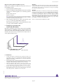
1
CD49b (DX5) MicroBeads
mouse
Order No. 130-052-501
Magnetic cell sorting
Index
1.3 Reagent and instrument requirements
1.
Description
●
1.1 Principle of MACS® separation
Buffer (degassed): Prepare a solution containing PBS (phosphate
buffered saline) pH 7.2, 0.5% BSA (bovine serum albumin) and 2
mM EDTA by diluting MACS BSA Stock Solution (# 130-091-376)
1:20 in autoMACS™ Rinsing Solution (# 130-091-222). Keep buffer
cold (4−8 °C).
▲ Note: EDTA can be replaced by other supplements such as anticoagulant citrate
dextrose formula-A (ACD-A) or citrate phosphate dextrose (CPD). BSA can be
replaced by other proteins such as gelatine, mouse serum or fetal calf serum. Buffers
or media containing Ca2+ or Mg2+ are not recommended for use.
●
MACS Columns and MACS Separators: CD49b+ cells can be
enriched by using MS, LS or XS Columns (positive selection).
CD49b (DX5) MicroBeads can be used for depletion of CD49b+ cells
on LD, CS or D Columns. Cells which strongly express CD49b can
also be depleted using MS, LS or XS Columns. Positive selection or
depletion can also be performed by using the autoMACS Separator.
1.2 Background and product applications
1.3 Reagent and instrument requirements
2. Protocol
2.1 Sample preparation
2.2 Magnetic labeling
2.3 Magnetic separation
3. Example of a separation using CD49b (DX5) MicroBeads
4. References
1. Description
Components
2 m L C D 4 9 b ( DX 5 ) Mi c ro B e ads , mous e : MicroBeads conjugated to monoclonal anti-mouse
NK cell (DX5; isotype: rat IgM; clone: DX5)
antibody.
Size
For 2×10⁹ total cells, up to 200 separations.
Product format
CD49b (DX5) MicroBeads are supplied as a
suspension containing stabilizer and 0.05% sodium
azide.
Storage
Store protected from light at 4−8 °C. Do not freeze.
The expiration date is indicated on the vial label.
1.1 Principle of MACS® separation
First the CD49b+ cells are magnetically labeled with CD49b (DX5)
MicroBeads. Then the cell suspension is loaded onto a MACS® Column
which is placed in the magnetic field of a MACS Separator. The magnetically
labeled CD49b+ cells are retained on the column. The unlabeled cells run
through and this cell fraction is depleted of CD49b+ cells. After removal
of the column from the magnetic field, the magnetically retained NK
cells can be eluted as the positively selected cell fraction.
Column
140-000-103.03
●
Separator
Positive selection
2×10⁸
MS
107
MiniMACS, OctoMACS, VarioMACS, SuperMACS
LS
108
2×10⁹
MidiMACS, QuadroMACS,
VarioMACS, SuperMACS
XS
109
2×10¹⁰
SuperMACS
Depletion
5×10⁸
LD
108
CS
2×10⁸
D
109
MidiMACS, QuadroMACS,
VarioMACS, SuperMACS
VarioMACS, SuperMACS
SuperMACS
Positive selection or depletion
autoMACS
2×10⁸
4×10⁹
autoMACS
▲ Note: Column adapters are required to insert certain columns into VarioMACS™
1.2 Background and product applications
Mouse CD49b (DX5) MicroBeads were developed for the isolation
of mouse NK cells from single-cell suspensions of lymphoid and nonlymphoid tissue, as well as from peripheral blood or body fluids. The
MicroBeads were formerly called Anti-NK Cell (DX5) MicroBeads.
CD49b is expressed on NK cells and a small population of T cells
(CD4+CD3+TCRαβ+). These cells can be depleted prior to NK cell
enrichment by using CD90 (Thy1.2) MicroBeads or CD5 MicroBeads.
CD49b is less mouse strain-specific than other NK cell markers (e.g.
NK1.1) and is expressed by the most common inbred mouse strains.
Examples of applications
● Isolated NK cells were used for the evaluation of NK cell
cytotoxicity in normal and immunodeficient mice in vitro1 or
after adoptive transfer in vivo.2
max. number max. number
of labeled cells of total cells
Separator or SuperMACS™ Separator. For details, see MACS Separator data sheets.
●
(Optional)
Fluorochrome-conjugated
CD49b
antibodies,
e.g. CD49b (DX5)-FITC (# 130-091-814), CD49b (DX5)-PE
(# 130-091-816) or CD49b (DX5)-APC (# 130-091-813).
●
(Optional) PI (propidium iodide) or 7-AAD for flow cytometric
exclusion of dead cells.
●
(Optional) Pre-Separation Filters (# 130-041-407) to remove cell
clumps.
Isolation of NK cells to study their role in experimental mouse
models of immune-mediated liver injury3 or chronic infection
with parasitic helminths4.
www.miltenyibiotec.com
Miltenyi Biotec GmbH
Friedrich-Ebert-Str. 68
51429 Bergisch Gladbach, Germany
Phone +49-2204-8306-0 Fax +49-2204-85197
Miltenyi Biotec Inc.
2303 Lindbergh Street, Auburn, CA 95602, USA
Phone 800 FOR MACS, 530 888-8871 Fax 530 888-8925
page 1/3
Order No. 130-052-501
2. Protocol
2.3 Magnetic separation
2.1 Sample preparation
Prepare a single-cell suspension from lymphoid organs, non-lymphoid
tissue or peripheral blood using standard methods (see "General
Protocols" in the User Manuals or visit www.miltenyibiotec.com).
▲ Dead cells may bind non-specifically to MACS MicroBeads. In case of high
numbers of dead cells we recommend to remove dead cells by density gradient
centrifugation or using the Dead Cell Removal Kit (# 130-090-101).
2.2 Magnetic labeling
▲ Work fast, keep cells cold, and use pre-cooled solutions. This will
prevent capping of antibodies on the cell surface and non-specific cell
labeling.
▲ Volumes for magnetic labeling given below are for up to 107 total
cells. When working with fewer than 107 cells, use the same volumes as
indicated. When working with higher cell numbers, scale up all reagent
volumes and total volumes accordingly (e.g. for 2×10⁷ total cells, use
twice the volume of all indicated reagent volumes and total volumes).
▲ For optimal performance it is important to obtain a singlecell suspension before magnetic separation. Pass cells through
30 µm nylon mesh (Pre-Separation Filters # 130-041-407) to remove cell
clumps which may clog the column.
1.
Determine cell number.
2. Centrifuge cell suspension at 300×g for 10 minutes. Pipette off
supernatant completely.
3.
Resuspend cell pellet in 90 µL of buffer per 107 total cells.
4.
Add 10 µL of CD49b (DX5) MicroBeads per 107 total cells.
5. Mix well and incubate for 15 minutes at 4−8 °C.
▲ Note: Working on ice may require increased incubation times. Higher temperatures
and/or longer incubation times lead to non-specific cell labeling.
6. (Optional) Add a fluorochrome-conjugated CD49b antibody
according to manufacturer's recommendation, and incubate for 5
minutes at 4−8 °C.
7.
Wash cells by adding 1−2 mL of buffer per 107 cells and centrifuge at
300×g for 10 minutes. Pipette off supernatant completely.
8. Resuspend up to 108 cells in 500 µL of buffer.
▲ Note: For higher cell numbers, scale up buffer volume accordingly.
▲ Note: For depletion with LD Columns, resuspend cell pellet in 500 µL of buffer
for up to 1.25×10⁸ cells.
9. Proceed to magnetic separation (2.3).
▲ Choose an appropriate MACS Column and MACS Separator according
to the number of total cells and the number of CD49b+ cells (see table in
section 1.3).
Magnetic separation with MS or LS Columns
1.
Place column in the magnetic field of a suitable MACS Separator
(see "Column data sheets").
2. Prepare column by rinsing with appropriate amount of buffer:
MS: 500 µL
LS: 3 mL.
3.
Apply cell suspension onto the column.
4.
Collect unlabeled cells which pass through and wash column
with appropriate amount of buffer. Perform washing steps by
adding buffer three times, each timeonce the column reservoir is
empty.
MS: 3×500 µL
LS: 3×3 mL.
Collect total effluent. This is the unlabeled cell fraction.
5. Remove column from the separator and place it on a suitable
collection tube.
6. Pipette appropriate amount of buffer onto the column. Immediately
flush out fraction with the magnetically labeled cells by firmly
applying the plunger supplied with the column.
MS: 1 mL
LS: 5 mL.
▲ Note: To increase the purity of the magnetically labeled fraction, it can be passed
over a new, freshly prepared column.
Magnetic separation with XS Columns
For instructions on the column assembly and the separation, refer to the
"XS Column data sheet".
Depletion with LD Columns
1.
Place LD Column in the magnetic field of a suitable MACS Separator
(see "LD Column data sheet").
2. Prepare column by rinsing with 2 mL of buffer.
3.
Apply cell suspension onto the column.
4.
Collect unlabeled cells which pass through and wash column with
2×1 mL of buffer. Collect total effluent. This is the unlabeled cell
fraction.
Depletion with CS Columns
1.
Assemble CS Column and place it in the magnetic field of a suitable
MACS Separator (see "CS Column data sheet").
2. Prepare column by filling and rinsing with 60 mL of buffer. Attach
a 22G flow resistor to the 3-way-stopcock of the assembled column
(see "CS Column data sheet").
3.
Apply cell suspension onto the column.
4.
Collect unlabeled cells which pass through and wash column with 30
mL buffer from the top. Collect total effluent. This is the unlabeled
cell fraction.
Depletion with D Columns
140-000-103.03
For instructions on column assembly and separation, refer to the
"D Column data sheet".
This MACS® product is for in vitro research use only
and not for diagnostic or therapeutic procedures.
page 2/3
Order No. 130-052-501
Magnetic separation with the autoMACS™ Separator
▲ Refer to the "autoMACS™ User Manual" for instructions on how to use
the autoMACS Separator.
1.
Prepare and prime autoMACS Separator.
2. Place tube containing the magnetically labeled cells in the autoMACS
Separator. For a standard separation, choose following separation
programs:
Positive selection: "Possel"
Depletion: "Depletes"
▲ Note: Program choice depends on the isolation strategy, the strength of magnetic
labeling and the frequency of magnetically labeled cells. For details see autoMACS
User Manual: "autoMACS Cell Separation Programs".
3.
When using the program "Possel", collect positive fraction (outlet
port "pos1"). This is the purified CD49b+ cell fraction.
When using the program "Depletes", collect unlabeled fraction (outlet
port "neg1"). This is the CD49b- cell fraction.
Warnings
Reagents contain sodium azide. Under acidic conditions sodium azide yields hydrazoic
acid, which is extremely toxic. Azide compounds should be diluted with running water
before discarding. These precautions are recommended to avoid deposits in plumbing
where explosive conditions may develop.
Warranty
The products sold hereunder are warranted only to be free from defects in workmanship
and material at the time of delivery to the customer. MILTENYI BIOTEC GmbH makes
no warranty or representation, either expressed or implied, with respect to the fitness
of a product for a particular purpose. There are no warranties, expressed or implied,
which extend beyond the technical specifications of the products. MILTENYI BIOTEC
GmbH’s liability is limited to either replacement of the products or refund of the
purchase price. MILTENYI BIOTEC GmbH is not liable for any property damage,
personal injury or economic loss caused by the product.
MACS® is a registered trademark of Miltenyi Biotec GmbH.
3. Example of a separation using
CD49b (DX5) MicroBeads
Relative cell number
NK cells were isolated from a mouse spleen cell suspension using CD49b
(DX5) MicroBeads, a MiniMACS™ Separator and an MS Column. The
cells are fluorescently stained with CD49b (DX5)-FITC (# 130-091-814).
Cell debris and dead cells were excluded from the analysis based on
scatter signals and PI fluorescence.
CD49b (DX5)+ cells
CD49b (DX5)- cells
Spleen cells before separation
CD49b (DX5)-FITC
4. References
1.
Tomasello, E; Desmoulins, P-O; Chemin, K; Guia, S; Cremer, H; Ortaldo, J; Love,
P; Kaiserlian, D; Vivier, E (2000) Combined Natural Killer Cell and Dendritic
Cell Functional Deficiency in KARAP/DAP12 Loss-of-Function Mutant Mice.
Immunity 13: 335–364. [945]
2.
Shi, FD; Takeda, K; Akira, S; Sarvetnick, N; Ljunggren, HG (2000) IL-18 Directs
Autoreactive T Cells and Promotes Autodestruction in the Central Nervous System
Via Induction of IFN-γ by NK Cells. J. Immunol. 165: 3099–3104. [1021]
3.
Mühlen KA, Schümann J, Wittke F, Stenger S, van Rooijen N, van Kaer L, Tiegs
G (2004) NK cells but not NKT cells, are involved in Pseudomonas aeruginosa
exotoxin a-induced hepatotoxicity in mice. J. Immunol. 172: 3034–3041. [4274]
4.
Hsieh GC, Loukas A, Wahl AM, Bhatia M, Wang Y, et al. (2004) A secreted protein
from the human hookworm necator americanus binds selectively to NK cells and
induces IFN-γ production. J. Immunol. 173: 2699–2704. [4275]
140-000-103.03
This MACS® product is for in vitro research use only
and not for diagnostic or therapeutic procedures.
page 3/3