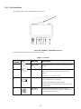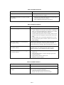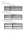
1
CARDELL® 9500 HD
Veterinary Monitor
User’s Guide
For Veterinary Use Only
003-2566-00 Rev. C (4/4/13)
Product Information
Product model: CARDELL® 9500 HD
Product name: Veterinary monitor
2
Cardell® 9500 HD User’s Guide
Explanation of Symbols
The following symbols appear on the monitor and its packaging.
Table 1 Monitor and Packaging Symbols
Symbol
ECG/RESP
NIBP
Description
Symbol
Description
Keep dry
Caution, consult
accompanying
documents
Atmospheric pressure
limitation
Temperature limitation
Keep upright
Humidity limitation
Recovery
Fragile, handle with care
Recyclable
Maximum stacking
Waveform Freeze key
On/Standby key
Alarm Silence key
Print key
NIBP history key
NIBP key
AC Power LED
Charging LED
ECG connector
NIBP cuff connector
SpO2
CO2
SpO2 probe connector
CO2 connector
Table 1 Monitor and Packaging Symbols (Continued)
Symbol
TEMP
4
Description
Symbol
Description
TEMP connector
Dangerous Voltage
CF applied part:
including F applied part
(float/insulation) and
defibrillation-proof
function.
BF applied part:
including F applied part
(float/insulation) and
defibrillation-proof
function.
Reserved port
Fuse
Protective grounding
Serial Number
Equipotential grounding
Manufacturer address
Date of manufacture
Configuration number
Batch code
Consult Instructions for
Use
Catalogue number
Compliance with WEEE
standard
Protective grade
USB port/
USB Export Menu
Cardell® 9500 HD User’s Guide
Safety Conventions
The manual uses the following conventions for Notes, Cautions, and Warnings.
Note — A Note calls attention to an important point in the text.
Caution
A Caution calls attention to a condition or possible situation that could damage or destroy the product or
the user’s work.
Warning
A Warning calls attention to a condition or possible situation that could cause injury to the user and/or
patient.
Safety Requirements
Note — The safety indications in this chapter apply to general monitor use. Safety indications in other chapters
apply to specific monitor measurements.
Follow the instructions in this user manual when using the monitor. However, conventional medical practices
always supersede this document. Significance of safety requirements set forth here in this manual is not in order of
reading sequence.
Warning
The monitor is not for home use.
The monitor is not for diagnostic or therapeutic use.
To avoid mixture, clear all historical data of the last patient before monitoring a new patient.
Ensure that the veterinary monitor is in good working condition and is placed in proper position before
clinical use.
Never rely exclusively on the alarm system for veterinary monitoring. You must periodically check that
monitor alarms are working properly. The most reliable method of veterinary monitoring combines close,
personal surveillance along with the correct operation of the monitor.
Never use the monitor during MRIs or CT scans.
Never use cables with exposed conductors. Only use undamaged cables and connectors to avoid personal or
patient injury.
Never simultaneously touch the patient and the input or output terminal of the monitor.
The monitor can be used with electrosurgical devices. To avoid burns or death, ensure that the
electrosurgical circuitry is properly connected.
Ensure that the patient is not connected to any metal, the surface of any conductors, or grounded parts of
any devices when a defibrillator is in use. Never touch the animal, table, or instruments during defibrillation.
Never use the monitor in an environment of uncontained anesthetic gases.
Disconnect the AC power supply and remove the battery before disassembling the monitor.
Caution
To avoid personal or patient injury, only use accessories and parts produced or recommended by the
manufacturer. Otherwise, damage to the monitor can occur.
Position power cords and other cables to prevent tangling, patient contact, or electrical interference.
Always use a three-wire receptacle in order to properly ground the monitor. Use the monitor on battery
power only if you suspect any problem with external grounding or wiring.
The battery will discharge over time when stored inside a monitor with no AC power. Keep the monitor
plugged in when storing it and perform a monthly check of battery power.
If multiple devices are connected to a patient, the sum of the leakage currents may cause a dangerous
situation. Therefore, qualified service representatives should always perform current leakage tests before
setting up the connections. This ensures that leakage is within limits and prevents personal or patient injury
and environmental damage. If any question remains, consult the manufacturer for correct usage.
Unknown electromagnetic interference could be caused by radio transmitters or nearby televisions. Move
veterinary monitors away from these devices or insert shielding materials around the monitor.
If patient data is lost, closely monitor the patient or replace the monitor immediately until the monitor
restores normal function again.
When the monitor is powered off, patient monitoring will be stopped and user configurations will be
saved.
To reduce the hazard of burns in high-frequency surgical neutral connections, the leads and connectors
must be located away from the surgical site. Keep cables away from other devices.
Periodically check all reusable accessories for damage. Replace and dispose of damaged accessories when
necessary according to your local hospital waste disposal regulations.
ECG electrodes are disposable accessories. Always properly dispose of ECG electrodes according to your
local hospital waste disposal regulations.
Clean and sterilize the monitor and accessories according to local requirements. Turn off the monitor and
disconnect the power cords before cleaning or sterilization.
Keep all monitor packing materials away from children, or dispose of them in accordance with your local
environmental regulations.
Always properly dispose of the monitor and all accessories at the end of their service life. Dispose of
batteries according to your local regulations. Never incinerate batteries or expose them to high
temperatures.
Ensure that no water condenses into or on the monitor. Condensation can occur from changes in
temperature or exposure to humidity.
6
Cardell® 9500 HD User’s Guide
Contents
1 Overview
1.1 Indications for Use - - - - - - - - - 1.2 Intended Use- - - - - - - - - - - - 1.3 CARDELL® 9500 HD Configurations
1.3.1 Main Components - - - - - -
-
-
-
-
-
-
-
-
-
-
-
-
-
-
-
-
-
-
-
-
-
-
-
-
- - - - - - - - - - - - -- - - - - -- - - - - - -
1-1
1-1
1-2
1-2
2.1 Before You Begin - - - - - - - - - - - - - - - - - - - - 2.2 Setting up the Monitor - - - - - - - - - - - - - - - - - - 2.2.1 Power Supply - - - - - - - - - - - - - - - - - - 2.2.2 Inspecting the Monitor - - - - - - - - - - - - - - 2.2.3 Checking Your Software Release - - - - - - - - - 2.3 Product Description - - - - - - - - - - - - - - - - - - - 2.3.1 The Front Panel - - - - - - - - - - - - - - - - - 2.3.2 The Rear Panel - - - - - - - - - - - - - - - - - 2.3.3 The Side Panels - - - - - - - - - - - - - - - - - 2.4 The Screen Display - - - - - - - - - - - - - - - - - - - 2.5 Menus - - - - - - - - - - - - - - - - - - - - - - - - - - 2.6 Configuring the Display - - - - - - - - - - - - - - - - - 2.6.1 Using the Large Font Screen - - - - - - - - - - 2.6.2 Changing the Waveform Display - - - - - - - - - 2.6.3 Changing the Waveform Speed - - - - - - - - - - 2.6.4 Freezing the Waveforms - - - - - - - - - - - - - 2.7 Changing the Monitor Settings - - - - - - - - - - - - - - 2.7.1 System Maintenance - - - - - - - - - - - - - - - 2.7.2 Changing the Date and Time - - - - - - - - - - - 2.8 Changing the Volume and Brightness - - - - - - - - - - - 2.8.1 Adjusting the HR and PR Volume - - - - - - - - 2.8.2 Adjusting the Brightness - - - - - - - - - - - - - 2.9 Managing Patients - - - - - - - - - - - - - - - - - - - - 2.10 Exporting ECG Waveform and Trend Data to a USB Disk -
-
-
-
-
-
-
-
-
-
-
-
-
-
-
-
-
-
-
-
- 2-1
- 2-1
- 2-2
- 2-2
- 2-2
- 2-2
- 2-3
- 2-4
- 2-5
- 2-6
- 2-7
- 2-8
- 2-8
- 2-8
- 2-8
- 2-9
- 2-9
- 2-9
- 2-9
- 2-10
- 2-10
- 2-10
- 2-10
- 2-11
-
-
-
-
-
-
-
-
-
-
-
-
-
-
-
-
-
-
-
-
2 Basic Operation
3 Alarms
3.1 Alarm Types - - - - - - - - - - - 3.2 Alarm Levels - - - - - - - - - - 3.2.1 High Priority Alarms - - - 3.2.2 Medium Priority Alarms - 3.2.3 Low Priority Alarms - - - 3.3 Visual Alarms - - - - - - - - - - 3.3.1 LED Flashing Alarms - - 3.3.2 Flashing Icons - - - - - - 3.3.3 Alarm Messages - - - - - 3.4 Audible Alarms - - - - - - - - - 3.4.1 Turning on Audible Alarms
3.4.2 Turning off Audible Alarms
3.4.3 Silencing Alarms - - - - 3.4.4 Adjusting Alarm Volume - 3.5 Setting the Alarm Mode - - - - - 3.5.1 Automatic - - - - - - - - 3.5.2 Standard - - - - - - - - - Cardell® 9500 HD User’s Guide
-
-
-
-
-
-
-
-
-
-
-
-
3-1
3-1
3-1
3-1
3-2
3-2
3-2
3-2
3-2
3-2
3-3
3-4
3-4
3-4
3-5
3-5
3-5
Contents-1
3.6 Setting Alarm Limits - - - - - - - - - - - - - - - - - - - - - - - - - - - - - - - - - - - - - - - - 3-5
3.6.1 Setting Individual Alarm Limits - - - - - - - - - - - - - - - - - - - - - - - - - - - - - - 3-5
3.6.2 Restoring Default Alarm Limits- - - - - - - - - - - - - - - - - - - - - - - - - - - - - - - 3-6
4 Monitoring ECG
4.1 Before Monitoring - - - - - - - - - - - - - - - - 4.1.1 Skin Preparation for Electrode Placement - 4.1.2 Placing the Electrodes - - - - - - - - - - 4.1.3 Changing a Lead Set - - - - - - - - - - - 4.1.4 Lead Recommendations for Surgical Patients
4.1.5 Connecting ECG Cables - - - - - - - - - 4.2 The ECG Display - - - - - - - - - - - - - - - - 4.2.1 ECG Waveform and Numeric Pane - - - - 4.3 Configuring the ECG Display - - - - - - - - - - - 4.3.1 Changing the Size of the ECG Waveform - 4.3.2 Changing the Speed of the ECG Waveform 4.4 Performing 1 mV Calibration - - - - - - - - - - - 4.5 Changing the Heart Rate Volume - - - - - - - - - 4.6 Changing the ECG Mode - - - - - - - - - - - - - 4.7 Changing HR Alarm Settings - - - - - - - - - - - 4.8 Configuring HR/PR Priority - - - - - - - - - - - 4.9 Monitoring Pacemaker Patients - - - - - - - - - 4.10 Safety Information - - - - - - - - - - - - - - - -
-
-
-
-
-
-
-
-
-
-
-
-
-
-
-
-
-
-
-
-
-
-
-
-
4-1
4-1
4-1
4-3
4-3
4-4
4-4
4-4
4-5
4-5
4-5
4-5
4-6
4-6
4-6
4-6
4-7
4-8
5.1 Lead Placement for Monitoring Respiration - - - - - - - 5.1.1 Selecting a Respiration Lead - - - - - - - - - - 5.2 The Respiration Display - - - - - - - - - - - - - - - - 5.3 Changing the Respiration Display - - - - - - - - - - - - 5.3.1 Changing the Respiration Waveform Size - - - - 5.3.2 Changing the Respiration Waveform Speed - - - 5.3.3 Changing the Respiration Waveform Display Mode
5.4 Changing the Respiration Alarm Settings - - - - - - - - 5.5 Changing the Apnea Alarm Settings- - - - - - - - - - - 5.6 Safety Information - - - - - - - - - - - - - - - - - - - -
-
-
-
-
-
-
-
-
-
-
-
-
-
-
-
-
-
-
-
-
-
5-1
5-1
5-2
5-2
5-2
5-2
5-3
5-3
5-3
5-3
-
-
-
-
-
-
-
-
- - - - - - - - - - - - - 6-1
- - - - - - - - - - - - - 6-1
- - - - - - - - - - - - - 6-2
- - - - - - - - - - - - - 6-2
- - - - - - - - - - - - - 6-2
- - - - - - - - - - - - - 6-3
- - - - - - - - - - - - - 6-3
- - - - - - - - - - - - - 6-3
- - - - - - - - - - - - - 6-3
- - - - - - - - - - - - - 6-3
- - - - - - - - - - - - - 6-3
- - - - - - - - - - - - - 6-4
- - - - - - - - - - - - - 6-4
5 Monitoring Respiration
6 Monitoring SpO2
6.1 SpO2 Monitoring Theory - - - - - - - - - - - - - - 6.2 Placing an SpO2 - - - - - - - - - - - - - - - - - 6.3 Connecting SpO2 Cables - - - - - - - - - - - - - - 6.4 Measuring SpO2 - - - - - - - - - - - - - - - - - 6.5 Assessing a Suspicious SpO2 Reading - - - - - - - - 6.6 The SpO2 Display - - - - - - - - - - - - - - - - - 6.7 Changing the SpO2 Display - - - - - - - - - - - - - 6.7.1 Changing the SpO2 Waveform Speed - - - - 6.7.2 Changing the SpO2 Waveform Display Mode 6.8 Configuring HR/PR Priority - - - - - - - - - - - - 6.9 Changing the SpO2 Alarm Settings - - - - - - - - - 6.10 Changing the PR Alarm Settings - - - - - - - - - - 6.11 Safety Information - - - - - - - - - - - - - - - - -
Contents-2
-
-
Cardell® 9500 HD User’s Guide
7 Monitoring NIBP
7.1 Selecting and Placing an NIBP Cuff - - - - - - - - - - - - - - - - - - - - - - - - - - - - - - - - 7-1
7.1.1Cuff Placement for Cat- - - - - - - - - - - - - - - - - - - - - - - - - - - - - - - - - - - - - 7-1
7.1.2 Cuff Placement for Dog - - - - - - - - - - - - - - - - - - - - - - - - - - - - - - - - - - - 7-2
7.1.3 Cuff Placement for Large Animal- - - - - - - - - - - - - - - - - - - - - - - - - - - - - - 7-3
7.1.4 Cuff Size Selection - - - - - - - - - - - - - - - - - - - - - - - - - - - - - - - - - - - - - 7-3
7.2 Connecting the Cuff - - - - - - - - - - - - - - - - - - - - - - - - - - - - - - - - - - - - - - - - 7-4
7.3 Measurement Limitations - - - - - - - - - - - - - - - - - - - - - - - - - - - - - - - - - - - - - 7-4
7.4 Measuring the NIBP - - - - - - - - - - - - - - - - - - - - - - - - - - - - - - - - - - - - - - - - 7-4
7.5 The NIBP Display - - - - - - - - - - - - - - - - - - - - - - - - - - - - - - - - - - - - - - - - - 7-5
7.6 Changing the NIBP Cuff Size - - - - - - - - - - - - - - - - - - - - - - - - - - - - - - - - - - - 7-5
7.7 Selecting NIBP Measurement Methods - - - - - - - - - - - - - - - - - - - - - - - - - - - - - - - 7-5
7.8 Selecting the NIBP Interval Time - - - - - - - - - - - - - - - - - - - - - - - - - - - - - - - - - - 7-6
7.9 Changing the NIBP Unit - - - - - - - - - - - - - - - - - - - - - - - - - - - - - - - - - - - - - - 7-7
7.10 Viewing the NIBP Measurement Data - - - - - - - - - - - - - - - - - - - - - - - - - - - - - - - 7-7
7.11 Changing the NIBP Alarm Settings - - - - - - - - - - - - - - - - - - - - - - - - - - - - - - - - 7-8
7.12 Calibrating NIBP - - - - - - - - - - - - - - - - - - - - - - - - - - - - - - - - - - - - - - - - - 7-8
7.13 Safety Information - - - - - - - - - - - - - - - - - - - - - - - - - - - - - - - - - - - - - - - - 7-8
8 Monitoring Temperature
8.1 Selecting and Connecting Temperature Probe 8.2 Influencing Factors - - - - - - - - - - - - 8.3 Monitoring Temperature - - - - - - - - - - 8.4 Changing the Temperature Unit - - - - - - 8.5 Changing the Temperature Alarm Settings - 8.6 Safety Information - - - - - - - - - - - - - -
-
-
-
-
-
-
-
-
-
-
-
-
-
-
-
-
-
-
-
-
-
-
-
-
-
-
-
8-1
8-1
8-1
8-2
8-2
8-2
9.1 Selecting CO2 Accessories - - - - - - - - - - - - 9.2 Zeroing the Sensor - - - - - - - - - - - - - - - - 9.3 Measuring Carbon Dioxide - - - - - - - - - - - - 9.3.1 Capnostat® 5 Mainstream CO2 Sensor- - - 9.3.2 LoFlo SideStream CO2 Sensor- - - - - - - 9.4 The CO2 Display - - - - - - - - - - - - - - - - 9.5 Changing the CO2 Display - - - - - - - - - - - - 9.5.1 Changing the CO2 Waveform Speed - - - 9.5.2 Changing the CO2 Waveform Display Mode
9.6 Changing the CO2 Alarm Settings - - - - - - - - 9.7 Safety Information - - - - - - - - - - - - - - - - -
-
-
-
-
-
-
-
-
-
-
-
-
-
-
-
-
-
-
-
- - - - -- - - - - - -
-
- - - - - - - - - -
9-1
9-1
9-2
9-2
9-3
9-5
9-5
9-5
9-5
9-5
9-6
9 Monitoring Carbon Dioxide
10 Managing Data
10.1 Viewing Trends - - - - - - - - - - - - - - - - - - - - - - - - - - - - - - - - - - - - - - - - - 10-1
10.2 Viewing Data List - - - - - - - - - - - - - - - - - - - - - - - - - - - - - - - - - - - - - - - - 10-2
10.2.1 Viewing All Measurement Data - - - - - - - - - - - - - - - - - - - - - - - - - - - - - - 10-2
11 Printing
11.1 About the Printer - - - - - - - - - 11.2 Loading the Printer Paper - - - - - 11.3 Printing - - - - - - - - - - - - - 11.4 Turning on Alarm-triggered Printing
11.5 Setting Timed Printing - - - - - - 11.6 Setting the Print Time- - - - - - - 11.7 Setting the Advance Time- - - - - Cardell® 9500 HD User’s Guide
-
-
-
-
-
-
-
-
-
-
-
-
-
-
-
-
-
-
-
-
-
-
-
-
-
-
-
-
-
- - - 11-1
- - -11-1
- - 11-2
- - 11-2
- - 11-3
- - 11-3
- - 11-3
Contents-3
11.8 Selecting the Print Mode 11.9 Changing the Print Speed 11.10 Sample Print Strip
11.11 Printer Paper Guidelines -
-
-
-
-
-
-
-
-
-
-
-
-
-
-
-
-
-
-
-
-
-
-
-
-
-
-
-
-
-
-
-
-
-
-
- - 11-3
- - 11-4
- - 11-4
- - -11-4
12 Cleaning and Care
12.1 General Guidelines - - - - - - - - - - - - - - - - - - - - - - - - - - - - - - - - - - - - - - - - 12-1
12.2 Cleaning the Monitor - - - - - - - - - - - - - - - - - - - - - - - - - - - - - - - - - - - - - - - 12-1
12.2.1 Cleaning the Display - - - - - - - - - - - - - - - - - - - - - - - - - - - - - - - - - - - 12-2
12.3 Cleaning the Accessories - - - - - - - - - - - - - - - - - - - - - - - - - - - - - - - - - - - - - 12-2
12.3.1 Cleaning the Cables - - - - - - - - - - - - - - - - - - - - - - - - - - - - - - - - - - - - 12-2
12.3.2 Cleaning the SpO2 Sensor - - - - - - - - - - - - - - - - - - - - - - - - - - - - - - - - - 12-2
12.3.3 Cleaning the NIBP Cuff - - - - - - - - - - - - - - - - - - - - - - - - - - - - - - - - - - 12-2
12.3.4 Cleaning the Temperature Probe - - - - - - - - - - - - - - - - - - - - - - - - - - - - - - 12-3
12.3.5 Cleaning the ECG Clips - - - - - - - - - - - - - - - - - - - - - - - - - - - - - - - - - - 12-2
12.3.6 Cleaning the CO2 Accessories - - - - - - - - - - - - - - - - - - - - - - - - - - - - - - - 12-3
12.4 Cleaning the Print Head - - - - - - - - - - - - - - - - - - - - - - - - - - - - - - - - - - - - - - - 12-4
12.5 Disinfecting the Monitor - - - - - - - - - - - - - - - - - - - - - - - - - - - - - - - - - - - - - 12-4
12.6 Sterilizing the Monitor - - - - - - - - - - - - - - - - - - - - - - - - - - - - - - - - - - - - - - 12-4
13 Maintaining the Battery
13.1 Charging LED - - - - - - - - - - - 13.2 Battery Indicator - - - - - - - - - - 13.3 Installing the Battery - - - - - - - - 13.4 Recharging the Battery - - - - - - - 13.5 Optimizing the Battery - - - - - - - 13.5.1 Conditioning the Battery - - 13.5.2 Checking Battery Performance
13.6 Disposing of a Battery- - - - - - - - 13.7 Safety Information - - - - - - - - - -
-
-
-
-
-
-
-
-
-
-
-
-
-
-
-
-
-
-
-
-
-
-
-
-
-
-
-
-
-
- 13-1
- 13-1
- 13-2
- 13-3
- 13-3
- 13-3
- 13-3
- 13-3
- 13-4
14.1 Inspecting the Equipment and Accessories
14.2 Maintenance Task and Test Schedule - 14.3 Troubleshooting - - - - - - - - - - - 14.4 Disposing of the Monitor - - - - - - - -
-
-
-
-
-
-
-
-
-
-
-
-
-
-
-
-
-
-
-
-
-
-
-
-
-
-
-
-
- 14-1
- 14-1
- 14-2
- 14-5
14 Maintenance and Troubleshooting
15 Warranty/Service
15.1 Limited Warranty- - - - - - - - - - - - - - - - - - - - - - - - - - - - - - - - - - - - - - - - - 15-1
15.2 After-sale Service - - - - - - - - - - - - - - - - - - - - - - - - - - - - - - - - - - - - - - - - - 15-2
Contents-4
Cardell® 9500 HD User’s Guide
A. Specifications
A.1 ECG Specifications - - - - - - - - - - - A.2 Respiration Specifications - - - - - - - - A.3 SpO2 Specifications - - - - - - - - - - - A.4 Temperature Specifications - - - - - - - A.5 Non-invasive Blood Pressure SpecificationsA.6 Carbon Dioxide Specifications - - - - - - A.7 Power Specifications- - - - - - - - - - - A.8 Display Specifications - - - - - - - - - - A.9 Printer Specifications - - - - - - - - - - A.10 Physical Specifications - - - - - - - - - A.11 Environmental Specifications - - - - - - -
-
-
-
-
-
-
-
-
-
-
-
-
-
-
-
-
-
-
-
-
-
-
-
-
-
-
- - A-1
- - A-2
- - A-2
- - A-3
- - A-4
- - A-4
- - A-5
- - A-5
- - A-6
- - A-6
- - A-6
-
-
-
-
-
-
-
-
-
-
-
-
-
-
-
-
-
-
-
-
-
-
-
-
-
-
-
B. Alarm Specifications
B.1 Alarm Classifications
B.2 Physiological Alarms
B.3 Technical Alarms - B.4 Default Alarm Limits
-
-
-
-
-
-
-
-
-
-
-
-
B-1
B-1
B-3
B-4
C.1 System Default Settings - - - - - - - - - - - - - - - - - - - - - - - - - - - - - - - - - - - - - C.2 Alarm Default Settings - - - - - - - - - - - - - - - - - - - - - - - - - - - - - - - - - - - - - - C.3 ECG Default Settings - - - - - - - - - - - - - - - - - - - - - - - - - - - - - - - - - - - - - - C.4 Respiration Default Settings - - - - - - - - - - - - - - - - - - - - - - - - - - - - - - - - - - - C.5 SpO2 Default Settings - - - - - - - - - - - - - - - - - - - - - - - - - - - - - - - - - - - - - - C.6 NIBP Default Settings - - - - - - - - - - - - - - - - - - - - - - - - - - - - - - - - - - - - - - C.7 Temperature Default Settings - - - - - - - - - - - - - - - - - - - - - - - - - - - - - - - - - - C.8 Carbon Dioxide Default Settings - - - - - - - - - - - - - - - - - - - - - - - - - - - - - - - - - C.9 Printer Default Settings - - - - - - - - - - - - - - - - - - - - - - - - - - - - - - - - - - - - - - -
C-1
C-1
C-1
C-2
C-2
C-3
C-3
C-3
C-4
C. Default Settings
D. Glossary
D.1 Terminology Definitions- - - - - - - - - - - - - - - - - - - - - - - - - - - - - - - - - - - - - - D-1
E. Tips
E.1 Blood Pressure E.2 ECG- - - - - E.3 SpO2 - - - - E.4 CO2 - - - - - E.5 Temperature - -
-
-
-
-
-
-
-
-
-
- - - - - - - - - - - - - - - - - - - - - - - - - - - - - - - - - - - - - - - - - - - - - - - - - - - - - - - - - - - - - - - - -- - - - - - - - - - - - - - - - - - - - - - - - - - - - - - - - - - - - - - - - - - - - - - - - - - - - - - - - - - - - - - - - - - - - - - - - - - - - - - - - - - - - - - - - - - - - - - - -
E -1
E -1
E -2
E -2
E -2
F. Accessories - - - - - - - - - - - - - - - - - - - - - - - - - - - - - - - - - - - - - - - - - - - - F - 1
1 Index
Cardell® 9500 HD User’s Guide
Contents-5
1
Overview
The CARDELL® 9500 HD veterinary monitor is used for monitoring, recording, and alarming of multiple
physiological parameters of animals. Your monitor stores data in trend database. You can view measurement
trend graphs to help you identify changes in the monitored animal's physiological condition.
®
Figure 1-1 CARDELL 9500 HD Monitor
This Instructions for Use is an integral part of the monitor and provides detailed introductions on how to use and
maintain it. Keep the document next to the monitor for reference. In addition, read and keep any documents that
come with any accessories, as these contain important information not in this guide.
This guide describes all of the monitor features and options. Your monitor may not have all of them. What you see
on the screen, including the menus, depends on the way it has been configured for your hospital.
1.1 Indications for Use
The CARDELL® 9500 HD veterinary monitor is for use by healthcare professionals whenever there is a need for
monitoring the physiological parameters of animals.
1.2 Intended Use
The CARDELL® 9500 HD veterinary monitor is intended to be used in special laboratory and other veterinary
hospital or clinic where veterinary monitoring is needed. Additionally, the monitor is also intended for use in
transport situations.
1-1
Indications for Use
1.3 CARDELL® 9500 HD
Configurations
The CARDELL® 9500 HD veterinary monitor includes several configurations.
1.3.1 Main Components
The monitor can contain the following components:
•
Main unit
•
ECG
•
RESP
•
NIBP
•
•
SpO2
TEMP
•
•
CO2
Printer
The following table lists the available configurations, the standard features in each monitor, as well as optional
features. In the table, a solid circle indicates a standard feature.
Table 1-1 Product Configurations
Parameter
CARDELL® 9500 HD
ECG
•
RESP
•
NIBP
•
SpO2
•
TEMP
•
CO2
• (with optional sensor)
Printer
•
USB Port
•
Battery
• (One battery)
Display Screen
7”
1-2
2
Basic Operation
This chapter describes how to set up and begin using the CARDELL® 9500 HD veterinary monitor.
2.1 Before You Begin
Before veterinary monitoring, inspect the monitor to ensure it is in good condition and used in proper
environment.
1. Unpack the equipment and ensure that you have the following:
– One veterinary monitor
– Instructions for Use
– AC power cord (country-specific)
– Set of accessories
2. Keep the packing case for future transport or storage. If it is damaged, immediately contact the courier
company.
3. Examine the monitor for cleanliness and general physical condition. Ensure that:
– The housing is not cracked or broken.
– Power plugs and cords are in good condition and plug prongs are not bent.
– The accessory cables are intact.
Caution
If the equipment, accessories or their packaging show signs of damage, do not use the equipment.
2.2 Setting up the Monitor
Place and use the monitor in a location that conforms to the environmental specifications outlined
on page A-6. Locate the monitor in an area that:
•
is convenient for observation and operation.
•
is free from movement, dust, corrosive or explosive gases.
•
has a five-centimeter space around to ensure good ventilation and smooth heat dissipation.
You can put the monitor on a flat surface.
Warning Never attempt to open the monitor case. Only qualified personnel should service the monitor.
Never block the monitor's air vent during operation.
2-1
2.2.1 Power Supply
You can power the monitor with either an AC power source or its internal battery. You can switch back and forth
between supplies without powering off the monitor.
When the monitor is connected to an AC power source, you can turn on and operate the veterinary monitor.
When powered on, the monitor is under normal working condition. It can be used to measure vital signs and
charge the battery.
When it is standby, the monitor can only charge the battery.
When AC power is lost, a battery powered monitor continues to run without interruption on battery power. For
more information on battery power, see Chapter 13, Maintaining the Battery on page 13-1.
If the monitor is only powered by an AC power source, the monitor will be powered off automatically when the
AC power is lost; when powered on again, the monitor is in normal working condition.
2.2.2 Inspecting the Monitor
Before you start using the monitor, ensure that:
1. There is no damage to the monitor or the accessories. Before turning on the power, check the monitor and all
the cables, connectors and accessories and ensure that the extension cables are correctly connected to the
fittings.
2. The AC power supply is in conformity with standards. Plug one end of the specified power cord into an AC
power supply with a protection-grounded wire. Plug the other end into the power connector on the monitor.
When the power cord is correctly connected, the AC power LED is yellow. If the monitor is powered by
battery, verify that adequate power remains and the battery power gauge is full.
3. The monitor is properly grounded. Connect the small end of the grounding cable to the grounding post on the
back of the monitor. The large end of the grounding cable should be connected to any metal surface or copper
pipe.
4. The accessories are correctly connected. For further testing of individual measurement alarms, perform the
measurement on a simulator (for example, SpO2). Adjust alarm limits and check that alarm behavior is
observed.
5. All settings like cuff size, alarm limits and so on, are correct. Ensure that the monitor is in good working
condition and that all functions of the monitor meet the technical requirements mentioned in the Instructions
for Use.
6. When you press the On/Standby key on the front panel, the main screen appears.
2.2.3 Checking Your Software Release
To check the software release of the monitor:
1. Rotate the navigation knob when no menu displays on the monitoring screen.
2. Rotate the navigation knob to the System menu and press the knob. The version number is shown in the
top left corner, displaying as Version: Vn.nn.
3. Rotate the navigation knob to Exit and press the knob to quit the menu.
2.3 Product Description
The monitor has a color TFT screen that displays waveforms, menus, alarm status and physiological
measurements. This section describes all the monitor features and options. Your monitor may not have all of them.
2-2
2.3.1 The Front Panel
The following figure shows the front panel of the monitor:
10
123
4 5
6
7
8
9
®
Figure 2-1 CARDELL 9500 HD Front Panel
The following table describes the controls on the monitor.
Table 2-1 Controls
Item
Number
Icon
Meaning
Description
1
On/Standby
key
Press and hold for three seconds to turn the monitor on
or off.
2
Power LED
Green: the monitor is powered on.
Yellow: the monitor is off and connected to an AC
power.
Off: there is no AC power source.
3
Charging LED
Flashing green: the monitor is connected to AC
power, and the battery is charging.
Steady green: the battery is fully charged.
Off: the monitor is not connected to an AC power or no
battery loaded.
4
Press to freeze/unfreeze the waveform. After 30
seconds, the wave unfreezes (if frozen) and the monitor
returns to the previous screen.
Waveform
Freeze key
2-3
Table 2-1 Controls (Continued)
Item
Number
Icon
Meaning
Description
5
NIBP key
Press to start or stop an NIBP measurement.
6
Alarm Silence
key
Press to turn the alarm audio off for 2 minutes. The
monitor resumes alarm audio when the counter reaches
00:00.
Press again when alarms are paused to restart alarm
audio.
7
Print key
Press to start or stop printing.
8
NIBP History
key
Press to view NIBP history on the monitoring screen.
9
Navigation
knob
When a menu is displayed, rotate and press the
navigation knob to select and change the settings;
When there is no menu on the screen, press to enter the
Data Review (history) window.
Alarm LED
10
Flashing red: indicates a high priority alarm or lifethreatening situation.
Flashing yellow: indicates a medium priority alarm.
Steady yellow: indicates a low priority alarm.
2.3.2 The Rear Panel
The following figure shows the rear panel of the monitor. Your monitor may not have all of the following items.
8
1
2
3
4
5
6
®
7
Figure 2-2 CARDELL 9500 HD Rear Panel
2-4
The following table describes each item on the rear panel.
Table 2-2 Rear Panel
Item
Number
Item
Name
Description
1
Audio outlet (speaker)
Enables audio output
2
USB port
3
Reserved port
4
Signal input/output
Software update port only, DB9
connector
5/6
Fuse socket
T1.6A, 250V fuse
7
Equipotential grounding lug
Connects to the grounding system
8
External AC power connector
Connects to external AC power supply
2.3.3 The Side Panels
The following figure shows the left and right side panels of the CARDELL® 9500 HD monitor. Your monitor may
not have all of the following items.
1
2
3
4
5
6
®
7
Figure 2-3 CARDELL 9500 HD Side Panels
2-5
The following table describes each item on the side panels.
Table 2-3 Side Panel
Item
Number
Symbol
Description
1
TEMP
TEMP connectors
2
CO2
CO2 connector
3
ECG/RESP
ECG connector
4
SpO2
SpO2 probe connector
5
NIBP
NIBP cuff connector
6
Battery compartment cover
7
Printer
2.4 The Screen Display
The monitor screen displays parameters, waveforms and relevant information about patient measurements. The
main screen display might look different, depending on the configuration of your monitor.
The following figure shows a typical screen display.
II
X1
25 mm/s
Monitor
HR/PR bpm
T
o
C
25 mm/s
1
SPO2 %
LA-RL
X1
25 mm/s
0: 07: 00
NIBP kPa
RR rpm
Large
EtCO2
InCO2
Auto
4
3
®
Figure 2-4 CARDELL 9500 HD Typical Screen Display
2-6
The following table describes the screen display.
Table 2-4 Screen Display Description
Item
Number
Description
Item
Number
Description
1
Waveform area
2
Numeric panes
3
Menu bar and message area
4
Information bar
2.5 Menus
Main menu and the submenu are two typical menus in the monitor.
When no menu displays on the screen:
1. Rotate the navigation knob to access the Main menu.
2. Scroll down the menus to highlight the menu you want and then press the navigation knob to select the
menu and make necessary changes.
The following table describes the Main menu indicators:
Table 2-5 Main Menu
Icon
Description
Icon
Description
Parameter menu
Trend Display menu
System menu
Volume & Brightness
Menu
Print menu
NIBP History List
Large Font
USB Export Menu
Exit
Submenu is the menu under the Main menu.
1. Rotate the navigation knob to access a submenu, then press the navigation knob to a menu item and press
the knob.
2. Make your changes then rotate the navigation knob to Exit and press the knob to return to the previous
menu.
2-7
2.6 Configuring the Display
You can configure the display to a variety of layouts.
2.6.1 Using the Large Font Screen
Select the Large Font option to enlarge the information on the parameter panes on the screen. This provides a
better view of parameters for remote monitoring.
The following figure shows an example of the screen display when Large Font is selected:
Figure 2-5 Large Font Screen
To display or exit the Big Number screen:
1. Rotate the navigation knob to access the Main menu.
2. Rotate the navigation knob to highlight Big Number and press the knob to confirm.
3. Press the navigation knob again to exit the Big Number screen.
Note — The parameter and layout of the Large Font screen depends on the parameters monitored.
2.6.2 Changing the Waveform Display
The monitor has three modes of waveform display: single line, bold line and filled. Waveforms display in a single
line by default. When Enhanced is selected in Waveform Mode, waveforms display in a bold line.
To change the waveform display:
1. Rotate the navigation knob to access the Main menu.
2. Rotate the navigation knob to the Parameter menu and press the knob to enter a submenu.
3. Rotate the navigation knob to a parameter (except ECG) and press the knob.
4. Rotate the navigation knob to Waveform Mode and press the knob.
5. Rotate the navigation knob to an option (Enhanced, Fill or Standard) and press the knob to confirm the
selection.
6. Rotate the navigation knob to Exit and press the knob to exit the menu.
2.6.3 Changing the Waveform Speed
You can configure the speed at which physiological waveforms are scanned. The unit of waveform speed is mm/s.
Increasing the waveform speed spreads the waveform scale in the channel for more detailed observation. Reducing
2-8
the waveform speed compresses the waveform display so that waveform segments display for longer period of
time.
To change the waveform speed:
1. Rotate the navigation knob to access the Main menu.
2. Rotate the navigation knob to the Parameter menu and press the knob.
3. Rotate the navigation knob to a parameter and press the knob.
4. Rotate the navigation knob to Waveform Speed and press the knob.
5. Rotate the navigation knob to an option and press the knob to confirm the selection.
6. Rotate the navigation knob to Exit and press the knob to exit the menu.
For information on the default waveform speed of each parameter, see Chapter C, Default Settings on page C-1.
2.6.4 Freezing the Waveforms
You can freeze the real-time waveforms to check measurements more closely.
When monitoring:
•
Press the Waveform Freeze key on the front panel to freeze waveforms.
After 30 seconds, the waveforms automatically unfreeze and the monitor returns to the previous screen.
•
To unfreeze waveforms, press the Waveform Freeze key on the front panel.
2.7 Changing the Monitor Settings
Warning Check for the correct cuff size and alarm settings before changing the monitor settings.
2.7.1 System Maintenance
Use System Maintenance menu to access “Demo” mode for use in training and simulation.
The password is “8727”
2.7.2 Changing the Date and Time
The monitor displays the current date and time in the bottom right corner of the monitoring screen. Once it is set,
the monitor saves the information in its internal clock even when powered off. All patient data, including
waveforms, trends, and measurements are stored with the preset timestamp.
To change date and time:
1. Rotate the navigation knob to access the Main menu.
2. Rotate the navigation knob to the System menu and press the knob.
3. Rotate the navigation knob to Date/Time and press the knob to confirm the selection.
4. Rotate the navigation knob to set a date and time and press the knob to confirm. Date and time is shown as
mm-dd-yy.
5. Rotate the navigation knob to Exit and press the knob to exit the menu.
2-9
Warning Changing the Date and Time deletes all data and affects the storage of trends.
2.8 Changing the Volume and Brightness
When the monitor is being transported or using battery power, you can lower the brightness to save battery power.
2.8.1 Adjusting the HR and PR Volume
You can configure the HR and PR volume through the Volume & Brightness menu.
To configure the HR and PR volume:
1. Rotate the navigation knob to access the Main menu.
2. Rotate the navigation knob to the Volume & Brightness menu and press the knob to confirm.
3. Rotate the navigation knob to adjust the volume to an option and press the knob to confirm the selection.
4. Press three times the navigation knob to exit the menu.
Note — The sound depends on the HR/PR Priority configuration. If it is set to ECG, the monitor sounds HR
tones. Otherwise, it sounds PR tones.
2.8.2 Adjusting the Brightness
To adjust the monitor brightness:
1. Rotate the navigation knob to access the Main menu.
2. Rotate the navigation knob to the Volume & Brightness menu and press the knob to confirm.
3. Press two times the navigation knob to show the Brightness icon and rotate the navigation knob to adjust
the brightness to an option and press the knob to confirm.
2.9 Managing Patients
For better management and storage of the patient data, the patient information needs to be properly administered
and documented. The monitor displays physiological data and stores it in the trends as soon as a patient is
connected. This lets you monitor a patient not yet admitted. It is however important to admit patients properly so
that you can identify your patient on recordings, reports and so on.
Patient Name: modifies the patient information.
New Patient: adds a new patient but this clears all old patient information.
To enter a patient:
1. Rotate the navigation knob to access the Main menu.
2. Rotate the navigation knob to the System menu and press the knob.
3. Rotate the navigation knob to Patient Name and press the knob to enter the text field.
4. To enter data (up to 20 characters), press the navigation knob again and rotate the knob to a letter or
number (numbers appear after the last letter) and press the knob to select each letter (or number).
2-10
5. Rotate the navigation knob to enter the next letter or number and repeat until you have entered the patient’s
name.
6. Rotate the navigation knob to the end of the text field to exit text input.
7. Rotate the navigation knob to Exit and press the knob to exit the System menu.
When the patient information is configured, the related information is shown at the bottom of the monitoring
screen.
Note — The patient information is saved when there is a power loss.
2.10 Exporting ECG Waveform and Trend Data to a USB Stick
You can export some patient data to a USB stick by connecting the USB stick to the USB port on the rear panel
of the monitor. The data is stored in two files whose names consist of seven numbers and one letter. The file
name format is as below.
Take 8101320E for example.
The first number indicates the month. 1 to 9 = January to September; A = October; B = November and C =
December. In this case, 8 means August.
The second and third numbers indicate the date. In this case, 10 means the tenth day of August.
The fourth and fifth numbers indicate the hour. In this case, 13 means 1:00 pm.
The sixth and seventh numbers indicate the minute. In this case, 20 means the 20th minute.
The last letter E or T indicates the data is on ECG Waveform or Trend.
To export data to a USB stick:
1. Rotate the navigation knob to access the Main menu.
2. Rotate the navigation knob to the USB Export menu and press the knob.
A confirmation window appears to ensure that a USB stick is connected.
3. Rotate the navigation knob to OK and press the knob, the monitor automatically exports data to the USB
stick. Select Cancel to cancel data export and exit the menu.
2-11
3
Alarms
This chapter describes how to configure alarms, including alarm indicators, and alarm limits. The alarm
information in this chapter applies to all measurements. Alarms for specific measurements are described in their
respective chapters.
Alarms sound when the monitor senses an irregular condition either in monitor operation or alarm limits.
Alarms can include:
•
audible alarms
•
visual alarms
3.1 Alarm Types
Alarms consist of physiological alarms and technical alarms. For more information on physiological and technical
alarms, see Chapter B, Alarm Specifications.
Physiological alarms are triggered by physiological values exceeding the limits or by a patient's abnormalities.
Technical alarms are triggered by equipment malfunction or improper equipment use. When a technical alarm
occurs, there might be problems with readability of patient data, however measurement is not interrupted.
3.2 Alarm Levels
Both physiological and technical alarms are classified by priority:
•
High
•
Medium
•
Low
3.2.1 High Priority Alarms
High priority alarms indicate a life-threatening or other serious situation; for example, cardiac arrest. These alarms
require immediate action from the clinician.
High priority alarms can be:
•
asystole
•
low differentiation between SYS and DIA
•
apnea
•
SpO2 ultra-low
3.2.2 Medium Priority Alarms
Medium priority alarms indicate a physiological or technical situation that requires timely handling. These alarms
can be exceeded values or system irregularity. Most medium priority alarms are triggered by values exceeding
alarm limits.
3-1
3.2.3 Low Priority Alarms
Low priority alarms are typically equipment malfunctions. These alarms require less immediate handling. For
example, a low priority alarm might indicate low battery power.
3.3 Visual Alarms
The monitor uses the following visual alarm indicators: flashing LED, flashing icons and alarm messages.
3.3.1 LED Flashing Alarms
There is an alarm LED on the front panel of the monitor. When the alarm is activated, the alarm LED changes
according to the alarm level. The following table describes the alarm LED colors:
Table 3-1 Alarm LED Color
Alarm Priority
Color
Frequency
High
Flashing red
Every 1/2 second
Medium
Flashing yellow
Every 2 second
Low
Steady yellow
-
Note — If more than one alarm condition is active, the monitor signals the most severe.
3.3.2 Flashing Icons
When a parameter value exceeds an alarm limit, the numeric value is underlined and the corresponding alarm
icon flashes. When battery power is low, the power indicator flashes every second.
3.3.3 Alarm Messages
When an alarm is triggered, the alarm message appears in the corresponding waveform area and alarm area. NIBP
alarm messages display in the NIBP parameter list.
Higher priority alarm messages pre-empt other priority alarm messages for the same parameter.
For more information on alarm messages, see Chapter B, Alarm Specifications.
3.4 Audible Alarms
When audible alarms are turned on, the audible alarm patterns are repeated at certain intervals. If alarms of more
than one priority are triggered, the monitor sounds the most severe. The audible alarms of your monitor are
configurable. You can turn on or off the alarm sound, pause the alarm sound or adjust the alarm volume.
3-2
Note — Changing the alarm sound does not affect visual alarm indicators. You cannot turn off the audible alarms
for low battery messages.
The alarm sound depends on the alarm priority. The following table describes the alarm sound in Auto alarm
mode.
Table 3-2 Alarm Sound
Warning
Alarm Priority
Alarm Sound
Frequency
High
Double high volume beeps 5
times.
Every 10 seconds.
Medium
Single medium volume beep 3
times.
Every 25 seconds.
Low
Single low volume beep.
Every 30 seconds.
Do not rely exclusively on the audible alarm system for veterinary monitor. Adjustment of alarm volume to a
low level or off during patient monitoring may result in patient danger. Remember that the most reliable
method of patient monitoring combines close personal surveillance with correct operation of monitoring
equipment.
3.4.1 Turning on Audible Alarms
When all audible alarms are turned on, the alarm-on icon displays in the middle of the screen. To turn on all audible
alarms:
1. Rotate the navigation knob to access the Main menu.
2. Rotate the navigation knob to the System menu and press the knob.
3. Rotate the navigation knob to Alarm ON/OFF and press the knob to confirm the selection.
4. Rotate the navigation knob to ON and press the knob to save the setup.
5. Rotate the navigation knob to Exit and press the knob to exit the menu.
To turn on an individual audible alarm:
1. Rotate the navigation knob to access the Main menu.
2. Rotate the navigation knob to the Parameter menu and press the knob.
3. Rotate the navigation knob to a parameter submenu (ECG, SPO2, CO2, RESP-TEMP or NIBP) and press the
knob.
4. Rotate the navigation knob to the corresponding alarm setup item and press the knob to confirm the
selection.
5. Rotate the navigation knob to ON and press the knob to save the setup.
6. Rotate the navigation knob to Exit and press the knob to exit the menu.
3-3
3.4.2 Turning off Audible Alarms
When all audible alarms are turned off, the alarm-off icon displays in the middle of the screen. When an individual alarm
is off, the icon displays beside the parameter value on the numeric pane.
To turn off all audible alarms:
1. Rotate the navigation knob to access the Main menu.
2. Rotate the navigation knob to the System menu and press the knob.
3. Rotate the navigation knob to Alarm ON/OFF and press the knob to confirm the selection.
4. Rotate the navigation knob to OFF and press the knob to save the setup.
5. Rotate the navigation knob to Exit and press the knob to exit the menu.
Note — When all audible alarms are off, pressing once the Alarm Silence key on the front panel turns on alarm
sound again. The Alarm Off configuration is not saved when the monitor is turned off. When you turn the monitor
on again, audible alarms are on by default.
To turn off an individual audible alarm:
1. Rotate the navigation knob to access the Main menu.
2. Rotate the navigation knob to the Parameter menu and press the knob.
3. Rotate the navigation knob to a parameter submenu and press the knob.
4. Rotate the navigation knob to the corresponding alarm setup item and press the knob to confirm the
selection.
5. Rotate the navigation knob to OFF and press the knob to save the setup.
6. Rotate the navigation knob to Exit and press the knob to exit the menu.
Warning When all alarms are turned off, there are no audible alarms for any alarm-triggered event. However, visual
alarm indicators and messages continue to appear. You cannot turn these off.
3.4.3 Silencing Alarms
To silence all audible alarms, press the Alarm Silence key on the front panel.
Audible alarms silence for two minutes and resume when the timeout counter reaches 00:00. When all audible
alarms are silenced, the monitor displays the alarm silenced icon in the middle of the screen.
To activate all audible alarms before the timeout counter reaches 00:00, press the Alarm Silence key again.
Note — Audible alarms reactivate when new alarm-triggered event occur during an alarm pause. These audible
alarms sound according to the alarm priority.
3.4.4 Adjusting Alarm Volume
You can adjust the alarm volume ranged from one to seven. The minimum volume is one.
To adjust the alarm volume:
1. Rotate the navigation knob to access the Main menu.
2. Rotate the navigation knob to the Volume & Brightness menu and press the knob.
3-4
3. Press the navigation knob once to access the alarm volume item and rotate the navigation knob to an
option and press the knob to confirm.
4. Press the navigation knob once to exit the menu.
3.5 Setting the Alarm Mode
The monitor has two types of alarm mode: automatic and standard. Changing the alarm mode only affects audible
alarms.
To change the alarm mode:
1. Rotate the navigation knob to access the Main menu.
2. Rotate the navigation knob to the System menu and press the knob.
3. Rotate the navigation knob to Alarm Mode and press the knob to confirm the selection.
4. Rotate the navigation knob to an option (Auto or Standard) and press the knob to save the setup.
5. Rotate the navigation knob to Exit and press the knob to exit the menu.
3.5.1 Automatic
When the alarm mode is set to Auto, and alarms are on, the monitor automatically activates an audible alarm with
appropriate messages in the corresponding area for any alarming event.
•
•
If a high priority alarm or limit violation (excluding NIBP limits) occurs, the monitor sounds a high
priority audible alarm until the alarm is acknowledged.
If an alarming event (Leads Off, for example) or limit violation for NIBP occurs, the monitor sounds a
medium priority audible alarm and the alarm LED flashes until the alarm is acknowledged.
3.5.2 Standard
When the alarm mode is set to Standard and alarms are on, the monitor sounds audible alarms with appropriate
messages in the corresponding area for any alarming event. The alarm continues until acknowledged.
3.6 Setting Alarm Limits
You can set alarm limits based on an individual patient’s vital signs measurements. These limits determine the
conditions that trigger alarms based on the high and low limits set. You set alarm limits in the specific parameter
alarm setup menu. See the chapters in this guide for setting specific parameter alarm limits.
Warning
Always check that the alarm settings are appropriate for your patient before you start monitoring.
3.6.1 Setting Individual Alarm Limits
Warning
For certain parameter, for example SpO2, alarm limits range from 0 to 100. Setting the upper limits to 100 or
setting the lower limit to zero is equivalent to no alarm limit, but without the alarm off icon.
3-5
To set an individual alarm limit:
1. Rotate the navigation knob to access the Main menu.
2. Rotate the navigation knob to the Parameter menu and press the knob to confirm the selection.
3. Rotate the navigation knob to a parameter and press the knob.
4. Rotate the navigation knob to the upper or lower limit of that parameter and press the knob.
5. Rotate the navigation knob to set an appropriate value for the limit and press the knob.
6. Repeat step 4 and 5 until you complete the alarm limits setup.
7. Rotate the navigation knob to Exit and press the knob to exit the menu.
Note — Both alarm limits remain in the system until they are changed, including when you turn the monitor on
and off, or after a power loss.
3.6.2 Restoring Default Alarm Limits
You can restore the default alarm limits when you need to restore the factory settings.
To restore default alarm limits:
1. Rotate the navigation knob to access the Main menu.
2. Rotate the navigation knob to the System menu and press the knob.
3. Rotate the navigation knob to Default Setup and press the knob. A confirmation window appears.
4. Rotate the navigation knob to OK and press the knob to confirm and resume the monitoring screen;
rotate the navigation knob to Cancel, and press the knob to cancel the operation.
For more information on default alarm limits, see Default Alarm Limits on page B-4.
3-6
4
Monitoring ECG
This chapter describes ECG monitoring on the CARDELL® 9500 HD veterinary monitor.
An electrocardiogram (ECG) monitors the electrical activity of the heart. The veterinary monitor processes these
electrical signals and presents an ECG waveform on the screen with its numeric on the numeric pane. The monitor
can:
•
Display an ECG waveform
•
•
Compute and display heart rate (HR) values
Detect and filter pacemaker-generated signals
4.1 Prior to Monitoring
4.1.1 Skin Preparation for Electrode Placement
Good electrode-to-skin contact is important for a good ECG signal because the skin is a poor conductor of
electricity. To obtain valid signals, the patient skin must be cleaned before applying any electrodes.
To prepare the skin:
1. Select sites with intact skin and no impairment of any kind.
2. Clip or shave hair from sites as necessary.
4.1.2 Placing the Lead Wires
The labels and colors of the ECG lead wires differ according to the standards. The lead wire placement illustrations
in this guide use the AAMI and IEC labels and colors. The following table describes the AAMI and IEC labels and
lead colors.
Table 4-1 Lead Description
AAMI
IEC
Lead
Color
Lead
Color
RA
White
R
Red
LA
Black
L
Yellow
LL
Red
F
Green
Table 4-1 Lead Description
AAMI
IEC
Lead
Color
Lead
Color
RL
Green
N
Black
V
Brown
C
White
4-1
When placing lead wires, choose a flat, non-muscular site where the signal will not be interfered with by movement
or bones. Correct lead placement is important for accurate diagnosis, especially in the precordial leads, which are
close to the heart, QRS morphology can be greatly altered if an electrode is moved away from its correct location.
The following figure shows the 3-lead ECG electrode locations:
RA
LA
LA
RA
R
R
L
LL
F
F
LL
Figure 4-1 Three-Lead Locations
The following figure shows the 5-lead ECG electrode locations:
RA
LA
V
RL
L
R
C
LL
N
Figure 4-2 Five-Lead Locations
4-2
L
F
4.1.3 Changing a Lead Set
A pair of lead wires forms a lead (electrical pathway). Each lead provides a different view of the same cardiac
activity. You can select a proper lead mode and an ECG lead. The selected lead mode and ECG lead displays on top
of the ECG waveform.
To change the ECG lead wire set:
1. Remove the lead wires and then replace them as required.
2. Reselect the ECG Lead Mode in the System menu and ECG Lead in the ECG menu.
Select the appropriate ECG Lead Mode based on the number of lead wires you are using. If you remove lead wires
and do not select the appropriate ECG Lead Mode, the monitor displays a Leads Off alarm message. Select the
appropriate ECG Lead Mode and the alarm message disappears.
To select an ECG lead mode (lead wires):
1. Rotate the navigation knob to access the Main menu.
2. Rotate the navigation knob to the System menu and press the knob.
3. Rotate the navigation knob to ECG Lead Mode and press the knob.
4. Rotate the navigation knob to a lead mode option (Five Lead or Three Lead) and press the knob.
5. Rotate the navigation knob to Exit and press the knob to exit the menu.
To select an ECG lead (electrical pathway):
1. Rotate the navigation knob to access the Main menu.
2. Rotate the navigation knob to the Parameter menu and press the knob.
3. Rotate the navigation knob to ECG and press the knob.
4. Rotate the navigation knob to ECG Lead and press the knob.
5. Rotate the navigation knob to a lead set option (see Table 4-2) and press the knob.
6. Rotate the navigation knob to Exit and press the knob to exit the menu.
Table 4-2 ECG Lead Mode
When Lead Mode is
The options are
Three Lead
I, II, III
Five Lead
I, II, III, V, aVR, aVL, aVF
I, II and III are bipolar extremity leads. aVR, aVL and aVF are voltage-added extremity leads. V is the chest lead.
4.1.4 Lead Recommendations for Surgical Patients
The lead location depends on the type of surgical operation. For example, for chest surgery, the lead wires should
be attached to the side or the back of the chest.
When HF electrosurgical equipment is used, patient leads and wires should be away from the surgical operation
site and other devices. This reduces the burning risk due to a poor connection of the neutral electrode.
Warning Electrosurgical equipment must be properly grounded to avoid a current inflow hazard. This can cause
interference on the ECG signal.
4-3
4.1.5 Connecting ECG Cables
To connect ECG cables:
1.
Attach the lead wires to the ECG cable. Plug the ECG cable into the ECG connector on the side panel of
the monitor (see CARDELL® 9500 HD Side Panels on page 2-5).
2. Attach the alligator clips or snaps to the lead wires before placing them. If you are not using pre-gelled
electrodes, apply electrode gel to the alligator clips before placement.
3. Place the alligator clips/lead wires on the patient according to the selected lead placement.
Warning To protect the monitor from damage during defibrillation, to catch accurate ECG information, and to protect
against noise and other interference, use only ECG electrodes and cables approved by manufacturer.
4.2 The ECG Display
When ECG is being monitored, the monitor displays the ECG waveform and its numeric HR values.
4.2.1 ECG Waveform and Numeric Pane
The following figure shows a typical ECG waveform and numeric pane:
1
2
II
X1
3
4
Monitor
25mm/s
7
5
8
Figure 4-3 Typical ECG Waveform
1-- ECG lead
2-- ECG gain
3-- ECG waveform speed
4-- Pacemaker status
5-- ECG mode
6-- Heart rate/Pulse rate label(unit)
7-- 1 mV calibration bar
8-- ECG waveform
4-4
6
HR/PR bpm
4.3 Configuring the ECG Display
By changing the ECG display, you can get a screen effect as needed.
4.3.1 Changing the Size of the ECG Waveform
If the displayed ECG wave is too small or it is too tall, use the ECG Gain menu to adjust the size of the waveform.
Changing the ECG gain only affects the visual appearance of the ECG wave on the screen. It does not affect the
ECG signal analyzed by the monitor.
To change the ECG gain:
1. Rotate the navigation knob to access the Main menu.
2. Rotate the navigation knob to the Parameter menu and press the knob.
3. Rotate the navigation knob to ECG and press the knob.
4. Rotate the navigation knob to ECG Gain and press the knob.
5. Rotate the navigation knob to an option (X1/4, X1/2, X1, X2, X4 or Auto) and press the knob.
6. Rotate the navigation knob to Exit and press the knob.
Selecting different gains displays the ECG waveform in different scales. In addition, a 1 mV calibration bar
displays on the left of the ECG channel. The following table describes the ECG scale:
Table 4-3 ECG Scale
When the ECG Gain is X
The Scale Height
1/4
is 2.5 mm.
1/2
is 5 mm.
1
is 10 mm.
2
is 20 mm.
4
exceeds the ECG display area.
Auto
varies in step with the ECG gain.
4.3.2 Changing the Speed of the ECG Waveform
The waveform speed setting in the ECG menu determines the speed at which the waveform is drawn across the
screen. For more information on changing this setting, see Changing the Waveform Speed on page 2-8.
The ECG waveform speed options are:
•
•
•
50 mm/s
25 mm/s
12.5 mm/s
The selected waveform speed displays on top of the wave.
4.4 Performing 1 mV Calibration
During ECG monitoring, select the 1mV Calibration button to display a 1 mV square wave on the ECG
waveform. Compare the waveform with the square wave to estimate the signal strength.
4-5
To perform 1 mV calibration:
1. Rotate the navigation knob to access the Main menu.
2. Rotate the navigation knob to the Parameter menu and press the knob.
3. Rotate the navigation knob to ECG and press the knob.
4. Rotate the navigation knob to the 1mV Calibration button and press the knob.
5. Wait until the calibration completes and the button recovers.
6. Rotate the navigation knob to Exit and press the knob to exit the menu.
4.5 Changing the Heart Rate Volume
To change the HR volume, see Adjusting the HR and PR Volume on page 2-10 for more information.
4.6 Changing the ECG Mode
The monitor has three ECG modes for different monitoring environments: Monitor, Operation and Diagnosis.
The current ECG mode displays on top of the ECG channel.
To change the ECG mode:
1. Rotate the navigation knob to access the Main menu.
2. Rotate the navigation knob to the Parameter menu and press the knob.
3. Rotate the navigation knob to ECG and press the knob.
4. Rotate the navigation knob to ECG Mode and press the knob.
5. Rotate the navigation knob to an option among Monitor, Operation and Diagnosis and press the knob.
In Monitor mode, false alarms are filtered.
In Operation mode, the monitor reduces false readings and interference. Use this only in locations (operating
room, for example) where there is significant external interference that might cause ECG waveform distortion.
In Diagnosis mode, the original (unfiltered) ECG waveform displays.
6. Rotate the navigation knob to Exit and press the knob to exit the menu.
4.7 Changing HR Alarm Settings
To turn on the HR alarm sounding, see Turning on Audible Alarms on page 3-3.
To turn off the HR alarm sounding, see Turning off Audible Alarms on page 3-4.
For more information on HR alarm settings, see Setting Individual Alarm Limits on page 3-5.
4.8 Configuring HR/PR Priority
Configure the HR/PR priority according to your site’s needs. The monitor provides tones, measurement values and
color of the heart rate indicator in step with the HR/PR priority configuration.
4-6
To configure the HR/PR priority:
1. Rotate the navigation knob to access the Main menu.
2. Rotate the navigation knob to the Parameter menu and press the knob.
3. Rotate the navigation knob to ECG (or SPO2) and press the knob.
4. Rotate the navigation knob to HR/PR Priority and press the knob.
5. Rotate the navigation knob to an option (ECG or SPO2) and press the knob.
6. Rotate the navigation knob to Exit and press the knob to exit the menu.
The monitor provides the following tones when the HR/PR priority is set:
•
When ECG is the first priority and a heart rate is detected, the monitor sounds HR tones; if a heart rate is not
detected, the monitor sounds the PR tones of the parameter which is second priority.
•
When SPO2 is the first priority and the SpO2 pulse rate is detected, the monitor sounds SpO2 pulse rate
tones; if SpO2 pulse rate is not detected, the monitor sounds HR tones of the parameter which is second
priority.
Table 4-4 HR/PR Priority
Priority
HR Source
ECG Numeric
Pane Displays
Monitor
Provides
HR Indicator and
Numeric Color is
ECG
HR detected
HR
HR tone
same as ECG waveform
PR detected and
HR not
PR
PR tone
same as SpO2 waveform
no HR or PR
detected
--
None
None
PR detected
PR
PR tone
same as SpO2 waveform
HR detected and
PR not
HR
HR tone
same as ECG waveform
no PR or HR
detected
--
None
None
SpO2
4.9 Monitoring Patients with Pacemakers
Pacemaker detect function must be switched on for paced patients. The paced status indicator displays on top of
the ECG channel
When pacemaker-generated signal is detected, the pace pulse markers (green short lines)display on the ECG wave
to suggest the position where the pace pulse is detected.
To select a certain ECG lead for the pacemaker detection:
1. Rotate the navigation knob to access the Main menu.
2. Rotate the navigation knob to the Parameter menu and press the knob.
3. Rotate the navigation knob to ECG and press the knob.
4. Rotate the navigation knob to Pacemaker Det. and press the knob.
5. Rotate the navigation knob to OFF and press the knob to turn the pacemaker detection off.
6. Rotate the navigation knob to an option(ON I, ON II, ON III or ON V)to turn the pacemaker detection on.
ON I: use lead I signal for pacemaker detection.
ON II: use lead II signal for pacemaker detection.
ON III: use lead III signal for pacemaker detection.
4-7
ON V: use lead V signal for pacemaker detection.
7. Rotate the navigation knob to Exit and press the knob to exit the menu.
Warning Some pace pulses can be difficult to detect. When this happens, the pulses are counted as a QRS complex,
and could result in an incorrect HR and failure to detect cardiac arrest or some arrhythmias. Keep
pacemaker patients under close observation. When changing configurations and at admission/discharge,
always check that paced status is correct for the patient.
With a properly placed three-lead connection, use signals from the channel in conformity with the ECG lead
as the pacemaker detect signal. With a properly placed 5-lead connection, use leads I, II, III and V with
comparatively high amplitude signals as a pacemaker detect signal.
4.10 Safety Information
Warning Use only ECG cables or leads specified by the manufacturer. Other ECG cables and leads can cause
improper performance and/or provide inadequate protection during defibrillation.
Using electrodes of dissimilar metal materials can cause over polarization or accelerated polarization.
Ensure that the ECG connectors or electrodes never come into contact with other conductive parts, or with
grounding cable. In particular, ensure that all of the ECG electrodes are attached to the patient.
When a defibrillator is used, ensure that the electrodes and patient cables are not in contact with metal or
other conductive surface or device grounds.
Do not use non-defibrillation cables to perform defibrillation on a patient.
Never entangle the electrosurgical (ES) cables with ECG cables. When using ES equipment, place the ECG
electrodes halfway between the ES grounding plate and the ES knife to avoid burning.
When using electrosurgical equipment, never place ECG electrodes near the grounding plate of the ES
devices, as this can cause interference on the ECG signal.
Properly ground electrosurgical equipment to reduce hazards from current inflow which might cause
interference to the ECG signals. When using ES equipment, place the patient lead wires and cables far way
from the operating room table and other devices.
Note — ECG cables can be damaged when connected to a patient during defibrillation. Check cables that have
been connected to a patient during defibrillation for functionality before using them again.
Monitoring with the pace detection feature enabled does not normally affect the monitoring of non-pacemaker
patients. However, in some instances, if the patient does not have a pacemaker, it can be desirable to turn the
detection function off so that artifacts in the waveform are not mistaken for a pacemaker signal.
When the ECG circuitry is overloaded or any part of the amplifier saturated, the top or the bottom of the ECG
waveform appears dashed or becomes a straight, broken-line.
Thorough skin cleaning plays an important role in minimizing electrostatic interference. Before using HF devices,
properly clean the skin and attach electrodes correctly to patient site. This reduces the hazard of burns in case of a
defective neutral electrode at the HF device.
4-8
Line isolation monitor transients may resemble actual cardiac waveforms and thus inhibit heart rate alarms. Such
transients may be minimized by proper electrode and cable placement, as specified in this manual and electrode
directions for use.
When the monitor is connected to a paced patient or other electric generators, or when the patient has an
arrhythmia problem, incorrect HR measurements could occur. In this case, ECG signals might still appear after the
patient is dead. Keep the paced patient under close observation in order to make correct diagnoses.
4-9
5
Monitoring Respiration
This chapter describes respiration monitoring on the CARDELL® 9500 HD veterinary
monitor. Respiration can be measured using one of the following methods:
•
ECG. The monitor measures the thoracic impedance between two ECG electrodes on the patient’s chest.
Changes in the impedance due to thoracic movement produce the Resp waveform on the monitor screen.
The monitor counts the waveform cycles to calculate the respiration rate (RR).
•
CO2. You can measure a patient’s airway respiration rate (awRR). The awRR value is calculated by directly
measuring air movement in and out of the patient’s airway. This is the preferred method of measurement as
it is measuring breaths not chest movement. (see Monitoring CO2 section 9)
5.1 Lead Placement for Monitoring Respiration with ECG
Correct patient skin preparation techniques for electrode placement are important for respiration measurement. See
Before Monitoring on page 4-1.
Cardiac Overlay: Cardiac activity that affects the respiration waveform is called cardiac overlay. Correct electrode
placement can help to reduce cardiac overlay: avoid the liver area and the ventricles of the heart in the line
between the respiratory electrodes. This is particularly important for small patients.
Lateral Chest Expansion: Some patients, especially small patients, expand their chests laterally. In this case it is
best to place the two respiratory electrodes in the right mid axillary and left lateral chest areas at the patient’s
maximum point of breathing movement to optimize the respiratory wave.
Abdominal Breathing: Some patients with restricted chest movement breathe mainly abdominally. In these cases,
you may need to place the left leg electrode on the left abdomen at the point of maximum abdominal expansion to
optimize the respiratory wave.
5.1.1 Selecting a Respiration Lead
You can select a respiration lead as necessary. Resp is detected from impedance changes between the selected
ECG lead wires.
To select a respiration lead:
1. Rotate the navigation knob to access the Main menu.
2. Rotate the navigation knob to the Parameter menu and press the knob.
3. Rotate the navigation knob to RESP-TEMP and press the knob.
4. Rotate the navigation knob to RESP Lead and press the knob.
5. Rotate the navigation knob to a lead set option and press the knob.
– When ECG Lead Mode is set to Three Lead, the RESP Lead options are: RA-LA, RA-LL.
– When ECG Lead Mode is set to Five Lead, the RESP Lead options are: RA-LA, RA-LL, LA-RL, LLRL.
6. Rotate the navigation knob to Exit and press the knob to exit the menu.
5-1
5.2 The Respiration Display
The respiration measurement is displayed on the monitor as a continuous wave and a numeric respiration rate. The
following figure shows an example of the respiration waveform and numeric pane:
1
LA-RL
2
3
4
5
6
RR rpm
X1 25 mm/s
Figure 5-1 Resp Waveform
1 -- Respiration lead
2 -- Respiration gain
3 -- Respiration waveform speed
4 -- Respiration waveform
5 -- Respiration indicator (unit)
6 -- Respiration value
5.3 Changing the Respiration Display
You can configure the monitor respiration display by changing the respiration settings.
5.3.1 Changing the Respiration Waveform Size
You can configure the respiration waveform size by selecting a respiration gain to increase/decrease the waveform
height.
To change the respiration gain:
1. Rotate the navigation knob to access the Main menu.
2. Rotate the navigation knob to the Parameter menu and press the knob.
3. Rotate the navigation knob to RESP-TEMP and press the knob.
4. Rotate the navigation knob to RESP Gain and press the knob.
5. Rotate the navigation knob to an option (X 1/2, X 1, or X 2) and press the knob.
6. Rotate the navigation knob to Exit and press the knob to exit the menu.
5.3.2 Changing the Respiration Waveform Speed
The speed of waveform scanning on the screen is determined by the configured waveform speed. For more
information on changing the speed of waveform, see Changing the Waveform Speed on page 2-8.
The options for waveform speed are: 25 mm/s, 12.5 mm/s and 6.25 mm/s.
5-2
5.3.3 Changing the Respiration Waveform Display Mode
To change the respiration waveform mode, see Changing the Waveform Display on page 2-8 for more information.
5.4 Changing the Respiration Alarm Settings
To turn on the respiration alarm, see Turning on Audible Alarms on page 3-3. To turn off the respiration alarm, see
Turning off Audible Alarms on page 3-4.
For more information on respiration alarm limits, see Setting Individual Alarm Limits on page 3-5.
5.5 Changing the Apnea Alarm Settings
The apnea alarm is a high priority alarm used to detect apneas. The respiration measurement does not recognize
obstructive and mixed apneas. It only indicates an alarm when a user-defined time has elapsed since the last
detected breath.
To turn apnea alarm on or off:
1. Rotate the navigation knob to access the Main menu.
2. Rotate the navigation knob to the Parameter menu and press the knob.
3. Rotate the navigation knob to RESP-TEMP and press the knob.
4. Rotate the navigation knob to APN Alarm Time and press the knob.
5. Rotate the navigation knob to an option and press the knob to save the configuration.
Apnea Alarm Time is the preset time for apnea alarm detection. It ranges from 5 seconds to 120 seconds at 5
seconds intervals. OFF indicates that the apnea alarm is turned off.
6. Rotate the navigation knob to Exit and press the knob to exit the menu.
5.6 Safety Information
Warning Radiated electromagnetic signals from sources external to the patient and might cause inaccurate respiration
readings. Keep patients under close surveillance when monitoring respiration.
Excessive patient movement could result in incorrect respiration measurements. Pay close attention to
patients who move excessively.
Note — When the ECG lead wires are applied to patient’s forelimbs, respiration impedance monitoring is not
available.
5-3
6
Monitoring SpO2
This chapter describes how to monitor SpO2. The monitor measures the functional arterial oxygen saturation. This
is the percentage of oxygenated hemoglobin in relation to the sum of oxyhemoglobin and deoxyhemoglobin.
6.1 SpO2 Monitoring Theory
SpO2 monitoring is determined by spectrophotometric principles allowing a clinician to measure oxygen saturation
without drawing blood. The monitor uses an SpO2 sensor that clips onto a patient’s tongue, for example. The probe
(sensor) determines SpO2 and pulse rate by passing two kinds of light: red light (660 nm) and infrared (940 nm)
through the body tissue to a photo detector. The photoelectricity detector converts the red light and infrared into
digital signals and the pulse rate of the photoelectricity signal is in step with the PR. Therefore, PR can be
calculated together with a signal cycle.
6.2 Placing an SpO2
Follow the SpO2 sensor’s instructions for use, adhering to all warnings and cautions.
To correctly apply the sensor:
•
Ensure that the sensor is applied to smooth skin sites with sufficient arterial blood flow and that no excessive
patient movement and/or vibration occurs.
•
Ensure that the sensor is not on extremities with an arterial catheter or intra vascular venous infusion line.
•
Ensure that the sensor is the appropriate size. The sensor should not be so loose as to fall off, nor should it
be too tight.
•
Ensure that the light emitter and the photo-detector are directly opposite each other. All light from the
emitter must pass through the patient’s tissue.
Warning At elevated ambient temperatures be careful with measurement sites that are not well perfused, because this
can cause severe burns after prolonged application.
6-1
6.3 Connecting SpO2 Cables
Connect one end of the sensor cable to the SpO2 probe connector on the side panel of the monitor (see The Side
Panels on page 2-5) and apply the other end (or via an extension cable) to patient site.
Warning Position the sensor cable and connector away from power cables to avoid electrical interference.
6.4 Measuring SpO2
Note —Clean the sensor by gently wiping it with an agent such as 70%
Isopropyl Alcohol, before and after use. Never immerse the sensor in liquid.
To start monitoring SpO2:
1. Connect the SpO2 cables and ensure that the connection is good.
2. Select appropriate sized clip and apply sensor to patient
3. Confirm proper SpO2 alarm limits. See Setting Individual Alarm Limits on page 3-5.
4. The monitor displays the SpO2 waveform and the SpO2 value when patient SpO2 is detected.
During measurement, the following factors can influence SpO2 readings:
•
•
Pigmented skin
Injected dyes like methylene blue or intra vascular dyshemoglobins (methemoglobin and
carboxyhemoglobin).
•
Strong ambient lighting and patient motion.
•
Misplacing the sensor.
•
Poor perfusion.
Warning Inspect the application site every two to three hours to ensure skin quality and correct optical alignment. If
the skin quality changes, move the sensor to another site. Change the sensor position at least every hour.
6.5 Assessing a Suspicious SpO2 Reading
Inaccurate readings may be caused by patient motion or weak signals, especially for small patients. Observe the
following guidelines when monitoring small patients:
•
•
There should be a six-second uninterrupted non-artifact SpO2 waveform.
A stable SpO2 value should remain for at least six seconds.
When pulse rate is very low, or serious arrhythmia occurs, the SpO2/Pleth pulse rate may differ from the heart rate
calculated from ECG. However, this does not indicate an inaccurate SpO2 value.
6-2
If you suspect the measured SpO2 value is in accurate, use the waveform and perfusion indicator to assess the signal
quality.
6.6 The SpO2 Display
The following figure shows an example of an SpO2 waveform and numeric pane.
1
2
3
4
SPO2%
25 mm/s
5
Figure 6-1 Pleth Wave and SpO2 Numeric Pane
1 -- SpO2 waveform speed
2 -- SpO2 waveform
3 -- SpO2 saturation unit (100%)
4 -- SpO2 perfusion indicator bar
5 -- SpO2 value
6.7 Changing the SpO2 Display
You can configure the monitor SpO2 display by changing the SpO2 settings.
6.7.1 Changing the SpO2 Waveform Speed
The speed of waveform scanning on the screen is determined by the waveform speed configured. For more
information on changing the speed of waveform, see Changing the Waveform Speed on page 2-8.
The options for waveform speed are: 50 mm/s, 25 mm/s and 12.5 mm/s.
6.7.2 Changing the SpO2 Waveform Display Mode
To change the SpO2 waveform mode, see Changing the Waveform Display on page 2-8 for more information.
6.8 Configuring HR/PR Priority
To configure HR/PR priority, see Configuring HR/PR Priority on page 4-6 for more information.
6.9 Changing the SpO2 Alarm Settings
To turn on the SpO2 alarm, see Turning on Audible Alarms on page 3-3. To turn off the SpO2 alarm, see Turning
off Audible Alarms on page 3-4.
For more information on SpO2 alarm limits, see Setting Individual Alarm Limits on page 3-5.
6-3
6.10 Changing the PR Alarm Settings
To turn on the PR alarm, see Turning on Audible Alarms on page 3-3. To turn off the PR, see Turning off Audible
Alarms on page 3-4.
To configure the PR alarm limits:
1. Rotate the navigation knob to access the Main menu.
2. Rotate the navigation knob to the Parameter menu and press the knob.
3. Rotate the navigation knob to SPO2 and press the knob.
4. Rotate the navigation knob to PR Upper Limit and press the knob; rotate the navigation knob to set an
alarm limit.
5. Rotate the navigation knob to PR Lower Limit and press the knob; rotate the navigation knob to set an
alarm limit.
6. Rotate the navigation knob to Exit and press the knob to exit the menu.
6.11 Safety Information
Warning Continuous monitoring might make skin red, blistered, or cause compression necrosis. This is especially true
for small patients or patients with perfusion disorder or unhealthy skin. Periodically inspect the sensor
application site and change the application site at least every hour.
Avoid placing the sensor on extremities with an arterial catheter, or intravascular venous infusion line.
If a sensor is too loose, it might compromise the optical alignment or fall off. If it is too tight, for example
because the application site is too large or becomes too large due to edema, excessive pressure may be
applied. This can result in venous congestion distal from the application site, leading to interstitial edema,
hypoxemia and tissue malfunction.
Caution
The sensor consists of sensitive electronic components which may be damaged if misused. Keep sharp-edged
objects away from the cable.
Improper cleaning of the sensor could compromise the SpO2 sensor life span.
Note — Use only the specified sensor for your monitor.
Do not use a sensor if you see any signs of damage.
6-4
7
Monitoring NIBP
This chapter describes NIBP monitoring on the CARDELL® 9500 HD veterinary monitor.
The 9500HD uses oscillometric principles to calculate the systolic, diastolic, and mean arterial pressure (MAP)
values. The MAP is measured at the lowest cuff pressure that provides the maximum cuff oscillations. Therefore,
MAP is the largest signal received and is the most accurate reading using oscillometric methods. Systolic pressure is
calculated as the cuff pressure at which an increase in cuff oscillations is perceived. The diastolic pressure is
the cuff pressure when oscillations are no longer decreasing as pressure is released from the cuff. It uses the changes
of the oscillometric amplitude under different cuff pressures to identify mean pressure and calculate the systolic
and diastolic pressure. Special veterinary specific algorithms have been designed to ensure reliable and accurate
measurements from kittens to horses.
7.1 Selecting and Placing an NIBP Cuff
Caution
Only accessories recommended by Midmark should be used.
Note — Place the patient on a padded surface to provide comfort, and warmth. Shivering will inhibit the monitor
from making a determination
7.1.1 Cuff Placement for a Cat
A cat may be left in its owner’s lap to keep it calm. Measurements are best done in an area of the hospital away
from noise and bright lights. The animal may be held so that the front limbs are free for cuff placement. In
conscious patients, the tail may be the most appropriate location for placement of the cuff. Cats may be most
comfortable in sternal recumbency making the tail a more preferable site. For the median artery on the foreleg,
place the cuff. around the forelimb, between the elbow and carpus. It is not necessary to center the cuff over the
artery which is on the medial side of the leg because of the fully encircling bladder design. Hair need not be
clipped except when heavily matted. In cats less than five (5) pounds when measurements are difficult to obtain,
place the cuff around the leg above the elbow to obtain measurements from the brachial artery. Measurements
from the coccygeal artery may be used by placing the cuff around the base of the tail but not in anesthetized
patients.
7-1
7.1.2 Cuff Placement for a Dog
For measurements in dogs, it is preferable to use the right lateral, sternal or dorsal recumbent
positions. That is not a problem in anesthetized patients, but it may be difficult to get large
dogs to cooperate for proper positioning. If the dog is in a sitting position, place the front paw
on the operator’s knee and take measurements from the metacarpus. Sites for cuff placement
are the metacarpus, metatarsus and anterior tibial. In anesthetized patients, most surgeries are
done on the posterior part of the body so the metacarpal area of the forelimb is most
convenient. In situations where this is not possible, the cuff should be wrapped around the
metatarsus just proximal to the tarsal pad or around the hind leg just distal to the hock. The
tail site should not be used for cuff placement during anesthesia. It is not necessary to center
the cuff over the artery because of the fully encircling bladder design. If the hair over the
artery site is too thick or matted for good contact, it should be clipped.
Figure 7-1 NIBP Cuff Placement
7-2
7.1.3 Cuff Placement for Large Animals
A large animal such as a horse should be in a stock, standing still, or lying down. For horses
and cows, the cuff can be wrapped around the base of the tail using the coccygeal artery on
the ventral surface.
Note — To achieve the most accurate readings, it is important to keep the cuff on a horizontal
plane with the heart.
Use the cuff selector to measure limb where cuff will be placed and determine appropriate size cuff for patient.
7.1.4 Cuff Size Selections
The widest cuff that can be placed on the patient, without extending beyond the joint, should
be selected. Appropriate sized cuffs may be selected based on published guidelines that cuff
width should be 40 – 60% of limb circumference. The cuff should be wrapped for a snug fit.
Overlapping the cuff will not affect measurement results. Make sure the hook and loop
sections of the cuff are fully engaged when it is wrapped around the limb. If not fully
engaged, the cuff will detach during bladder inflation. If that happens, select the next size
bigger cuff. Adhesive tape or other material should not be used to secure the cuff. Use the
following table as a guide to select the correct size.
References:
Pedersen KM, Butler MA, Ersboll AK, Pedersen HD (2002). Evaluation of an oscillometric blood pressure monitor for use in anesthetized cats.
JAVMA 221: 646-650.
Sawyer DC, Guikema AH, Siegel EM (2004). Evaluation of a new oscillometric blood pressure monitor in isoflurane anesthetized dogs. Vet
Anaesth Analg 31: 27 – 39.
7-3
Warning
When monitoring over an extended period of time, or at frequent intervals, periodically observe the patient’s
limb to make sure that the circulation is not impaired for a prolonged period of time.
If the cuff is too small or too tight, the result will be higher than the normal value, and vice versa. Select
a proper cuff for different patients and correctly place the cuff to collect reliable results.
For small patients, use only the NIBP cuff recommended by the manufacturer to avoid overloaded inflation
pressure.
7.2 Connecting the Cuff
Connect the selected cuff to the hose and the hose to the NIBP cuff connector on the side panel of the monitor, as
shown in the CARDELL® 9500 HD Side Panels on page 2-5. The hose clicks into place when seated correctly.
Note — Do not compress the hose or restrict the pressure.
7.3 Measurement Limitations
Measurements are impossible with pulse rate extremes of less than 20 bpm or greater than 265 bpm,
The measurement may be inaccurate or impossible:
•
If a regular arterial pressure pulse is hard to detect.
•
With cardiac arrhythmias.
•
With excessive and continuous patient movement such as shivering or convulsions.
•
With rapid blood pressure changes.
•
With severe shock or hypothermia that reduces blood flow to the peripheries.
•
With obesity, where a thick layer of fat surrounding a limb dampens the oscillations coming from the artery.
•
On an edematous extremity.
7.4 Measuring the NIBP
To measure the NIBP:
1.
Select an NIBP cuff of proper size and wrap the cuff around the patient’s limb at heart level. For detailed
information, see Selecting and Placing an NIBP Cuff on page 7-1.
2. Connect the selected cuff. See Connecting the Cuff on page 7-2.
3. Select a proper NIBP cuff size and a measurement mode and set the appropriate alarm limits.
4. Press the NIBP key on the front panel of the monitor to start NIBP measurement. When NIBP is being
measured, press the NIBP key again to stop NIBP measurement.
7-4
7.5 The NIBP Display
The following figure shows an example of the NIBP numeric pane.
Figure 7-2 NIBP Numeric Pane
1 -- NIBP label (unit)
2 -- Systolic pressure value
3 -- Cuff size
4 -- Diastolic pressure value
5 -- Work mode (Auto mode in this example)
6 -- Interval time (7 minutes in this example)
7 -- Mean arterial pressure value
7.6 Changing the NIBP Cuff Size
Set an appropriate cuff size before taking an NIBP measurement. The current cuff size displays in the NIBP
numeric pane. Use Small for patients using the white vinyl cuffs (SV1-SV5), and Large when using the nylon cuffs
(SV8 & SV10).
To change the NIBP cuff size:
1. Rotate the navigation knob to access the Main menu.
2. Rotate the navigation knob to the Parameter menu and press the knob.
3. Rotate the navigation knob to NIBP and press the knob.
4. Rotate the navigation knob to Cuff Size and press the knob.
5. Rotate the navigation knob to a proper option (Large or Small) and press the knob.
6. Rotate the navigation knob to Exit and press the knob to exit the menu.
7.7 Selecting NIBP Measurement Methods
There are three methods of measuring NIBP:
•
•
Manual
Auto
•
Stat
Manual
Selecting Manual lets you manually take NIBP readings. When you press the NIBP key on the front panel, the
monitor starts/stops an NIBP measurement. When an NIBP measurement is started, the monitor inflates the cuff to
a certain pressure and then deflates it to get an NIBP value.
7-5
Auto
Selecting Auto specifies that the monitor take NIBP measurements at regular intervals. The monitor inflates the
cuff at the start of each measurement cycle and collects NIBP values. In Auto mode, if the patient blood pressure is
high, the monitor adjusts the maximum inflation pressure by re-inflating the cuff. NIBP measurements
automatically stop if NIBP values cannot be correctly collected in Auto mode.
Stat
Selecting Stat specifies that the monitor continuously measure patient NIBP for 5 minutes. Use Stat for continuous
NIBP monitoring of supervised patients in special cases. The monitor displays 0: 05: 00 in the NIBP parameter area
and starts to count down after starting the first NIBP measurement.
When Stat mode times out or Auto mode pauses due to a measurement error or technical malfunction, the monitor
switches to Manual mode.
To change the NIBP measurement mode:
1. Rotate the navigation knob to access the Main menu.
2. Rotate the navigation knob to the Parameter menu and press the knob.
3. Rotate the navigation knob to NIBP and press the knob.
4. Rotate the navigation knob to Work Mode and press the knob.
5. Rotate the navigation knob to an option (Manual, Auto or Stat) and press the knob.
6. Rotate the navigation knob to Exit and press the knob to exit the menu.
Note — When monitoring NIBP, the initial inflation pressure is adjusted (except in Manual mode) according to
the previous measurement value. The initial inflation pressure specifies the maximum amount that the cuff will
inflate for the first NIBP measurement. The factory default value for initial inflation pressure is 150 mmHg.
In Auto mode, the interval time displays in the NIBP numeric pane. The monitor starts to count down after an NIBP
measurement completes. When the measurement is paused, it reverts to the preset time display until the next
measurement.
During induction and following patient alarms for hypotension and bradycardia it is recommended to cycle NIBP
inStat mode until the patient is stabilized
7.8 Selecting the NIBP Interval Time
An interval is measured from the end of one NIBP measurement to the start of the next measurement and displays
in the NIBP numeric pane. For example, 0: 01 is displayed as 0: 01: 00 in the numeric pane. You can configure the
NIBP interval time only in Auto mode. During anesthetic procedures the recommended interval time is 3-5
minutes.
To change the NIBP interval time:
1. Rotate the navigation knob to access the Main menu.
2. Rotate the navigation knob to the Parameter menu and press the knob.
3. Rotate the navigation knob to NIBP and press the knob.
4. Rotate the navigation knob to Interval Time and press the knob.
5. Rotate the navigation knob to an option and press the knob to confirm the selection. The options are: 0: 01
to 0: 10 at one-minute intervals, 0: 15, 0: 30, 1: 00, 1: 30, 2: 00, 3: 00, 4: 00 and 8: 00.
6. Rotate the navigation knob to Exit and press the knob to exit the menu.
7-6
Warning In Auto or Stat mode, prolonged measurements could increase friction between the cuff and patient skin
which could cause purpura, ischemia, and neuropathy. Inspect the application site regularly to ensure skin
quality and inspect the extremity of the cuffed limb for normal color, warmth and sensitivity. If any
abnormality occurs, place the cuff on another site or stop NIBP monitoring.
7.9 Changing the NIBP Unit
You can configure the NIBP unit to mmHg or kPa. The unit displays in the NIBP numeric pane.
To change the NIBP unit:
1. Rotate the navigation knob to access the Main menu.
2. Rotate the navigation knob to the Parameter menu and press the knob.
3. Rotate the navigation knob to NIBP and press the knob.
4. Rotate the navigation knob to Unit and press the knob.
5. Rotate the navigation knob to an option (mmHg or kPa) and press the knob to confirm the selection.
6. Rotate the navigation knob to Exit and press the knob to exit the menu.
Note — When you change the NIBP unit, the change displays wherever pressure parameters are displayed on the
monitor.
7.10 Viewing the NIBP Measurement Data
After a complete NIBP measurement, the NIBP measurement value displays in the NIBP numeric pane.
The NIBP values measured are listed in the NIBP history. Measurement data is shown in the list in time
sequence, with the latest one being on top.
When no menu is shown on the monitoring screen, you can view the NIBP list by pressing the NIBP History key
on the front panel of the monitor.
The NIBP list appears in the SpO2 waveform channel and the RESP waveform channel.
The following figure shows a typical NIBP list:
1
2
Figure 7-3 Example of NIBP List
1 -- NIBP list title
2 -- NIBP measurement data
7-7
7.11 Changing the NIBP Alarm Settings
To turn on the NIBP alarm, see Turning on Audible Alarms on page 3-3. To turn off the NIBP alarm, see Turning
off Audible Alarms on page 3-4.
For more information on NIBP alarm limit settings, see Setting Individual Alarm Limits on page 3-5.
You can configure an NIBP alarm for pressure difference between the systolic value and the diastolic value. When
the difference is less than the set value, the monitor sounds an alarm.
To set the alarm:
1. Rotate the navigation knob to access the Main menu.
2. Rotate the navigation knob to the Parameter menu and press the knob.
3. Rotate the navigation knob to NIBP and press the knob.
4. Rotate the navigation knob to SYS-DIA Alarm and press the knob.
5. Rotate the navigation knob to an option and press the knob to confirm the selection.
6. Rotate the navigation knob to Exit and press the knob to exit the menu.
7.12 Calibrating NIBP
Calibration of NIBP is not typically necessary during the warranty period. NIBP calibration is not user-configured.
Calibration is included as part of the Preventative Maintenance service recommended every two years post
warranty.
7.13 Safety Information
Warning Continual NIBP measurements can cause injury to the patient being monitored. Weigh the advantages of
frequent measurement and/or use of Stat mode against the risk of injury.
In some cases, rapid, prolonged cycling of an NIBP cuff has been associated with any or all of the following:
ischemia, purpura, or neuropathy. Apply the cuff according to the directions and check the cuff site and
cuffed extremity regularly when blood pressure is measured at frequent intervals or over extended periods of
time.
Check the patient’s limb to assure that circulation is not constricted, i.e. no discoloration or ischemia of the
extremities. Check the limb at regular intervals based on specific situation.
Never place the cuff on an area where circulation is compromised or has the potential to be compromised.
Never use the NIBP cuff on a limb with an intravenous infusion or arterial catheter in place. This could cause
tissue damage around the catheter when the infusion is slowed or blocked during cuff inflation.
Never apply the blood pressure cuff to the same extremity as the one to which an SpO2 sensor is attached
because the cuff inflation disrupts SpO2 monitoring and leads to nuisance alarms.
Select an appropriate cuff and ensure that the correct cuff size is selected before monitoring. Wrong cuff
sizes can overload inflation pressure on small patients.
Use clinical judgment before using NIBP monitoring on patients with serious blood clot disease due to the
risk of hematoma in the limb with the cuff.
7-8
8
Monitoring Temperature
This chapter describes how to monitor body temperature. The monitor measures temperature through the sensor
temperature and electrical impedance. The monitor can measure one temperature and displays the temperature
value in the numeric pane.
8.1 Selecting and Connecting Temperature Probe
Included in your accessory kit is an esophageal/rectal probe.
1. Connect the temperature cable to the TEMP connector (see CARDELL® 9500 HD Side Panels on page
2-5) on the monitor.
2. Place the other end of the cable on the patient site.
The numeric pane of the monitor displays the temperature value collected.
8.2 Influencing Factors
The following factors can affect temperature measurements:
•
Improper position of the temperature probe.
•
Poor heat balance, heat dissipation.
•
Patient temperature fluctuation.
•
Site specific influences, including air temp during esophageal use and presence of stool during rectal use.
8.3 Monitoring Temperature
Warning Before taking temperature measurements, ensure that the temperature probe is not in contact with a heat
source.
To measure the temperature:
1. Connect the probe to the monitor. See Selecting and Connecting Temperature
Probe on page 8-1.
2. Apply the probe to the correct location on the patient.
3. Set the temperature unit and appropriate alarm limits
4. Start temperature monitoring.
The temperature value displays in the numeric pane.
Note — Clean the probe before and after each use.
8-1
8.4 Changing the Temperature Unit
You can configure the temperature unit to °C or °F. The unit displays in the temperature numeric pane.
To change the temperature unit:
1. Rotate the navigation knob to access the Main menu.
2. Rotate the navigation knob to the Parameter menu and press the knob.
3. Rotate the navigation knob to RESP-TEMP and press the knob.
4. Rotate the navigation knob to Unit and press the knob.
5. Rotate the navigation knob to an option (°C or °F) and press the knob to confirm the selection.
6. Rotate the navigation knob to Exit and press the knob to exit the menu.
8.5 Changing the Temperature Alarm Settings
To turn on the temperature alarm, see Turning on Audible Alarms on page 3-3. To turn off the temperature alarm,
see Turning off Audible Alarms on page 3-4.
For more information on temperature alarm limits, see Setting Individual Alarm Limits on page 3-5.
8.6 Safety Information
Warning Use only the specified probes for your monitor.
When the probe is disconnected or loose, the monitor stops measuring the temperature with no audible
alarm. To ensure patient safety, check the probe connection and position periodically.
Do not use a probe if you see any signs of damage.
8-2
9
Monitoring Carbon Dioxide
This chapter describes CO2 monitoring on the CARDELL® 9500 HD veterinary monitor.
The carbon dioxide (CO2) measurement principle is infrared transmission, where the intensity of infrared light
passing the respiratory gas is measured with a photo detector. As some of the infrared light is absorbed by the CO2
molecules, the amount of light passing the gas probe depends on the concentration of the measured CO2.
Use the CO2 measurement to monitor a patient’s respiratory and ventilation status. The CO2 measurement
produces:
•
•
•
•
a CO2 waveform.
an end tidal CO2 (etCO2) value: the CO2 value measured at the end of the expiration phase.
an inspired minimum CO2 (inCO2) value: the smallest value measured during inspiration.
an airway respiration rate (awRR): the number of breaths per minute, calculated from the CO2 waveform.
9.1 Selecting CO2 Accessories
The monitor measures partial pressure of carbon dioxide in a patient’s expired gas using the mainstream
(Capnostat®) or sidestream (LoFlo®) method.
9.2 Zeroing the Sensor
Zeroing is required when a sensor is used for the first time or whenever you suspect incorrect CO2 readings.
Zeroing the sensor will temporarily interrupt the CO2 waveform. This does not affect the CO2 measurement.
Generally, after an airway adapter is installed, it is only required to be zeroed once.
To zero the sensor manually:
1. Expose the sensor to room air and keep it away from all sources of CO2 including the ventilator, the
patient’s breath and your own.
1. Rotate the navigation knob to access the Main menu.
2. Rotate the navigation knob to the Parameter menu and press the knob.
3. Rotate the navigation knob to CO2 and press the knob.
4. Rotate the navigation knob to Zero Adapter.
5. Press the navigation knob to start zeroing. When the monitor is not ready, the screen shows Not Ready Try
Later. If the monitor is ready, when you press the navigation knob the monitor displays Zero In Progress.
When the zeroing process completes, the monitor shows Zero Successfully.
6. Rotate the navigation knob to Exit and press the knob to exit the menu.
Note — Other measurement functions are not available during CO2 zeroing.
9-1
9.3 Measuring Carbon Dioxide
To start a CO2 measurement:
1. Set correct CO2 alarm limits. See Changing the CO2 Alarm Settings on page 9-4.
2. Insert the CO2 sensor connector into the receptacle on the side panel of the monitor (see The Side Panels on
page 2-5). Make sure the arrows on the connector are at the top of the connector and line up the two keys of
the connector with the receptacle and insert.
3.
4.
5.
Choose the appropriate airway adapter/filter line and connect it to the sensor head.
The airway adapter/filter line clicks into place when seated correctly.
Wait two minutes to allow sensor to warm up.
Zero the transducer, if necessary. See Zeroing the Sensor on page 9-1. When the zero calibration is finished
you can begin monitoring.
6. Install the airway adapter/filter line at the proximal end of the circuit between the endotracheal tube and the
Wye section.
To remove the connector, grasp the body portion of the connector back and remove. Do not remove by pulling
cable.
Note — Always connect the airway adapter to the sensor before inserting the airway adapter into the breathing
circuit. In reverse, always remove the airway adapter from the breathing circuit before removing the sensor.
The purple airway adapter/filter line is for use with 4.0 mm and smaller ET tubes. The clear airway
adapter/filter line is for use with 4.5mm and larger ET tubes.
9.3.1 Capnostat® 5 Mainstream CO2 Sensor
CO2 sensor
window
Wye
Elbow
Airway adapter
Patient connector
Large
9-2
Warning To prevent stress on the endotrachial tube, support the sensor and airway adapter.
Position sensor cables and tubing carefully to avoid entanglement or potential strangulation. Do not apply
excessive tension to any cable.
Replace the airway adapter, if excessive moisture or secretions are observed in the tubing or if the CO2
waveform changes unexpectedly without a change in patient status.
To avoid infection, use only sterilized, disinfected or disposable airway adapters.
Inspect the airway adapters prior to use. Do not use the airway adapter if you see any damage to the airway
adapter.
9.3.2 LoFlo® Sidestream CO2 Sensor
Connect the airway adapter or sample line to the sensor. It will click into place when
seated correctly.
Install the airway adapter at the proximal end of the circuit, between the endotracheal tube and the Wye
section.
For intubated patients with an integrated airway adapter in the breathing circuit,
connect the mail luer connector on the straight sample line to the female port on the
airway adapter.
9-3
Warning
Position sensor cables and tubing carefully to avoid entanglement or potential strangulation. Do not apply
excessive tension to any cable.
Replace the filter line, if excessive moisture or secretions are observed in the tubing or if the CO2
waveform changes unexpectedly without a change in patient status.
Filter lines should be replaced after 3-4 months of use to keep moisture from entering the sensor.
Inspect the airway adapters prior to use. Do not use the airway adapter if you see any damage to the airway
adapter.
Using the Sidestream Sensor Holder:
The sidestream sensor holder can be used to clamp the sensor onto an IV pole or a shelf.
1. Push the sensor into the holder until it clicks into position.
2. Clamp the holder onto an IV pole, a shelf, or another appropriate location.
3. To remove the sensor from the holder, release the clip and pull the sensor out of the holder.
Removing Exhaust Gases from the System:
Use an exhaust tube to remove the sample gas to a scavenging system. Attach it to the
sidestream sensor at the outlet connector.
Warning
Regarding Anesthetics: When using the sidestream CO2 measurement on
patients who are receiving or have recently received anesthetics, connect the outlet to a
scavenging system, to avoid exposing the veterinary staff to anesthetics.
9-4
9.4 The CO2 Display
Note — CO2 waveform displays in the RESP waveform channel when CO2 is connected.
The following figure shows an example of the CO2 waveform and numeric pane.
1 -- CO2 waveform label
Figure 9-1 CO2 Waveform
2 -- CO2 waveform speed
3 -- CO2 waveform
4 -- EtCO2 indicator
5 -- EtCO2 value
6 -- InCO2 indicator
7 -- InCO2 value
9.5 Changing the CO2 Display
The CO2 display can be changed for waveform speed and mode.
9.5.1 Changing the CO2 Waveform Speed
The speed of waveform scanning on the screen is determined by the waveform speed configured. For more
information on changing the speed of waveform, see Changing the Waveform Speed on page 2-8.
The options for waveform speed are: 25 mm/s, 12.5 mm/s and 6.25 mm/s.
9.5.2 Changing the CO2 Waveform Display Mode
To change the CO2 waveform mode, see Changing the Waveform Display on page 2-8 for more information.
9.6 Changing the CO2 Alarm Settings
CO2 alarms include the etCO2 alarms, the inCO2 alarms and respiration rate alarms.
To turn on CO2 audible alarms, see Turning on Audible Alarms on page 3-3. To turn off CO2 audible alarms, see
Turning off Audible Alarms on page 3-4.
For more information on CO2 alarm limits, see Setting Individual Alarm Limits on page 3-5
9-5
9.7 Safety Information
Warning Use only approved accessories to ensure accurate CO2 measurements.
Never use CO2 measurements in the presence of flammable uncontained anaesthesia gases mixed with air
and/or Oxygen or Nitrous Oxide.
Never measure CO2 in the presence of pharmaceuticals in aerosols.
EtCO2 measurement accuracy may decrease temporarily while performing electro-surgery or defibrillation.
This does not affect patient or equipment safety.
Leakages in the breathing system or sampling system may cause the displayed etCO2 values to be too low.
Always connect all components securely and check for leaks according to standard clinical procedures.
9-6
10
Managing Data
This chapter describes how to manage and view patient data on the CARDELL® 9500 HD veterinary monitor.
The monitor can store measurement data in the trend database. You can view the trend information in graphic form
through the trend display.
Measurements (for example, SpO2 and temperature) are captured and stored in the trend database every 4 minutes
or at the time that the measurement is started.
10.1 Viewing Trends
Trends display the patient data in graphic form. The Trend Display window opens the most recent data in the past
48 hours.
To enter the Trend Display window:
1. Rotate the navigation knob to access the Main menu.
2. Rotate the navigation knob to the Trend Display menu and press the knob.
3. Rotate the navigation knob to Exit and press the knob.
SPO2
HR/PR bpm
Label
Up/Down
Page
Trend Display
12-20-11 16:08:33
Figure 10-1 Trend Display
In the window, the parameter values of the trend display on the left of the graphics. The buttons at the bottom of
the screen change the displaying time scale and the trend segments.
Parameter values are plotted vertically along the y-axis of the display. A time range (48 hours) displays along the
horizontal x-axis. The window displays 24 hours of data per page. The most recent measurements appear on the
right side of the graph and the data scrolls to the left so that the oldest data appears on the far left.
10-1
In the Trend Display window, you can check trend data:
1. Rotate the navigation knob to Label and press the knob to confirm the selection. Rotate the navigation
knob to check trends and press the knob again.
2. Rotate the navigation knob to Up/Down and press the knob to confirm the selection. Rotate the navigation
knob to check trends of different parameters and press the knob again.
3. Rotate the navigation knob to Page and press the knob to confirm the selection. Rotate the navigation
knob to check trends on different pages.
10.2 Viewing Data List
The monitor stores and allows you to check no less than 600 groups of the data in a list.
10.2.1 Viewing All Measurement Data
In the Data List window, you can recall all the measurement data. The monitor shows the stored parameter values
in 4-minute intervals. NIBP measurement data is saved in real-time.
The following figure shows an example of the data list window:
Date Time SYS DIA SPO2 HR/PR RR TEMP1 TEMP2 EtCO2 InCO2
12-19-11 16:15:08
To check all measurement data:
1. Press the navigation knob when only the Main menu icon is on the main screen to enter the data list
window.
2. Press the navigation knob again to exit the data list window.
10-2
11
Printing
This chapter describes how to print waveforms and data on the CARDELL® 9500 HD
veterinary monitor.
The monitor Printer prints real time parameter values and waveforms. The speed, content, time length, timing
interval, and mode of printing are all configurable. You can print up to three waveforms at the same time. If the
monitor is configured to print when an alarm occurs, the system automatically prints the related alarm data.
11.1 About the Printer
The following figure shows the built-in printer of the monitor.
1
2
3
Figure 11-1 Printer
1 -- Outlet slot for printer paper
2 -- Printer latch
3 -- Printer door
11.2 Loading the Printer Paper
Use only thermal paper with a width of 50 mm.
To load the printer paper:
1. Insert your finger into the printer latch (marked OPEN) and lift it to open the printer door.
Figure 11-2 Opening the Printer Door
11-1
2. Insert a new roll on the holder so that the end comes from under the roll, extending beyond the Printer
door.
Figure 11-3 Loading Paper
3. Pull at least 25 mm of the paper out of the door, removing any slack.
4. Firmly close the printer door.
5. To test the printer and ensure that the paper is correctly loaded, start printing. If no printout generated, the
paper might be loaded backwards. Try reloading the paper.
Caution
Use only the printer paper specified by the manufacturer. Using wrong paper can damage the printer.
11.3 Printing
To turn on or off the printer:
•
Press the Print key on the front panel to turn on recording manually.
• To stop printing, press the Print key again when recording is ongoing.
If the alarm-triggered printing or the timed printing is enabled, the Printer starts to print at a specific starting
point and stops when the configured print time is reached.
Note — Printing stops if the Printer paper runs out or for technical problems.
11.4 Turning on Alarm-triggered Printing
The alarm-triggered recording only prints waveforms. If the alarm trigger menu is enabled, the Printer
automatically prints waveforms when an upper or lower alarm limit is exceeded. Alarm-triggered recordings are
based on the Print Time setting configured.
To turn on the alarm-triggered recording:
1. Rotate the navigation knob to access the Main menu.
2. Rotate the navigation knob to the Print menu and press the knob.
3. Rotate the navigation knob to Alarm Trigger menu and press the knob to confirm the selection.
11-2
4. Rotate the navigation knob to an option (ON or OFF) and press the knob.
5. Rotate the navigation knob to Exit and press the knob to exit the menu.
11.5 Setting Timed Printing
Note — Timed Printing is only available for waveform printing.
With timed printing enabled, the monitor generates a recording of waveforms when a preset time occurs.
To turn on Timed Printing:
1. Rotate the navigation knob to access the Main menu.
2. Rotate the navigation knob to the Print menu and press the knob.
3. Rotate the navigation knob to Timed Printing menu and press the knob to confirm the selection.
4. Rotate the navigation knob to an option (5 minutes,10 minutes,15 minutes, 30 minutes, 1 Hour, 4 Hours,
8 Hours or 12 Hours) to turn on timed printing and set the interval.
5. Rotate the navigation knob to Exit and press the knob to exit the menu.
11.6 Setting the Print Time
When you set alarm-triggered or timed printing, the monitor generates printouts according to the preset alarm print
time.
To set the alarm print time:
1. Rotate the navigation knob to access the Main menu.
2. Rotate the navigation knob to the Print menu and press the knob.
3. Rotate the navigation knob to Print Time menu and press the knob to confirm the selection.
4. Rotate the navigation knob to an option (8s, 16s or 32s ) and press the knob to confirm.
5. Rotate the navigation knob to Exit and press the knob to exit the menu.
11.7 Setting the Advance Time
When alarm-triggered or timed printing is enabled, you can set an advance time that generates a printout
containing the waveforms that occurred from a short period of time (2 seconds or 5 seconds) before the Print key
was pressed.
11.8 Selecting the Print Mode
The monitor has three print modes: Data, Waveform and Timed.
When it is configured to Data mode, the monitor prints data list. When it is configured to Waveform or Timed
mode, the monitor prints waveforms.
Data: the monitor prints the most recent 10 groups of data in the data list window.
11-3
Waveform: the monitor prints real-time parameter waveforms for a maximum of 90 seconds, or until you press the
Print key again.
Timed: the monitor prints according to the length of time specified in the Print Time menu. For example, if Print
Time is set to 8 seconds, the Printer prints eight seconds of waveforms after the Print key is pressed.
Note — When Waveform print mode is selected, the Printer creates a 90-second recording and stops if no user
interaction is taken. This does not include alarm-triggered printing and the timed printing.
11.9 Changing the Print Speed
To change the print speed:
1. Rotate the navigation knob to access the Main menu.
2. Rotate the navigation knob to the Print menu and press the knob.
3. Rotate the navigation knob to Print Speed menu and press the knob.
4. Rotate the navigation knob to an option (12.5 mm/s, 25 mm/s or 50 mm/s) and press the knob to confirm
the selection.
5. Rotate the navigation knob to Exit and press the knob to exit the menu.
11.10 Sample Recording Strip
The following figure shows an example of a recording printouts.
Figure 11-4 Sample Recording Printout
11.11 Printer Paper Guidelines
Keep the Printer paper properly for future review. For long term storage, make copies of the printouts.
Observe the following precautions when keeping the printouts:
•
•
Keep the Printer paper in a dry, dark and cool place.
Avoid exposure to:
11-4
•
– high temperature, humidity, direct sunshine, and fluorescent lighting and radiation.
– ambient temperatures over 50 °C.
– liquids.
– starch, adhesive agent, oily paper or carbon paper.
– ethane, oxide, plastic rubber, adhesive tape, fluorescent ink or red inks.
Never squeeze, apply friction or scratch the paper.
•
Never place two printing surfaces together.
11-5
12
Cleaning and Care
This chapter gives general guidelines on the cleaning and care of your monitor and accessories. Use only the
approved cleaning methods and agents listed in this chapter. The warranty does not cover damage caused by using
unapproved substances. In addition, qualified professionals should perform periodic calibration and maintenance of
the monitor.
12.1 General Guidelines
Keep the monitor, cables and accessories free of dust and dirt. After cleaning and disinfecting, check the
equipment carefully. Do not use if you see signs of deterioration or damage. If you need to return any equipment to
the manufacturer, decontaminate it first.
Observe these general precautions:
Caution
•
Always dilute cleaning agents according to the instructions in this chapter or use the lowest possible
concentration.
•
•
Never allow liquid to enter the case.
Never immerse any part of the equipment in liquid or allow any liquid to enter electrical contacts.
•
Never use abrasive material (such as steel wool or silver polish).
•
Never use bleach on electrical contacts or connectors.
•
Never use strong solvents such as acetone or acetone-based compounds.
•
Never autoclave, steam sterilize, or ultrasonically clean the monitor or cables.
Use the equipment according to instructions accompanied with the sensors.
If you spill liquid on the exterior of the monitor, use a clean cloth to dry the monitor. If you believe the
liquid may have gotten inside the monitor, power off the monitor and contact your service representative.
Use the accessories according to instructions accompanied.
12.2 Cleaning the Monitor
To clean the monitor:
1. Turn off the veterinary monitor and unplug the power cord.
2. Clean the monitor at room temperature. Use a soft cloth and any of the following cleaning agents:
– Isopropyl alcohol (70% solution in water)
– Mild soap and water
– Sodium hypochlorite (chlorine bleach), 3% solution in water
3. Dry the cleaned parts with a cloth.
12-1
Note — Do not permit any liquid to enter the monitor case and avoid pouring liquids on the monitor while
cleaning. Do not allow water or cleaning solution to enter the measurement connectors.
12.2.1 Cleaning the Display
Occasionally, as needed, clean the display window using a soft, lint-free cloth sprayed wit an alcohol free glass
cleaner. Do not use either isopropyl alcohol or solvent to clean thisdisplay. Use of these cleaners can cause damage
to the display. The use of paper towels is not recommended as it may scratch the surface.
Caution Use care when cleaning the display. Do not use a paper towel to clean the display as this may cause scratches.
12.3 Cleaning the Accessories
When possible, clean the accessories according to the accessory manufacturer’s instructions. Use the general
guidelines in this chapter if you do not have specific product cleaning instructions.
12.3.1 Cleaning the Cables
As necessary, the cables should be cleaned with a mild detergent and water to remove excess bioburden.
When necessary, the cables may be disinfected using a soft cloth saturated with a 10% (1:10) solution of chlorine
bleach in tap water or 70% isopropyl alcohol. Dry the cables with a soft cloth.
12.3.2 Cleaning the SpO2 Sensor
To clean the SpO2 sensor:
1. The sensor may be surface-cleaned by wiping it with an agent such as 70% Isopropyl Alcohol.
2. The clip may be cleaned by either wiping it with, or soaking it for ten (10) minutes in, 70% Isopropyl Alcohol.
If the clip is soaked, be sure to rinse it with water and air-dry it prior to use on the next patient.
3. Then dry all sensor and cable surfaces with a clean, dry cloth. Set the sensor aside allowing it to dry
completely
Note — After each cleaning and prior to each use, inspect the sensor and cable for fraying, cracking,breakage,
or other damage. Inspect the clip for cracking or breakage, or loss of spring tension that would allow slippage
or movement of the sensor from its proper position.
12.3.3 Cleaning the NIBP Cuff
Prior to each patient use, inspect the blood pressure cuff and its hose for damage.
To clean the NIBP cuff:
1. Clean the cuff with detergent (mild soap water) or dilute bleach solution (1-2%),
2. Wipe down with soft cloth or airdry the cuff.
Note — Midmark does not recommend submersion of the cuff. Liquid should not be permitted to enter the cuff
bladder because instrument damage may occur. The cuff should be allowed to thoroughly dry before use.
12-2
12.3.4 Cleaning the Temperature Probe
As necessary, the probe should be cleaned with a mild detergent and water to remove excess bioburden.
When necessary, the probe may be disinfected using a soft cloth saturated with a 10% (1:10) solution of chlorine
bleach in tap water or 70% isopropyl alcohol. Dry the probe with a soft cloth.
Caution
Check the temperature probe and do not use if you see signs of deterioration or damage.
12.3.5 Cleaning the ECG clips
The clips may be cleaned with 70% isopropyl alcohol.
Note —The conductivity of the ECG clips will be reduced if electrolyte gel or other material is allowed to dry and
build up on clips
12.3.6 Cleaning the CO2 Accessories
Cleaning the outside of the Capnostat 5 or LoFlo CO2 sensor :
Caution
1.
Use a cloth dampened with isopropyl alcohol 70%, a 10% aqueous solution of sodium hypochlorite (bleach),
or mild soap. Take care not to allow moisture to enter the LoFlo sensor.
2.
Wipe down with a clean water-dampened cloth to rinse and dry before use.
Make certain that the sensor windows are clean and dry before reuse. Only use lens cleaner or
70% alcohol with lens paper to clean sensor windows or irreparable damage may occur to the CO2
windows.
Cleaning Capnostat airway adapters:
1.
2.
3.
Clean by rinsing in a warm soapy solution followed by soaking in a liquid disinfectant such as 70% isopropyl
alcohol, a 10% aqueous solution of sodium hypochlorite (bleach), a gluteraldehyde 2.4% solution such as
Cidex®, Steris System 1® or ammonia. It should then be rinsed out with sterile water and dried.
May be disinfected using the methods listed below:
Immerse and soak in Cidex® or equivalent 2.4 gluteraldehyde solution for a 10 hour soak.
Immerse and soak in Perasafe® or equivalent peracetic acid .26% solution for a 10 minutes.
Before reusing the adapter, ensure the windows are dry, residue free and that the adapter has not been
damaged during handling or the cleaning/disinfecting process.
Note —Capnostat airway adapters are intended for single patient use in human medicine, but in the veterinary
setting, may be reused as long as any moisture is allowed to dry between uses. It is estimated that with fairly
regular use, the adapters should be replaced every 60 to 90 days.
Refer to the manufacturer’s instructions enclosed with each sensor for more information.
Caution
DO NOT insert any object, such as a brush, into the CAPNOSTAT 5 CO2 airway adapter. Irreparable
damage may occur to the adapter windows.
Note —The LoFlo filter lines should not be cleaned other than to wipe any debris off of the adapter piece
12-3
12.4 Cleaning the Print Head
After long term use, deposits of paper debris may collect on the print head making recordings unevenly fainter in
horizontal stripes on the printouts, and shortening the life of the print head and roller.
To clean the print head:
1. Use electrostatic discharge (ESD) precautionary measures.
2. Power off the monitor and open the Printer door to remove the paper roll from the Printer.
3. Soak a cotton swab in alcohol (70%) and gently wipe the surface of the thermal printhead.
4. Clean the roller using the same method above.
5. Reload the paper roller after the alcohol is dried and close the Printer door.
Note — Do not use any hard metal or sharp tool (for example, a screwdriver) to remove debris on the print head.
12.5 Disinfecting the Monitor
To avoid long-term damage to the equipment, disinfect the equipment as determined by hospital’s policy.
Warning Never mix disinfecting solutions (such as bleach and ammonia) as hazardous gases may result.
Always shut off the monitor and clean it before performing any disinfection.
Clean the equipment before disinfecting. Wipe the monitor after disinfection.
You can use the following disinfectants:
•
Ethanol (up to 70%), 1- and 2- Propanol (up to 70%)
•
Glutaraldehyde (up to 3.6%)
Note — Disinfect the accessories according to the accessory manufacturer’s instructions. Use the general
guidelines in this chapter if you do not have specific product cleaning instructions
12.6 Sterilizing the Monitor
Sterilization is not recommended for this monitor, related products, accessories or supplies unless otherwise
indicated in the Instructions for Use that accompany the accessories and supplies.
12-4
13
Maintaining the Battery
This chapter describes how to charge and maintain the battery on the CARDELL® 9500 HD
veterinary monitor. You can power the monitor with either an AC power source or its internal
battery.
Warning Only use batteries specified by the manufacturer. Other batteries can cause monitor damage or personal injury.
13.1 Charging LED
A battery symbol on the front panel indicates the charging LED.
•
•
Flashing green: the monitor is connected to an AC power source and charging the battery.
Steady green: the monitor is fully charged.
•
Off: the monitor is not connected to an AC power source.
When the monitor is charging the battery, the charging LED flashes. The battery needs 8 hours before it is fully
charged. After the charging is complete, the charging LED is steady green.
Note — Always leave the monitor plugged into an AC power source when not in use. This ensures that the battery
is charged. Check battery status at least once a month.
13.2 Battery Indicator
When the monitor is powered by the internal battery, a battery power gauge displays on the bottom right corner of
the screen and shows the remaining battery capacity. More lines inside the indicator mean a larger capacity
remains.
The battery capacity depends on the method and frequency of use. A new, fully charged battery can support a
continuously working monitor for at least 1.5 hours. However, NIBP measurements and printing might use more
battery power.
When the gauge has only one or no lines, the gauge flashes and the monitor provides an alarm, suggesting low
battery. The monitor will shut off in 5 minutes if you do not plug it into an AC power source to charge the battery.
Note — When the battery does not have enough power to support normal monitor use, the monitor shuts off and
will not function until you plug it into an AC power source.
13-1
13.3 Installing the Battery
Caution
Always remove the battery when transporting the monitor.
To remove the battery:
1. Power off the monitor and unplug the power cable or other wires.
2. Open the battery cover.
Figure 13-1 Open the Battery Cover
3. Rotate the battery holder counterclockwise to release the battery from the monitor.
Figure 13-2 Remove the Battery
To install the battery:
1. Check the polarity indicators on the battery cover and insert the new battery according to the indicators.
2. Push the battery into the monitor and rotate the battery holder clockwise to hold it in place.
4.
Install the battery cover.
Warning Ensure that the battery is completely inserted into the monitor, the battery holder is implace and that the
battery door is firmly closed. An improperly installed battery could result in serious injury to small patients if it
falls out of the monitor.
13-2
13.4 Recharging the Battery
Before putting the monitor into service, fully charge the battery. You can charge a battery in a monitor used to
monitor patients.
To charge the battery:
1. Insert the battery into the monitor. See Installing the Battery on page 13-2.
2. Connect the monitor to an AC power source.
The charging LED flashes, indicating the charging status.
3. Charge the battery at least 8 hours. When the charging LED is steady green and the battery power gauge is
filled, continue charging the battery for at least 2 more hours.
13.5 Optimizing the Battery
The battery life depends on the time and frequency of usage. Correctly maintain and use the battery to extend the
battery life span.
13.5.1 Conditioning the Battery
Before putting a new battery into use, perform at least two complete reconditioning cycles. This improves the
battery life and increases performance specifications. A reconditioning cycle is complete (charging and
discharging the battery) when the monitor powers off from lack of battery power. Perform a recondition when
battery performance is shortened or when the battery is stored for 2 months or more.
To condition the battery:
1. Disconnect the monitor from the patient and turn it off.
2. Place the battery you want to condition into the monitor battery compartment (see Installing the Battery on
page 13-2).
3. Connect the monitor to AC power and continuously charge the battery for more than 8 hours.
4. Disconnect the monitor from AC power and power the monitor with the battery until the monitor turns off.
5. Connect the monitor to AC power and continuously charge the battery for more than 8 hours.
Conditioning is complete.
13.5.2 Checking Battery Performance
Battery performance decreases with increased use. To monitor battery performance:
1. Disconnect the monitor from the patient and turn it off.
2. Connect the monitor to AC power and continuously charge the battery for more than 8 hours.
3. Disconnect the monitor from AC power and power the monitor with the battery until the monitor turns off.
4. Observe how long the battery works. The length of time the battery can power the monitor indicates the
current battery performance.
If the battery does not work according to specifications, replace the battery or contact your service representative.
13.6 Disposing of a Battery
When a battery displays signs of damage or when it no longer charges, replace it. Dispose of the used battery in
accordance with local laws and regulations.
13-3
13.7 Safety Information
Warning Never open a battery or incinerate it. The battery could ignite, explode, leak or heat up, causing personal
injury if not disposed of properly.
Never heat the battery above 104°F.
Keep the battery out of the reach of children and in its original package until you are ready to use it.
Under extreme conditions, the battery could leak, causing corrosive liquids to enter eyes and burn skin. If
this occurs, flush with water and seek immediate medical attention.
13-4
14
Maintenance and Troubleshooting
This chapter describes how to maintain and troubleshoot the CARDELL® 9500 HD veterinary monitor.
Warning Failure on implementing a satisfactory maintenance schedule for this equipment could cause undue
equipment failure and potential health hazards.
If you discover a problem with any of the equipment, contact your service personnel.
14.1 Inspecting the Equipment and Accessories
Perform a visual inspection before every use.
1. Examine the unit exterior for general physical condition. Make sure that the monitor is clean and the
housings and connectors are not cracked or broken.
2. Inspect all accessories (cables, transducers, sensors and so on). If any show signs of damage, do not use.
3. Examine all system cables, the power plug and cord for damage. Make sure that the prongs of the plug do
not move in the casing. If damaged, replace it with an appropriate one.
4. Inspect the patient cables, leads and their strain reliefs for general condition. Make sure there are no breaks in
the insulation. Make sure that the connectors are properly engaged at each end to prevent rotation or other
strain.
14.2 Maintenance Task and Test Schedule
The following tasks are for qualified service professionals only. Ensure that these tasks are carried out as indicated
by the monitor’s maintenance schedule, or as specified by local laws. Clean and disinfect equipment to
decontaminate it before testing or maintaining it. There is no required maintenance during warranty period.
Table 14-1 Maintenance Task and Test Schedule
Maintenance and Test
Schedule
Frequency
Safety checks
Once every two years and after repairs where the
unit has been opened (front and back separated) or
the monitor has been damaged by impact.
NIBP calibration
Once every two years, or more often if specified by
local laws.
14-1
Table 14-1 Maintenance Task and Test Schedule (Continued)
Maintenance and Test
Schedule
Frequency
CO2 calibration and
performance test
First calibration at 1,200 hours of continuous use.
After first calibration, once every two years or after
4,000 hours-whichever comes first. Perform
following any instrument repairs or the replacement
of any instrument parts. Replace entire CO2 module
after 20,000 hours of continuous use.
Performance assurance for any
other measurements
Once every two years, or if you suspect the
measurement is incorrect.
Battery
See Chapter 13, Maintaining the Battery.
14.3 Troubleshooting
•
If you suspect a problem with an individual measurement, read the related section in this guide and double
check the measurement settings.
•
If you suspect an intermittent, system-wide problem, call Midmark
Use the following tables to troubleshoot problems with the monitor and accessories..
Table 14-2 ECG Problems
Problem
Solution
Inaccurate HR measurements
•
•
•
•
•
The lead wires are connected but there is no
ECG waveform and the screen displays Leads
Off or No Signal.
• Ensure that all alligator clips are in good contact
with the patient body.
• Check all the extension parts of the ECG lead wires.
The circuit between the plugs and pins of the 5 ECG
extension cables are open. Replace the lead wire if the
circuit is open.
• Check the ECG lead wires and replace the lead
wires after long-term use.
• The ECG might have a problem communicating with
the host. Turn off and restart the monitor. If the
problem continues, contact your supplier.
• The ECG lead mode might be set on 5 instead of 3
The monitor does not show the ECG baseline
waveform consistently on the screen and the
waveform moves out of the display area at
times.
• Ensure that the environment is dry and that the monitor
is not wet. If there is dampness in the environment or
monitor, run the monitor continuously for 24 hours to
dry it out.
• Check the quality of the lead wire/clip and whether
the patient site where the clip is attached is clean.
Replace the lead wire/clip or clean the contact site.
14-2
Check the patient ECG signal.
Check/adjust lead position.
Check/re-clean skin.
Check/replace ECG electrode.
Check ECG wave amplitude (GAIN).
Table 14-3 RESP Problems
Problem
Solution
There is RESP interference or noise
Turn off and restart the monitor. If the problem continues,
contact Midmark technical support.
The RESP waveform is too weak for stable
observation.
• Check the ECG lead wires/clips for correct placement.
• Check the patient site for cleanness.
• Select a RESP lead suitable for the patient.
Table 14-4 SpO2 Problems
Problem
Solution
There is no SpO2 waveform or values.
• Check the position of the sensor on the patient.
• Check whether the red light of the probe is flashing. If
there is no red light flashing, there might be poor contact.
Check the extension cable and the connector.
• Check whether the patient’s limb is under pressure. Never
take blood pressure and SpO2 measurements on the same
limb.
• Check whether the environmental temperature is too low.
Do not expose the patient’s limb to cold air since this can
affect readings.
The SpO2 values turn on and off during
SpO2 monitoring.
In long term monitoring or operation, patient movement may
result in SpO2 interruptions. Keep the patient stabilized.
Patient limb motion resulting in SpO2 interruptions is normal.
No Signal displays in the SpO2
The SpO2 function has a problem with the host
communication. Turn off and restart the monitor. If the
problem continues, contact Midmark technical support.
waveform channel
Search Timeout displays in the SpO2
waveform channel.
• Check for a loose SpO2 probe or move the probe to
another site.
• If the problem remains, reconnect the SpO2 probe and
reboot the monitor.
• If the problem remains, contact your
Midmark technical support.
Table 14-5 NIBP Problems
Problem
Air Leak displays in the NIBP channel.
Measurement Error displays in the NIBP
channel.
Solution
• Check the cuff or hose for leakage.
There is a malfunction during measurement and the monitor
cannot process the measurement data.
• Check the patient’s status and connection.
• Replace the cuff and restart the measurement.
14-3
Table 14-5 NIBP Problems (Continued)
Problem
Solution
• Ensure the proper cuff size is being used.
• Check the cuff position.
• Check the extension tube for breakage and if the tube is
damaged, replace it.
Cuff Loose or Cuff Position Err.
displays in the NIBP numeric pane.
Overpressure displays in the NIBP
Check the hose for kinks or jams and restart the
measurement.
Pressure range exceeded.
A blood pressure reading has exceeded the pressure range.
The monitor cannot take any more NIBP readings until the
alarm is acknowledged.
Measurement Error displays in the NIBP
This error can be caused by: system self-testing, excessive
patient and/or limb movement, or air leakage. Consult
Midmark technical support.
channel
numeric pane.
Table 14-6 TEMP Problems
Problem
Solution
There is no temperature displayed.
The TEMP sensor might be off the patient or the monitor.
• Check the sensor connection.
• If you cannot solve the problem, replace the sensor.
• If the problem continues, consult Midmark
technical support.
Table 14-7 CO2 Problems
Problem
Solution
• Check whether the accessories are connected
correctly then reboot the monitor.
• If the problem continues, consult Midmark
technical support.
There is no CO2 waveform or EtCO2/InCO2
displayed.
Table 14-8 Printer Problems
Problem
The Printer does not work.
Solution
• Make sure the Printer has paper.
• If the Printer has paper, reboot the monitor.
• If the problem continues, consult your service
representative.
• Make sure the paper is loaded correctly
14-4
Table 14-9 USB Exporting Problems
Problems
The USB stick is not detected by the
monitor or an error occurs during data
exporting.
Solutions
•
Check whether the USB stick is connected correctly.
•
Remove the USB stick from the monitor and reconnect it.
•
Format the USB stick to FAT 16 or FAT 32, and then
run data exporting.
•
If the problem continues, try a new USB stick
formatted in FAT 16 or FAT 32, and then run data
exporting again.
•
If the problem continues, consult Midmark
technical support.
If you suspect an intermittent, system-wide problem, call Midmark technical support.
14.4 Disposing of the Monitor
Warning To avoid contaminating or infecting personnel, the environment or other equipment, make sure you disinfect
and decontaminate the monitor appropriately before disposing of it in accordance with your country’s laws
for equipment containing electrical and electronic parts. For disposal of accessories, where not otherwise
specified, follow local regulations regarding disposal of hospital waste.
Do not dispose of waste electrical and electronic equipment as unsorted municipal waste. Collect it separately, so
that it can be safely and properly reused, treated, recycled, or recovered.
14-5
15
Warranty/Service
15.1 Limited Warranty
REGISTRATION
Register your monitor at midmark.com/vet-register to:
•
•
•
•
Log your product warranty with Midmark
Keep you informed on important warranty information and product updates
Provide you with faster, more convenient service in the event you experience a problem
Enhance customer service benefits tailored to your product and account
SCOPE OF WARRANTY
Midmark Corporation (“Midmark”) warrants to the original purchaser its Veterinary products and components (except for components not
warranted under “Exclusions”) manufactured by Midmark to be free from defects in material and workmanship under normal interior use and
service. Midmark’s obligation under this warranty is limited to the repair or replacement, at Midmark’s option, of the parts or the product defects
of which are reported to Midmark within the applicable warranty period and which, upon examination by Midmark, prove to be defective.
APPLICABLE WARRANTY PERIOD
The applicable warranty period, measured from the date of delivery to the original user, shall be as follows:
®
• The Cardell 9500HD unit is warranted against defect in material and workmanship for a period of two years from the time of delivery.
Monitor Accessories are warranted against defect in material and workmanship for a period indicated below from the time of delivery:
• C-Stat Mainstream and LoFlo Sidestream CO2 Modules carry a 2 year warranty
• Battery and temperature probes carry a 1 year warranty
• V-Sat SpO2 sensors carry a 9 month warranty
• Blood pressure cuffs, CO2 sidestream sampling lines, CO2 mainstream adapters and ECG cable/wire sets shall be free of defects at the
time of delivery to the customer.
EXCLUSIONS
This warranty does not cover and Midmark shall not be liable for the following; (1) repairs and replacements required because of misuse, abuse,
negligence, alteration, accident, freight damage, or tampering; (2) changes in color caused by natural or artificial light; (3) products which are not
installed, used, and properly cleaned as required in the Midmark written “User” and or “Quick Reference” Guide applicable for this product; (4)
products considered to be of a consumable nature such as light bulbs and surge suppression product; (5) accessories or parts not manufactured by
Midmark; (Any warranties on these items are extended directly by the manufacturer of these items to the original purchaser. Information on these
manufacturers’ warranties will be enclosed with the applicable products. Also, Midmark will furnish copies of any of the warranties extended by
any such manufacturers upon request.) (6) specially manufactured products; (7) damage caused by animals; (8) charges by anyone (including
Midmark’s authorized dealers) for adjustments, repairs, replacement parts, installation, or other work performed upon or in connection with such
products which is not expressly authorized in writing in advance by Midmark.
EXCLUSIVE REMEDY
Midmark’s only obligation under this warranty is the repair or replacement of defective parts. Midmark shall not be liable for any direct, special,
indirect, incidental, exemplary or consequential damages or delay including, but not limited to, damages for loss of profits or loss of use.
NO AUTHORIZATION
No person or firm is authorized to create for Midmark any other obligation or liability in connection with the products.
THIS WARRANTY IS MIDMARK’S ONLY WARRANTY AND IS IN LIEU OF ALL OTHER WARRANTIES, EXPRESSED OR IMPLIED. MIDMARK
MAKES NO IMPLIED WARRANTIES OF ANY KIND INCLUDING ANY WARRANTIES OF MERCHANTABILITY OR FITNESS FOR ANY
PARTICULAR PURPOSE. THIS WARRANTY IS LIMITED TO THE REPAIR OR REPLACEMENT OF DEFECTIVE PARTS.
15.2 After-sale service
To obtain service or product support, please contact Midmark at 800-643-6275 or visit the website at midmark.com. Have the following
information available:
•
Model and serial number of the equipment
•
Date of purchase and distributor name
15-1
ECG Specifications
A
Specifications
This appendix describes the specifications of the CARDELL® 9500 HD veterinary monitor.
A.1 ECG Specifications
The following table indicates the ECG specifications.
Table A-1 ECG Specifications
Parameter
Specifications
CMRR (common mode rejection ratio)
≥ 90 db
HR measurement and alarm range
15 bpm - 300 bpm
HR accuracy
±1 bpm or ±1%, whichever is greater
ECG channel
1 channel
ECG leads
3-lead and 5-lead, user selectable
Alarm indication
Visual and audible indicators
QRS indicator
Audible tone and heart symbol flashes
ECG speed
12.5 mm/s, 25mm/s, 50 mm/s
ECG gain
X 1/4, X 1/2, X 1, X 2, X4, Auto
Bandwidth
Monitor mode: 0.5 Hz - 40 Hz
Operation mode: 1 Hz - 25 Hz
Diagnosis mode: 0.5 Hz - 100 Hz
Heart rate alarm response time
< 12 seconds
Defibrillator protected
Tested with 5 kV
Recovery time following defibrillation
< 5 seconds
Signal delay
≤ 0.5 seconds
Auxiliary current (leads off detection)
Active electrode: < 100 nA
Reference electrode: < 900 nA
Response time of heart rate meter to
change in heart rate
HR change from 80 bpm to 120 bpm: 8 seconds
maximum
HR change from 80 bpm to 40 bpm: 10 seconds
maximum
A-1
Respiration Specifications
Table A-1 ECG Specifications (Continued)
Parameter
Specifications
Time to alarm for tachycardia
Vent tachycardia (1 mVpp, 206 bpm):
Gain 0.5, range 5.5 to 8.5 seconds, average 7.2 seconds
Gain 1.0, range 6.1 to 7.5 seconds, average 6.5 seconds
Gain 2.0, range 5.9 to 6.7 seconds, average 6.3 seconds
Vent tachycardia (2 mVpp, 195 bpm):
Gain 0.5, range 5.4 to 6.2 seconds, average 5.8 seconds
Gain 1.0, range 5.7 to 6.5 seconds, average 6.1 seconds
Gain 2.0, range 5.3 to 6.1 seconds, average 5.7 seconds
A.2 Respiration Specifications
The following table indicates the respiration specifications.
Table A-2 Respiration Specifications
Parameter
Specification
Work mode
Thoracic impedance
Respiration rate measurement and alarm range
0 rpm - 150 rpm
Apnea alarm duration
5 seconds - 120 seconds
Respiration accuracy
± 2 rpm
Respiration speed
6.25 mm/s, 12.5 mm/s, 25 mm/s
Respiration gain
X 1/2, X 1, X 2
A.3 SpO2 Specifications
The update rate for the SpO2 value and pulse rate is 6 to 7 seconds. Data averaging and other signal processing on
the displayed and transmitted data values of SpO2 and pulse rate is not more than 20 seconds. Depending on the
magnitude of difference between the alarm limit and the displayed value, the alarm signal generation delay may be
from 1 second to 20 seconds.
Note — It is not necessary to have an SpO2 calibration when the monitor is in use.
A-2
Temperature Specifications
The following table indicates the SpO2 specifications.
Table A-3 SpO2 Specifications
Parameter
Specification
SpO2 measurement and alarm range
1% - 100%
SpO2 average
8 beat average
Accuracy
+/-2% (70-100%), +/-3% (50-69%)
(Tested basing on Nellcor V-SAT digital lingual
sensor provided with small and large clip)
SpO2 pulse rate range
30 bpm -300 bpm
SpO2 pulse rate average
8 seconds
SpO2 pulse rate accuracy
+/-3% ±1 digit
SpO2 pulse rate sound
PR tone
Wavelength range1
Red light: 660nm
Infrared light: 900nm
Maximum optical output power
15 mW for all specified sensors
Pulse rate display
Numeric
1. Information about wavelength ranges can be useful for clinicians performing photo
dynamic therapy.
A.4 Temperature Specifications
The following table indicates the temperature specifications.
Table A-4 Temperature Specifications
Parameter
Specification
Temperature measurement and alarm range
0°C - 50°C (32°F - 122°F)
Unit
°C or °F
Temperature accuracy
± 0.1°C
Resolution
0.1°C
Updating time
Approximately 1 second
A-3
Non-invasive Blood Pressure Specifications
A.5 Non-invasive Blood Pressure Specifications
The following table indicates the NIBP specifications.
Table A-5 NIBP Specifications
Parameter
Specification
Measuring method
Oscillometric method
Measuring parameter
SYS, DIA, MAP, PR
Unit
mmHg or kPa
Work mode
Manual, Auto, Stat
Cycle period
1-10, 15, 30, 60, 90, 120, 180, 240, 480 minutes
Measurement range
20 mmHg - 265 mmHg (2.7 kPa - 35.3 kPa)
NIBP accuracy
Max. Std. Deviation: 8 mmHg (1.1 kPa)
Max. Mean Error: ±5 mmHg (±0.7 kPa)
Cuff pressure range
0 mmHg - 280 mmHg (0 kPa - 37.3 kPa)
Initial inflation pressure
150 mmHg (20 kPa)
Subsequent cuff inflation
30 mmHg above the last measured Systolic value
Measuring time
Manual/Auto: 25 seconds
Stat: 20 seconds
PR measuring range
30 bpm - 300 bpm
PR accuracy
± 5% or ± 5 bpm, whichever is greater
A.6 Carbon Dioxide Specifications
The following table indicates the CO2 specifications.
Table A-6 CO2 Specifications
Parameter
Specification
CO2 measurement and alarm range
0 mmHg - 150 mmHg (0 kPa - 20 kPa)
Unit
mmHg or kPa
CO2 accuracy
0 mmHg - 40 mmHg (0 kPa - 5.33 kPa): ± 2 mmHg (± 0.3 kPa)
41 mmHg - 70 mmHg (5.5 kPa - 9.3 kPa): ± 5 % of reading
71 mmHg - 100 mmHg (9.4 kPa - 13.3 kPa): ± 8% of reading
101 mmHg - 150 mmHg (13.4 kPa - 20 kPa): ± 10% of reading
CO2 resolution
0 mmHg - 69 mmHg (0 kPa - 8 kPa): 0.1 mmHg
70 mmHg - 150 mmHg (7.3 kPa - 20 kPa): 0.25 mmHg
RR range
0 rpm - 150 rpm
RR accuracy
1 rpm
A-4
Power Specifications
Table A-6 CO2 Specifications
Parameter
Specification
RR resolution
1 rpm
Waveform speed
6.25 mm/s, 12.5 mm/s, 25 mm/s
A.7 Power Specifications
The following table indicates the power specifications.
Table A-7 Power Specifications
AC Power Parameter
Specification
Mains power input
AC 100 V - 240 V, 50 Hz/60 Hz
Power input
115 VA
Fuse
T1.6 A, 250 V
Battery Power Parameter
Specification
Capacity
Lead acid: 12 V/2.3 Ah per piece
Charging time
≥ 8 hours
Typical operating time
1.5 hours (monitoring ECG, SpO2 and NIBP measured every 15 minutes)
Charge mode
Automatic (with charge protection feature) when the monitor is
connected to an AC power source.
Discharge protection
When the monitor is powered by battery, the monitor powers off if the
battery power is almost depleted.
A.8 Display Specifications
The following table indicates the display specifications.
Table A-8 Display Specifications
Parameter
Specification
Type
Color TFT LCD
Size
7” (15.41 cm x 8.66 cm)
Channel
3 channels
A-5
Recorder Specifications
A.9 Printer Specifications
The following table indicates the Printer specifications.
Table A-9 Printer Specifications
Parameter
Specification
Type
Thermal array Printer
Mode
Timed, Data and Waveform
Channel
3 channels
Speed
12.5 mm/s, 25 mm/s, 50 mm/s
A.10 Physical Specifications
The following table indicates the physical specifications.
Table A-10 Physical Specifications
Parameter
Specification
Net weight
4.0 kg (8.8 lbs)
Gross weight
7.0 kg (16.4 lbs)
Monitor dimensions
225 mm (w) X 170 mm (d) X 230 mm (h)
(8.86” (w) X 6.69” (d) X 9.05” (h))
A.11 Environmental Specifications
The following table indicates the environmental specifications.
Table A-11 Environmental Specifications
Parameter
Specification
Temperature
Operating: 5 °C - 40 °C (41 °F - 104 °F)
Storage: -20 °C - 40 °C (-4 °F - 122 °F)
Relative humidity
Operating: ≤ 90% (with Printer: ≤ 80%)
Barometric pressure
Operating: 70.0 kPa - 106.0 kPa
Storage & transportation: 50.0 kPa - 106.0 kPa
A-6
Alarm Classifications
B
Alarm Specifications
This appendix describes the physiological and technical alarm messages for the CARDELL® 9500 HD veterinary
monitor. Each table provides alarm priority and descriptions for each alarm. The appendix also provides the
factory default alarm limits.
B.1 Alarm Classifications
All alarms (technical and physiological alarms) are classified into three levels for different priorities.
•
High (H)
•
Medium (M)
•
Low (L)
B.2 Physiological Alarms
Warning When a high priority alarm occurs, an alarm message appears on the screen. Middle and low priority alarms
do not display alarm messages on the screen. Instead, the line under the corresponding parameter value and
alarm icon flashes.
The following table lists physiological alarms alphabetically of the patient monitor.
Table B-1 Physiological Alarms
Alarm Message
Priority
Cause
HR: too high/too low1
M
HR value exceeds the alarm limits. Check the patient.
Asystole2
H
No QRS is detected. Check the patient, electrodes and
leads.
RESP: too high/too
low1
M
RESP value exceeds the alarm limits. Check the
patient.
Apnea3
H
A user-defined time has elapsed since the last detected
breath.
SpO2: too high/too low1
M
SpO2 value exceeds the alarm limits. Check the
patient.
ultra-low4
H
SpO2 value drops more than 10 points below the low
alarm limit. Check the patient.
SpO2: PR too high/too
low1
M
SpO2 PR value exceeds the alarm limits. Check the
patient.
SYS: too high/too low1
M
SYS value exceeds the alarm limits. Check the patient.
B-1
Physiological Alarms
Table B-1 Physiological Alarms (Continued)
Alarm Message
Priority
Cause
DIA: too high/too low1
M
DIA value exceeds the alarm limits. Check the patient.
SYS-DIA: out of range1
H
Difference between the Systolic and Diastolic blood
pressure values is lower than the preset value. Check
the patient.
Press. Rge. Exceed5
M
The blood pressure value exceeds the pressure range
limit. Check the tube and the patient.
Temp: too high/too
low1
M
Temp value exceeds the alarm limits. Check the
patient.
EtCO2: too high/too
low1
M
EtCO2 value exceeds the alarm limits. Check the
patient.
InCO2: too high/too
low1
M
InCO2 value exceeds the alarm limits. Check the
patient.
Apnea6
H
A user-defined time has elapsed since the last detected
breath.
1. When the alarm is activated, there is no alarm message, but there is a yellow underline under
the corresponding parameter value and the yellow underline flashes.
2. When the alarm is activated, the alarm message will display in the ECG waveform channel.
3. When the alarm is activated, the alarm message will display in the RESP waveform channel.
4. When the alarm is activated, the alarm message will display in the SpO2 waveform channel.
5. When the alarm is activated, the alarm message will display in the NIBP numeric pane.
6. When the alarm is activated, the alarm message will display in the CO2 waveform channel.
B-2
Technical Alarms
B.3 Technical Alarms
The following table lists technical alarms alphabetically of the veterinary monitor.
Table B-2 Technical Alarms
Alarm Message
Priority
Cause
No Signal1
M
There is an ECG module failure or communication
between the monitor and the module has failed.
Leads Off1
M
Not all the ECG electrodes are connected.
Leads Off2
M
Not all the ECG electrodes are connected.
No Signal3
M
There is a SpO2 module failure or communication
failure between the monitor and the module.
Probe Off3
M
The SpO2 sensor is not on the patient site. The
monitor or the sensor has failed, or an incorrect
sensor is being used.
Searching timeout3
M
The monitor is checking for SpO2 data.
Overpressure4
M
The NIBP tube may be blocked.
Cuff Position Err.4
M
The blood pressure cuff is in wrong position.
Air Leak4
M
There is an air leakage.
Measurement Error4
M
There is an error in the NIBP measurement.
Cuff Loose4
M
The blood pressure cuff is not connected or placed
correctly.
Battery alarm5
L
The monitor battery is low.
1. When the alarm is activated, the alarm message will display in the ECG waveform channel.
2. When the alarm is activated, the alarm message will display in the RESP waveform channel.
3. When the alarm is activated, the alarm message will display in the SpO2 waveform channel.
4. When the alarm is activated, the alarm message will display in the NIBP numeric pane.
5. Alarm message not displayed. Only battery indicator flashes.
B-3
Default Alarm Limits
B.4 Default Alarm Limits
The following table lists default alarm limits of the veterinary monitor.
Table B-3 Default Alarm Limits
Parameter
HR/PR
High
180
(bpm)
Low
60
SpO2
High
100
(%)
Low
90
SYS (NIBP)
High
160 (21.3)
(mmHg/kPa)
Low
70 (9.3)
DIA (NIBP)
High
100 (13.3)
(mmHg/kPa)
Low
40 (5.3)
SYS-DIA
Low
20 (2.7)
TEMP
High
104.0 (40)
(°F/°C)
Low
96.8 (36.0)
EtCO2
High
60 (8.0)
(mmHg/kPa)
Low
20 (2.7)
InCO2
High
10 (1.3)
(mmHg/kPa)
Low
0
RR (rpm)
High
55
Low
5
(mmHg/kPa)
APN Alarm Time
(s)
20
B-4
System Default Settings
C
Default Settings
This appendix documents the most important default settings of your monitors as it is delivered from the factory. If
your monitor has been ordered preconfigured to your requirements, the settings at delivery will be different from
those listed here.
C.1 System Default Settings
The following table describes the system default settings.
Table C-1 System Default Settings
Configuration
Default
Patient name
No Patient ID
HR/PR sound
5
Brightness
3
C.2 Alarm Default Settings
The following table describes the alarm default settings.
Table C-2 Alarm Default Settings
Configuration
Default
Alarm volume
5
Alarm mode
Auto
Alarm on/off
On
C.3 ECG Default Settings
The following table describes the ECG default settings.
Table C-3 ECG Default Settings
Configuration
Default
ECG lead
II
ECG gain
X1
Waveform speed
25 mm/s
C-1
Respiration Default Settings
Table C-3 ECG Default Settings (Continued)
Configuration
Default
Pacemaker detect
Off
Lead mode
Three lead
ECG mode
Operation
HR/PR priority
ECG
ECG alarm
On
C.4 Respiration Default Settings
The following table describes the respiration default settings.
Table C-4 Respiration Default Settings
Configuration
Default
RESP lead
RA-LA
RR alarm
On
RESP gain
X1/2
Waveform speed
12.5 mm/s
Waveform mode
Standard
Apnea alarm time
20 s
C.5 SpO2 Default Settings
The following table describes the SpO2 default settings.
Table C-5 SpO2 Default Settings
Parameter
Default
SpO2 alarm
On
Waveform speed
25 mm/s
Waveform mode
Standard
HR/PR priority
ECG
C-2
NIBP Default Settings
C.6 NIBP Default Settings
The following table describes the NIBP default settings.
Table C-6 NIBP Default Settings
Parameter
Default
Cuff Size
Small
NIBP alarm
On
Work mode
Manual
Unit
mmHg
Calibration
Auto
C.7 Temperature Default Settings
The following table describes the temperature default settings.
Table C-7 TEMP Default Settings
Parameter
Default
Temp alarm
On
Unit
°F
C.8 Carbon Dioxide Default Settings
The following table describes the CO2 default settings.
Table C-8 CO2 Default Settings
Parameter
Default
RR alarm
On
Waveform speed
12.5 mm/s
Waveform mode
Standard
Apnea alarm time
20 s
C-3
Recorder Default Settings
C.9 Printer Default Settings
The following table describes the Printer default settings.
Table C-9 Printer Default Settings
Parameter
Default
Recording
Off
Print speed
12.5 mm/s
Channel 1 waveform
ECG
Channel 2 waveform
SpO2
Channel 3 waveform
RESP/CO2
Print mode
Timed
Alarm-triggered recording
Off
Alarm print time
8s
Advance time setup
2s
Timed printing
Off
C-4
Terminology Definitions
D
Glossary
This appendix defines some terminology and abbreviations used in the monitor and this Instructions for Use.
D.1 Terminology Definitions
Table D-1 Terminology Definitions
Term
Definition
AO
Aortic pressure
ART
Arterial pressure
aVL
Left arm augmented lead
aVR
Right arm augmented lead
BSA
Body surface area
CO2
Carbon dioxide
DIA
Diastolic
ECG
Electrocardiograph
EtCO2
End-tidal carbon dioxide
FA
Femoral artery pressure
HR
Heart rate
InCO2
Inspiratory carbon dioxide
LA
Left arm
LL
Left leg
MAP
Mean arterial pressure
NIBP
Non-invasive blood pressure
PR
Pulse rate
RA
Right arm
RESP
Respiration
RL
Right leg
RR
Respiration rate
SpO2
Arterial oxygen saturation from pulse
oximetry
D-1
Terminology Definitions
Table D-1 Terminology Definitions (Continued)
Term
Definition
SYS
Systolic pressure
TEMP
Temperature
D-2
Terminology Definitions
E
Tips
E.1 Blood Pressure
All white vinyl cuffs are considered small cuffs and are to be used for small animals. The two dark colored nylon cuffs are for larger mammals.
A test run can be made by placing a small cuff around the base of your thumb holding it over your heart, but don't be concerned about the
numbers. For more accurate measurement, use the largest cuff.
Key points to keep in mind in order to achieve effective blood pressure readings:
• Determine appropriate cuff size by measuring the circumference of the limb. The width of the cuff should be about 40% of the circumference
of the limb. The 2.5cm size should work on most adult cats (2.0cm for small cats). For awake cats, the cuff can be placed around the base of
®
the tail or on the forelimb between the carpus and elbow. For measurements during anesthesia, do not use the tail site. Use the Cardell Cuff
Selector™ as a guideline.
• Wrap the cuff snugly. Make sure it doesn't slip during readings. (See section 7 on cuff and site selection.) If during the measurement, the monitor
indicates an effort or pulse rate is abnormally low, reposition the cuff and make sure it is securely wrapped.
• If the Velcro fails, check that the cuff is snug, that the monitor is set to the proper size, and if the Velcro is worn, replace the cuff, as using
tape can result in inaccurate readings.
• The cuff position should be at the approximate level of the right atrium. Other than in very
large dogs, this is not an issue with anesthetized patients. However, when assessing blood pressures in awake animals, the cuff should be on
a horizontal plane with the heart. Keep the animal comfortable and minimize movement of the appendage where the cuff is placed.
• Allow the patient to relax for a few minutes with the cuff in place. Then make a couple of measurements so that the animal can become
accustomed to the procedure.
• Using a subdued environment- no bright lights, no white coats, and not in a noisy part of the hospital or around other animals, take 4 or 5
readings at 1-2 minute intervals, throw out the "wild one" and average the others.
E.2 ECG
• 3-Lead systems- Use all 3 leads for ECG recordings.
• 5-Lead systems - Remove the exploring lead (brown) for ECG monitoring unless being used for detailed diagnostic
measurements.
• Standard right forelimb lead is negative and standard left hind leg is positive in Lead II. If these leads are properly placed
and the monitor is set to Lead II, an upright complex should be displayed.
• It is not necessary for hair to be clipped over the lead site. If hair is thickly matted, only lightly
clip, but not down to skin.
• Prepare the lead sites with alcohol, and then use the supplied electrode paste/gel for better contact.
• For cats and small dogs, place a thin layer of alcohol-saturated cotton over the site, apply gel and attach leads.
• Avoid allowing the metal clips to come in contact with conductive surfaces such as the table or
other parts of the body.
• Other monitors and motors should not be close to the Cardell as can cause interference. Fan motors and circulating
water pumps are a common source.
• If interference is encountered, turn off each motor one-by-one to determine the source, and try moving the Cardell to
different locations. Depending on the model, you can change the filtering selection to remove some of the baseline
"noise" or interference.
• Always be sure to keep the clips clean of hair and debris, and inspect for any breaks in the wire
prior to use.
E-1
Terminology Definitions
E.3 SPO2
• Extend the life of the sensor by hanging it (not winding or wrapping it) for storage and be sure to unclip the sensor for
removal or before moving the patient.
• In small patients, values may drop below 90% due to restriction of blood flow to the measuring site by the probe. Reposition
the sensor periodically to avoid this problem.
• Do not use on an extremity with a blood pressure cuff or catheter in place.
• Cover site with opaque material (like blue surgical towels) to minimize interference from bright lights directed toward the
patient.
• If slippage occurs on the tongue, use one layer of gauze around the tongue and then replace the sensor.
• Alternative sites include lip, ear, prepuce, vulva, webbing between toes in non-pigmented skin.
• For long term use, it is best to move sensor to new site every few hours to preserve skin integrity.
• Clean sensor and clips with70% isopropyl alcohol before using on another patient.
Note that some vasoconstrictive drugs (alpha-2s, for instance), may limit the ability to obtain readings on peripheral areas.
You may need to reposition the sensor or try another site.
E.4 CO2
• Wipe clean the sensor/adapter after each use and during long procedures. However, do not submerge them or clean them with
any clorohexadine or other abrasive cleaners. This may cause the plastic to become clouded in appearance and also may harm
the line itself.
• For the LoFlo the CO2 filter lines
should be switched out between patients and hung up to air dry completely. Continuing to
2
use the same line after each patient could result in a build up of condensation and cause inaccurate readings or occlusion alarms.
Replace the filter lines approximately every 3 months to ensure safe, accurate and reliable readings.
• Check the sensor by breathing through the opening. Inspired reading should be nearly zero and expired values should be in the 30's. A large
expired breath will bring it closer to 40 mrnHg.
• When attaching the sensor between the endotracheal tube adapter and breathing tube of the anesthetic unit, make sure the fitting is tight
and secure.
• When evaluating rneasuremen1s, any rise in inspired carbon dioxide values should be considered abnormal. This may indicate a problem with
ET tube placement, rebreathing of CO2 from the anesthesia circuit, depleted CO2 absorbent, etc.
• For cats and small dogs, the exotic/neonatal adapter should be used to minimize dead space imposed by the sensor.
E.5 Temperature
• The temperature probe can be used esophageally or rectally, but be sure to flag the probe with a label indicating which site has
been designated to avoid contamination in subsequent uses.
• Use gauze with 70% isopropyl alcohol to clean the probe. Do not saturate or submerge your probe in solutions.
• To store the probe between uses, hang it rather than winding or wrapping it.
E-2
Terminology Definitions
F
Accessories
The following items are included in the standard monitor kit and can be reordered from your
distributor or directly from Midmark using the associated reorder codes:
Reorder number
SV1
SV2
SV3
SV4
SV5
SV8
SV10
01-03-0162
V-SAT
01-02-0183
016-1602-00
016-1604-00
ECG-A3
590004
PAPER-4F
PL-200
BATTERY-7C
01-02-0395
015-3091-00
Description
2.0 cm bp cuff, for limb circumference 3-6cm (white vinyl fabric)
2.5 cm bp cuff, for limb circumference 4-8cm (white vinyl fabric)
3.5 cm bp cuff, for limb circumference 6-11cm (white vinyl fabric)
4.0 cm bp cuff, for limb circumference 7-13cm (white vinyl fabric)
5.0 cm bp cuff, for limb circumference 8-15cm (white vinyl fabric)
8.0 cm bp cuff, for limb circumference 13-20cm (green nylon fabric)
10.2 cm bp cuff, for limb circumference 18-26cm (blue nylon fabric)
6 ‘ bp hose with quick disconnect
6’ Nellcor SpO2 lingual sensor and clips (large and small)
10’ Nellcor extension cable
ECG Trunk Cable
3-lead ECG wire set
MAX-12/9500 ECG alligator clip
MAX-1/MAX-12/9500 Flexible Esophageal/Rectal Temp probe
Thermal Printer paper (4 rolls/tube)
Redux gel for ECG use
MAX-12/9500 rechargeable sealed lead acid battery
Power cord, domestic 10’
Grounding wire
The following are optional accessories for use with the 9500HD series:
Reorder number
C-Stat 5
6063-00
6312-00
LoFlo
3473INF-00
3473ADU-00
3475-00
MaxFast-1
8008-002
9A465002
9A465004
EP-XS
EP-S
EP-L
Description
Capnostat Mainstream CO2 probe (incl. airway adapters 6063 & 6312)
CO2 Small animal airway adapter (<5cc dead space)
CO2 Exotic animal airway adapter (<1cc dead space)
LoFlo Sidestream CO2 module (incl. sampling lines 3473INF & 3473ADU
LoFlo airway adapter w/filter – small/neonate
LoFlo airway adapter w/filter – adult/pediatric
LoFlo sampling line w/filter and male leur adapter
Nellcor MaxFast Reflectance sensor & posey wrap
9500HD Rolling Stand w/basket and mounting plate
9500 Mount for Matrx VMS Plus anesthesia machine
9500 Pole mount
MAX-12/9500 series small esophageal probe (ecg, temp, resp.)
MAX-12/9500 series small esophageal probe (ecg, temp, resp.)
MAX-12/9500 series small esophageal probe (ecg, temp, resp.)
Included
1
2
3
3
2
1
1
1
1
1
1
1
3
1
1
1
1
1
1
Terminology Definitions
ECG, C-1
NIBP, C-3
Printer, C-4
RESP, C-2
SpO2, C-2
system, C-1
TEMP, C-3
disinfect
monitor, 12-4
A
advance time, 11-3
alarm
adjust volume, 3-4
apnea setting, 5-3
default limit, 3-6
flashing icons, 3-2
flashing LED, 3-2
individual limit, 3-5
levels, 3-1
limit range, B-4
messages, 3-2
mode, 3-5
pause, 3-4
physiological, B-1
PR setting, 6-4
technical, B-3
types, 3-1
auditory alarm, 3-2
turn off, 3-4
turn on, 3-3
ECG
1mV calibration, 4-5
cable, 4-4
lead mode, 4-3
mode, 4-6
waveform and numeric, 4-4
waveform gain, 4-5
waveform speed, 4-5
electrode
place, 4-1
B
H
battery
charging LED, 13-1
check performance, 13-3
condition, 13-3
dispose, 13-3
optimize, 13-3
recharge, 13-3
big number screen, 2-8
brightness, 2-10
HR/PR priority, 4-6
E
I
inspect
monitor and accessory, 14-1
M
main components, 1-2
maintenance
task and schedule, 14-1
manage patient, 2-10
measure limit, 7-2
menu, 2-7
monitor
dispose, 14-5
front panel, 2-3
inspect, 2-2
power supply, 2-2
screen display, 2-6
setting, 2-1
side panel, 2-5
version, 2-2
monitor configurations, 1-2
C
clean
accessory, 12-2
cable, 12-2
guideline, 12-1
monitor, 12-1
NIBP cuff, 12-2
Printer printhead, 12-4
SpO2 sensor, 12-2
temperature probe, 12-3
CO2
accessory, 9-1
measure, 9-2
waveform and numeric, 9-5
waveform mode, 9-5
waveform speed, 9-5
CO2 sensor
zero, 9-1
components, main, 1-2
configurations, monitor, 1-2
N
NIBP
alarm setting, 7-8
calibrate, 7-8
cuff, 7-1
interval time, 7-6
measure, 7-4
measurement data, 7-7
mode, 7-5
cuff size, 7-3
unit, 7-7
waveform and numeric, 7-7
D
data list, 10-2
date and time, 2-9
default
alarm setting, C-1
CO2, C-3
Index-1
Index
P
paced patient, 4-7
print
mode, 11-3
print time, 11-3
R
Printer paper
load, 11-1
recording
alarm triggered, 11-2
speed, 11-4
strip, 11-4
RESP
abdominal breathing, 5-1
cardiac overlay, 5-1
lateral chest expansion, 5-1
lead, 5-1
waveform and numeric, 5-2
waveform gain, 5-2
waveform mode, 5-3
waveform speed, 5-2
trend, 10-1
troubleshooting, 14-2
V
volume
HR and PR, 2-10
W
waveform
freeze, 2-9
mode, 2-8
speed, 2-8
S
skin preparation, 4-1
specification
CO2, A-4
display, A-5
ECG, A-1
environmental, A-6
NIBP, A-4
physical, A-6
power, A-5
Printer, A-6
RESP, A-2
SpO2, A-2
TEMP, A-3
SpO2
apply sensor, 6-1
cable, 6-2
measure, 6-2
suspicious reading, 6-2
waveform and numeric, 6-3
waveform speed, 6-3
sterilize
monitor, 12-4
system maintenance, 2-9
T
TEMP
influencing factor, 8-1
measure, 8-1
unit, 8-2
terminology, D-1
timed printing
turn on, 11-3
Index-2