1
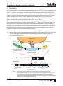
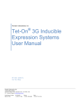
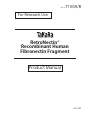
Cat. #
T100A/B
For Research Use
RetroNectin®
Recombinant Human
Fibronectin Fragment
Product Manual
v201303
RetroNectin®
(Recombinant Human Fibronectin Fragment)
Cat. #T100A/B
v201303
Table of Contents
I.
Description........................................................................................................................3
II.
Components.....................................................................................................................4
III. Storage................................................................................................................................4
IV. Materials Required but not Provided......................................................................4
V.
Protocol...............................................................................................................................5
1. Preparation of RetroNectin Coated Plates.....................................................5
2. Gene Transduction..................................................................................................5
A. RetroNectin-bound Virus (RBV) Infection Method...............................6
B. Supernatant (SN) Infection Method...........................................................8
VI. References..........................................................................................................................9
VII. Related Products.............................................................................................................9
2
URL:http://www.takara-bio.com
RetroNectin®
(Recombinant Human Fibronectin Fragment)
I.
Cat. #T100A/B
v201303
Description
RetroNectin reagent is a recombinant human fibronectin fragment (rFN-CH-296) composed of three
functional domains: the cell-binding domain (C-domain), heparin-binding domain (H-domain),
and CS-1 sequence. The fragment enhances retroviral-mediated gene transduction by aiding the
co-localization of target cells and virions. Specifically, virus particles bind RetroNectin reagent via
interaction with the H-domain, and target cells bind mainly through the interaction of cell surface
integrin receptor VLA-5 and VLA-4 with the fibronectin C-domain and CS-1 site, respectively.
Through facilitating close proximity, RetroNectin reagent can enhance retroviral-mediated gene
transfer to target cells expressing integrin receptors VLA-4 and/or VLA-5*1.
There are two RetroNectin-mediated infection protocols: the supernatant (SN) infection method
and the RetroNectin-bound virus (RBV) infection method 5), *2. With the SN infection method, cells
are mixed with virus supernatant and loaded on a RetroNectin-coated plate. In the RBV method,
the retrovirus is first bound to the RetroNectin coated plate, and cells are added after removing the
retrovirus supernatant. Removal of the supernatant reduces inhibitory molecules (e.g., molecules
secreted from the producer cells such as proteoglycans and/or viral envelope proteins) that can
reduce the efficiency of viral-mediated gene transduction.
*1 : RetroNectin can also enhance lentiviral-mediated gene transfer.
*2 : Both methods can be used for efficient gene transduction. Although the RBV infection method
is widely applicable, some modification might be required depending on the target cells,
vectors, and/or target genes.
TargetCell
receptor
VLA-5(α5β1)
VLA-4(α4β1)
CS-1
Cell-bindingdomain
(C-Domain;RGDS)
Heparin-binding
domain(H-Domain)
RGDS
RetroNectin(CH-296)
RGDS
SH
1
Fibrin Collagen
Heparin
9 10
Cell
Fibronectin
NH2
8
2
3
4
5
DNA
6
7
8
CS-1
12 13 14
Heparin
CS-1
9 10 11 12 13 14
Cell
Blue:bindingdomain
SH
SS
COOH
Heparin Cell
Fibrin
(IIICS)
Figure 1. The hypothesized mechanism of RetroNectin-mediated transduction. The cell binds to the CS-1 site via VLA-4, and to the C-domain via VLA-5. The
viral particle can bind to the H-domain of RetroNectin. These interactions
increase the localized concentrations of cells and viral particles, an effect that is
thought to enhance gene transduction.
URL:http://www.takara-bio.com
3
RetroNectin®
(Recombinant Human Fibronectin Fragment)
Cat. #T100A/B
v201303
II. Components
RetroNectin (Cat. #T100A)
RetroNectin (Cat. #T100B)
0.5 mg (0.5 ml)
2.5 mg (2.5 ml)
Note: RetroNectin is provided as a sterile 1 mg/ml solution.
III. Storage
-20℃
Caution: Freezing and thawing can be repeated up to 10 times.
Do not mix the solution vigorously. Do not vortex.
IV. Materials Required but not Provided
[Equipment]
• Non-treated tissue culture plates or dishes
• Electric pipetter
• Pipetter
• Sterile pipettes
• Sterile tips with filters
• Safety cabinet or clean workstation
• Microscope
• CO2 incubator
• Microplate centrifuge
[Reagents]
• Sterile PBS ( - ) (Phosphate Buffered Saline)
• HBSS/Hepes (Hank's Balanced Salt Solution supplemented with 2.5% (v/v) 1 M Hepes)
• 2% BSA (BSA Fraction V)/PBS Solution
4
URL:http://www.takara-bio.com
RetroNectin®
(Recombinant Human Fibronectin Fragment)
Cat. #T100A/B
v201303
V. Protocol
1.Preparation of RetroNectin Coated Plates
Coat a plate using 20 - 100 μg/ml RetroNectin with a volume corresponding to 4 - 20 μg/cm2 plate area.
(1) Prior to coating, prepare a RetroNectin solution (20 - 100 μg/ml*1) by diluting with
sterile PBS.
*1 : Example of calculating amount of RetroNectin reagent :
When 2.25 ml of RetroNectin solution at a concentration of 20 μg/ml is placed into
a 35 mm diameter dish (9 cm2), the concentration used for coating is 5 μg/cm2.
Note: To avoid loss of RetroNectin fragment, do not filter sterilize RetroNectin solution
diluted with PBS.
(2) Dispense an appropriate volume*2 of sterile RetroNectin solution into each plate and
allow the plate to stand for 2 hours at room temperature or at 4℃ overnight.
*2 : Dispense 0.5 ml into each well of a 24-well plate or 2 ml into each well of a 6-well
plate.
Note : Non-treated, cell culture-grade tissue culture plates or dishes should be used in
this step.
(3) Remove the RetroNectin solution and then block with an appropriate volume*3 of sterile
2% bovine serum albumin (BSA, Fraction V) in PBS. Allow the plate to stand at room
temperature for 30 minutes.
*3 : Use 0.5 ml for each well of a 24-well plate or 2 ml for each well of a 6-well plate.
(4) Remove the BSA solution, and wash the plate once with an appropriate volume of
HBSS/Hepes or PBS. After removing the wash solution, the plate is ready for use. The
RetroNectin coated plate can be sealed with Parafilm and stored at 4℃ for up to one week.
2.Gene Transduction
There are two methods of gene transduction using RetroNectin reagent: RetroNectin-bound
virus (RBV) infection method (Section A) and supernatant (SN) infection method (Section B).
In the RBV infection method, retroviral particles are first bound to the plate coated with
RetroNectin reagent, and the target cells are added after removing the virus supernatant. In
the SN infection method, the virus solution and target cells are mixed and then added to the
RetroNectin coated plate.
When virus solution is directly used for virus infection without purification, the gene
transduction efficiency may be reduced because of contaminating molecules that inhibit
infection. In such cases, the RBV infection method is recommended. With this method,
inhibitory molecules can be removed by binding viruses to RetroNectin reagent and
removing the supernatant.
Note: Using a transient production system, such as the Retrovirus Packaging Kit Eco/
Ampho (Cat. #6160/6161)* and the pDON-AI-2 vector (Cat. #3654/3653), retroviral
vectors can be prepared in one week.
* : Not available in all geographic locations. Check for availablity in your region.
URL:http://www.takara-bio.com
5
RetroNectin®
(Recombinant Human Fibronectin Fragment)
Cat. #T100A/B
v201303
A. RetroNectin Bound Virus (RBV) Infection Method
A-1. Preparation of virus bound plate without centrifugation
1. Add retrovirus supernatant at 125 - 250 μl/cm2 to a RetroNectin coated
plate or dish.
2. Incubate for 4 to 6 hours at 32℃ or 37℃ in a 5% CO2 incubator to promote
binding of the virus particles with RetroNectin reagent.
3. Discard the supernatant, but do not allow the plate to dry. Wash the plate
with an appropriate volume of PBS or PBS containing 0.1 - 2% albumin (BSA
or HSA). After washing, perform infection according to A-3.
A-2. Preparation of virus binding plate by centrifugation
If the virus titer is high enough, binding of viruses with RetroNectin reagent
can be accomplished without centrifugation as described in A-1.
However, if the titer is low, or if you require higher gene transduction
efficiency, binding the virus by centrifugation is preferable. With this method
the time required to bind the retrovirus is significantly less (2 hours versus 4 6 hours for the non-centrifugation method). A plate, such as a non-treated cell
culture plate, that can tolerate centrifugation at 1,000 - 2,000X g for 2 hours at
32℃ is required for this method. In addition, please note there is a possibility of
aerosol formation.
1. Add the retrovirus stock solution or diluted solution at 125 - 500 μl/cm2 to
the RetroNectin coated plate.
Note: The volume that can be added to the plate varies. For a 6-well plate,
the upper limit is 3 ml (320 μl/cm2). In this method, some infection
inhibitory molecules may not be removed by washing with PBS. For
this reason the efficiency of gene transduction might be reduced.
In such a case, it is recommended that the virus stock solution be
used after dilution with growth medium. Optimization is required to
determine the suitable dilution rate.
2. Place the plate in a centrifuge pre-warmed to 32℃ and centrifuge for 2 hours at 32℃ at 1,000 - 2,000X g to facilitate binding of virus particles
with RetroNectin reagent.
3. Discard the supernatant, but do not allow the plate to dry. Wash the plate
with an appropriate volume of PBS or PBS containing 0.1 - 2% albumin (BSA
or HSA). Then perform virus infection according to A-3.
6
URL:http://www.takara-bio.com
RetroNectin®
(Recombinant Human Fibronectin Fragment)
Cat. #T100A/B
v201303
A-3. Virus Infection
Prepare the target cells while the retrovirus particles are binding to the
RetroNectin reagent coated plate. It is important that the target cells be in
logarithmic growth phase and express integrin receptors VLA-4 and/or VLA5. When using hematopoietic stem cells, pre-stimulation with cytokine may
be necessary. The cytokine type should be determined based on your specific
research protocols. Examples are cited in references 3 and 5.
1. Collect the target cells and count the number of living cells. Then suspend
the cells in the growth medium at a concentration of 0.2 - 1 x 10⁵ cells/ml.
2. Remove the wash solution from the virus bound plates prepared by A-1 or
A-2. Do not allow the plate to dry. Immediately add target cells at a density
of 0.5 - 2.5 x 10⁴ cells/cm2. Although the optimal cell density depends on
cell size and growth rate, the initial cell density should allow the cells to
be actively growing or nearly confluent when analyzed 2 - 3 days after
transduction. When infecting more cells, you may increase the cell density,
but the cells will need to be subcultured after gene transduction.
Note: To promote contact between the target cells and viral particles,
plates can be centrifuged after adding the cells.
3. Incubate in a 37℃, 5% CO2 incubator for 2 - 3 days.
4. Collect both non-adherent and adherent cells:
(1) Transfer the supernatant to a centrifuge tube.
(2) Recover remaining non-adherent cells by washing the plate with PBS.
(3) Dissociate adherent cells from the plate with Cell Dissociation Buffer (Life
Technologies), an enzyme free solution, or trypsin-EDTA following the
manufacturer's instructions.
Note: For many cell types, adherent cells may be collected by pipetting
only.
(4) Combine the cells obtained from steps (1)-(3) in the same tube, and
centrifuge to recover the cells.
(5) Wash the cells with HBSS/Hepes twice, collecting the cells by
centrifugation. Suspend the cells in HBSS/Hepes* for further analysis.
* : Any buffer or medium suitable for downstream application of the
cells can be also used for resuspension.
URL:http://www.takara-bio.com
7
RetroNectin®
(Recombinant Human Fibronectin Fragment)
Cat. #T100A/B
v201303
B. Supernatant (SN) Infection Method
When the virus stock solution is used, the RBV method described in A is
recommended, but if a 4-fold dilution or more is used, either the RBV or SN method
may be used as equivalent gene transduction efficiency will be obtained. The time
required for virus infection is much shorter with the SN method than with the RBV
method.
1. Suspend the target cells in virus solution that has been diluted with growth
medium to prepare the cell suspension.
2. Add the cell suspension to the RetroNectin coated plate at a density of 0.5 - 2.5 x 10⁴ cells/cm2. Although the optimal cell density depends on cell size and
growth rate, the initial cell density should allow the cells to be actively growing or
nearly confluent when gene expression is analyzed 2 - 3 days after transduction.
When infecting more cells, you may increase the cell density, but the cells will
need to be subcultured after gene transduction.
Note: To promote contact between the target cells and virus vectors, the plate
can be centrifuged after adding the cells.
3. Incubate in a 37℃, 5% CO2 incubator for 2 - 3 days.
4. Collect both non-adherent and adherent cells:
(1) Transfer the supernatant to a centrifuge tube.
(2) Recover remaining non-adherent cells by washing the plate with PBS.
(3) Dissociate adherent cells from the plate with Cell Dissociation Buffer (Life
Technologies), an enzyme free solution, or trypsin-EDTA following the
manufacturer’ s instructions.
Note: For many cell types, adherent cells may be collected by pipetting only.
(4) Combine the cells obtained from steps (1)-(3) in the same tube, and centrifuge
to recover the cells.
(5) Wash the cells with HBSS/Hepes twice, collecting the cells by centrifugation.
Suspend the cells in HBSS/Hepes* for further analysis.
* : Any buffer or medium suitable for downstream application of the cells can
be also used for resuspension.
8
URL:http://www.takara-bio.com
RetroNectin®
(Recombinant Human Fibronectin Fragment)
Cat. #T100A/B
v201303
VI. References
1) Kimizuka F, Taguchi Y, Ohdate Y, Kawase Y, Shimojo T, Hashino K, Kato I, Sekiguchi K,
and Titani K. (1991) Production and characterization of functional domains of human
fibronectin expressed in Escherichia coli. J. Biochem . 110:284-291.
2) Hanenberg H, Xiao XL, Dilloo D, Hashino K, Kato I, and Williams DA. (1996)
Colocalization of retrovirus and target cells on specific fibronectin fragments increases
genetic transduction of mammalian cells. Nat Med . 2:876-882.
3) Hanenberg H, Hashino K, Konishi H, Hock RA, Kato I, and Williams DA. (1997)
Optimization of fibronectin-assisted retroviral gene transfer into human CD34+
hematopoietic cells. Hum . Gene Ther . 8:2193-2206.
4) Pollok KE, Hanenberg H, Noblitt TW, Schroeder WL, Kato I, Emanuel D, and Williams
DA. (1998) High-efficiency gene transfer into normal and adenosine deaminasedeficient T lymphocytes is mediated by transduction on recombinant fibronectin
fragments. J. Virol . 72:4882-4892.
5) Chono, H., Yoshioka, H., Ueno, M. and Kato, I. (2001) Removal of inhibitory substance
with recombinant fibronectin-CH-296 plates enhances the retroviral transduction
efficiency of CD34+CD38- bone marrow cells. J. Biochem . 130:331-334.
VII. Related Products
Retroviral vectors
pDON-AI-2 Neo DNA
pDON-AI-2 DNA
(Cat. #3653)
(Cat. #3654)
Preparation of Recombinant Retroviral Particles
Retrovirus Packaging Kit Ampho Retrovirus Packaging Kit Eco Retro-X™ System
Retro-X™ Universal Packaging System (Cat. #6161)*
(Cat. #6160)*
(Clontech, Cat. #631508)
(Clontech, Cat. #631530)
Lentiviral Vectors and Vector Systems
Lenti-X™ Expression System Lenti-X™ Expression System (EF1alpha Version) (Clontech, Cat. #632164)
(Clontech, Cat. #631253)
Preparation of Recombinant Lentiviral Particles
Lenti-X™ 293T Cell Line
Lenti-X™ HTX Packaging System
Lenti-X™ HTX Ecotropic Packaging System (Clontech, Cat. #632180)
(Clontech, Cat. #631247)
(Clontech, Cat. #631251)
Other
RetroNectin® Pre-coated Dish RetroNectin® (GMP)
Retrovirus Titer Set (for Real Time PCR) Retro-X™ qRT-PCR Titration Kit
Lenti-X™ qRT-PCR Titration Kit (Cat. #T110A)
(Cat. #T201)*
(Cat. #6166)*
(Clontech, Cat. #631453)
(Clontech, Cat. #631235)
* : Not available in all geographic locations. Check for availability in your region.
URL:http://www.takara-bio.com
9
RetroNectin®
(Recombinant Human Fibronectin Fragment)
10
Cat. #T100A/B
v201303
URL:http://www.takara-bio.com
RetroNectin®
(Recombinant Human Fibronectin Fragment)
Cat. #T100A/B
v201303
NOTICE TO PURCHASER: LIMITED LICENSE
[L9] RetroNectin®
A method to increase the efficiency of retrovirus mediated gene transfer (covered by the claims of
U.S. Patent No. 5,686,278, 6,033,907, 7,083,979, and 6,670,177 and their foreign counterpart patent
claims)is licensed to TAKARA BIO INC. exclusively and worldwide.
[M69] RetroNectin® Expansion Method
This product is covered by the claims of Japanese Patent No. 4406566 and its foreign counterpart
patent claims.
URL:http://www.takara-bio.com
11
RetroNectin®
(Recombinant Human Fibronectin Fragment)
Cat. #T100A/B
v201303
NOTE : This product is for research use only. It is not intended for use in therapeutic or diagnostic
procedures for humans or animals. Also, do not use this product as food, cosmetic, or
household item, etc.
Takara products may not be resold or transferred, modified for resale or transfer, or used
to manufacture commercial products without written approval from TAKARA BIO INC.
If you require licenses for other use, please contact us by phone at +81 77 543 7247 or
from our website at www.takara-bio.com.
Your use of this product is also subject to compliance with any applicable licensing
requirements described on the product web page. It is your responsibility to review,
understand and adhere to any restrictions imposed by such statements.
All trademarks are the property of their respective owners. Certain trademarks may not be
registered in all jurisdictions.
12
URL:http://www.takara-bio.com