1
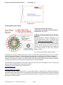
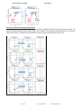
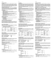
ver. 8-2013-02-22 ExoQuick™ Exosome Precipitation Solution Cat. # EXOQ__A-1 Contents I. II. III. IV. V. VI. VII. VIII. Overview 2 Exosome Isolation Protocol 3 Exosome analysis A. RNA analysis 4 B. Protein analysis 4 Example Data and Applications 8 Appendix……………………………………………………12 References………………………………………………….12 Technical Support…………………………………………14 Licensing and Warranty………………………………….15 List of Components Item Catalog # Reactions ExoQuick exosome precipitation solution (20 ml) ExoQuick exosome precipitation solution (5 ml) EXOQ20A-1 300 reactions EXOQ5A-1 75 reactions The ExoQuick™ kits are shipped at room temperature or on blue ice and should be stored at +4°C upon receipt. Properly stored kits are stable for 1 year from the date received. The reaction size is based on using 250 µl serum for exosome isolation. Examples of precipitating exosomes from various biofluids can be seen in the Table below. Bio-fluid Serum Ascites fluid Sample volume 250 l ExoQuick volume 63 l 250 l 63 l To isolate exosomes from tissue culture media or urine, we recommend using the ExoQuick-TC reagent (cat# EXOTC10A-1 or EXOTC50A-1) which is a distinct formulation from the original ExoQuick reagent detailed in this manual. 888-266-5066 (Toll Free) 650-968-2200 (outside US) Page 1 System Biosciences (SBI) User Manual ExoQuick Exosome Precipitation I. Overview Exosomes are 60 –150 nm membrane vesicles secreted by most cell types in vivo and in vitro. Exosomes are found in blood, urine, amniotic fluid, malignant ascite fluids and contain distinct subsets of microRNAs depending upon the tumor from which they are secreted. SBI's ExoQuick exosome precipitation reagent makes microRNA and protein biomarker discoveries simple, reliable and quantitative. Enrich for circulating exosomal microRNAs with ExoQuick™ and accurately profile them using SBI’s SeraMir™ qPCR arrays. Further, selective exosome capture is also possible using SBI’s Exo-Flow kits that purify exosomes on magnetic beads based upon exosome surface protein marker capture. * No time-consuming ultracentrifugation * Less expensive than costly antibodies and beads * More effective than any other method * Use as little as 100 µl of serum or bio-fluid Page 2 ver. 9-2013-08-22 www.systembio.com ExoQuick™ Exosome Precipitation Solution Cat. # EXOQ__A-1 Exosome Precipitation Protocol II. Isolate exosomes with ExoQuick 1. Collect biofluid and centrifuge at 3000 × g for 15 minutes to remove cells and cell debris. 2. Transfer supernatant to a sterile vessel and add the appropriate volume of ExoQuick Exosome Precipitation Solution to the bio-fluid. Some examples are shown in the Table below. Mix well by inverting or flicking the tube. *Note when precipitating exosomes from plasma, the resulting pellet may be difficult to resuspend due to precipitated fibrin and other Incubation Sample ExoQuick fibrinogens. For plasma, please Time Bio-fluid volume volume refer to the Appendix for 30 minutes Serum* 63 l 250 l additional preparation steps. Overnight 63 l Ascites fluid 250 l 3. Refrigerate overnight (at least 12 hours) for ascites fluid or 30 minutes for serum. The tubes do not need to be rotated during the incubation period. 4. Centrifuge ExoQuick/biofluid mixture at 1500 × g for 30 minutes. Centrifugation may be performed at either room temperature or 4°C with similar results. After centrifugation, the exosomes may appear as a beige or white pellet at the bottom of the vessel. 5. Aspirate supernatant. Spin down residual ExoQuick solution by centrifugation at 1500 × g for 5 minutes. Remove all traces of fluid by aspiration, taking great care not to disturb the precipitated exosomes in pellet. 6. Resuspend exosome pellet in 1/10 of original volume using sterile or nuclease-free water. If the pellet is difficult to resuspend, add slightly more water to the pellet to further dilute the salt. III. A. Using Precipitated Exosomes for RNA Extraction For RNA extraction, we recommend following the protocol outlined in the SeraMir Kit user manual as shown here (Catalog #: RA800A-1, RA805A-1, RA806A-1, RA810A-1, and RA820A-1). 1. 2. 3. 4. 5. 6. 7. 8. Thaw serum sample on ice Exosome Combine 500µl serum + 120 µl ExoQuick Isolation Mix well by inversion three times and Lysis Place at 4ºC for 30 minutes Centrifuge at 13,000 rpm for 2 minutes Remove supernatant, keep exosome pellet Add 350 µl LYSIS Buffer to exosome pellet and vortex 15 seconds Place at room temperature for 5 minutes (to allow complete lysis) --- optional--- add 5µl of SeraMir control RNA spike-in (cat#RA805A-1) 9. 10. 11. 12. Add 200µl of 100% Ethanol, vortex 10 seconds Assemble spin column and collection tube Transfer all (600µl) to spin column exoRNA Centrifuge at 13,000 rpm for 1 minute Purification (check to see that all flowed through, otherwise spin longer) 13. Discard flow-through and place spin column back into collection tube 888-266-5066 (Toll Free) 650-968-2200 (outside US) Page 3 System Biosciences (SBI) 14. 15. 16. 17. User Manual Add 400µl WASH Buffer Centrifuge at 13,000 rpm for 1 minute Repeat steps 13 to 15 once again (total of 2 Washes) Discard flow-through and centrifuge at 13,000 rpm for 2 minutes to dry (IMPORTANT !) 18. Discard collection tube and assemble exoRNA spin column with a fresh, Elution RNase-free 1.5ml elution tube (not provided) 19. Add 30µl ELUTION Buffer directly to membrane in spin column 20. Centrifuge at 2,000 rpm for 2 minutes (loads buffer in membrane) 21. Increase speed to 13,000 rpm and centrifuge for 1 minute (elutes exoRNAs) 22. You should have recovered 30-40µl exosome RNA The yield of RNA from isolated exosomes is different depending on the starting biofluid or the type of cells that were grown in culture. Different cell types secrete varying levels of exosomes. For serum, the level of RNA isolated from 500 µl is usually in the 500ng range and can be measured using a Agilent Bioanalyzer or a NanoDrop Spectrophotometer. B. Using Precipitated Exosomes for Protein Extraction ELISA analysis SBI offers three ELISA kits (Catalog#: ExoELISA-63, ExoELISA-9, ExoELISA-81) for fast and quantitative analysis of wellcharacterized exosomal protein markers: CD63, CD9 and CD81. 1. 2. 3. 4. 5. 6. 7. 8. 9. 10. 11. 12. If frozen, thaw culture media or urine sample on ice Combine 500µl serum + 120 µl ExoQuick Mix well by inversion three times Place at 4ºC for 30 minutes (or up to 12 hours) Centrifuge at 1500 × g for 30 minutes Remove supernatant, keep exosome pellet Centrifuge at 1500 × g for 5 minutes to remove all traces of fluid (take great care not to disturb the pellet) Add 200 µl Exosome Binding buffer to exosome pellet and vortex 15 seconds Incubate at 37 ºC temperature for 20 minutes to liberate exosome proteins Centrifuge at 1500 × g for 5 minutes to remove all residual precipitation solution Transfer supernatant to new centrifuge tube on ice Exosome protein is now ready for immobilization onto micro-titer plate Please refer to the ExoELISA manual for the complete protocol. Western blot analysis 1 For Western blotting analysis, we recommend resuspending the exosome pellet in 1XRIPA buffer with the appropriate protease inhibitor cocktail. SBI offers a Western blot antibody detection kit (Catalog# ExoAB-KIT-1) which includes four exosomal marker antibodies: CD63, CD9, CD81, HSP70 and a Goat anti-Rabbit IgG HRP conjugated secondary antibody specifically tested for use in exosomal protein analysis. Page 4 ver. 9-2013-08-22 www.systembio.com ExoQuick™ Exosome Precipitation Solution Cat# Description EXOAB‐CD9A‐1 EXOAB‐C63A‐1 EXOAB‐CD81A‐1 EXOAB‐Hsp70A‐1 EXOAB‐KIT‐1 EXOEL‐CD9A‐1 EXOEL‐CD63A‐1 EXOEL‐CD81A‐1 Cat. # EXOQ__A-1 Size Anti‐CD9 Antibody (rabbit anti‐human) with goat anti‐ 25 ul rabbit HRP secondary antibody Anti‐CD63 Antibody (rabbit anti‐human) with goat anti‐ 25 ul rabbit HRP secondary antibody Anti‐CD81 Antibody (rabbit anti‐human) with goat anti‐ 25 ul rabbit HRP secondary antibody Anti‐Hsp70 Antibody (rabbit anti‐human) with goat 25 ul anti‐rabbit HRP secondary antibody ExoAb Antibody Kit (CD9, CD63, CD81, Hsp70 antibodies, rabbit anti‐human) with goat anti‐rabbit 25 ul each HRP secondary antibody 96 reactions Exosome ELISA Complete Kit (CD9 detection) Exosome ELISA Complete Kit (CD63 detection) 96 reactions Exosome ELISA Complete Kit (CD81 detection) 96 reactions 1. 2. 3. 4. 5. 6. 7. 8. 9. 10. 11. 12. 13. 14. 15. 16. If frozen, thaw culture media or urine sample on ice Combine 500µl serum + 120 µl ExoQuick Exosome Mix well by inversion three times Isolation and Place at 4ºC for 30 minutes (or up to12 hours) lysis Centrifuge at 1500 × g for 30 minutes Remove supernatant, keep exosome pellet Centrifuge at 1500 × g for 5 minutes to remove all traces of fluid (take great care not to disturb the pellet) 1 Add 200 µl RIPA buffer to exosome pellet and vortex 15 seconds Place at room temperature for 5 minutes (to allow complete lysis) Add Laemmli buffer2 (with Beta-mercaptoethanol) and heat at 95⁰C for 5 minutes. Chilled on ice for 5 minutes before loading onto gel Perform standard SDS-PAGE electrophoresis and Western transfer onto PVDF membrane Block with 5% dry milk in Tris Buffered Saline + 0.05% Tween (TBS-T) for 1 hour Incubate blot overnight at 4°C with SBI's exosome specific antibody (e.g. CD9) at 1:1000 dilution (5% dry milk in TBS-T) Wash 3X with TBS-T Incubate one hour at room temperature with SBI's Goat anti-Rabbit-HRP antibody at 1:20,000 dilution (5% dry milk in TBS-T) 17. Wash 3X with TBS-T 18. Incubate blot with chemi-luminescence substrate and visualize on film or other imaging equipment 1 1X RIPA buffer contains: 25mM Tris-HCl pH 7.6 150mM NaCl 1% NP-40 1% sodium deoxycholate 0.1% SDS 2 2X Laemmli buffer contains: 4% SDS 20% glycerol 10% 2mercaptoethanol 0.004% bromphenol blue 0.125 M Tris-HCl pH 6.8 888-266-5066 (Toll Free) 650-968-2200 (outside US) Page 5 System Biosciences (SBI) IV. User Manual Example Data and Applications 1. Protein Yield from Exosomes precipitated with ExoQuick versus other Extraction Methods a. b. a. The quantity of protein was determined by the Bradford microassay method (Bio-Rad Laboratories) using BSA as a standard. b. Proteins from each exosome isolate were standardized to the original sample volume and equal volumes were applied per lane of a 12.5% SDS-PAGE gel. Western immunoblotting was performed to analyze the presence of the specific marker protein, placental alkaline phosphatase (PLAP). The SDS-PAGE gel was transferred to a nitrocellulose membrane, the membrane blocked for 1 hour at room temperature with non-fat dried milk, and probed overnight at 4°C with primary antibody. The bound immune complexes were visualized by enhanced chemiluminescence (ECL, Amersham Life Sciences) and quantitated by densitometry (Un-Scan-it Software, Silk Scientific Corp). 2. MicroRNA Yield from Exosomes precipitated with ExoQuick versus other Extraction Methods Page 6 ver. 9-2013-08-22 www.systembio.com ExoQuick™ Exosome Precipitation Solution Cat. # EXOQ__A-1 The RNA quality and yield was accessed using a GeneQuant II. Small RNAs were analyzed with the Agilent 2100 Bioanalyzer Lab-on-a-Chip instrument system (Agilent Technologies), using the Agilent Small RNA chip and reagent kit. Approximately 100ng of isolated total RNA in 1μl was applied to each run. The manufacturer’s recommended protocol was strictly followed to obtain Bioanalyzer profiles for the size range 6 to 150 nucleotides (nt). The profiles were calibrated for size (nt) using the small RNA ladder supplied with the kit, containing markers of 20, 40, 60, 80, and 150 nt in size, as reference. The instrument software quantitated the peak area between 0 and 150 nt as small RNA region, the area within 10 to 40 nt as microRNA region, and provides percentages of miRNA detected for each sample. 888-266-5066 (Toll Free) 650-968-2200 (outside US) Page 7 System Biosciences (SBI) User Manual 4. Activity Assays: Track Exosomes using Cyto-Tracers SBI has created a line of lentivector-based Cyto-Tracers™ that utilize GFP-fusion proteins to mark cellular compartments, organelles, vesicles and structures to enable more long-term and more in-depth experimentation. The Cyto-Tracers can be used in transfections as well as packaged into virus to create stable GFP tracer cell lines in primary cells, tumor cell lines and stem cells. The Tetraspanin CD63 protein is a common biomarker for exosomes. With the pCT-CD63-GFP construct you can make you cells of interest secrete exosomes that glow green for downstream functional delivery studies (Cat. # CYTO120-PA-1). 1. NanoSight The NanoSight LM10 instrument is based on a conventional optical microscope and uses a laser light source to illuminate nano-scale particles within a 0.3 ml sample introduced to the viewing unit with a disposable syringe. Enhanced by a near perfect black background, particles appear individually as point-scatterers moving under Brownian motion. The image analysis Nanoparticle Tracking Analysis (NTA) software suite allows users to automatically track and size nanoparticles on an individual basis. Results are displayed as a frequency size distribution graph and output to spreadsheet. ExoQuick serum exosome analysis Normal human serum from 50 pooled samples was used. Only 250ul serum was combined with 63ul ExoQuick to pellet the exosomes in 30 minutes. The exosome pellet was resuspended in 100ul PBS, diluted 1:10,000 and visualized on the NanoSight LM10 instrument. The analysis shows that ExoQuick isolated 90nm exosomes with a recovery of 2.74 x 10^12 particles/ml. Page 8 ver. 9-2013-08-22 www.systembio.com ExoQuick™ Exosome Precipitation Solution Cat. # EXOQ__A-1 Data acquisition and analysis These experiments were done in collaboration with Particle Characterization Laboratories, Inc (PCL). 2. Magnetic Beads and Exosome Surface Markers SBI has developed a magnetic streptavidin 9.1 um Exo-Flow bead system. The 9.1 um diameter of the beads enables more exosome capture per volume added when compared to 4 um Dynabeads. This is significant in that some exosome subpopulations that are desired may only be present in very low numbers. The increased surface area enables the more efficient capture of these rare exosomes. Bigger is better. Fig. 1: Exo-Flow streptavidin magnetic beads Depending upon the specific exosomes you wish to purify, a particular biotinylated antibody may be used to couple to the Exo-Flow streptavidin beads. The Exo-Flow kits are modular, thus you can select from various pre-validated capture antibody kits, or utilize your own biotinylated capture antibody corresponding to the exosome surface marker specific for the exosomes of interest in your model system. SBI has thoroughly tested a variety of capture antibodies that work quite well to flow-sort exosomes from either serum or cell culture samples. The data below were generated on a BD LSR II instrument and are meant as examples of how to set the gate settings and perform data analysis using FlowJo software. Information on FlowJo software can be viewed online. http://www.flowjo.com/ Exo-Flow software gate settings The forward and side scatter data for the 9.1 um Exo-Flow beads are shown below for samples containing no captured exosomes stained with Exo-FITC (left panel) and then data for serum CD9-captured exosomes stained with Exo-FITC. Set the gate primarily on the majority bead singlets (outlined in a black oval, red arrow pointing to the gate setting) prior to full flow analysis. 888-266-5066 (Toll Free) 650-968-2200 (outside US) Page 9 System Biosciences (SBI) User Manual Exo-Flow exosome flow-exometry example data Bead flow separation data for the various capture antibodies coupled with Exo-FITC staining are shown below. The data are graphed showing forward scatter versus FITC intensity. The first panel depicts beads with no exosomes then with exosomes. The degree of flow separation is shown on the right side for each capture set. Page 10 ver. 9-2013-08-22 www.systembio.com ExoQuick™ Exosome Precipitation Solution V. Cat. # EXOQ__A-1 Appendix PURIFIED THROMBIN PLASMA REAGENT FOR EXOQUICK Catalog #s TMEXO-1 and EXOQ5TM-1 1) Purified Thrombin is at [500U/mL] in PBS. 2) Add 5 uL of [500U/mL] Thrombin per 0.5 mL plasma to a final concentration of 5U/mL. 3) Incubate at room temperature for 5 minutes while mixing (gently flicking tube). 4) Centrifuge in a standard microfuge at 10,000 rpm, 5 minutes. 5) There should be a visible fibrin pellet at the bottom of the tube. 6) Transfer supernatant to new clean tube. 7) Treat serum-like supernatant with ExoQuick to precipitate exosomes, 30-60 minutes at 5°C. VI. References As featured in: Exosome Isolation for Proteomic Analyses and RNA Profiling Douglas D. Taylor, Wolfgang Zacharias and Cicek Gercel-Taylor, Serum/Plasma Proteomics, Methods in Molecular Biology, 2011, Volume 728, Part 4, 235-246, (PDF) » Tae Hoon Lee, Esterina D'Asti, Nathalie Magnus, Khalid Al-Nedawi, Brian Meehan and Janusz Rak. Review: Microvesicles as mediators of intercellular communication in cancer—the emerging science of cellular 'debris'. Seminars in Immunopathology DOI: 10.1007/s00281-011-0250-3. (PDF) » Technical References Adachi T, Nakanishi M, Otsuka Y, Nishimura K, Hirokawa G, Goto Y, Nonogi H, Iwai N. Plasma microRNA 499 as a biomarker of acute myocardial infarction. Clin Chem. 2010 Jul;56(7):1183-5. De Smaele E, Ferretti E, Gulino A. MicroRNAs as biomarkers for CNS cancer and other disorders. Brain Res. 2010 Jun 18;1338:100-11. Mitchell PS, Parkin RK, Kroh EM, Fritz BR, Wyman SK, Pogosova-Agadjanyan EL, Peterson A, Noteboom J, O'Briant KC, Allen A, Lin DW, Urban N, Drescher CW, Knudsen BS, Stirewalt DL, Gentleman R, Vessella RL, Nelson 888-266-5066 (Toll Free) 650-968-2200 (outside US) Page 11 System Biosciences (SBI) User Manual PS, Martin DB, Tewari M. Circulating microRNAs as stable blood-based markers for cancer detection. Proc Natl Acad Sci U S A. 2008 Jul 29;105(30):10513-8. Laterza OF, Lim L, Garrett-Engele PW, Vlasakova K, Muniappa N, Tanaka WK, Johnson JM, Sina JF, Fare TL, Sistare FD, Glaab WE. Plasma MicroRNAs as sensitive and specific biomarkers of tissue injury. Clin Chem. 2009 Nov;55(11):1977-83. Valadi H, Ekström K, Bossios A, Sjöstrand M, Lee JJ, Lötvall JO. Exosome-mediated transfer of mRNAs and microRNAs is a novel mechanism of genetic exchange between cells. Nat Cell Biol. 2007 Jun;9(6):654-9. Pegtel DM, Cosmopoulos K, Thorley-Lawson DA, van Eijndhoven MA, Hopmans ES, Lindenberg JL, de Gruijl TD, Wordinger T, Middeldorp JM. Functional delivery of viral miRNAs via exosomes. Proc Natl Acad Sci USA; 2010 Apr 6; 107(14):6328-33. Mathivanan, S. and Simpson, R.J. ExoCarta: A compendium of exosomal proteins and RNA. Proteomics. 2009.21, 4997-5000. Thery C, Ostrowski M, Segura E. Membrane vesicles as conveyors of immune responses. Nat Rev Immunol. 2009. 8, 581-93. Michael A, Bajracharya SD, Yuen PS, Zhou H, Star RA, Illei GG, Alevizos I. Exosomes from human saliva as a source of microRNA biomarkers. Oral Dis; 2010 Jan; 16(1):34-8. Luo SS, Ishibashi O, Ishikawa G, Ishikawa T, Katayama A, Mishima T, Takizawa T, Shigihara T, Goto T, Izumi A, Ohkuchi A, Matsubara S, Takeshita T, Takizawa T. Human villous trophoblasts express and secrete placentaspecific microRNAs into maternal circulation via exosomes. Biol Reprod; 2009 Oct; 81(4):717-29. Taylor DD, Gercel-Taylor C. MicroRNA signatures of tumor-derived exosomes as diagnostic biomarkers of ovarian cancer. Gynecol Oncol; 2008 Jul; 110(1):13-21. Simpson RJ, Lim JW, Moritz RL, Mathivanan S. Exosomes: proteomic insights and diagnostic potential. Expert Rev Proteomics. 2009 Jun;6(3):267-83. Review. VII. Technical Support For more information about SBI products and to download manuals in PDF format, please visit our web site: http://www.systembio.com For additional information or technical assistance, please call or email us at: System Biosciences (SBI) 265 North Whisman Road. Mountain View, CA 94043 Phone: (650) 968-2200 (888) 266-5066 (Toll Free) Fax: (650) 968-2277 E-mail: General Information: [email protected] Technical Support: [email protected] Ordering Information: [email protected] Page 12 ver. 9-2013-08-22 www.systembio.com ExoQuick™ Exosome Precipitation Solution Cat. # EXOQ__A-1 II. Licensing and Warranty Statement Limited Use License TM Use of the ExoQuick Exosome Precipitation Solution (i.e., the “Product”) is subject to the following terms and conditions. If the terms and conditions are not acceptable, return all components of the Product to System Biosciences (SBI) within 7 calendar days. Purchase and use of any part of the Product constitutes acceptance of the above terms. The purchaser of the Product is granted a limited license to use the Product under the following terms and conditions: The Product shall be used by the purchaser for internal research purposes only. The Product is expressly not designed, intended, or warranted for use in humans or for therapeutic or diagnostic use. The Product may not be resold, modified for resale, or used to manufacture commercial products without prior written consent of SBI. This Product should be used in accordance with the NIH guidelines developed for recombinant DNA and genetic research. SBI has pending patent applications related to the Product. For information concerning licenses for commercial use, contact SBI. Purchase of the product does not grant any rights or license for use other than those explicitly listed in this Licensing and Warranty Statement. Use of the Product for any use other than described expressly herein may be covered by patents or subject to rights other than those mentioned. SBI disclaims any and all responsibility for injury or damage which may be caused by the failure of the buyer or any other person to use the Product in accordance with the terms and conditions outlined herein. Limited Warranty SBI warrants that the Product meets the specifications described in this manual. If it is proven to the satisfaction of SBI that the Product fails to meet these specifications, SBI will replace the Product or provide the purchaser with a refund. This limited warranty shall not extend to anyone other than the original purchaser of the Product. Notice of nonconforming products must be made to SBI within 30 days of receipt of the Product. SBI’s liability is expressly limited to replacement of Product or a refund limited to the actual purchase price. SBI’s liability does not extend to any damages arising from use or improper use of the Product, or losses associated with the use of additional materials or reagents. This limited warranty is the sole and exclusive warranty. SBI does not provide any other warranties of any kind, expressed or implied, including the merchantability or fitness of the Product for a particular purpose. SBI is committed to providing our customers with high-quality products. If you should have any questions or concerns about any SBI products, please contact us at (888) 266-5066. © 2013 System Biosciences (SBI), All Rights Reserved 888-266-5066 (Toll Free) 650-968-2200 (outside US) Page 13